Review

Robotic Bronchoscopy: A Comprehensive Review

by
Bharat Singh Bhandari
1,*,
Akshita Jain
2,
Soumit Sharma
2,
Gunjan Rana
3 and
Bruce Fernando Sabath
1
1
Department of Pulmonary Medicine, The University of Texas MD Anderson Cancer Center, Houston, TX 77030, USA
2
North DMC Medical College and Hindu Rao Hospital, Delhi 110005, India
3
Saint Vincent Hospital, Worcester, MA 01608, USA
*
Author to whom correspondence should be addressed.
J. Respir. 2024, 4(2), 128-139; https://doi.org/10.3390/jor4020011
Submission received: 9 February 2024 / Revised: 1 April 2024 / Accepted: 9 May 2024 / Published: 30 May 2024

Abstract

:
Lung cancer, a major global cause of cancer-related deaths, demands continual advancements in diagnostic methodologies. This review delves into the transformative role of Robotic-Assisted Bronchoscopy (RAB) in redefining lung cancer diagnostics. As lung cancer screenings intensify, leading to a surge in pulmonary nodule diagnoses, navigational bronchoscopy, notably electromagnetic navigational bronchoscopy (ENB), faces persistent limitations. Examining key RAB platforms—Monarch™, Ion™ and the Galaxy System™—reveals their distinctive features, with RAB demonstrating superior diagnostic yields over traditional biopsy methods. However, challenges include CT-to-body divergence (CBCT) and divergent findings in diagnostic yield studies and a lack of head-to-head comparisons with non-RAB modalities. Future directions should explore RAB’s potential therapeutic applications, shaping the landscape of both diagnostics and therapeutics in lung cancer management.

1. Introduction

Globally, lung cancer exhibits the highest mortality rate among all cancers, standing as the primary cause of cancer-related deaths (18% of total cancer deaths). The incidence of lung cancer is nearly on par with its mortality, with approximately 2.20 million new cases and over 1.79 million associated deaths occurring annually worldwide [1]. The 5-year survival rate for lung cancer is 23% in the United States. This is markedly different from the more favorable survival rates seen in breast cancer (90%), colon cancer (65%) and prostate cancer (nearly 100%) [2]. After the advent of lung cancer screening following the national lung cancer screening trial in 2013, pulmonary nodules are being diagnosed at a rapid rate [3]. In the United States, more than 10 million CT scans of the chest are performed annually. With the increased use of low-dose CT scans (LDCT) for screening along with new-generation CT picking up very small nodules, the diagnosis of lung nodules is predicted to rise [2].
Up-to-date algorithms and guidelines have been designed to manage the diagnosed lung nodules, recommending biopsy in high-risk nodules [4]. Lung cancers detected at an early stage via screening boast a remarkable cure rate exceeding 90%, in contrast to the mere 15% cure rate for cancers that are diagnosed after the manifestation of symptoms. The 5-year survival rate of lung cancer patients drops from 82% for stage IA to 6% for stage IV. Multiple trials have shown significant mortality reduction and increased five-year survival rates with the early diagnosis of lung cancer [3,5,6].
Different modalities and approaches are now used to biopsy and diagnose lung nodules. Conventionally CT-guided transthoracic percutaneous needle lung biopsy (CT-PTNB) has been better in terms of diagnostic yield when compared to traditional flexible bronchoscopy (FFB), even after the advent of electromagnetic navigation. However, it is important to note the increased risk of pneumothorax and bleeding associated with CT-PTNB [7]. We discuss robotic bronchoscopy/robot-assisted bronchoscopy (RAB) in this review, which may be a better alternative for lung biopsy in terms of its better diagnostic yield and safety profile.

2. History

We have come a long way since bronchoscopy was discovered by Gustav Killian in 1876, when he removed a pork bone from a farmer’s airway using an esophagoscope [8]. Conventional diagnostic methods for solitary pulmonary nodules (SPN’s) rely on a flexible bronchoscope, primarily effective for central larger lesions but a diagnostic yield that is limited in peripheral areas [9]. The radial EBUS probe worked as an adjunct with the flexible bronchoscope after its introduction in 1996 and improved lesion localization, but navigating to the site of the lesson still posed an imminent challenge [10].
The genesis of navigational bronchoscopy arose in response to recognized limitations. The initial navigational systems comprised electromagnetic navigational bronchoscopy (ENB) platforms. The concept was that if the volume within the electromagnetic field could be mapped to align with a 3D reconstruction of the patient’s anatomy from CT imaging, the sensor could then be systematically guided through this volume. The sensor’s position would be reflected on a virtual 3D map of the lung. This differs from virtual bronchoscopy, which utilizes a chest CT scan to generate an airway rendering but lacks a positioning function. Virtual bronchoscopy was swiftly surpassed by the ENB platform which came out in 2004. In simple terms, ENB can be likened to navigating with a global positioning system, while virtual bronchoscopy is akin to navigating with a roadmap [11,12].
Early investigations into ENB displayed promising outcomes, with initial prospective studies indicating diagnostic yields ranging between 60% and 80% in diverse research settings, and approximately 75% in the case of peripheral SPN, albeit with only 57% of these measuring less than 2 cm. However, subsequent research yielded less optimistic results. The NAVIGATE trial, which studied ENB using the SuperDimension system, showed a 12-month diagnostic yield of ~70% but demonstrated negative predictive values ranging from 46% to 64%. Subsequently the multicenter AQuiRE registry exhibited a diagnostic yield of 38.5% for ENB alone, which rose to 47% for ENB and radial EBUS combined, underscoring the presence of notable publication bias [12,13,14].
Other limitations of ENB include maintaining stability, addressing physician fatigue and navigating difficult airways, thus setting the stage for the emergence of RAB.
Posing a striking contrast to its predecessors, robotic bronchoscopy offered a multitude of advantages. It completely alleviated the need for the physician’s hands and improved navigation to smaller and more peripheral lung lesions, with initial cadaveric studies showing a 100% success rate [15]. Studies showed improved access to almost twice the generation of bronchioles, as much as the 8th vs. 4th generation of bronchioles. The diagnostic yield was at 80% even with the initial systems that came into play, clearly making RB a frontrunner in the field of interventional pulmonology [16,17].

3. Robotic Bronchoscopy

RAB platforms utilize a proprietary omnidirectional bronchoscope on a robotic arm, guided by pre-procedure CT scans. Despite substantial costs encompassing the initial purchase, maintenance and processing fees, RB, akin to robotic surgery, reduces reliance on the physician’s manual dexterity. Currently, there are three available RAB platforms on the market (Table 1).
  • Monarch™ Platform (FDA 2018);
  • Ion™ Endoluminal RB Platform (FDA 2019);
  • Galaxy System™ (FDA 2023).
All three broadly follow a common process:
  • The planning phase—The systems require data from thin-slice CT scans to plan the pathway and navigate to the desired target using specialized software. This phase is typically performed on the same day or the day before the actual procedure.
  • The guidance and biopsy phase—The bronchoscope is systematically guided and advanced through the bronchial branches until it reaches the target lesion, following a predetermined pathway generated by specialized software utilizing the initial CT scan data. Subsequently, based on procedural requirements, conventional biopsy methods like Transbronchial Needle Aspiration (TBNA), Cryobiopsy and/or forceps may be employed for biopsy of the identified lesion.

4. MONARCH™ by Auris Health

The Monarch platform (Figure 1), marking the initial venture into navigation bronchoscopy, comprises a bronchoscope with a 4-way steering control and an outer sheath. Controlled by distinct robotic arms, both the bronchoscope and sheath offer independent movement. Additionally, the outer sheath can be advanced and articulated up to 130 degrees, while the inner sheath can achieve articulation up to 180 degrees, allowing movement in any direction.
It works on the same principles as electromagnetic navigation (EMN) using sensors placed on the chest of the patient, and the system monitor brings together vision, navigation, rEBUS and CT overlay. Patients are positioned within an electromagnetic field, facilitating the use of ENB for navigation guidance. The operator utilizes a handheld controller, resembling a gaming controller, to control the bronchoscope. Upon reaching the target, verification of the location can be achieved through fluoroscopy or radial endobronchial ultrasound (rEBUS). Subsequently, the entire bronchoscope system is locked to maintain a static position during the biopsy procedure.
The first feasibility study using the Monarch™ platform concluded with samples from 93% of the patients and a 0% rate of pneumothorax or significant bleeding [17]. Cadaveric studies such as the ACCESS trial showed diagnostic yields of 94% (TBNA) and 97% (TBBX) [15]. In contrast to ENB, which exhibited notable publication bias during real-world testing in its early stages, patient studies with RAB demonstrated successful navigation to 88.6% of lung nodules, with a significant portion (70.7%) located in the outer third of the lung [18]. Chaddha et al., in their multicenter retrospective analysis of 165 patients employing the Monarch robotic system, revealed an 88% success rate in navigation, with the diagnostic yield reaching up to 77% [19].
In the BENEFIT study, successful lesion localization was achieved in 96.2% of cases and a diagnostic yield of 74% was reported, with an adverse event rate comparable with conventional bronchoscopic procedures [20]. Interim findings from the large multicenter prospective real-world observation study TARGET concluded that RAB with the MONARCH™ platform generated high navigation success (97.5%) and nodule localization by rEBUS (91%) (https://clinicaltrials.gov/study/NCT04182815) (accessed on 10 Dec 2019).

5. ION ™ ENDOLUMINAL SYSTEM by Intuitive Surgical

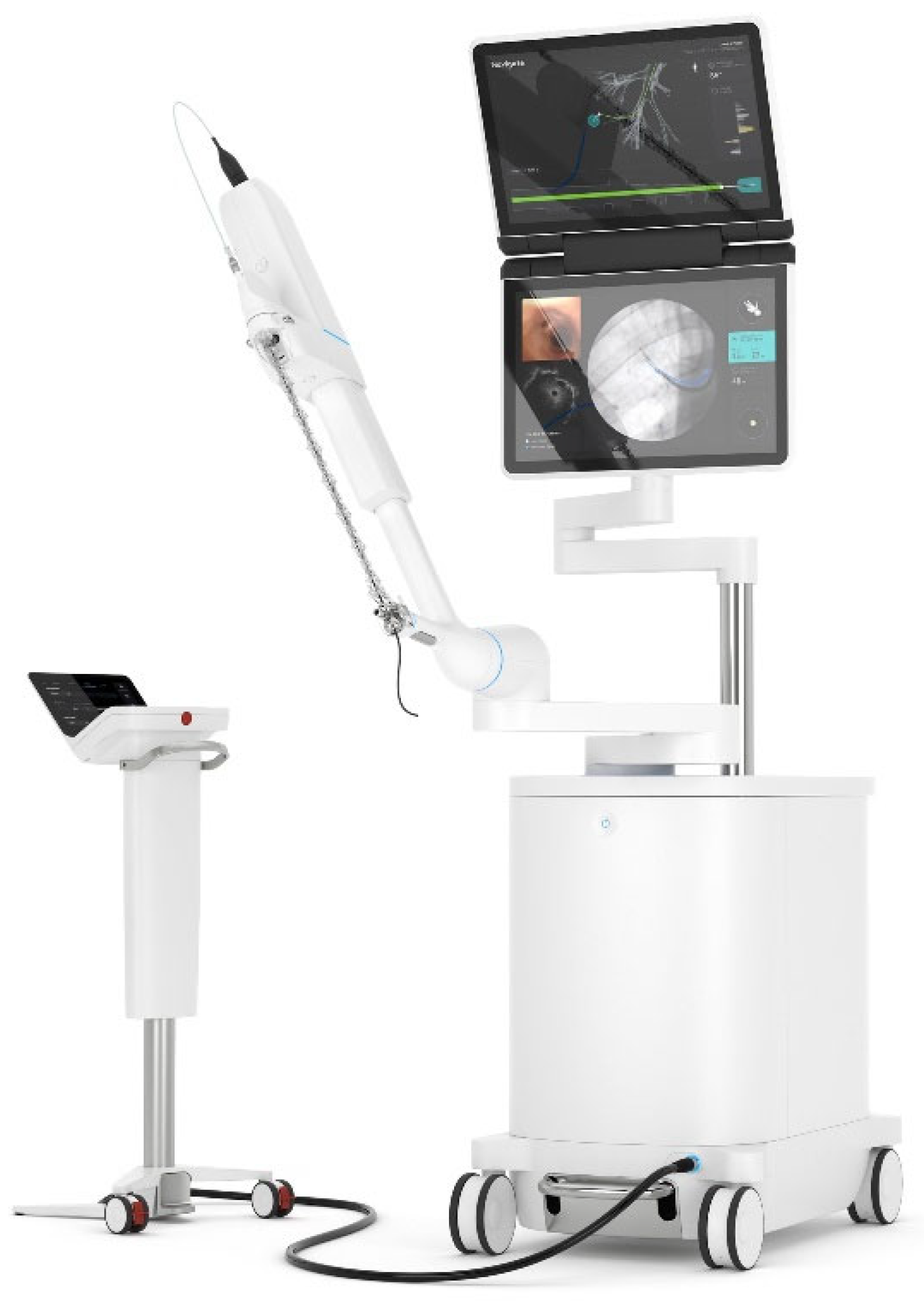
In comparison to the MONARCH system, Ion systems (Figure 2) consist of a single ultrathin 3.5 mm bronchoscope with shape-sensing technology that helps it to navigate to target lesions in the airways. The catheter can be articulated to 180 degrees in any direction. Navigation pathway generation software with the Ion™ system has been shown to be superior to EMN in both the distance from the terminal end of the navigation pathway to target lesions (9.4 mm for robotic bronchoscopy vs. 14.2 mm for tip-tracked electromagnetic navigation vs. 17.2 mm for catheter-based electromagnetic navigation) and the generation of complete distal airway maps [21].
The Ion system allows the simultaneous visualization of navigation and fluoroscopy, either with vision or radial endobronchial ultrasound (rEBUS). Due to a single working channel, switching between vision and rEBUS requires the removal of one probe for the insertion of the other. In the Precision-1 cadaver-based study with the Ion system, a remarkable 100% lesion localization rate was observed, surpassing EMN alone by 15% and conventional methods by 35%. Notably, successful lesion biopsy was achieved in 80% of cases, marking a significant 35% increase compared to EMN alone and an impressive 55% improvement over conventional methods [16]. Human studies also demonstrated favorable results, with one study reporting a diagnostic yield of 88% [22]. There are other studies showing similar results, which are described in Table 2.

6. GALAXY SYSTEM™ by Noah Medical

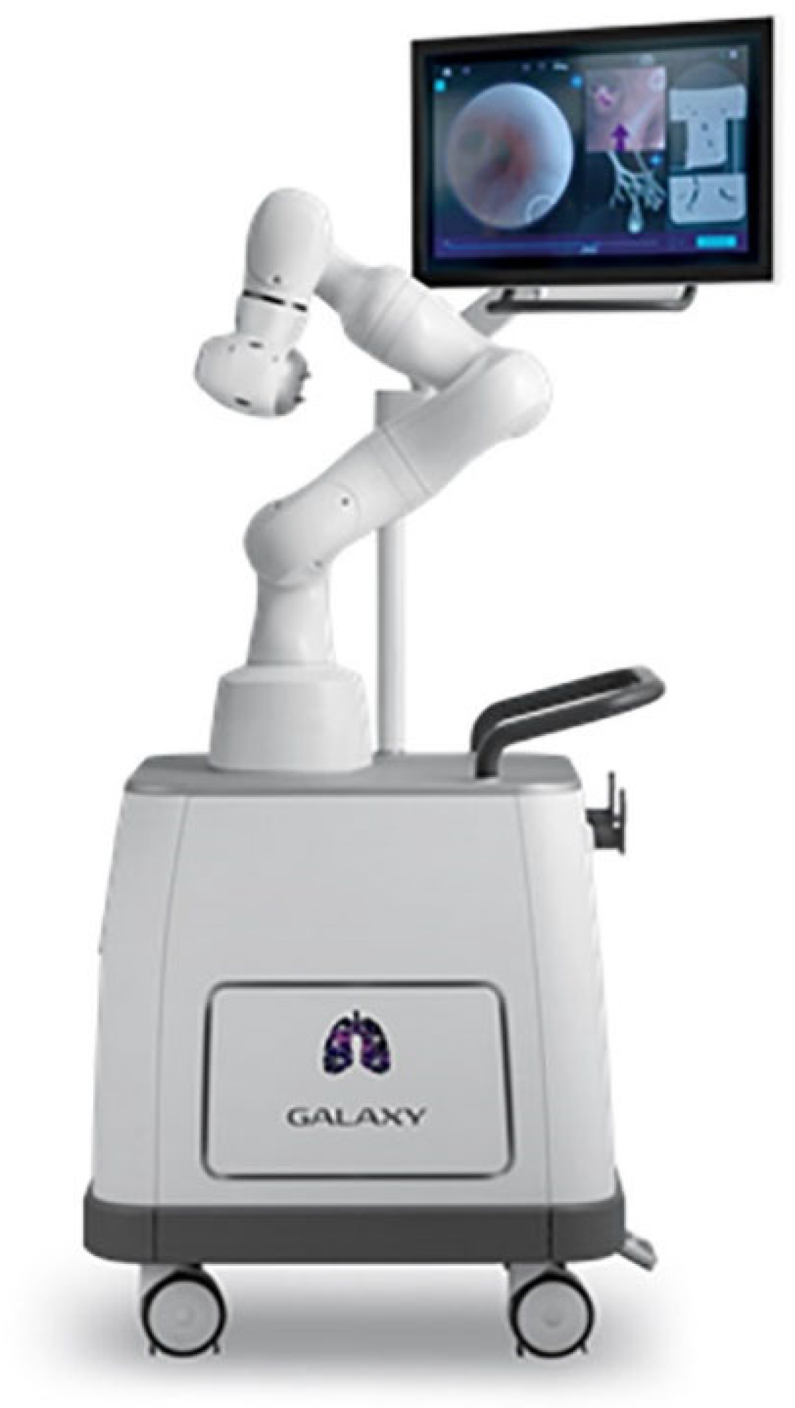
The Galaxy System from Noah Medical (Figure 3) is an innovative robotic endoluminal platform that integrates EMN with tomosynthesis technology and augmented fluoroscopy. This integration is strategically designed to harness the advantages of robotic bronchoscopy while addressing the challenge of CT-to-body divergence through unique “tool-in-lesion technology” (TiLT) confirmation. Digital tomosynthesis is an imaging method akin to CT scanning, involving the capture of a sequence of X-ray images to create a 3D image. In contrast to CT scans, digital tomosynthesis acquires a reduced number of images from a more limited angle range (15–60 degrees, as opposed to CT’s 180 degrees), enabling it to be conducted using a standard C-arm rather than a specialized CT scanner [26]. The Galaxy uses guidance with EMN to navigate to within 2 cm of the lesion and then switches to TiLT, i.e., digital tomosynthesis. This helps in overcoming the localization limitations of EMN. Cost is the major disparaging factor for this system, especially as it utilizes single-use disposable bronchoscopes and extremely specialized equipment and technology. The cost-to-benefit ratio has also been called into question, as shape-sensing RB appeared to have a similar diagnostic yield to that of digital tomosynthesis-assisted EMN (77% for ssRB to 80% for DT-EMN) in the one prospective comparative study that was conducted; however, whether the addition of CBCT or other advanced imaging to shape-sensing bronchoscopy would change this comparison is unknown [27].

7. Advantages over Other Biopsy Modalities

Robotic bronchoscopy represents a notable advancement in medical procedures, and may offer improved lesion localization and biopsy methods compared to traditional approaches. Particularly effective for peripheral and small lung nodules, robotic bronchoscopes provide extended reach beyond conventional tools, which will help in enhancing diagnostic capabilities [17]. The advantages are numerous, offering physicians improved distal dexterity, seamless maneuverability, comprehensive endobronchial visualization and the capacity to effectively utilize both hands. These attributes collectively serve to alleviate physician fatigue.
A recent meta-analysis evaluating the diagnostic efficacy and safety of RAB analyzed 12 articles (1409 patients), which showed a pooled diagnostic yield (81.9%) surpassing conventional methods. The Ion Intuitive and Auris Monarch platforms were commonly used. Complication rates, notably pneumothorax (1.18%) and bleeding (0.04%), were low [25]. Another retrospective multicenter study compared the efficacy and diagnostic performance of RAB to CT-PTNB for diagnosing pulmonary nodules suspected of lung cancer. The overall diagnostic yield was comparable between RAB (87.6%) and CT-PTNB (88.4%), and the complication rate was significantly lower for RAB compared to CT-PTNB (4.4% vs. 17%; p = 0.002) [28].
Both Monarch and ION systems have reported a low rate of pneumothorax ranging from 0 to 5.8%. Out of this small cohort of patients, only half required chest tube placement. Bleeding rates in these patients were also as low as 2.4–3.2% [16,23,28,29,30].
Furthermore, the integration of robotic bronchoscopy minimizes intraoperative forces on the patient, thereby reducing the likelihood of associated complications. The procedure’s efficiency is significantly heightened through improved navigation, addressing the common challenge of “getting lost in the airways”. Studies have unequivocally demonstrated a notable decrease in the mean time required to navigate to the lesion, with a mere 7 min compared to the protracted 25.0 min associated with ENB-specific navigation and sampling. However, it is important to note the mean procedure time (catheter in to catheter out), which was 63.1 ± 29.7 min [31].
The significant advantage of Robotic Bronchoscopy lies in its capacity to streamline and consolidate multiple procedures for high-risk nodules. It enables visualization, sampling, diagnosis and staging in a single setting. A strategic approach involves an initial biopsy of lymph nodes contralateral to the lesion and those farthest from the site of the lesion to assess for atypia or malignancy. This sequential method allows for informed decision-making during the procedure. This streamlined process not only reduces procedure time but also minimizes the duration of patient sedation, contributing to an overall safer medical procedure. In essence, Robotic Bronchoscopy emerges as a beacon of progress, revolutionizing diagnostic approaches with its multifaceted advantages.

8. Limitations

Notwithstanding robust endorsement for RAB, characterized by its precise localization capabilities and minimal complication rates, the existing body of literature on diagnostic yield presents divergent findings. Feasibility studies conducted on Monarch and Ion revealed diagnostic yields ranging from 69% to 79%, while disparate reports from singular centers documented exceptionally high rates of 96% [31]. In comparison to the pre-robotic bronchoscopy era, in the NAVIGATE trial, which reported a diagnostic yield of 72.9%, the observed distinctions appear less pronounced [13]. The integration of RAB with advanced imaging modalities, such as cone-beam computed tomography (CBCT) and fluoroscopy, has demonstrated a notable enhancement in diagnostic yield, reaching percentages between 86% and 94% [23,32,33]. However, a conspicuous void exists in head-to-head comparisons with non-RAB modalities.
This nuanced landscape prompts contemplation of the authentic clinical benefits and cost-effectiveness of RAB, areas that warrant comprehensive investigation and scrutiny. As the discipline of bronchoscopy continues to evolve, a deeper understanding of the comparative advantages and economic implications of RAB vis à vis conventional approaches is essential for informed clinical decision-making.
A recurrent challenge in various studies lies in the lack of consistent definition regarding diagnostic yield. Broadly, diagnostic yield is defined as the probability of a test confirming a diagnosis, calculated by dividing the number of biopsies with a diagnosis by the total number of biopsies. The primary focus is typically on discerning between benign and malignant conditions. However, the classification of histopathological findings, such as non-specific inflammation, non-diagnostic atypia or normal lung tissue, as indicative of non-malignant diseases can inadvertently inflate the diagnostic yield. Limited data are available on the negative predictive value (NPV) for RAB; hence, if the suspicion for malignancy is high, usually, a negative RAB biopsy may be followed by a CT-guided transthoracic percutaneous needle lung biopsy (CT-PTNB) or repeat Chest CT to ensure resolution of the pulmonary nodule.
Moving forward, it is imperative to establish a universally accepted and consistent definition of diagnostic yield. This standardization is essential for ensuring precision in comparing diagnostic yields across diverse studies. By adopting a uniform definition, researchers can enhance the reliability and interpretability of findings, ultimately advancing the collective understanding of diagnostic outcomes in the context of various medical investigations [14,34].
Both shape-sensing and electromagnetic navigation (EMN) bronchoscopy systems are susceptible to CT-to-body divergence (CTBD). CTBD denotes the inconsistency between the electronic virtual target and the actual anatomical location of the peripheral lung lesion. Differences in lung volumes during the preprocedural scan and bronchoscopy lead to CTBD. Respiratory variations can result in target lesion movement, averaging up to 14 mm, at times exceeding the lesion size itself [35]. The secretion of mucus, pleural effusion, anatomical changes from atelectasis and movement are also factors that can play a contributing role in this error.
A study demonstrated that the primary endpoint of three-dimensional target overlap was attained in 59.6% of cases (28/47) before location correction and significantly improved to 83.0% (39/47) after correction when utilizing electromagnetic navigation bronchoscopy (ENB) assisted by a tomosynthesis-based software algorithm. This outcome underscores the notable margin of error observed between the target lesion and the bronchoscope endpoint, emphasizing the efficacy of the correction process in enhancing precision during the procedure [36]. In Benn et al.’s study, which initially reported an 86% diagnostic yield with RAB and cone-beam CT, it was notable that proceduralists had to reposition their bronchoscopes in 15% of cases to confirm tool placement within the lesion [23].
To address CTBD, bronchoscopists often integrate advanced imaging tools such as digital tomosynthesis, cone beams and O-arms. While cone-beam CT is considered the gold standard for intraoperative imaging, challenges related to accessibility and cost limit its widespread adoption.
Other solutions to CTBD include paralytics during the procedure, expeditious intubation, increasing positive end-expiratory pressure during bronchoscopy to 10–12, increasing tidal volumes, the avoidance of excess suction, breath holds for CBCT spins and using a low fraction of inspired oxygen to reduce absorptive atelectasis [37]. The I-LOCATE trial puts the time from intubation to significant atelectasis at 30 min. Reducing the time from intubation to successful biopsy under this specific time can also help reduce CTBD [38]. These solutions need to be extensively studied in prospective research to establish standardized clinical practice.
A specific limitation associated with the use of the Monarch and Galaxy systems, employing EMN, is their restricted application in patients with cardiac pacemakers and defibrillators due to concerns about potential signal interference. However, a study involving 24 patients with these cardiac devices undergoing ENB yielded reassuring results. No instances of arrhythmias or alterations in pacemaker function were seen during the procedure. The study concluded that ENB could be safely conducted in all cases without encountering complications related to the presence of cardiac devices [39].
The other disadvantages of RAB are common to all robotic procedures, and include cost, the loss of tactile feedback and the delay in the surgeon controlling a vascular catastrophe due to the rigid structure placed on the patient. No research is available on topics like the cost efficacy of RAB, which in the future, will be needed to predict RAB use in low to middle socioeconomic areas. Cost will also be a key concern when combining already expensive imaging modalities like cone-beam CT or fluoroscopy to RAB. As previously elucidated, the prevalence of incidentally detected pulmonary nodules is on the rise, necessitating careful clinical consideration regarding the decision to pursue biopsy. This determination typically relies upon the nuanced integration of clinical judgment or the utilization of risk stratification calculators [40]. Moreover, the expanding accessibility of RAB across healthcare institutions signifies a broader capacity for comprehensive evaluation, ensuring that patients presenting with such nodules receive thorough assessment not solely relegated to tertiary care centers.

9. Future Directions

Though studies have been mixed regarding the accuracy and diagnostic yield of RAB, it is certain that the use of RAB will only continue to grow in the future. The therapeutic applications of RAB have been under investigation, along with some innovative ideas for how we can use its high stability and lesion localization to our advantage.
One such method is tattooing the peripheral lung nodules with a dye or contrast agent before surgery using IGB techniques, which can help surgeons easily locate the nodule intraoperatively at the time of surgical resection [41]. This enhancement in the visualization of nodules can be very useful in minimally invasive thoracic surgery.
Another innovative modality is fiducial marker placement, in which we can place markers into the peripheral lung malignancies. Using these markers, we can later selectively target the lesion using stereotactic ablative radiotherapy with promising results [42].
As it increases our stability inside the airway, RAB has potential for investigations into therapeutic procedures like cryotherapy; brachytherapy; radiofrequency and microwave ablation; and thermal, laser and Cryospray ablation directly to the malignant pulmonary nodule. The advancement of these techniques could pave the way for a variety of bronchoscopic treatments for conditions such as dehiscence, stenosis and tracheomalacia, using methods like bronchoscopic tracheoplasty. They may also open doors to the development of thoracic natural orifice transluminal endoscopic surgery (NOTES), expanding the scope of minimally invasive surgery options. These are, however, not possible with the current technology and available systems.
Following the remarkable findings of JCOG 0802, which demonstrated the superiority of segmentectomy over lobectomy for stage IA non-small-cell lung cancer, RAB holds the potential to offer a comprehensive single-procedure approach for the diagnosis, staging and treatment of early-stage lung cancer [43].
Looking ahead, addressing the issue of divergence is crucial for the advancement of RAB. In addition to the previously mentioned strategies to minimize divergence, exploring the integration of advanced imaging technologies such as cone-beam and advanced fluoroscopy with RAB could be instrumental in effectively managing this challenge.
It was initially believed that RAB, similar to robotic mitral valve repairs, would have a more challenging learning curve compared to conventional surgical techniques. However, certain robotic procedures, such as robotic-assisted laparoscopic prostatectomy, hysterectomy and cholecystectomy, have demonstrated a relatively shorter learning curve when contrasted with their traditional open surgery counterparts.
RAB integrates various technologies commonly used by bronchoscopists, yet mastering it requires a considerable learning process. The time required to achieve competency is likely to vary widely. This variability in the learning curve has been observed among both fellows and experienced bronchoscopists in the context of EMN [44]. To provide a more precise assessment of the learning curve and associated factors of RAB, further research is needed.

Funding

This research received no external funding.

Conflicts of Interest

Bruce Sabath is employed in the following capacities: 1. As a consultant for Intuitive Surgical and 2. As research support for Seimens. However, the authors declare that this research was conducted in the absence of any commercial or financial relationships that could be construed as a potential conflict of interest.

References

  1. Sung, H.; Ferlay, J.; Siegel, R.L.; Laversanne, M.; Soerjomataram, I.; Jemal, A.; Bray, F. Global Cancer Statistics 2020: GLOBOCAN Estimates of Incidence and Mortality Worldwide for 36 Cancers in 185 Countries. CA Cancer J. Clin. 2021, 71, 209–249. [Google Scholar] [CrossRef] [PubMed]
  2. American Cancer Society. Annual Cancer Facts & Figures; American Cancer Society: Atlanta, GA, USA, 2023. [Google Scholar]
  3. Church, T.R.; Black, W.C.; Aberle, D.R.; Berg, C.D.; Clingan, K.L.; Duan, F.; Fagerstrom, R.M.; Gareen, I.F.; Gierada, D.S.; Jones, G.C.; et al. Results of initial low-dose computed tomographic screening for lung cancer. New Engl. J. Med. 2013, 368, 1980–1991. [Google Scholar] [PubMed]
  4. Gould, M.K.; Donington, J.; Lynch, W.R.; Mazzone, P.J.; Midthun, D.E.; Naidich, D.P.; Wiener, R.S. Evaluation of individuals with pulmonary nodules: When is it lung cancer? Diagnosis and management of lung cancer, 3rd ed: American College of Chest Physicians evidence-based clinical practice guidelines. Chest 2013, 143 (Suppl. 5), e93S–e120S. [Google Scholar] [CrossRef] [PubMed]
  5. Forde, P.M.; Spicer, J.; Lu, S.; Provencio, M.; Mitsudomi, T.; Awad, M.M.; Felip, E.; Broderick, S.R.; Brahmer, J.R.; Swanson, S.J.; et al. Neoadjuvant Nivolumab plus Chemotherapy in Resectable Lung Cancer. New Engl. J. Med. 2022, 386, 1973–1985. [Google Scholar] [CrossRef] [PubMed]
  6. De Koning, H.J.; Van Der Aalst, C.M.; De Jong, P.A.; Scholten, E.T.; Nackaerts, K.; Heuvelmans, M.A.; Lammers, J.-W.J.; Weenink, C.; Yousaf-Khan, U.; Horeweg, N.; et al. Reduced Lung-Cancer Mortality with Volume CT Screening in a Randomized Trial. New Engl. J. Med. 2020, 382, 503–513. [Google Scholar] [CrossRef]
  7. Wang, J.; Zhang, T.; Xu, Y.; Yang, M.; Huang, Z.; Lin, J.; Xie, S.; Sun, H. Comparison between percutaneous transthoracic co-axial needle CT-guided biopsy and transbronchial lung biopsy for the diagnosis of persistent pulmonary consolidation. Insights Into Imaging 2023, 14, 80. [Google Scholar] [CrossRef] [PubMed]
  8. Gustav, F.Z. Killian, father of bronchoscopy. Arch. Otolaryngol. 1965, 82, 656–659. [Google Scholar]
  9. Baaklini, W.A.; Reinoso, M.A.; Gorin, A.B.; Sharafkaneh, A.; Manian, P. Diagnostic yield of fiberoptic bronchoscopy in evaluating solitary pulmonary nodules. Chest 2000, 117, 1049–1054. [Google Scholar] [CrossRef] [PubMed]
  10. Ali, M.S.; Trick, W.; Mba, B.I.; Mohananey, D.; Sethi, J.; Musani, A.I. Radial endobronchial ultrasound for the diagnosis of peripheral pulmonary lesions: A systematic review and meta-analysis. Respirology 2017, 22, 443–453. [Google Scholar] [CrossRef] [PubMed]
  11. Vining, D.J.; Liu, K.; Choplin, R.H.; Haponik, E.F. Virtual bronchoscopy. Relationships of virtual reality endobronchial simulations to actual bronchoscopic findings. Chest 1996, 109, 549–553. [Google Scholar] [CrossRef] [PubMed]
  12. Gildea, T.R.; Mazzone, P.J.; Karnak, D.; Meziane, M.; Mehta, A.C. Electromagnetic navigation diagnostic bronchoscopy: A prospective study. Am. J. Respir. Crit. Care Med. 2006, 174, 982–989. [Google Scholar] [CrossRef] [PubMed]
  13. Folch, E.E.; Pritchett, M.A.; Nead, M.A.; Bowling, M.R.; Murgu, S.D.; Krimsky, W.S.; Murillo, B.A.; LeMense, G.P.; Minnich, D.J.; Bansal, S.; et al. Electromagnetic Navigation Bronchoscopy for Peripheral Pulmonary Lesions: One-Year Results of the Prospective, Multicenter NAVIGATE Study. J. Thorac. Oncol. 2019, 14, 445–458. [Google Scholar] [CrossRef] [PubMed]
  14. Ost, D.E.; Ernst, A.; Lei, X.; Kovitz, K.L.; Benzaquen, S.; Diaz-Mendoza, J.; Greenhill, S.; Toth, J.; Feller-Kopman, D.; Puchalski, J.; et al. Diagnostic Yield and Complications of Bronchoscopy for Peripheral Lung Lesions. Results of the AQuIRE Registry. Am. J. Respir. Crit. Care Med. 2016, 193, 68–77. [Google Scholar] [CrossRef] [PubMed]
  15. Chen, A.C.; Pastis, N.J.; Machuzak, M.S.; Gildea, T.R.; Simoff, M.J.; Gillespie, C.T.; Mahajan, A.K.; Oh, S.S.; Silvestri, G.A. Accuracy of a Robotic Endoscopic System in Cadaver Models with Simulated Tumor Targets: ACCESS Study. Respir. Int. Rev. Thorac. Dis. 2020, 99, 56–61. [Google Scholar] [CrossRef] [PubMed]
  16. Yarmus, L.; Akulian, J.; Wahidi, M.; Chen, A.; Steltz, J.P.; Solomon, S.L.; Yu, D.; Maldonado, F.; Cardenas-Garcia, J.; Molena, D.; et al. A Prospective Randomized Comparative Study of Three Guided Bronchoscopic Approaches for Investigating Pulmonary Nodules: The PRECISION-1 Study. Chest 2020, 157, 694–701. [Google Scholar] [CrossRef] [PubMed]
  17. Chen, A.C.; Gillespie, C.T. Robotic Endoscopic Airway Challenge: REACH Assessment. Ann. Thorac. Surg. 2018, 106, 293–297. [Google Scholar] [CrossRef] [PubMed]
  18. Agrawal, A.; Hogarth, D.K.; Murgu, S. Robotic bronchoscopy for pulmonary lesions: A review of existing technologies and clinical data. J. Thorac. Dis. 2020, 12, 3279–3286. [Google Scholar] [CrossRef] [PubMed]
  19. Chaddha, U.; Kovacs, S.P.; Manley, C.; Hogarth, D.K.; Cumbo-Nacheli, G.; Bhavani, S.V.; Kumar, R.; Shende, M.; Egan, J.P.; Murgu, S. Robot-assisted bronchoscopy for pulmonary lesion diagnosis: Results from the initial multicenter experience. BMC Pulm. Med. 2019, 19, 243. [Google Scholar] [CrossRef] [PubMed]
  20. Chen, A.C.; Pastis, N.J., Jr.; Mahajan, A.K.; Khandhar, S.J.; Simoff, M.J.; Machuzak, M.S.; Cicenia, J.; Gildea, T.R.; Silvestri, G.A. Robotic Bronchoscopy for Peripheral Pulmonary Lesions: A Multicenter Pilot and Feasibility Study (BENEFIT). Chest 2021, 159, 845–852. [Google Scholar] [CrossRef] [PubMed]
  21. Akulian, J.; Molena, D.; Wahidi, M.; Chen, A.; Yu, D.; Maldonado, F.; Lee, H.; Vachani, A.; Yarmus, L. A Direct Comparative Study of Bronchoscopic Navigation Planning Platforms for Peripheral Lung Navigation: The ATLAS Study. J. Bronchol. Interv. Pulmonol. 2022, 29, 171–178. [Google Scholar] [CrossRef] [PubMed]
  22. Fielding, D.I.; Bashirzadeh, F.; Son, J.H.; Todman, M.; Chin, A.; Tan, L.; Steinke, K.; Windsor, M.N.; Sung, A.W. First Human Use of a New Robotic-Assisted Fiber Optic Sensing Navigation System for Small Peripheral Pulmonary Nodules. Respir. Int. Rev. Thorac. Dis. 2019, 98, 142–150. [Google Scholar] [CrossRef] [PubMed]
  23. Benn, B.S.; Romero, A.O.; Lum, M.; Krishna, G. Robotic-Assisted Navigation Bronchoscopy as a Paradigm Shift in Peripheral Lung Access. Lung 2021, 199, 177–186. [Google Scholar] [CrossRef]
  24. Kalchiem-Dekel, O.; Connolly, J.G.; Lin, I.-H.; Husta, B.C.; Adusumilli, P.S.; Beattie, J.A.; Buonocore, D.J.; Dycoco, J.; Fuentes, P.; Jones, D.R.; et al. Shape-Sensing Robotic-Assisted Bronchoscopy in the Diagnosis of Pulmonary Parenchymal Lesions. Chest 2022, 161, 572–582. [Google Scholar] [CrossRef] [PubMed]
  25. Pyarali, F.F.; Hakami-Majd, N.; Sabbahi, W.; Chaux, G. Robotic-assisted Navigation Bronchoscopy: A Meta-Analysis of Diagnostic Yield and Complications. J. Bronchol. Interv. Pulmonol. 2024, 31, 70–81. [Google Scholar] [CrossRef] [PubMed]
  26. Rickman, O.B.; Mahajan, A.K.; Hogarth, D.K.; Bhadra, K. “Tool-in-lesion” Accuracy of Galaxy System—A Robotic Electromagnetic Navigation BroncHoscopy with Integrated Tool-in-lesion-Tomosynthesis Technology: The MATCH Study. J. Bronchol. Interv. Pulmonol. 2024, 31, 23–29. [Google Scholar] [CrossRef]
  27. Low, S.W.; Lentz, R.J.; Chen, H.; Katsis, J.; Aboudara, M.C.; Whatley, S.; Paez, R.; Rickman, O.B.; Maldonado, F. Shape-Sensing Robotic-Assisted Bronchoscopy vs Digital Tomosynthesis-Corrected Electromagnetic Navigation Bronchoscopy: A Comparative Cohort Study of Diagnostic Performance. Chest 2023, 163, 977–984. [Google Scholar] [CrossRef] [PubMed]
  28. Lee-Mateus, A.Y.; Reisenauer, J.; Garcia-Saucedo, J.C.; Abia-Trujillo, D.; Buckarma, E.H.; Edell, E.S.; Grage, R.A.; Bowman, A.W.; Labarca, G.; Johnson, M.M.; et al. Robotic-assisted bronchoscopy versus CT-guided transthoracic biopsy for diagnosis of pulmonary nodules. Respirology 2023, 28, 66–73. [Google Scholar] [CrossRef] [PubMed]
  29. Rojas-Solano, J.R.; Ugalde-Gamboa, L.; Machuzak, M. Robotic Bronchoscopy for Diagnosis of Suspected Lung Cancer: A Feasibility Study. J. Bronchol. Interv. Pulmonol. 2018, 25, 168–175. [Google Scholar] [CrossRef] [PubMed]
  30. Agrawal, A.; Ho, E.; Chaddha, U.; Demirkol, B.; Bhavani, S.V.; Hogarth, D.K.; Murgu, S. Factors Associated with Diagnostic Accuracy of Robotic Bronchoscopy with 12-Month Follow-up. Ann. Thorac. Surg. 2023, 115, 1361–1368. [Google Scholar] [CrossRef] [PubMed]
  31. Reisenauer, J.; Simoff, M.J.; Pritchett, M.A.; Ost, D.E.; Majid, A.; Keyes, C.; Casal, R.F.; Parikh, M.S.; Diaz-Mendoza, J.; Fernandez-Bussy, S.; et al. Ion: Technology and Techniques for Shape-sensing Robotic-assisted Bronchoscopy. Ann. Thorac. Surg. 2022, 113, 308–315. [Google Scholar] [CrossRef] [PubMed]
  32. Simoff, M.J.; Pritchett, M.A.; Reisenauer, J.S.; Ost, D.E.; Majid, A.; Keyes, C.; Casal, R.F.; Parikh, M.S.; Diaz-Mendoza, J.; Fernandez-Bussy, S.; et al. Shape-sensing robotic-assisted bronchoscopy for pulmonary nodules: Initial multicenter experience using the Ion™ Endoluminal System. BMC Pulm. Med. 2021, 21, 322. [Google Scholar] [CrossRef] [PubMed]
  33. Kalchiem-Dekel, O.; Fuentes, P.; Bott, M.J.; Beattie, J.A.; Lee, R.P.; Chawla, M.; Husta, B.C. Multiplanar 3D fluoroscopy redefines tool-lesion relationship during robotic-assisted bronchoscopy. Respirology 2021, 26, 120–123. [Google Scholar] [CrossRef] [PubMed]
  34. Leonard, K.M.; Low, S.W.; Echanique, C.S.; Swanner, B.A.; Johnson, J.; Dahlberg, G.; Paez, R.; Ratwani, A.P.; Shojaee, S.; Rickman, O.B.; et al. Diagnostic Yield vs Diagnostic Accuracy for Peripheral Lung Biopsy Evaluation: Evidence Supporting a Future Pragmatic End Point. Chest 2023, in press.
  35. Pritchett, M.A. Prospective Analysis of a Novel Endobronchial Augmented Fluoroscopic Navigation System for Diagnosis of Peripheral Pulmonary Lesions. J. Bronchol. Interv. Pulmonol. 2021, 28, 107–115. [Google Scholar] [CrossRef] [PubMed]
  36. Pritchett, M.A.; Bhadra, K.; Mattingley, J.S. Electromagnetic Navigation Bronchoscopy with Tomosynthesis-based Visualization and Positional Correction: Three-dimensional Accuracy as Confirmed by Cone-Beam Computed Tomography. J. Bronchol. Interv. Pulmonol. 2021, 28, 20. [Google Scholar] [CrossRef] [PubMed]
  37. Pritchett, M.A.; Bhadra, K.; Calcutt, M.; Folch, E. Virtual or reality: Divergence between preprocedural computed tomography scans and lung anatomy during guided bronchoscopy. J. Thorac. Dis. 2020, 12, 1595–1611. [Google Scholar] [CrossRef]
  38. Sagar, A.E.S.; Sabath, B.F.; Eapen, G.A.; Song, J.; Marcoux, M.; Sarkiss, M.; Arain, M.H.; Grosu, H.B.; Ost, D.E.; Jimenez, C.A.; et al. Incidence and Location of Atelectasis Developed during Bronchoscopy under General Anesthesia: The I-LOCATE Trial. Chest 2020, 158, 2658–2666. [Google Scholar] [CrossRef] [PubMed]
  39. Khan, A.Y.; Berkowitz, D.; Krimsky, W.S.; Hogarth, D.K.; Parks, C.; Bechara, R. Safety of pacemakers and defibrillators in electromagnetic navigation bronchoscopy. Chest 2013, 143, 75–81. [Google Scholar] [CrossRef] [PubMed]
  40. Elicker, B.M. Lung Nodule Risk Calculator and Cost-Effectiveness of Different Lung Cancer Screening Algorithms. Radiol. Cardiothorac. Imaging 2021, 3, e210050. [Google Scholar] [CrossRef] [PubMed]
  41. Marino, K.A.; Sullivan, J.L.; Weksler, B. Electromagnetic Navigation Bronchoscopy for Identifying Lung Nodules for Thoracoscopic Resection. Ann. Thorac. Surg. 2016, 102, 454–457. [Google Scholar] [CrossRef] [PubMed]
  42. Casutt, A.; Kinj, R.; Ozsahin, E.-M.; von Garnier, C.; Lovis, A. Fiducial markers for stereotactic lung radiation therapy: Review of the transthoracic, endovascular and endobronchial approaches. Eur. Respir. Rev. 2022, 31, 210149. [Google Scholar] [CrossRef] [PubMed]
  43. Saji, H.; Okada, M.; Tsuboi, M.; Nakajima, R.; Suzuki, K.; Aokage, K.; Aoki, T.; Okami, J.; Yoshino, I.; Ito, H.; et al. Segmentectomy versus lobectomy in small-sized peripheral non-small-cell lung cancer (JCOG0802/WJOG4607L): A multicentre, open-label, phase 3, randomised, controlled, non-inferiority trial. Lancet 2022, 399, 1607–1617. [Google Scholar] [CrossRef] [PubMed]
  44. Lee, H.J.; Argento, A.C.; Batra, H.; Benzaquen, S.; Bramley, K.; Chambers, D.; Desai, N.; Dincer, H.E.; Ferguson, J.S.; Kalanjeri, S.; et al. A Multicenter Study Assessing Interventional Pulmonary Fellow Competency in Electromagnetic Navigation Bronchoscopy. ATS Sch. 2022, 3, 220–228. [Google Scholar] [CrossRef] [PubMed]
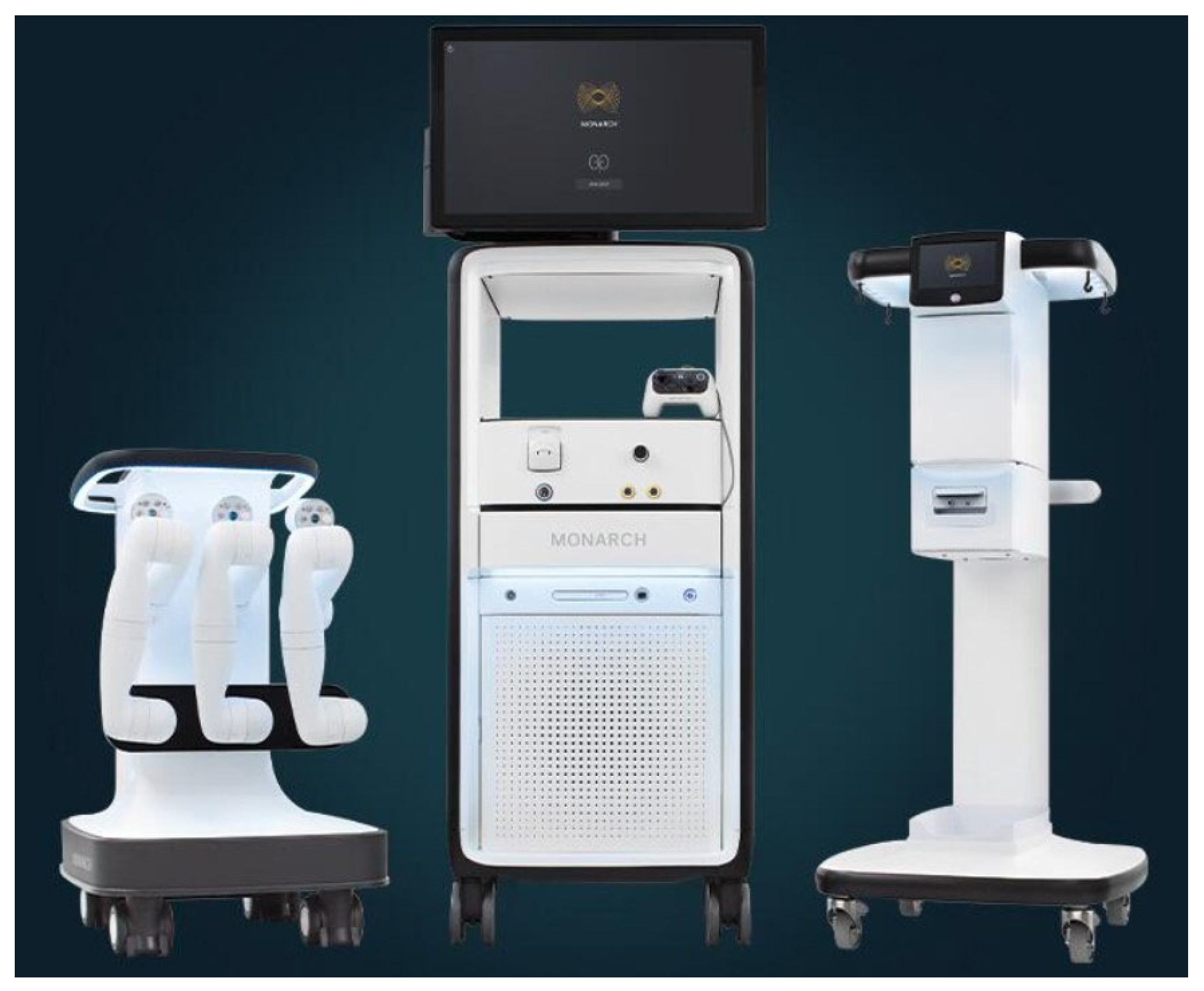
Figure 1. Monarch by Auris Health.
Jor 04 00011 g001
Figure 2. Ion by Intuitive Surgical.
Jor 04 00011 g002
Figure 3. Galaxy by Noah Medical.
Jor 04 00011 g003
Table 1. Comparing the three robotic systems.
Monarch Robotic Bronchoscopy System (Auris Health)Ion Robotic Bronchoscopy System (Intuitive Surgical)The Galaxy System (Noah Medical)
FDA ApprovalMarch 2018February 2019March 2023
Technology usedElectromagnetic NavigationShape-sensing technologyTilt technology/real-time navigation:
electromagnetic navigation, digital tomosynthesis
Bronchoscope specificationsOuter sheath: 6.0 mm
Inner scope: 4.4 mm OD
Working Channel: 2.1 mm
Integrated working camera: Yes

Peripheral vision available during biopsy
Scope: 3.5 mm OD
Working Channel: 2.0 mm
Vision probe which goes through working channel.
Shape-sensing fibers providing feedback

Peripheral vision is not available during biopsy
Scope: 4 mm OD
Working Channel: 2.1 mm

Integrated vision
ControllerGaming controllerTrack ball and scroll wheelGaming controller
Cone-beam CT/rEBUS/fluoroscopy compatibilityYes
(EMN is sensitive to metal)
Yes
(shape sensing not sensitive to metal)
Yes
(EMN is sensitive to metals, but system has inbuilt digital tomosynthesis)
ScopeReusableReusableDisposable
Table 2. Brief overview of existing data.
#StudyPlatformNo. of PatientsFollow UpNavigation SuccessfulBronchus SignAdjuvant ImagingReported Diagnostic Yield
1Chadda 2019
BMC Pulm Med [19]
Monarch1656 months88.6%63%rEBUS, 2D Fluoro69–77%
2Chen 2020 (BENEFIT STUDY)
Chest [20]
Monarch551 year96.2%60%rEBUS, 2D
Fluoro
74%
3Benn 2021
Lung [23]
Ion525–16 months85.0%46%Cone-beam CT86%
100.0%
4Fielding 2019
Respiration [22]
Ion296 months96.5%59%rEBUS, 2D Fluoro79%
93.0%
5Dekel 2021
Chest [24]
Ion1311 year98.7%63%rEBUS, 2D, 3D
Fluoro
81.7%
6Pyarali 2024 (meta analysis)
JOBIP [25]
Ion/Monarch1409
(23 studies)
VariableNA25–70%rEBUS, cone-beam
CT
81.90%
Disclaimer/Publisher’s Note: The statements, opinions and data contained in all publications are solely those of the individual author(s) and contributor(s) and not of MDPI and/or the editor(s). MDPI and/or the editor(s) disclaim responsibility for any injury to people or property resulting from any ideas, methods, instructions or products referred to in the content.