Article

Is Bronchiectasis (BE) Properly Investigated in Patients with Severe Asthma? A Real-Life Report from Eight Italian Centers

by
Giovanna Elisiana Carpagnano
1,
Vitaliano Nicola Quaranta
1,*,
Claudia Crimi
2,
Pierachille Santus
3,
Francesco Menzella
4,
Corrado Pelaia
5,
Giulia Scioscia
6,
Cristiano Caruso
7,
Elena Bargagli
8,
Nicola Scichilone
9 and
Eva Polverino
1,10
1
Department of Basic Medical Sciences, Neuroscience and Sense Organs, Section of Respiratory Disease, University “Aldo Moro” of Bari, 70124 Bari, Italy
2
Department of Internal Medicine and Specialistic Medicine, Section of Respiratory Diseases, University of Catania, 95100 Catania, Italy
3
Department of Clinical and Biomedical Sciences, Università degli Studi di Milano, Division of Respiratory Diseases, “L. Sacco” University Hospital, ASST Fatebenefratelli-Sacco, 20019 Milan, Italy
4
Pulmonology Unit, S. Valentino Hospital, Montebelluna (TV), AULSS 2 Marca Trevigiana, 31044 Montebelluna, Italy
5
Department of Health Sciences, Section of Respiratory Disease, University ‘‘Magna Græcia’’ of Catanzaro, 31044 Catanzaro, Italy
6
Department of Medical and Surgical Sciences, University of Foggia, University Hospital Policlinico Riuniti of Foggia, 71100 Foggia, Italy
7
UOSD DH Internal Medicine and Digestive Diseases, Policlinico Gemelli Foundation IRCCS, 00100 Rome, Italy
8
Respiratory Diseases Unit, Department of Medical, Surgical Sciences and Neurosciences, University of Siena, 53100 Siena, Italy
9
PROMISE Department, University of Palermo, 90100 Palermo, Italy
10
Servei de Pneumologia, Hospital Universitari Vall d’Hebron (HUVH), Institut de Recerca Vall d’Hebron (VHIR), Universitat Autonoma de Barcelona, 08001 Barcelona, Spain
*
Author to whom correspondence should be addressed.
J. Respir. 2023, 3(4), 178-190; https://doi.org/10.3390/jor3040017
Submission received: 13 August 2023 / Revised: 23 September 2023 / Accepted: 27 September 2023 / Published: 2 October 2023

Abstract

:
Introduction: Asthma and bronchiectasis are often partners in a complex but uneven relationship with asthma receiving more attention. The aim of this study is to describe how bronchiectasis is investigated in some Severe Asthma (SA) Centers, scattered throughout the Italian territory. Materials and Methods: We enrolled 92 patients with SA and bronchiectasis from eight Italian SA Centers and recorded diagnostic approaches to investigate SA and bronchiectasis at the time of enrollment (T0), at the 6-month (T1), and at the 12-month (T2) follow-up visits. Results: A statistically significant heterogeneous diagnostic approach emerged across the centers under study. In fact, while, as expected, all involved centers made an in-depth investigation of SA, only a few of them provided a complete investigation of bronchiectasis in order to provide specific treatment. Discussion: This real-life multicenter study confirmed that patients with coexistent SA and bronchiectasis are mainly investigated for pheno-endotyping asthma but rarely for the complete assessment of bronchiectasis. We believe that the diagnostic flowchart of SA patients with suspicion or confirmed bronchiectasis needs to be clarified and implemented as the association of these conditions strongly influences the final outcome and management of these patients.

1. Introduction

Asthma and bronchiectasis are often partners in a complex relationship and this interplay has important clinical implications in terms of more frequent exacerbations, chronic infections, increased disease severity and related healthcare costs, and, finally, poor quality of life (QoL).
Bronchiectasis is present in 18–80% of patients affected by severe asthma [1,2,3,4], whereas asthma is apparently less frequent in patients with bronchiectasis and, according to the European bronchiectasis registry (EMBARC), this association can reach 30.5% when “self-reported” [5,6].
Despite the presence of symptoms that can lead to suspect bronchiectasis, such as recurrent exacerbations with pathogenic isolates from sputum, sometimes, SA subjects undergo diagnostic tests for bronchiectasis.
Over the last few years, the importance of bronchiectasis in SA has been recognized and several case series have already been reported. For instance, a total of 696 patients from the Severe Asthma Network in Italy (SANI) registry were reviewed for co-presence of bronchiectasis and SA and the diagnosis of bronchiectasis was confirmed in 108 (15.5%, BE+) [7].
Many studies have shown that the association of bronchiectasis and SA frequently occurs in older, non-atopic asthmatic subjects with more severe airway obstruction and higher rates of chronic expectoration and infection [8]. Most of those patients exhibit low IgG, likely a consequence of chronic corticosteroid therapy or because of an unknown immune deficit [8].
Investigating the presence of bronchiectasis in patients with SA has the potential to have therapeutic implications. Patients who have both severe asthma and bronchiectasis were identified as a unique subgroup, characterized by differences in disease severity, microbiological profiles, and asthma phenotypes. Utilizing high-resolution computed tomography (HRCT) scans and sputum cultures can aid in the identification of individuals within this group. These findings have the potential to facilitate early detection and tailored treatment for these patients [9].
An increased awareness of the existence of a large group of severe asthmatics with bronchiectasis has resulted in greater attention, especially in specialized centers, and particularly in those dedicated to SA.

2. Materials and Methods
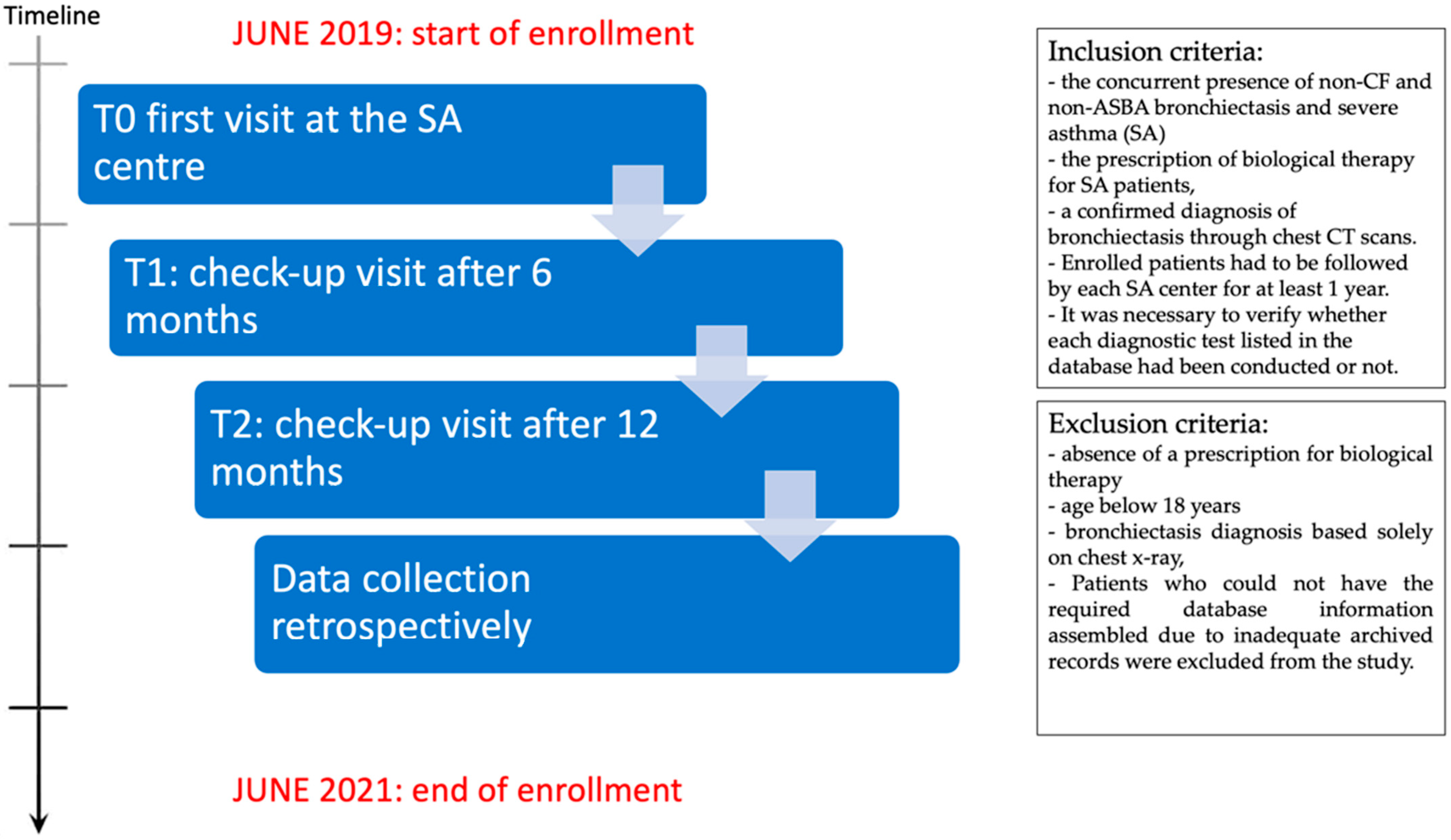
The primary goal of this study is to make a real-life overview of diagnostic workout in some SA centers scattered throughout the Italian territory to describe the level of attention given to the investigation of bronchiectasis across these centers.
The secondary objective was to analyze and compare the variability in diagnostic practices among different SA Centers. This encompassed evaluating various aspects, including lung function tests, radiological assessments, microbiological investigations, asthma evaluations, asthma scoring, bronchiectasis assessments, and bronchiectasis scoring.
A retrospective longitudinal observational study was constructed. We compared 8 SA centers in terms of diagnostic and monitoring approaches to the study of SA and bronchiectasis at the time of enrollment (T0), at 6-month (T1), and at 12-month follow-up visits (T2) (Figure 1).

2.1. Population

A total of 92 patients, already diagnosed with non-cystic fibrosis (non-CF) and non-allergic bronchopulmonary aspergillosis (non-ASBA) bronchiectasis, were enrolled retrospectively from 8 SA centers scattered throughout the Italian territory from June 2019 to June 2021.
Each center was provided with a predetermined database where they could record the diagnostic tests conducted and the therapies administered to their patients at three specific time points: baseline (T0), 6 months after enrollment (T1), and 12 months after enrollment (T2). Database fields to be inserted are those described in Table 1 and Table 2 and in Figure 2 and Figure 3. The baseline measurement (T0) encompassed either the initial visit when patients were prescribed biologic therapy or the first visit to an SA center for patients already receiving biologic therapy prescribed elsewhere. Due to the retrospective nature of the study, the various centers used individual, variable databases for data entry during the T0, T1, and T2 visits. Instead, data was retrospectively entered using information archived in accordance with clinical practice after these visits occurred. The monitoring schedule was not established before T0, but it was retroactively determined (as indicated in the study flowchart). The SA centers of Siena, Bari, Foggia, Ferrara, Catania, Palermo, Naples, Milano, Rome, and Reggio Emilia participated in this study.
The inclusion criteria comprised the concurrent presence of non-CF and non-ASBA bronchiectasis and severe asthma (SA), the prescription of biological therapy for SA patients, and a confirmed diagnosis of bronchiectasis through chest CT scans. Enrolled patients had to be followed by each SA center for at least 1 year. Additionally, it was necessary to verify whether each diagnostic test listed in the database had been conducted or not. Morphological criteria for identifying bronchiectasis on CT scans included bronchial dilation relative to the accompanying pulmonary artery, the absence of bronchial tapering, and the presence of bronchi within 1 cm of the pleural surface [5].
Exclusion criteria encompassed the absence of a prescription for biological therapy, age below 18 years, a bronchiectasis diagnosis based solely on chest X-rays, and the presence of psychiatric comorbidities that could impede follow-up. Patients with notable absent data (as per protocol) in their local hospital database were excluded.

Methods

From the different centers, clinical features, comorbidities and chronic respiratory therapy were collected (Table 1). Additionally, each center provided diagnostic assessments for both asthma and bronchiectasis, including disease control questionnaires, quality of life assessments, and prognostic scores conducted at baseline, 6 months (T1), and 12 months (T2) during the follow-up period (see Table 2).
Table 1. Clinical features, comorbidities, and chronic respiratory therapy.
Clinical Features
Age (n = 92)58.8 ± 12.1
Female51.9%
Former smoker22.2%
Current smoker7.4%
Family History of Asthma75%
Atopy69.1%
Bilateral Bronchiectasis79.5%
Chronic bronchial Infection by P. Aeruginosa2.4%
NMT colonization0.0%
TBC0.0%
Comorbidities (88.9% of all patients)
Rhinitis63.1%
Nasal polyposis61.7%
Rhinosinusitis50.0%
GERD42.0%
Autoimmune disease 14.6%
ASA intolerance12.2%
Vasculitis9.8%
Urticaria9.8%
OSAS 7.3%
Dermatitis7.3%
Infertility0.0%
Main Diagnostic Evaluations
ACT (n = 83)14.21 ± 4.93
BSI (n = 34)5.64 ± 3.90
% FEV1 (n = 92)64.66 ± 21.60
% FVC (n = 92)82.35 ± 15.55
FeNO 50 (n = 33)70.15 ± 72.84
IgE (n = 75)672.53 ± 235.67
Blood Eosinophilia (n = 58)858.43 ± 640.00
Home Therapy
ICS-LABA100%
LAMA72.5%
OCS69.1%
Moderate dose ICS56.6%
High dose ICS43.4%
Anti-LTRA33.8%
Mucolytics26.8%
Number of antibiotic cycles/year2.87 ± 2.00
Omalizumab32.1%
Mepolizumab38.3%
Benralizumab29.6%
Abbreviations: ACT: Asthma control test; ASA sensibility: acetylsalicylic acid sensibility; BSI: Bronchiectasis Severity Index; CF: cystic fibrosis; FeNO 50: Fractional Exhaled Nitric Oxide 50; FEV1 Forced Expiratory Volume in the 1st second; FVC: forced vital capacity; GERD: Gastro-esophageal reflux disease; ICS: inhaled corticosteroids; LAMA: long-acting muscarinic antagonists; LTRA: Leukotriene Receptor Antagonists; NMT colonization: non-mycobacterial tuberculosis infection; OCS: oral corticosteroid; TBC: pulmonary tuberculosis. Autoimmune diseases: mixed connectivitis, Sjögren’s syndrome, systemic lupus erythematosus, systemic sclerosis, Hashimoto thyroiditis, rheumatoid arthritis, psoriatic arthritis; Mucolytics: N-acetylcysteine (NAC), carbocysteine.
Table 2. Diagnostic evaluations and questionnaires of disease control, quality of life, and prognostic scores performed at baseline, 6 months (T1), and 12 months (T2) of follow-up.
LUNG FUNCTION TESTS
T0T1T2
Spirometry%100.064.243.0
Bronchodilator Reversibility Testing%64.317.10.0
TLC%16.721.00.0
DLCO%16.013.68.9
RADIOLOGY
Chest X-ray%28.47.411.1
Chest CT scan%100.04.93.7
MICROBIOLOGY
Sputum culture, %29.63.73.7
BAL culture, %11.10.00.0
LABORATORY DIAGNOSTICS
Blood Count%63.027.227.2
Blood eosinophilia%63.027.227.2
Sputum eosinophilia%2.50.00.0
Total S-IgE%81.516.012.3
Nasal cytology%28.40.02.5
ESR%8.68.60.0
CRP%50.611.10.0
ANA, ANCA%20.00.00.0
FRACTIONAL EXHALED NITRIC OXIDE (FeNO) TEST
FeNO50%35.839.539.5
FeNO350%8.616.09.9
OTHER DIAGNOSTIC TOOLS
A1AT genotyping%17.30.00.0
Ciliary motility test%2.50.00.0
QUESTIONNAIRES
ACT%90.167.961.7
ACQ%28.40.00.0
AQLQ%21.019.825.9
Compliance to therapy%96.397.682.5
BSI%37.011.08.6
FACED score%9.90.07.4
Abbreviations: A1AT: α1-antitrypsine; ACQ: Asthma control questionnaire; ACT: Asthma control test; ANA: Antinuclear antibodies; ANCA: Anti-neutrophil cytoplasmic antibody; AQLQ: Asthma quality of life questionnaire; BAL: Broncho-Alveolar Lavage; BSI: Bronchiectasis Severity Index; CRP: c-reactive protein; DLCO: diffusing capacity; ESR: erythrocyte sedimentation rate; Faced score: F—FEV1, A—Age, C—Colonization, E—extension, D—dyspnoea. FeNO 350 Fractional Exhaled Nitric Oxide 350; FeNO 50: Fractional Exhaled Nitric Oxide 50; TLC: total lung capacity; VR: residual volume.
In particular, anthropometric data, past clinical history and comorbidities, functional data (global lung function and bronchodilator reversibility testing), imaging tests (High-Resolution Computed Tomography-HRCT), microbiological exams (sputum and broncho-alveolar lavage (BAL) cultures), etiological tests for bronchiectasis (alfa 1 antiTrypsine (A1AT) dosage, ciliary motility test, autoimmunity), endotyping tests for asthma (total IgE, Fractional Exhaled Nitric Oxide 50(FeNO 50), Fractional Exhaled Nitric Oxide 350 (FeNO 350), complete blood count (CBC), induced sputum cellularity, nasal cytology)) and questionnaires for asthma symptoms and quality of life (QoL) and Bronchiectasis (ASTHMA: Asthma Control Test (ACT) [10], Asthma Control Questionnaire (ACQ) [11], Asthma Quality of Life Questionnaire (AQLQ) [12], Test of the Adherence to Inhalers (TAI) [13]; BRONCHIECTASIS: Bronchiectasis Severity Index (BSI) score [14] and FACED [15] (an acronym for Exacerbations, FEV1, Age, chronic Pseudomonas aeruginosa bronchial infection Colonization, radiological Extension, and Dyspnoea) score) were collected.
This study was carried out according to the principles of the Declaration of Helsinki, was approved by the local Ethics Committee of the “Riuniti” Hospital of Foggia (Institutional Review Board approval number 17/CE/2014), and all recruited patients gave their written informed consent.

2.2. Statistical Analysis

Nominal and dichotomous variables were expressed as %. Nominal variables were compared with the Chi-square test and the Mantel–Haenszel test. Significance levels of p < 0.050 were assumed for all analyses. All analyses were conducted with SPSS 23 software.

3. Results

The median age of our study population was 58.8 ± 12.1 years. There were slightly more females than males (51.9% vs. 48.1%) and 70.4% of patients were nonsmokers.
Seventy-five percent of patients had a family history of asthma and 69.1% of them were atopic. Most patients exhibited bilateral bronchiectasis in 79.5% and mono-lateral bronchiectasis in the remaining cases. Comorbidities were described in 88.9% of cases, being the most common rhinitis and nasal polyposis, but gastroesophageal reflux disease (GERD) was also very common (42%), followed by autoimmune diseases (14.6%), acetylsalicylic acid (ASA) sensitivity (12.2%), vasculitis (9.8%), and urticaria (9.8%) (see Table 1).
All patients were on chronic therapy with LABA (Long-acting beta-adrenoceptor agonists)—ICS (Inhaled Corticosteroids) combination. Regarding inhaled corticosteroids, 56.6% of patients were on a moderate dose while the remaining 43.4% were on a high dose. In addition, 72.5% of patients were on oral corticosteroids, 69.1% on long-acting muscarinic antagonists (LAMA), 33.8% on antileukotrienes, and 26.8% on mucolytics (N-acetylcysteine (NAC), carbocysteine).
Additionally, all patients were receiving biological therapy: 26 (32.1%) were given omalizumab, 31 (38.3%) mepolizumab, and 24 (29.6%) on benralizumab.
About 10% of patients performed regular physiotherapy and some with the help of a cough assist device or incentive spirometer (1.2% each). Overall, the compliance to drug therapy performed with the Test of Adherence for Inhalers (TAI) [14] was 73.2%.
DIAGNOSTIC ASSESSMENT OVER THE STUDY PERIOD (T0, T1, T2)
Table 2 reports the diagnostic tools used for the study of asthma and bronchiectasis at times T0, T1, and T2.
LUNG FUNCTION TESTS. Forced spirometry was performed in all patients at baseline (T0) but less at 6 and 12 months; similarly, Bronchodilator Reversibility Testing was performed in most patients at baseline but usually not repeated at follow-up. Only a minority of patients underwent total lung capacity (TLC) and Diffusion (DLCO) tests.
RADIOLOGY. High-resolution computerized lungs of the lungs were performed in all patients at T0 time (100% at the first visit and <5% at 6 and 12 months) showing bilateral bronchiectasis in almost 80% of cases (n, 73) and monolateral bronchiectasis in the remaining cases. Additionally, a minority of patients also underwent conventional chest X-ray at the first visit (28.4%) or at follow-up (7–11% at 6–12 months).
MICROBIOLOGY. Only a minority of patients performed a microbiological investigation of respiratory samples at baseline; sputum was cultured in less than 29.6% of cases and bronchoalveolar lavage cultured in only 10 patients. the same tests were only anecdotic at follow-up. When performed, the respiratory cultures were positive (Pseudomonas Aeruginosa or Haemophilus Influenzae) in 37% of cases at T0 and in 40% at T1.
ASTHMA ASSESSMENT (ENDOTYPING). At baseline, 81.5% of patients underwent total S-IgE tests, 63% complete blood count (CBC), 35.8% FeNO50, 8.6% FeNO350, 2.5% induced sputum, and 28.4% nasal cytology. At follow-up (T1 and T2), only a minority of patients repeated these asthma-related tests, except for FeNO50%, which was repeated in a similar percentage of cases over the study period (35–40%, overall). A total of 10 patients underwent BAL.
ASTHMA SCORES. At baseline, ACT score was performed in over 90% of cases and was repeated in 68% and 62% of cases at T1 and T2, respectively. Both ACQ and AQLQ scores were used in a minority of cases at all time points. Compliance with therapy was assessed in the vast majority of patients (>80%) during the study.
BRONCHIECTASIS ASSESSMENT. At baseline, less than 3% of all patients underwent ciliary motility tests for primary ciliary dyskinesia and only 17.3% of all patients underwent AAT dosage to investigate a potential AAT deficit. About 20% of all patients were tested for autoimmune markers, being ANA and ANCA positive in 7.4%. None of the patients did any specific diagnostic test for bronchiectasis during follow-up.
BRONCHIECTASIS SCORES. Specific prognostic scores were used only in a minority of cases at baseline, being BSI used in 37% and FACED in 10% of patients. Less than 10% of patients repeated the assessment of prognostic scores at follow-up.
Comparison across The Centers
At each time point, the different centers were compared in terms of the performance of the different diagnostic tools, as shown in Figure 2 and Figure 3. Overall, a heterogeneous diagnostic approach emerges across the Centers under study.
With regards to asthma assessment, clearly, most centers perform a complete evaluation at baseline, including spirometry (100% of cases), bronchodilatation tests (50 to 100%) blood eosinophils (40 to 100%) and IgE levels (60 to 100%), and, in a more variable proportion, FeNO50 (0 to 100%) and the ACQ questionnaire (0 to 100%) (Figure 2).
Figure 2. Performance of the different tests or questionnaires for asthma evaluation across the different centers participating in the study. (A) Spirometry; (B) Bronchodilator test; (C) blood eosinophil count; (D) blood level of Total IgE; (E) FeNO50; and (F) ACQ. Abbreviations: ACQ: Asthma control questionnaire; BD test: bronchodilator reversibility test; Blood eos: blood eosinophilia; FeNO: exhaled nitric oxide; Spiro: spirometry. * = Presence of statistically significant difference between the different centers at a particular time T (T0 and/or T1 and/or T2).
Jor 03 00017 g002
In contrast, the assessment of Bronchiectasis was extremely heterogeneous and incomplete in most cases (Figure 3). At baseline, a CT scan was performed in all centers as per inclusion in the study. IgG blood level and ciliary function were performed in only two out of eight centers (100% of patients), while AAT blood level was performed sporadically in only four centers. Similarly, a huge variability was detected in the performance of sputum culture across different centers (range 0 to 66.7%). Furthermore, when performed, the sputum culture was positive at T0 in 37% of cases (Figure 3) and remained positive in 40% of cases at the 6-month visit, suggesting potential chronic infections and/or inadequate treatment.
Figure 3. Performance of the different tests or questionnaires for evaluation of bronchiectasis across the different centers participating in the study. (A) CT scan; (B) IgG blood level; (C) ciliary function tests; (D) Sputum/BAL culture; (E) execution of AAT blood level; (F) execution of the BSI questionnaire. Abbreviations: AAT: α1-antitrypsine; BAL: Broncho-Alveolar Lavage; BSI: Bronchiectasis Severity Index; CT scan: computed tomography scan. * = Presence of statistically significant difference between the different centers in that particular time period T (T0 and/or T1 and/or T2).
Jor 03 00017 g003
Follow-up visits (T1 and T2) did not include any further evaluation specific to bronchiectasis disease.

4. Discussion

The association between asthma and bronchiectasis has recently attracted increasing interest probably because bronchiectasis is one of the comorbidities with major impact on asthma severity and a real clinical challenge. The latest document of the Global Initiative for Asthma [16] states that the primary goal for achieving control of asthmatic patients is based on the identification and management of comorbidities, including bronchiectasis [17]. For this reason, we described for the first time how pulmonologists from eight Italian Centers for severe asthma, manage patients affected by severe asthma and bronchiectasis.
The main results of the present study are: (1) In severe asthma patients, the presence of bilateral bronchiectasis is common; (2) despite this, the assessment of bronchiectasis in a real-word scenario is heterogeneous; and (3) the asthma component in these SA patients has a more diligent follow-up compared with bronchiectasis.
The presence of bronchiectasis in patients with severe asthma has already been described by different authors worldwide. From an epidemiological point of view, this association has been described in 35.2% [18] to 67.5% of all cases of severe asthma [4]. Based on data from the “Severe Asthma Network Italy” (SANI) Registry, 15.5% of individuals with severe asthma receive a clinical-radiological diagnosis of bronchiectasis, and this figure can range from 25% to 40% in other case series [19,20]. In simpler terms, at least one-third of severe asthma patients also have bronchiectasis, and this concurrent presence of both conditions carries substantial clinical implications, imposing a significant social and economic burden on healthcare. Furthermore, if left unrecognized, this coexistence is likely to lead to treatment failures, worsened symptoms, increased frequency and severity of exacerbations, disease progression, and elevated healthcare costs [21]. Our study specifically enrolled severe asthma (SA) patients who had a confirmed diagnosis of bronchiectasis, and, therefore, we cannot determine the prevalence of bronchiectasis within the entire patient population across these centers. However, it is noteworthy that 80% of the included patients exhibited bilateral bronchiectasis, indicating a widespread extent of the disease with significant potential consequences. Considering the reported prevalence rates of bronchiectasis in SA patients, ranging from 15% to 40%, it is plausible that a substantial number of SA patients may remain undiagnosed for bronchiectasis due to a lack of suspicion.
Additionally, despite the clear presence of bronchiectasis at CT scan, most centers did not perform a complete etiological investigation in these patients. A reason for this could be the lack of knowledge or awareness of the clinical relevance of this workflow among asthma experts. In fact, some patients could have underlying conditions that require a specific intervention: this is the case of immunodeficiencies or primary ciliary dyskinesia or cystic fibrosis that could easily have clinical and functional manifestations of asthma.
Particularly, conducting ciliary function tests is crucial when symptoms first manifest during childhood or adolescence. Specialized treatments, such as IgG replacement therapy or CFTR modulators, have the potential to significantly enhance the outcomes and long-term prognosis of these patients. Hence, it is emphasized in all the current bronchiectasis guidelines [5] that a comprehensive etiological investigation should be conducted. However, it is possible that the adoption of these recommendations remains inadequate among physicians who are not experts in the field of respiratory infections and bronchiectasis.
Another potential reason for not performing a complete investigation of bronchiectasis is the general belief that these bronchial alterations are a natural consequence of prolonged airway inflammation due to asthma. However, today, there is no evidence to support this hypothesis. It is well known that severe asthma is associated with bronchiectasis. Current research provides conflicting data on severe inflammation or recurrent infections, as forerunners of bronchiectasis, in SA patients. Asthma patients and especially severe asthma patients have both virus and bacterial infections more often than non-asthmatic individuals. Further, especially high-dose inhaled steroids or some particular inhaled steroids are considered to predispose to respiratory infections [5]. In fact, bronchiectasis resulting from respiratory infections (such as pneumonia or NTM) or other etiologies, such as immunodeficiencies or CF or PCD, could facilitate bronchial hyper-responsiveness and the development of asthma in some cases, such as in the presence of atopy. Boyton et al. suggested that the cyclical presence of inflammation, infection, and/or antibiotic therapy in patients with bronchiectasis could disrupt the fragile balance of the lung microbial ecosystem, creating a dysfunctional microbiota. This would cause excessive activation of the Natural Killer cells, creating a high level of bacterial lung infection [22]. The presence of a pro-inflammatory background caused by bronchiectasis could lead to serious problems in controlling asthma.
On the other hand, severe asthma could lead to airway damage, including the development of bronchiectasis. The existence of a direct causal link between the two diseases mediated by mucus and inflammation is suggested by recent evidence [23]. These authors hypothesize that in the presence of stagnant mucus, pathogens at a small airway level and lower activity of phagocytosis, as usually described in asthmatic patients, bronchiectasis could develop through a vicious circle like the ‘Cole’ one but characterized by eosinophilic inflammation as primum movens for bronchial remodeling [24,25].
Ideally, future long-term longitudinal studies will be able to demonstrate if these hypotheses are valid or not but at the present time, we should possibly consider both possibilities as plausible, although clinical implications in terms of prevention of disease progression are not clear. While follow-up of asthma is well characterized in all patients and similarly across the different centers in the study, the follow-up of bronchiectasis is extremely poor.
Indeed, our study placed significant emphasis on inflammation, as evidenced by the widespread utilization of systemic inflammatory markers (such as CRP, WCC, and ESR) and FeNO at all observation points. However, the investigation into potential bronchial infections remained inadequate. The performance of sputum cultures across the centers was notably lower than anticipated, marked by extreme heterogeneity and inconsistent follow-up assessments. It is worth noting that the presence of potential pathogenic microorganisms is quite common in asthma patients who exhibit typical symptoms of productive cough and mucopurulent sputum [4,26]. Specifically, the repeated isolation of Haemophilus influenzae and, even more concerning, Pseudomonas aeruginosa can signify the presence of chronic bronchial infection, leading to poorer clinical outcomes. This includes an increased risk of exacerbations, decreased lung function, reduced quality of life, and diminished life expectancy in respiratory patients with such infections [27,28]. Unfortunately, there is currently no consensus definition of the relationship between asthma and bronchiectasis, which has resulted in a lack of clear therapeutic recommendations. This ambiguity may be a primary reason behind the insufficient microbiological investigation in asthma patients. Additionally, the use of inhaled antibiotics, which is recommended for certain bronchiectasis patients, may not be easily tolerated by severe asthma patients, further complicating their treatment.
In general, the best therapeutic management of asthma patients with bronchiectasis is still to be elucidated; for instance, the high doses of inhaled corticosteroids required for the management of severe asthma are theoretically associated with an increased risk of hospitalization for respiratory infections [1] and conceptually associated with asthma exacerbation; thus, creating a real vicious circle. So, stopping the use of oral corticosteroids is a priority in patients with asthma and bronchiectasis, especially in the more critically ill patients with severe asthma, anticipating, where possible, the step up with biologicals. This approach becomes more interesting today in the light of the identification of super-responders to biologicals that, according to the definition of Upham et al., are asthmatics that can reach an exceptional level of asthma control until a more ambitious remission of the disease [29,30,31]. Often, those super-responder patients present other comorbidities, such as TH2, as well as nasal polyposis or bronchiectasis that need to be taken into consideration in the clusterization of asthmatic patients, which is crucial to make precision medicine [32]
However, if phenotyping of asthma is considered fundamental for appropriate therapy, as demonstrated by the large use of specific tests, questionnaires, and biologic treatments in these patients, relatively poor knowledge is available in bronchiectasis; in fact, almost none of the patients received appropriate therapy or follow-up for bronchiectasis. Only in the last 2 years, research has approached the biological characterization of inflammation in bronchiectasis more seriously. Indeed, a significant U-shaped relationship has recently been observed between blood eosinophil count and the severity of exacerbations in bronchiectasis. This relationship was more prominent in the eosinopenic group. Notably, treatment with inhaled corticosteroids (IC) reduced both the frequency and severity of exacerbations, but this effect was observed only in the eosinophilic group [33]. Similarly, different drugs are being tested to contrast excessive neutrophilic inflammation [34]. In carrying out the assessment of bronchiectasis, the execution of diagnostic tests such as that of the AAT should be discussed with the patient. It is a hereditary disease without curative treatment and, thus, it may be that not all patients would wish to be examined [35].
One limitation of our study is that we did not conduct certain essential differential diagnostic tests for asthma during the enrollment process. These tests include assessments for immunodeficiencies, primary ciliary dyskinesia, congenital bronchiectasis, and collagen diseases. Consequently, we cannot definitively assert that all included patients are solely asthma cases.
Another limitation of the study is that we have no data on the type and location of bronchiectasis.
In summary, despite the increasing scientific interest in this topic, the current study highlights the existence of a diverse range of approaches in the management plans for patients with severe asthma and bronchiectasis, with significant variations observed among different healthcare centers. Moreover, no consensus exists today on the most appropriate clinical approach and, in clinical practice, the management of asthma associated with bronchiectasis is highly heterogeneous. For all these reasons, we consider that further efforts are required to improve awareness of the role of bronchiectasis in asthma and to achieve consensus and evidence-based recommendations for this challenging clinical entity.

Author Contributions

Conceptualization, V.N.Q., C.C. (Claudia Crimi), P.S., F.M., C.C. (Cristiano Caruso) and E.P.; Methodology, G.E.C., F.M., G.S. and E.B.; Software, V.N.Q.; Formal analysis, V.N.Q. and E.P.; Investigation, C.C. (Claudia Crimi); Data curation, V.N.Q., G.S. and E.B.; Writing—original draft, G.E.C., V.N.Q., P.S. and E.P.; Writing—review & editing, C.P., C.C. (Cristiano Caruso), E.B., N.S. and E.P.; Visualization, C.P., C.C. (Cristiano Caruso) and N.S.; Supervision, G.E.C., C.P., G.S., N.S. and E.P. All authors have read and agreed to the published version of the manuscript.

Funding

This research received no external funding.

Institutional Review Board Statement

This study was carried out according to the principles of the Declaration of Helsinki, was approved by the local Ethics Committee of the “Riuniti” Hospital of Foggia (Institutional Review Board approval number 17/CE/2014), and all recruited patients gave their written informed consent.

Informed Consent Statement

Informed consent was obtained from all subjects involved in the study.

Data Availability Statement

Not applicable.

Conflicts of Interest

The authors declare no conflict of interest.

References

  1. Crimi, C.; Ferri, S.; Crimi, N. Bronchiectasis and asthma: A dangerous liaison? Curr. Opin. Allergy Clin. Immunol. 2019, 19, 46–52. [Google Scholar] [CrossRef] [PubMed]
  2. Park, J.W.; Hong, Y.K.; Kim, C.W.; Choe, K.O.; Hong, C.S. High-resolution computed tomography in patients with bronchial asthma: Correlation with clinical features, pulmonary functions and bronchial hyperresponsiveness. J. Investig. Allergol. Clin. Immunol. 1997, 7, 186–192. [Google Scholar] [PubMed]
  3. Paganin, F.; Seneterre, E.; Chanez, P.; Daurés, J.P.; Bruel, J.M.; Michel, F.B.; Bousquet, J. Computed tomography of the lungs in asthma: Influence of disease severity and etiology. Am. J. Respir. Crit. Care Med. 1996, 153, 110–114. [Google Scholar] [CrossRef] [PubMed]
  4. Dimakou, K.; Gousiou, A.; Toumbis, M.; Kaponi, M.; Chrysikos, S.; Thanos, L.; Triantafillidou, C. Investigation of bronchiectasis in severe uncontrolled asthma. Clin. Respir. J. 2018, 12, 1212–1218. [Google Scholar] [CrossRef]
  5. Polverino, E.; Goeminne, P.C.; McDonnell, M.J.; Aliberti, S.; Marshall, S.E.; Loebinger, M.R.; Murris, M.; Cantón, R.; Torres, A.; Dimakou, K.; et al. European Respiratory Society guidelines for the management of adult bronchiectasis. Eur. Respir. J. 2017, 50, 1700629. [Google Scholar] [CrossRef]
  6. Porsbjerg, C.; Menzies-Gow, A. Comorbidities in severe asthma: Clinical impact and management. Respirology 2017, 22, 651–661. [Google Scholar] [CrossRef]
  7. Malipiero, G.; Paoletti, G.; Blasi, F.; Paggiaro, P.; Senna, G.; Latorre, M.; Caminati, M.; Carpagnano, G.E.; Crimi, N.; Spanevello, A.; et al. Clinical features associated with a doctor-diagnosis of bronchiectasis in the Severe Asthma Network in Italy (SANI) registry. Expert Rev. Respir. Med. 2021, 15, 419–424. [Google Scholar] [CrossRef]
  8. Luján, M.; Gallardo, X.; Amengual, M.J.; Bosque, M.; Mirapeix, R.M.; Domingo, C. Prevalence of bronchiectasis in asthma according to oral steroid requirement: Influence of immunoglobulin levels. Biomed. Res. Int. 2013, 2013, 109219. [Google Scholar] [CrossRef]
  9. Bendien, S.A.; van Loon-Kooij, S.; Kramer, G.; Huijgen, W.; Altenburg, J.; Ten Brinke, A.; Maitland-van der Zee, A.H. Bronchiectasis in Severe Asthma: Does It Make a Difference? Respiration 2021, 99, 1136–1144. [Google Scholar] [CrossRef]
  10. Thomas, M.; Kay, S.; Pike, J.; Williams, A.; Rosenzweig, J.R.C.; Hillyer, E.V.; Price, D. The Asthma Control Test (ACT) as a predictor of GINA guideline-defined asthma control: Analysis of a multinational cross-sectional survey. Prim. Care Respir. J. 2009, 18, 41–49. [Google Scholar] [CrossRef]
  11. Juniper, E.F.; O’Byrne, P.M.; Guyatt, G.H.; Ferrie, P.J.; King, D.R. Development and validation of a questionnaire to measure asthma control. Eur. Respir. J. 1999, 14, 902–907. [Google Scholar] [CrossRef] [PubMed]
  12. Juniper, E.F.; Guyatt, G.H.; Epstein, R.S.; Ferrie, P.J.; Jaeschke, R.; Hiller, T.K. Evaluation of impairment of health related quality of life in asthma: Development of a questionnaire for use in clinical trials. Thorax 1992, 47, 76–83. [Google Scholar] [CrossRef] [PubMed]
  13. Plaza, V.; Fernández-Rodríguez, C.; Melero, C.; Cosío, B.G.; Entrenas, L.M.; de Llano, L.P.; Gutiérrez-Pereyra, F.; Tarragona, E.; Palomino, R.; López-Viña, A.; et al. Validation of the ‘Test of the Adherence to Inhalers’ (TAI) for Asthma and COPD Patients. J. Aerosol. Med. Pulm. Drug Deliv. 2016, 29, 142–152. [Google Scholar] [CrossRef]
  14. Chalmers, J.D.; Goeminne, P.; Aliberti, S.; McDonnell, M.J.; Lonni, S.; Davidson, J.; Poppelwell, L.; Salih, W.; Pesci, A.; Dupont, L.J.; et al. The bronchiectasis severity index. An international derivation and validation study. Am. J. Respir. Crit. Care Med. 2014, 189, 576–585. [Google Scholar] [CrossRef] [PubMed]
  15. Martınez, M.; Gracia, J.; Vendrell, M.; Giron, R.; Maiz, L.; Carrillo, D.; Olveira, C. Multidimensional approach to BQNFQ the FACED score. Eur. Respir. J. 2014, 43, 1357–1367. [Google Scholar] [CrossRef] [PubMed]
  16. “Diagnosis and Management of Difficult-to-Treat and Severe Asthma”, Global Initiative for Asthma (GINA). Global Strategy for Asthma Management and Prevention 2019. Available online: http://www.ginasthma.org.GINA2019 (accessed on 27 June 2019).
  17. Israel, E.; Reddel, H.K. Severe and difficult-to-treat asthma in adults. N. Engl. J. Med. 2017, 377, 965–976. [Google Scholar] [CrossRef]
  18. Kim, S.; Lee, C.; Jin, K.; Cho, S.; Kang, H. Severe asthma phenotypes classified by site of airway involvement and remodeling via chest CT scan. J. Investig. Allergol. Clin. Immunol. 2018, 28, 312–320. [Google Scholar] [CrossRef]
  19. Bisaccioni, C.; Aun, M.V.; Cajuela, E.; Kalil, J.; Agondi, R.C.; Giavina-Bianchi, P. Comorbidities in severe asthma: Frequency of rhinitis, nasal polyposis, gastroesophageal reflux disease, vocal cord dysfunction and bronchiectasis. Clinics 2009, 64, 769–773. [Google Scholar] [CrossRef]
  20. Gupta, S.; Siddiqui, S.; Haldar, P.; Raj, J.V.; Entwisle, J.J.; Wardlaw, A.J.; Bradding, P.; Pavord, I.D.; Green, R.H.; Brightling, C.E. Qualitative analysis of high-resolution CT scans in severe asthma. Chest 2009, 136, 1521–1528. [Google Scholar] [CrossRef]
  21. Coman, I.; Pola-Bibian, B.; Barranco, P.; Vila-Nadal, G.; Dominguez-Ortega, J.; Romero, D.; Villasante, C.; Quirce, S. Bronchiectasis in severe asthma: Clinical features and outcomes. Ann. Allergy Asthma Immunol. 2018, 120, 409–413. [Google Scholar] [CrossRef]
  22. Boyton, R.J.; Reynolds, C.J.; Quigley, K.J.; Altmann, D.M. Immune mechanisms and the impact of the disrupted lung microbiome in chronic bacterial lung infection and bronchiectasis. Clin. Exp. Immunol. 2013, 171, 117–123. [Google Scholar] [CrossRef] [PubMed]
  23. Matsumoto, H. Bronchiectasis in severe asthma and asthmatic components in bronchiectasis. Respir. Investig. 2022, 60, 187–196. [Google Scholar] [CrossRef] [PubMed]
  24. Liang, Z.; Zhang, Q.; Thomas, C.M.; Chana, K.K.; Gibeon, D.; Barnes, P.J.; Chung, K.F.; Bhavsar, P.K.; Donnelly, L.E. Impaired macrophage phagocytosis of bacteria in severe asthma. Respir. Res. 2014, 15, 72. [Google Scholar] [CrossRef] [PubMed]
  25. Zhang, Q.; Illing, R.; Hui, C.K.; Downey, K.; Carr, D.; Stearn, M.; Alshafi, K.; Menzies-Gow, A.; Zhong, N.; Chung, K.F. Bacteria in sputum of stable severe asthma and increased airway wall thickness. Respir. Res. 2012, 13, 35. [Google Scholar] [CrossRef]
  26. Padilla-Galo, A.; Olveira, C.; Fernández de Rota-Garcia, L.; Marco-Galve, I.; Plata, A.J.; Alvarez, A.; Rivas-Ruiz, F.; Carmona-Olveira, A.; Cebrian-Gallardo, J.J.; Martinez-Garcia, M.A. Factors associated with bronchiectasis in patients with uncontrolled asthma; the NOPES score: A study in 398 patients. Respir. Res. 2018, 19, 43. [Google Scholar] [CrossRef]
  27. McDonnell, M.J.; Jary, H.R.; Perry, A.; MacFarlane, J.G.; Hester, K.L.; Small, T.; Molyneux, C.; Perry, J.D.; Walton, K.E.; De Soyza, A. Non cystic fibrosis bronchiectasis: A longitudinal retrospective observational cohort study of Pseudomonas persistence and resistance. Respir. Med. 2015, 109, 716–726. [Google Scholar] [CrossRef]
  28. Loebinger, M.R.; Wells, A.U.; Hansell, D.M.; Chinyanganya, N.; Devaraj, A.; Meister, M.; Wilson, R. Mortality in bronchiectasis: A long-term study assessing the factors influencing survival. Eur. Respir. J. 2009, 34, 843–849. [Google Scholar] [CrossRef]
  29. Upham, J.W.; Le Lievre, C.; Jackson, D.J.; Masoli, M.; Wechsler, M.E.; Price, D.B.; Panel, D. Defining a severe asthma super-responder: Findings from a Delphi process. J. Allergy Clin. Immunol. Pract. 2021, 9, 3997–4004. [Google Scholar] [CrossRef]
  30. Kavanagh, J.E.; D’ancona, G.; Elstad, M.; Green, L.; Fernandes, M.; Thomson, L.; Roxas, C.; Dhariwal, J.; Nanzer, A.M.; Kent, B.D.; et al. Real-world effectiveness and the characteristics of a “super-responder” to mepolizumab in severe eosinophilic asthma. Chest 2020, 158, 491–500. [Google Scholar] [CrossRef]
  31. Harvey, E.S.; Langton, D.; Katelaris, C.; Stevens, S.; Farah, C.S.; Gillman, A.; Harrington, J.; Hew, M.; Kritikos, V.; Radhakrishna, N.; et al. Mepolizumab effectiveness and identification of super-responders in severe asthma. Eur. Respir. J. 2020, 55, 1902420. [Google Scholar] [CrossRef]
  32. Rupani, H.; Hew, M. Super-Responders to Severe Asthma Treatments: Defining a New Paradigm. J. Allergy Clin. Immunol. Pract. 2021, 9, 4005–4006. [Google Scholar] [CrossRef] [PubMed]
  33. Martínez-García, M.Á.; Méndez, R.; Olveira, C.; Girón, R.; García-Clemente, M.; Máiz, L.; Sibila, O.; Golpe, R.; Rodríguez-Hermosa, J.L.; Barreiro, E.; et al. The U-Shaped Relationship Between Eosinophil Count and Bronchiectasis Severity: The Effect of Inhaled Corticosteroids. Chest 2023, 164, 606–613. [Google Scholar] [CrossRef] [PubMed]
  34. Keir, H.R.; Shoemark, A.; Dicker, A.J.; Perea, L.; Pollock, J.; Giam, Y.H.; Suarez-Cuartin, G.; Crichton, M.L.; Lonergan, M.; Oriano, M.; et al. Neutrophil extracellular traps, disease severity, and antibiotic response in bronchiectasis: An international, observational, multicohort study. Lancet Respir. Med. 2021, 9, 873–884. [Google Scholar] [CrossRef] [PubMed]
  35. Sanduzzi, A.; Ciasullo, E.; Capitelli, L.; Sanduzzi Zamparelli, S.; Bocchino, M. Alpha-1-Antitrypsin Deficiency and Bronchiectasis: A Concomitance or a Real Association? Int. J. Environ. Res. Public Health 2020, 17, 2294. [Google Scholar] [CrossRef]
Figure 1. Flow Chart of the Study.
Jor 03 00017 g001
Disclaimer/Publisher’s Note: The statements, opinions and data contained in all publications are solely those of the individual author(s) and contributor(s) and not of MDPI and/or the editor(s). MDPI and/or the editor(s) disclaim responsibility for any injury to people or property resulting from any ideas, methods, instructions or products referred to in the content.

