Review

The Multifaceted Profile of Thyroid Disease in the Background of DICER1 Germline and Somatic Mutations: Then, Now and Future Perspectives

by
Sule Canberk
1,2,3,†,
Marcelo Correia
1,2,†,
Ana Rita Lima
1,2,
Massimo Bongiovanni
4,
Manuel Sobrinho-Simões
1,2,5,6,
Paula Soares
1,2,5 and
Valdemar Máximo
1,2,5,*
1
Cancer Signaling and Metabolism Group, Instituto de Investigação e Inovação em Saúde (i3S), University of Porto, Rua Alfredo Allen 208, 4200-135 Porto, Portugal
2
Institute of Molecular Pathology and Immunology of the University of Porto (Ipatimup), Rua Júlio Amaral de Carvalho 45, 4200-135 Porto, Portugal
3
Abel Salazar Biomedical Sciences Institute (ICBAS), University of Porto, Rua Jorge de Viterbo Ferreira 228, 4050-313 Porto, Portugal
4
Synlab Pathology, Rue du Liseron 5, 1006 Lausanne, Switzerland
5
Department of Pathology, Faculty of Medicine of the University of Porto (FMUP), Alameda Professor Hernâni Monteiro, 4200-319 Porto, Portugal
6
Department of Pathology and Oncology, Centro Hospitalar São João, Alameda Professor Hernâni Monteiro, 4200-319 Porto, Portugal
*
Author to whom correspondence should be addressed.
These authors contributed equally to this work.
J. Mol. Pathol. 2022, 3(1), 1-14; https://doi.org/10.3390/jmp3010001
Submission received: 14 November 2021 / Revised: 19 December 2021 / Accepted: 29 December 2021 / Published: 11 January 2022
(This article belongs to the Special Issue Molecular Pathology in Solid Tumors)

Abstract

:
DICER1 protein is a member of the ribonuclease (RNAse) III family with a key role in the biogenesis of microRNAs (miRNA) and in microRNA processing, potentially affecting gene regulation at the post-transcriptional level. The role of DICER1 and its relevance to thyroid cellular processes and tumorigenesis have only recently been explored, following the acknowledgement that DICER1 germline and somatic changes can contribute not only to non-toxic multinodule goiter (MNG) lesions detected in individuals of affected families but also to a series of childhood tumours, including thyroid neoplasms, which can be identified from early infancy up until the decade of 40s. In a context of DICER1 germline gene mutation, thyroid lesions have recently been given importance, and they may represent either an index event within a syndromic context or the isolated event that may trigger a deeper and broader genomic analysis screening of individuals and their relatives, thereby preventing the consequences of a late diagnosis of malignancy. Within the syndromic context MNG is typically the most observed lesion. On the other hand, in a DICER1 somatic mutation context, malignant tumours are more common. In this review we describe the role of DICER protein, the genomic events that affect the DICER1 gene and their link to tumorigenesis as well as the frequency and pattern of benign and malignant thyroid lesions and the regulation of DICER1 within the thyroidal environment.

1. Introduction

DICER1 protein is a member of the ribonuclease (RNAse) III family, which has a key role in the biogenesis of microRNAs (miRNA), potentially affecting gene regulation at the post-transcriptional level. During the study of a large Canadian family [1] in 1986, with 18 cases of nontoxic multinodular goiter (MNG), a locus on chromosome 14q was identified and designated as ”‘multinodular goiter-1” (MNG1). Only later was it verified that it shares the same locus as DICER1 in the chromosome 14 (14q32) [1,2]. The evidence that DICER1 germline mutations represent a lead to distinctive and varied neoplasms was first reported by Hill et al. in 2009 [3] with a case involving pleuropulmonary blastoma (PPB). Subsequently, Slade et al. found DICER1 mutations in 25 individuals of 19 families, within a series of 823 unrelated patients with a broad range of tumours [4]. Based on the presence of a variety of childhood tumours (PPB, cystic nephroma, Sertoli–Leydig cell tumours, embryonal rhabdomyosarcoma, among others), the authors proposed the term “DICER1 pleiotropic tumours predisposition syndrome” for this entity. None of those pioneer studies highlighted the occurrence of thyroid lesions in that setting. It was Rio Frio [5] who in 2011 identified five different heterozygous DICER1 gene mutations in five families presenting autosomal dominant MNG with or without Sertoli–Leydig cell tumours. The results of the study by Rio Frio extended the tumours spectrum beyond lung, kidney and other well-described childhood tumours and amplified the interest in thyroid lesions.
DICER1 mutations can be seen in “non-hotspot” and “hotspot” fashions. The majority of the “non-hotspot” mutations of DICER1 are germline loss-of-function (LOF) mutations that can lead to DICER1 syndrome when a somatic hit occurs in the second DICER1 allele, as in the typical example of “Knudson’s two-hit” model [6]. Thyroid benign lesions are the most common phenotype in individuals who carry DICER1 germline mutations. However, knowing that the majority of DICER1-related non-thyroid cancers are not curable if left undiagnosed at early stages, thyroid manifestations may play a role as an “index” clinical marker for the early diagnosis of DICER1 syndrome, along with a well-characterized family history [2,7]. Moreover, a 16/18-fold increase in thyroid carcinoma (TC) risk has been found in the background of DICER1 germline mutations [8], pointing to the need to regularly assess the thyroid in daily clinical practice. On the other hand, “hotspot” DICER1 mutations are rare and affect the metal binding sites of the RNase IIIb domain [9]. Thus far, these have been exclusively detected in thyroid malignancies, including high-grade/primitive transformations [10,11] and are accepted as a driver event for paediatric thyroid nodules [12]. Based on data from The Cancer Genome Atlas (TCGA) project and MSK-IMPACT profiling, it is estimated that 1.5–3.7% of thyroid carcinomas harbour DICER1 mutations, of which most are still of unknown significance [2,13]. These mutations are most often accompanied by other gene mutations, particularly under the non-familial somatic context, with about 70% affecting oncogenic genes commonly known to be involved in TC [2].
From a clinical perspective, it is crucial to separate DICER1 syndromic/germline altered cases from non-syndromic/isolated somatic DICER1 mutations in order to identify those cases that justify genetic counselling and wide clinical screening [4,7,13,14]. On the other hand, when focusing on thyroid malignancies, data are still too scanty to accurately draw definite conclusions about the prognosis significance of DICER1 mutations in TC, as DICER1 function and its crosstalk with other molecular players within normal and tumour cells is being unravelled. Some published data indicate an indolent behaviour in paediatric patients with germline mutations later diagnosed with additional somatic mutations [14], while others suggest that we should not ignore the higher prevalence of bilaterality of the disease, with an increased risk of lymph node invasion and potential recurrence, regardless of the syndromic/non-syndromic context [2].
In this article, we have focused on DICER1 manifestations affecting the thyroid in both syndromic/germline and non-syndromic/isolated settings, along with the molecular mechanisms, prognostic importance and suggested follow-up recommendations.

2. Syndromic and Non-Syndromic DICER1 Alterations and Mutations in Relation to Thyroid Manifestations

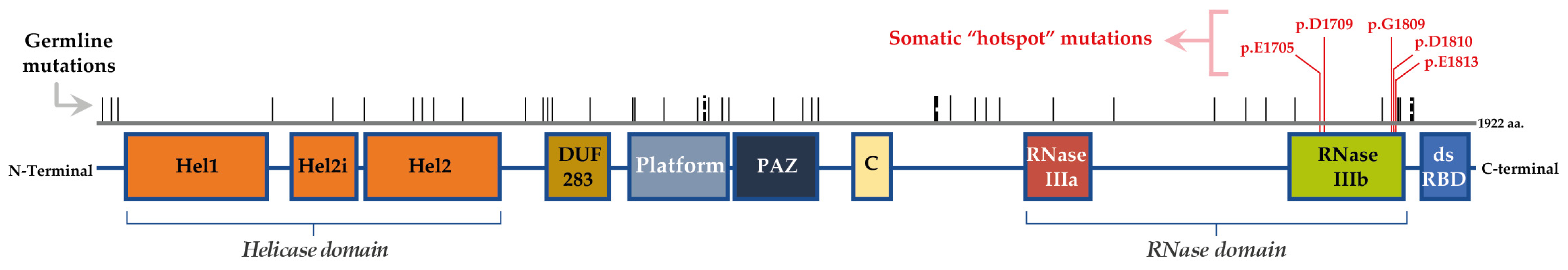
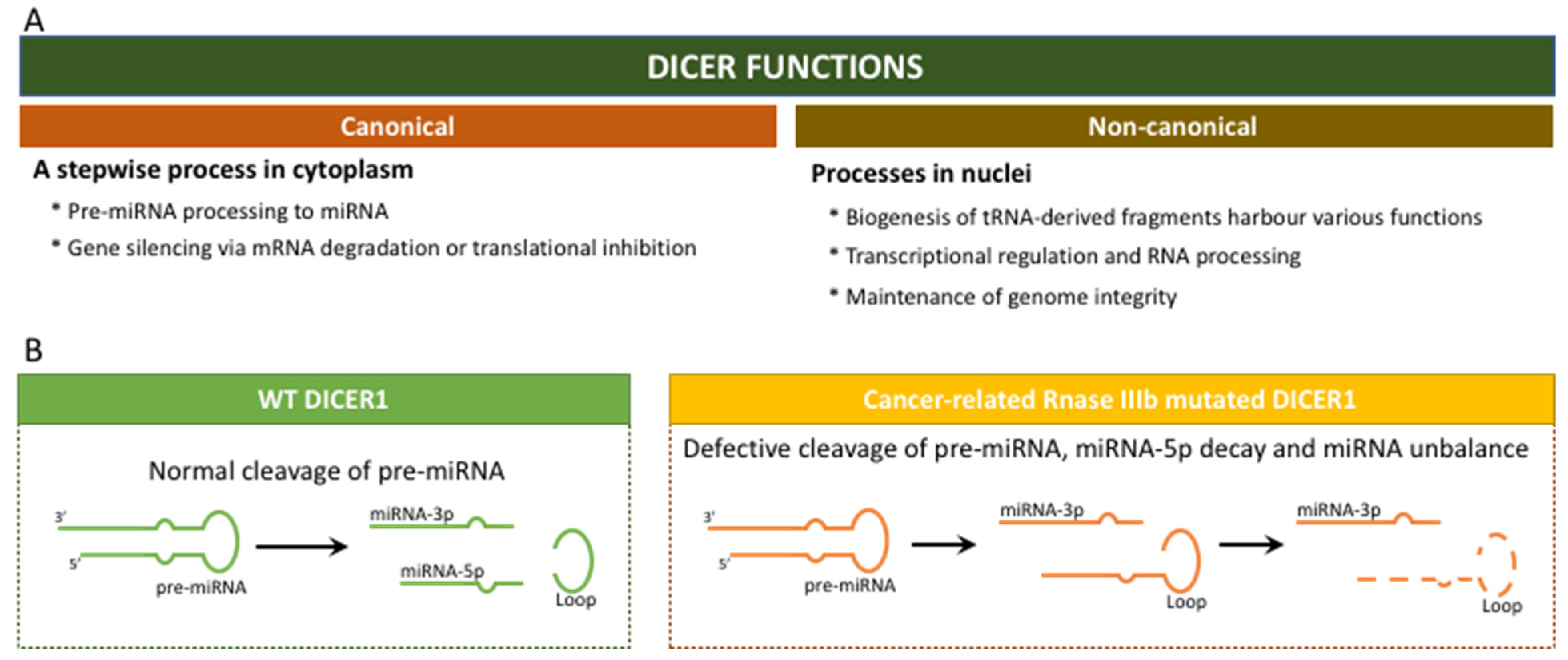
2.1. “The Gene DICER1” and the “Enzyme DICER”

The DICER1 gene is located on chromosome 14q32.13. It is composed of 27 exons and encodes a 1922-aminoacid protein with a molecular weight of approximately 200 kDa [6]. DICER1 encodes a multidomain enzyme that belongs to the RNase III family. DICER protein domains orderly locate from the N- to the C-terminus and include the following domains: Helicase (Hel1, Hel2i and Hel2), DUF283, Platform, PAZ (Piwi/Argonaut/Zwille), Connector helix, RNase IIIa and IIIb and dsRNA-binding domain (dsRBD) (Figure 1). Structurally, DICER has three rigid regions: RNase III, Platform-PAZ and helicase [6,15].
In a very simplistic description, the helicase domain allows the opening of the double-stranded RNA structures of the precursor microRNAs (pre-miRNAs) that are then cut by the RNase IIIa and IIIb domains in the 3p and 5p strand, respectively. The other domains are crucial to support these processing reactions from pre-miRNA to miRNA, namely the Platform, PAZ and Connector helix domains, which are important for the recognition, and the dsRBD domain needed for the binding of DICER1 to pre-miRNAs [13,16].
In the canonical path, the DICER multi-domain enzyme plays a crucial role in the biogenesis of the small RNAs, namely miRNAs (Figure 2) [15,17,18]. This is a cytoplasmic stepwise process that is incorporated into a specific pathway involving the large family of Argonaute (AGO) proteins and large multiprotein complexes termed RNA-induced silencing complexes (RISC) [17,18]. These processes guide for the sequence-specific silencing of genes through mRNA degradation, translational repression and heterochromatin formation. Beyond the canonical role of DICER via small RNA biogenesis in the cytoplasm, accumulated evidence suggests a non-canonical, non-endonuclease role of DICER in the nucleus. These processes are briefly summarized in Figure 2.

2.2. Mutations/Alterations of DICER1

Normal development and tumorigenesis of thyroid gland share many common pathways involved in cell proliferation and differentiation. The importance of miRNAs, as well as the role of DICER1 in processing precursor miRNA to mature ones, was evident in the maintenance of the thyroid tissue homeostasis, as well as in its involvement in tumorigenesis [19,20].
As will be discussed below, germline mutations in the DICER1 gene occur in any part of the gene, while somatic mutations are particularly frequent in the ribonuclease (ribonuclease IIIb) domain (Figure 1). The germline mutations are mostly inactivating mutations that cause DICER1 loss of function (LOF) and, as consequence, the downregulation of miRNA levels [9,20]. Mutations may be seen either in individuals in the context of DICER1 syndromic cases or in predisposed carriers. The LOF germline mutations can occur via deletion of the entire locus of the gene, as in- and out-of-frame intragenic deletions for one or more exons and also as somatic mosaicism. As supported by animal model studies, DICER1 does not act as the classical tumour suppressor genes or oncogenes [21]. To be able to promote tumorigenesis, the partial loss of DICER1 is required, indicating its role as a haploinsufficient tumour suppressor [2,9]. It was estimated that germline pathogenic variants of DICER1 may lead to LOF occurring in a ratio of 1:10,600 in the population [22]. Intriguingly, thyroid lesions seen in both germline and somatic mutations of the DICER1 gene were not found to be associated with other canonical thyroid genetic events in paediatric patients, such as BRAF, RAS or TERTp mutations [12].

2.3. Thyroid Neoplasm in the Context of DICER1 Syndromic and Non-Syndromic Mutations

Table 1 presents a compilation of studies carried out in the context of thyroid lesions that have been screened for the presence of alterations in the DICER1 gene.
The predisposition to the manifestation of thyroid lesions is increased when the individual presents germline syndromic mutations [23]. However, if the germline alteration is present in one allele only, the chance of developing a lesion in the thyroid gland is low. A second event is usually necessary. The second event can be the presence of a DICER1 somatic mutation in the second allele. In the context of DICER1 syndrome with previous clinical manifestations, screening for DICER1 somatic mutations is standard when thyroid lesions are identified in the individuals. Unlike what was previously reported [12], the second event can be unrelated to additional alterations in DICER1; it can be related with the presence of genetic alterations in other genes that increase the risk of the occurrence of thyroid lesions (benign and/or malignant). Examples of the latter were identified by our group and include mutations in BRAF, RAS and EIF1AX, among others [2].
Initially, DICER1-related thyroid manifestations were thought to be restricted to MNG or FA, but with the increase in case reports, large cohorts and population data-base studies on both DICER1 germline and somatic testing, a phenotypically diverse spectrum of thyroid entities, ranging from the most benign manifestations to the most aggressive tumours, was revealed [2]. MNG is frequently associated with DICER1 syndrome, which can be diagnosed from early ages until the fourth decade of life [8]. Despite being less frequent than MNG, differentiated thyroid tumours are also seen in DICER1 syndrome, with a higher incidence in the first decades of life [24]. Individuals who harbour a germline DICER1 alteration have a 16-fold increased risk of developing a thyroid tumour, and the incidence of MNG is higher in female than in male carriers [8]. While MNG is not a life-threatening lesion, differentiated thyroid carcinoma (DTC) or poorly differentiated thyroid carcinoma (PDTC) diagnosis, especially in young people, is associated with some risk of mortality [25]. For this reason, when a DICER1 syndrome or DICER1 alterations in an individual are known, a closer surveillance for thyroid lesions (as well as those of other organs) must follow, since early diagnosis can improve patient follow-up, and therefore lead to a more favourable prognosis. The diagnosis of any thyroid lesion, especially in early life, even in the absence of a familial history, should also be considered as a warning of the possibility of alterations in DICER1. So, molecular testing of DICER1 may be beneficial to the patient and, ultimately, to their relatives.
Recently, Stewart et al. studied the risk of the appearance of various types of neoplasms associated with DICER1 syndrome [26]. There is a range of ages with a high risk of certain types of neoplasms associated with DICER1 syndrome. For instance, the risk of PPB is high in the first years of life (up to 6 years), while the risk for Sertoli–Leydig cell tumours (SLCT) and other neoplasms (which include those of the thyroid) is distributed from early ages to 20 years of age (and there is still a slight increase up to around the age 40) [26]. If we focus the attention on TC, the authors found that there is a higher risk for the disease manifestation starting in the first decade of life until adulthood [26]. Findings from our previous study concerning TC also demonstrate a large range of ages for the occurrence of thyroid lesions [2]. If we take into account the nonproband carriers of DICER1 germline variants, TC appears as one of the most common events [26].
Thyroid gland lesions are the paediatric syndromic forms that have been reported the most in the context of DICER1 syndrome [23], but thyroid tumours are also found in the range of 20–40 years of age [26]. Thyroid lesions are less common in the oldest age groups, i.e., more than 40 years. Nonetheless, they are still represented. Two issues that are pertinent to the aforementioned age distribution profile deserves discussion. Firstly, the lesions may have been temporarily diagnosed long after their initial development and, due to this, the association of the age at diagnosis with the presence of DICER1 mutations may be somewhat biased. Secondly, the anticipated knowledge of the presence of DICER1 germline mutations in families/individuals in most studies leads to a closer clinical surveillance, supporting an early detection of lesions.
Two studies highlighted the results in paediatric thyroid tumours [11,27]. Chernock et al. studied six cases of paediatric PDTC and found that the presence of DICER1 somatic mutations in those aggressive TC is relatively common (four out of six, all of them with a hotspot in the RNAse IIIB domain), whereas germline mutations are less common (one out of six, affecting one region of splicing). These cases suggest that, in the presence of relatively more aggressive tumours at younger ages, the evaluation for the presence of mutations in the DICER1 gene could be beneficial, but further studies are needed in larger series [11]. Bae et al. studied a series of 41 paediatric follicular thyroid tumours (adenoma and carcinoma), whose patients had no previous history of DICER1 syndrome-associated lesions. Using NGS, the authors found that DICER1 somatic mutations were more common (9 out of 41 cases) than mutations in thyroid-related genes, such as NRAS (6 out of 41 cases), HRAS, PTEN, TSHR, RET and others. In that study, DICER1 and NRAS alterations were found to be mutually exclusive [27].
Knowing that DICER1 somatic mutations, although not very common, can occur and represent a tumorigenic event, it would be advisable to include DICER1 hotspot mutations analysis in the panels of genes studied for the molecular profiling of thyroid lesions. The study of mutations for DICER1 in commercial applications is already performed, namely in ThyroSeq v3 [28]. In a large study carried out by Chong et al., the authors found the presence of DICER1 hotspot mutations in 1.4% of the cohort studied (214 out of 14,993 FNAs). In the same study, Chong et al. found that, although not absent in DICER1 mutated lesions—as shown by our group [2]—changes in other genes that are related with thyroid tumorigenesis (RAS, BRAF, RET, TSHR, EIF1AX and TP53, among others) are less common in lesions that present DICER1 hotspot mutations [28]. Taking this fact into consideration, and the evidence of more recent studies demonstrating that DICER1 mutations are not restricted to benign thyroid lesions and may contribute biologically to more aggressive thyroid tumours (Table 1), the molecular testing for DICER1 could be recommended to patients whose thyroid lesions are diagnosed before the age of 40, when typically one would not expect to see genetic alterations in the most common thyroid cancer-related genes as often and where an individualized treatment and follow-up approach is required the most. Similarly, a more thorough molecular study on poorly differentiated TC and some variants of PTC may be of interest, given their apparent higher relative prevalence in DICER1-mutated patients. In such cases, when there is a clinical indication to look for the presence of hotspot mutations in the DICER1 gene, this can be done by PCR followed by Sanger sequencing. These are well-established and robust techniques that provide a good cost–benefit ratio for the screening of hotspot mutations.
When searching for mutations or variants in the entire DICER1 gene, the benefit of using different techniques must be considered. Since the gene is very large, it can be performed either by the laborious amplification of several amplicons by PCR/Sanger sequencing or by the use of more advanced techniques, such as NGS. While in cases of suspected DICER1 syndrome the second option seems to be the most obvious, given the lack of a predominant region for the appearance of germline alterations, when searching for somatic alterations the use of NGS should only be considered if hotspot mutations were not detected. Even so, the search for somatic mutations beyond the RNase IIIb hotspot needs additional supportive studies to ascertain its clinical relevance. Most studies reported hotspot mutations in thyroid lesions, mainly because of their screening focused on this region of the gene (Table 1). Studies on TCGA database did not find other somatic mutations either; however, a recent report using the MSK-IMPACT database described additional ones [2,13,29].
Table 1. Summary of DICER1 somatic and germline mutations reported in screened thyroid lesions.
Thyroid Lesion(s)Age (yo)Other Known Lesions (age)Germline MutationsSomatic Mutations (Thyroid Lesions)Ref
DNA Mutation(s)Protein Alteration(s)DNA Mutation(s)Protein Alteration(s)
Invasive FVPTC9PPB type II (1.9; 4.3); MNG (7)c.3505dupT
* mother carrier
p.S1169F,fs*8c.5439G>Tp.E1813D[30]
FVPTC7PPB type I (1.3); cataracts (6), CBME (6.1)c.3579_3580delCAp.N1193K,fs*41c.5438A>Gp.E1813G
Bilateral PTC within an FA11.5Type II PPB & CN (2.7)c.2379T>Gp.Y793Xc.5113G>Ap.E1705K
PTC within encapsulated follicular nodulesNASLCT, cystic nephroma, MNGc.5441C>Tp.S1814Lc.5126A>G p.D1709G[31]
PTC within encapsulated follicular nodulesNANAc.5425G>A p.G1809R
Follicular nodule with papillary hyperplasia, focal PTCNANAc.5126A>G (left node)
c.5428G>C (right node)
p.D1709G
p.D1810H
MNGNANAWithout hotspot mutations
Follicular hyperplasia18.0 (a)NAc.1329_1344_del16p.C443W,fs*10c.5438A>Gp.E1813G[8]
Nodular hyperplasia36.5 (a)NAc.1408G>Tp.E470*c.5126A>Gp.D1709G
Nodular hyperplasia13.7 (a)NAc.1525C>Tp.R509*c.5125G>Ap.D1709N
Nodular hyperplasia14.2 (a)NAc.1525C>Tp.R509*c.5428G>Cp.D1810H
MNG41.6 (a) c.1870C.Tp.R624c.5126A>G (2 lesions)
c.5429A>T (1 lesion)
c.5437G>C (1 lesion)
p.D1709G
p.D1810V
p.E1813Q
Nodular hyperplasia21.0 (a)NAc.2062C>Tp.R688*c.5428G>Tp.D1810Y
Nodular hyperplasia37 (a)NAc.2062C>Tp.R688*c.5429A>Tp.D1810V
Multinodular hyperplasia15.5 (a)NAc.2247C>Ap.Y749*c.5429A>Tp.D1810V
Hashimoto’s thyroiditis20.6 (a)NAc.2247C>Ap.Y749*NoneNone
Nodular hyperplasia13.6 (a)NAc.2650+1G>TSplice site variantNoneNone
Nodular hyperplasia21.0 (a)NAc.2830C>Tp.R944*c.5126A>Gp.D1709G
Nodular hyperplasia21.9 (a)NAc.3019C>Tp.Q1007*c.5113G>Ap.E1705K
Nodular hyperplasia with Hashimoto’s thyroiditis32.4 (a) c.3515_3525del11insA c.5126A>G (2 lesions)
c.5429A>T (1 lesion)
None (1 lesion)
p.D1709G
p.D1810V
-
Thyroid carcinoma, papillary, macrofollicular30.6 (a)NAc.3675C>Gp.Y1225c.5113G>A (1 lesion)
c.5126A>G (1 lesion)
p.E1705K
p.D1709G
PTC, follicular variant18.6 (a)PPB type II (4.1), MNG (18)c.3726C>Ap.Y1242c.5426G.Ap.G1809E
Nodular hyperplasia with Hashimoto’s thyroiditis60.9 (a)NAc.4812C>Ap.C1604*NoneNone
MNG15SLCT (13)c.4207-41_5364+1034delLoss of exons 23 and 24c.5113G>C+c.5114A>Tp.E1705Q +
p.E1705V
[32]
MNG15, 56NAc.5126A>Gp.D1709G
MNG13NAc.5429A>Tp.D1810V
MNG7, 26NAND-
MNG and DTC (papillary)70NANo-ND-
FVPTC13ERMS, SLCT, MNGc.5504_5507delATCCp.Y1835S,fs*2c.5113G>Ap.E1705K
Encapsulated cPTC16.5None c.2875A>T
c.5125G>A
p.K959 *
p.D1709N
[33]
Minimally invasive, encapsulated FVPTC14Nonec.1124C>Gp.P375R (rs148758903)c.5428G>T,
LOH (del chr14:94,043,795-104,822,229)
p.D1810Y
Infiltrative classical PTC11.7ALL, TBI, HSCT c.5439G>C
LOH (del chr14:78,529,021-100,616,514)
p.E1813D
Classical PTC with focal hobnail and tall cell change10None c.4260_4262delGGA (b)p.E1420del (rs544960260) (probably benign)
Minimally invasive solid-variant PTC15ALL, TBIc.2997T>Gp.L999L (rs12018992)Silent
Minimally invasive FVPTC and miPTC17.4ALL, c.20A>G (b)p.Q7R (rs117358479)
Follicular Nodular Disease, multifocal12Nonec.2535_2539del + insAATCAACTTCAAGCATTp.T847del + insNFKHSc.5438A>Gp.E1813G
Follicular Nodular Disease16Nonec.84dupTp.G29W,fs*11c.5125G>Ap.D1709N
FVPTC9PPB type II (2), PPB metastasis (4), MNG (7)c.3505insT
* mother carrier
p.S1169F,fs*8c.5438A>Gp.E1813G[14] (c)
NIFTP7PPB type I (1), CBME (6)c.3579_3580delCAp.N1193K,fs*41c.5438A>Gp.E1813G[14] (d)
PTC11PPB type II (2), CN (2), Askin tumour (13)c.2379T>Gp.Y793*c.5113G>Ap.E1705K
PDTC10Bilateral renal and lung cysts (2), pineoblastoma (7), bilateral SLCT (13,15), CBME (17)c.5437G>Cp.E1813QLOHLOH[14] (e)
FVPTC, NTH13Nonec.1363delp.V455fsIn 3 out of 5 lesions:
c.5126A>G (tumour 1)
c.5127T>G (tumour 2)
c.5113G>A (NTH)
p.D1709G
p.D1709E
p.E1705K
[14]
NIFTP, NTH17 (NIFTP)MNG (13)c.1363delp.V455fsIn 1 out of 2 NIFTP lesions: c.5427_5428del+insTTp.D1810Y
NIFTP15Lung cystsc.3999C>Ap.C1333*c.5437G>Ap.E1813K
PDTC14Nonec.2256+1G>CSplice variantc.5437G>Cp.E1813Q
FVPTC, NTH23 (FVPTC)Nonec.988G>Ap.Q330*c.5125G>A (T1a+T1b tumours)
c.5126A>G (T2 tumour+NTH1)
c.5437G>A (T3 tumor)
c.5438A>T (NTH2)
c.5428G>T (NTH3)
c.5429A>T (NTH4)
p.D1709N
p.D1709G
p.E1813K
p.E1813V
p.D1810Y
p.D1810V
FVPTC, NTH28 (FVPTC)Nonec.988G>Ap.Q330*c.5113G>A (NTH 1,2)
c.5126A>G (NTH 3,4)
c.5438A>T (tumour1+NTH 5–9)
c.5429A>T (NTH 10)
p.E1705K
p.D1709G
p.E1813V
p.D1810V
eaFTC2.4Nonec.3506C>Gp.S1169*c.5437G>Ap.E1813K[12]
wiFTC9.3PPB (2), Nodular hyperplasiac.3505dupTp.S1169fsc.5439G>Tp.E1813D
miFTC8.9Bronchogenic cyst or pulmonary parenchymal cyst; Nodular hyperplasia; FAc.5378delAp.E1793fsc.5125G>Ap.D1709N
wiFTC14None c.5437G>C
c.5465A>G
p.E1813Q
p.D1822G
miFTC14.6PNET in lung; FAc.2621C>A
Copy number loss (chr1:96–106Mb/chr10:pter-10Mb)
Copy number gain (chr9:121Mbqter)
p.S874*c.5126A>Gp.D1709G
miFTC18Lymphocytic thyroiditis c.5437G>Ap.E1813K
miFTC18.3Nodular hyperplasia c.3157dupT,
c.5437G>A
p.C1053fs,
p.E1813K
miFTC18.5Nodular hyperplasia c.4273G>T
c.5437G>A
LOH (chr9/chr21q)d
p.E1425*
p.E1813K
PDTC, encapsulated FVPTC14 (PDTC)NA c.5113G>Ap.E1705K[11]
PDTC, encapsulated FVPTC14 (PDTC)NA c.5125G>A,
c.3627dupA
p.D1709N,
p.P1210Tfs*25
PDTC19NA c.5137G>Tp.D1713Y
PDTC17NAc.735-8T>GSplicing site affectedc.5437G>Ap.E1813K
PDTC, PTC17 (PDTC)NA c.5437G>C, LOHp.E1813Q,
LOH
miFTC with multifocal capsular invasion; MNG12Sever’s diseaseNDNDc.5113G>Ap.E1705K[34]
miPTC, MNG37Heterozygous factor V Leiden mutation (24)NDNDc.5113G>Ap.E1705K
Minimally invasive FTC58Ovarian endometriosisNDNDc.5113G>Ap.E1705K
Adenomatoid nodules35Breast fibroadenoma and benign cystsNDNDc.5126A>Gp.D1709G
FTC with a focus on vascular invasion; MNG14UnknownNDNDc.5428G>Tp.D1810Y
PDTC17PPD+/CXR− (15)NDNDc.5428G>Tp.D1810Y
Classic PTC65Breast cancer (45), uterine cancer (52), schwannomaNDNDc.5428G>Tp.D1810Y
Classic PTC38NAc.-3T>CPromoter regionNot foundNot found[2]
OV-PTC44NAc.20A>Gp.Q7RNot foundNot found
OV-PTC65NAc.59C>Tp.A20VNot foundNot found
Classic PTC-A63NAc.1795A>Gp.T599ANot foundNot found
FVPTC53NAc.1887G>Ap.T629TNot foundNot found
FVPTC44NAc.1904A>Gp.N635SNot foundNot found
FVPTC25NAc.2512T>Gp.L838VNot foundNot found
OV-PTC27NAc.2557A>Gp.I853VNot foundNot found
Classic PTC-O45NAc.2614G>Ap.A872TNot foundNot found
Classic PTC30NAc.2951A>Cp.N984TNot foundNot found
Classic PTC31NANot foundNot found
FVPTC36NANot foundNot found
HV-PTC33NAc.3778G>Ap.V1260INot foundNot found
FVPTC88NAc.4260_4262delGGAp.E1420delNot foundNot found
OV-PTC51NAc.4680G>Ap.A1560ANot foundNot found
OV_PTC65NAc.4891T>Gp.S1631ANot foundNot found
OV-PTC26NAc.5013G>Cp.K1671NNot foundNot found
FVPTC31NANot foundNot foundc.5428G>Cp.D1810H
OV-PTC44NANot foundNot foundc.5438A>Gp.E1813G
Classic PTC64NAc.5507C>Tp.P1836HNot foundNot found
OV-PTC20NANot foundNot foundc.5718A>Cp.R1906S
Note: Only studies that screened the thyroid lesions for the presence of DICER1 alterations were included. CBME, ciliary body medulloepithelioma; DHL, dominant hyperplastic lesion or assumed hyperplastic nodules of thyroid (benign lesions); DTC, differentiated thyroid cancer; eaFTC, encapsulated angioinvasive FTC; ERMS, embryonal rhabdomyosarcoma; FA, follicular thyroid adenoma; FTC, follicular thyroid carcinoma; NIFTP, non-invasive follicular thyroid neoplasm with papillary-like nuclear features; NTH, nodular thyroid hyperplasia; miPTC, papillary thyroid microcarcinoma; MNG, multinodular goitre; PDTC, poorly differentiated thyroid carcinoma; PNET, primitive neuroectodermal tumour; PPB, pleuropulmonary blastoma; PTC, papillary thyroid carcinoma; SLCT: Sertoli–Leydig cell tumours. NA, Not available information. ND, Not done. (a) Age at surgery. (b) Germline or somatic origin unknown. (c) The case is the same case of the studies [30,35]. (d) These two cases are the same cases of the study [30]. (e) The case is the same case of the study [36].

2.4. DICER1 Somatic Hotspot Mutations and the Free Pass to Thyroid Malignancy

As mentioned above, DICER1-related tumours typically and most frequently harbour an “RNase IIIb hotspot” somatic mutation and less frequently LOH. The tumour-specific RNase IIIb hotspot mutations are missense mutations that normally occur in one of the five codons that encode for the protein catalytic domain residues (p.E1705, p.D1709, p.G1809, p.D1810 and p.E1813). These DICER1 hotspot mutations result in altered activity of DICER1 protein in microRNA processing and lead to a rapid and improper 5p miRNA cleavage and consequent degradation. Because of the 5p strand miRNAs loss, there is a consistent change in the microRNA landscape, as well as in the messenger RNA (mRNA) profiles mediated by the RISC complex. Somatic RNase hotspot mutations typically partner with a second alteration to promote tumorigenesis. They can couple in three fashions: somatic with germline, somatic with mosaic and somatic with somatic [9]. Although there is a remarkable miRNA imbalance/deregulation in the presence of somatic DICER1 mutation, this does not mean that it will always lead to malignancy. Indeed, MNG is the most frequent thyroid entity in DICER1 syndromic cases, but malignant entities ranging from the most innocent to the aggressive ones were more often associated with somatic mutations [37]. Oliver-Petit and al. [7], reported a series of eight families referred for childhood-onset of MNG or DICER1-related tumours with a familial history of MNG. The authors found that DICER1 somatic pathogenic mutations were present in both benign and malignant thyroid nodules, suggesting that the thyroid carcinogenesis pathway may be somehow unique in the background of DICER1-syndromic cases. The frequency of DICER1 mutations is higher in the background of LOF germline mutations, and RNase IIIb hotspot mutations were found to be more frequently associated with the presence of malignant tumours [9]. Noteworthily, more recent studies have documented a higher prevalence of DICER1 variants in an adolescent-onset PTC group [12,33]. In the study of Lee at el., although the number of FTC was far too low to draw any conclusion, all the cases of somatic mutations were observed in FTC only [12]. Based on this, the group draw the attention to the possible utility of somatic DICER1 testing in a young age group, particularly in the presence of a family history of MNG, thyroid surgery or associated embryonal tumour. Wasserman et al. noted the absence of thyroid autoimmunity lesions and local or distant metastasis in this group of PTCs, whereas Lee et al. attributed the favourable course of the cases to the predominance of miFTC cases in their series [12,33]. Nonetheless, these two studies pointed out the low risk of the malignant lesions in paediatric PTC and FTC groups. More recently, somatic mutations of DICER1 were also found to be related with PDTC and teratocarcinomas of the thyroid [6]. On this point, Agaimy et al. have reported two cases of DICER1 sporadic malignant teratoid thyroid tumours while thoroughly reviewing the clinicopathological and molecular characteristics of six additional cases previously reported [38]. In their report, the authors point out the highly aggressive course of this entity in comparison with the low malignant potential of other organ blastomas and proposed the term “thyroblastoma” for the disease. These cases tend to present at higher ages, without any family or personal history of other neoplasms, and have somatic mutations of DICER1, mostly a hotspot, thus supporting the concept of a DICER1 sporadic, non-syndromic form of this neoplasm and the need to differentiate it from the classical teratomas or carcinosarcomas [38].
Of those RNase IIIb hotspot mutations, there is a particular group of mutations called “mosaicism for RNase IIIb domain hotspot mutations” in DICER1 syndrome patients that deserves mention. According to Brenneman et al. and based on the study of 124 patients from the International PPB Registry (IPPBR), this group should be distinguished from the germline and mosaic LOF mutations due to their peculiar clinical characteristics—the disease tends to occur much earlier in life, and multisite disease is frequent. Shultz et al. [39] emphasized the importance of this subgroup, underlying two points: (a) First, whereas in the presence of a germline mutation (usually LOF) there is a need of a second hotspot mutation in the RNAse IIIb domain (limited to a very small target site(s)), in the presence of mosaicism hotspot mutations there is “only” a need for an additional alteration that causes LOF (whose probability of occurring is hundreds of times more likely); (b) Second, there is also the chance that the allele combination of hotspot and wild type together could be tumorigenic on its own. Therefore, a group of authors and working groups from Bakhuizen et al. suggested that the patients with mosaicism for hotspot should be under intensive and long-term surveillance. Today, the individuals with somatic mosaicism are known to show increased penetrance of DICER1 syndrome, including an earlier onset, higher number of disease foci and wider range of phenotypes [40].

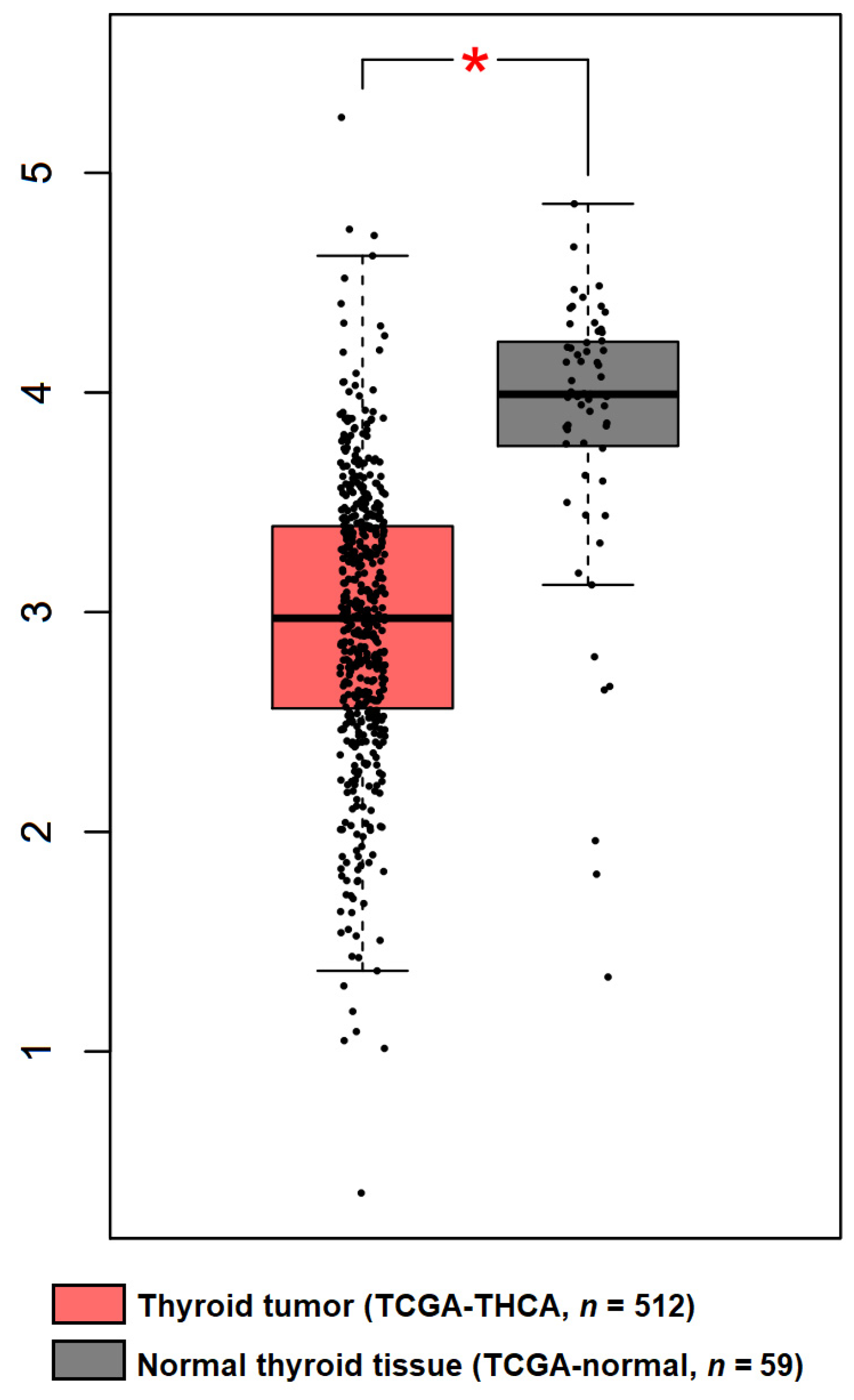
3. Regulation of DICER1 Expression in TC

The detection of mutations in the DICER1 gene (germline or somatic) has been the main focus of the studies identifying thyroid lesions (and other neoplasms) associated with the DICER1 syndrome. However, there is also a need to focus the attention on the role that DICER1 protein plays in signalling pathways that may promote the appearance or development of benign and malignant lesions.
Paulsson et al. observed that DICER1 mRNA expression is decreased relatively to normal thyroid tissues in FTC, but significantly higher in comparison with Hürthle cell thyroid carcinoma (HCC). At the protein level, DICER1 was also decreased in the FTC/HCC group when compared with normal thyroid tissues [41]. When searching for DICER1 expression in TC using the online GEPIA tool, a significant lower mRNA expression in TC was also evident in comparison with normal thyroid tissues (Figure 3).
Recently, the transcription factor GABPA was described to be involved in the transcription regulation of the DICER1 gene in papillary and follicular thyroid tumours [41,42]. A positive correlation between GABPA and DICER1 RNA expression was found in FTC cases [41]. A positive correlation between DICER1 and GABPA expression was also previously reported by Yuan et al. in PTC [42]. Mechanistically, both studies corroborated that GABPA regulates DICER1 expression. The silencing of GABPA reduced DICER1 expression levels [41,42], and the opposite was also observed, i.e., the overexpression of GABPA promoted an increase in DICER1 expression. This regulation was found to occur by a direct interaction of GABPA in DICER1 promoter: (1) a reduction in DICER1 promoter activity was seen when the GABPA binding site at the DICER1 promoter was mutated (-417A>C), both under basal conditions and with GABPA overexpression [42]; (2) DICER1 overexpression counteracts the effects mediated by GABPA depletion [42]; and (3) GABPA physically interacts with the DICER1 promoter region, as confirmed by chromatin immunoprecipitation (ChIP) [41]. The cellular studies exploring this molecular regulation suggested that it may provide cell advantages, namely in proliferation, viability, invasion and capacity of metastization in vitro. Whether this mechanism confers the same advantages in TC in vivo is a question that should also be studied.
The fact that the expression of two proteins (TERT and DICER1) that play an important role in thyroid tumorigenesis are regulated by the same transcription factor (GABPA) is curious. TERT is the catalytic subunit of telomerase, whose activity is known to be re-activated in up to 90% of human cancers [43,44]. The telomerase reactivation occurs mainly due to the re-expression of TERT. One of the most frequent mechanisms underlying TERT re-expression is the presence of the -124C>T and -146C>T mutations in the TERT promoter (TERTp) region [45,46], commonly found in TC [47,48]. This creates a new consensus-binding sites for the binding of E-twenty-six (ETS) transcription factors, especially GABPA [49].
Yuan et al., found that GABPA downregulation led to a reduction in TERT expression in TERTp-mutated cells [42]. However, when studying the cellular consequences of GABPA downregulation, they did not correlate with the mutational status of TERT. In contrast to what would be expected, an increased invasiveness of cells upon GABPA downregulation was seen. On this point, the authors found that this phenomenon correlates with the downstream effects of GABPA in DICER1 rather than those of TERT [42]. DICER1 was also implicated in TC cells’ proliferation and viability, i.e., the downregulation of DICER1 increased cell proliferation and viability [41].
Further studies are needed to understand whether a crosstalk between the regulation of TERT and DICER1 expression may occur or if these are completely independent regulatory mechanisms, despite the involvement of GABPA in both. The regulation of DICER1 expression by GABPA seems to be, at first sight, intrinsic to cell mechanisms that directly act on the GABPA regulation, dependent on cellular or extracellular regulatory signals (only). In the case of TERT, besides the same (extra)cellular signals, the occurrence of a mutation in the TERT promoter is needed so that, only in this case, GABPA can regulate its expression.
Besides DICER1, mutations in the microprocessor complex subunit DGCR8 (also referred to in the literature as DiGeorge syndrome critical region 8) and in DROSHA genes were recently reported [50,51,52,53]. These are genes that encode for proteins involved in microRNA processing machinery.
DGCR8 alterations were found in familial forms of MNG with schwannomatosis [52] and in FTCs [51], while DROSHA alterations were found in PDTC of one patient [50] and in FA and FVPTC [53]. Poma et al., found DROSHA1 germline synonymous mutations in patients who also harboured RAS mutations [53]. The microRNA processing machinery, which includes DROSHA, DGCR8 and DICER1, seems to be involved in thyroid tumorigenesis, but the knowledge in this field is still emerging. The need to disclose the alterations and dysfunctions of this processing machinery in the thyroid still exists to clarify its relative contribution to tumorigenesis.

4. Conclusions

DICER1 protein is one of several proteins involved in microRNA processing machinery affecting gene regulation at the post-transcriptional level. Although the link between germline mutations and the syndromic predisposition to MNG and childhood tumours is not new, the relative importance and biological meaning of thyroid lesions in the context of DICER1 mutations, associated with syndromic or non-syndromic cases, is very recent. Our review of the literature supports the concept that molecular investigation of thyroid lesions both in a paediatric-onset scenario and a young adulthood-onset remit, with or without a family history of “DICER1 pleiotropic tumours predisposition syndrome”, is of increasing importance; the study of DICER alterations in these circumstances goes hand in hand with the concept of secondary prevention in patients who are at increased risk of malignancy and poor outcomes, while also allowing the identification of index cases’ relatives whose TC risk would be otherwise unknown.

Author Contributions

Conceptualization, S.C., M.C. and V.M.; writing—original draft preparation, S.C., M.C. and A.R.L.; writing—review and editing, all authors (S.C., M.C., A.R.L., M.B., M.S.-S., P.S. and V.M.) contributed to this step; supervision, P.S. and V.M. All authors have read and agreed to the published version of the manuscript.

Funding

This work was supported by Portuguese funds through FCT—Fundação para a Ciência e a Tecnologia through a PhD grant to S.C. (SFRH/BD/147650/2019). The authors acknowledge the support fromFCT that funded the contract to MC through the project PTDC/MED-ONC/31438/2017 (The Other Faces of Telomerase: Looking beyond Tumor Immortalization). The project is supported by NORTE 2020 under the PORTUGAL 2020 Partnership Agreement, through the European Regional Development Fund (ERDF)/COMPETE 2020—Operational Program for Competitiveness and Internationalization (POCI) and by Portuguese funds through FCT. Additional funding was provided (in part) by the Programa Operacional Regional do Norte, and co-funding was given by the European Regional Development Fund under the project “The Porto Comprehensive Cancer Center” with the reference NORTE-01-0145-FEDER-072678—Consórcio PORTO.CCC—Porto. Comprehensive Cancer Center. Additional funding was provided by the Bolsa E. Limbert SPEDM/Genzyme em Patologia da Tiróide for the project “Diabetes mellitus and oncocytic tumors of thyroid: the mitochondrial connection”.

Institutional Review Board Statement

Not applicable.

Informed Consent Statement

Not applicable.

Data Availability Statement

Data sharing not applicable.

Conflicts of Interest

The authors declare no conflict of interest.

References

  1. Couch, R.M.; Hughes, I.A.; DeSa, D.J.; Schiffrin, A.; Guyda, H.; Winter, J.S. An autosomal dominant form of adolescent multinodular goiter. Am. J. Hum. Genet. 1986, 39, 811–816. [Google Scholar] [PubMed]
  2. Canberk, S.; Ferreira, J.C.; Pereira, L.; Batista, R.; Vieira, A.F.; Soares, P.; Sobrinho Simoes, M.; Maximo, V. Analyzing the Role of DICER1 Germline Variations in Papillary Thyroid Carcinoma. Eur. Thyroid. J. 2021, 9, 296–303. [Google Scholar] [CrossRef] [PubMed]
  3. Hill, D.A.; Ivanovich, J.; Priest, J.R.; Gurnett, C.A.; Dehner, L.P.; Desruisseau, D.; Jarzembowski, J.A.; Wikenheiser-Brokamp, K.A.; Suarez, B.K.; Whelan, A.J.; et al. DICER1 mutations in familial pleuropulmonary blastoma. Science 2009, 325, 965. [Google Scholar] [CrossRef] [PubMed] [Green Version]
  4. Slade, I.; Bacchelli, C.; Davies, H.; Murray, A.; Abbaszadeh, F.; Hanks, S.; Barfoot, R.; Burke, A.; Chisholm, J.; Hewitt, M.; et al. DICER1 syndrome: Clarifying the diagnosis, clinical features and management implications of a pleiotropic tumour predisposition syndrome. J. Med. Genet. 2011, 48, 273–278. [Google Scholar] [CrossRef] [Green Version]
  5. Rio Frio, T.; Bahubeshi, A.; Kanellopoulou, C.; Hamel, N.; Niedziela, M.; Sabbaghian, N.; Pouchet, C.; Gilbert, L.; O’Brien, P.K.; Serfas, K.; et al. DICER1 mutations in familial multinodular goiter with and without ovarian Sertoli-Leydig cell tumors. JAMA 2011, 305, 68–77. [Google Scholar] [CrossRef] [Green Version]
  6. Thunders, M.; Delahunt, B. Gene of the month: DICER1: Ruler and controller. J. Clin. Pathol. 2021, 74, 69–72. [Google Scholar] [CrossRef] [PubMed]
  7. Oliver-Petit, I.; Bertozzi, A.I.; Grunenwald, S.; Gambart, M.; Pigeon-Kerchiche, P.; Sadoul, J.L.; Caron, P.J.; Savagner, F. Multinodular goitre is a gateway for molecular testing of DICER1 syndrome. Clin. Endocrinol. 2019, 91, 669–675. [Google Scholar] [CrossRef]
  8. Khan, N.E.; Bauer, A.J.; Schultz, K.A.P.; Doros, L.; Decastro, R.M.; Ling, A.; Lodish, M.B.; Harney, L.A.; Kase, R.G.; Carr, A.G.; et al. Quantification of Thyroid Cancer and Multinodular Goiter Risk in the DICER1 Syndrome: A Family-Based Cohort Study. J. Clin. Endocrinol. Metab. 2017, 102, 1614–1622. [Google Scholar] [CrossRef]
  9. De Kock, L.; Wu, M.K.; Foulkes, W.D. Ten years of DICER1 mutations: Provenance, distribution, and associated phenotypes. Hum. Mutat. 2019, 40, 1939–1953. [Google Scholar] [CrossRef]
  10. Rooper, L.M.; Bynum, J.P.; Miller, K.P.; Lin, M.T.; Gagan, J.; Thompson, L.D.R.; Bishop, J.A. Recurrent DICER1 Hotspot Mutations in Malignant Thyroid Gland Teratomas: Molecular Characterization and Proposal for a Separate Classification. Am. J. Surg. Pathol. 2020, 44, 826–833. [Google Scholar] [CrossRef]
  11. Chernock, R.D.; Rivera, B.; Borrelli, N.; Hill, D.A.; Fahiminiya, S.; Shah, T.; Chong, A.S.; Aqil, B.; Mehrad, M.; Giordano, T.J.; et al. Poorly differentiated thyroid carcinoma of childhood and adolescence: A distinct entity characterized by DICER1 mutations. Mod. Pathol. Off. J. U. S. Can. Acad. Pathol. Inc 2020, 33, 1264–1274. [Google Scholar] [CrossRef]
  12. Lee, Y.A.; Im, S.W.; Jung, K.C.; Chung, E.J.; Shin, C.H.; Kim, J.I.; Park, Y.J. Predominant DICER1 Pathogenic Variants in Pediatric Follicular Thyroid Carcinomas. Thyroid Off. J. Am. Thyroid Assoc. 2020, 30, 1120–1131. [Google Scholar] [CrossRef]
  13. Vedanayagam, J.; Chatila, W.K.; Aksoy, B.A.; Majumdar, S.; Skanderup, A.J.; Demir, E.; Schultz, N.; Sander, C.; Lai, E.C. Cancer-associated mutations in DICER1 RNase IIIa and IIIb domains exert similar effects on miRNA biogenesis. Nat. Commun. 2019, 10, 3682. [Google Scholar] [CrossRef] [PubMed]
  14. van der Tuin, K.; de Kock, L.; Kamping, E.J.; Hannema, S.E.; Pouwels, M.M.; Niedziela, M.; van Wezel, T.; Hes, F.J.; Jongmans, M.C.; Foulkes, W.D.; et al. Clinical and Molecular Characteristics May Alter Treatment Strategies of Thyroid Malignancies in DICER1 Syndrome. J. Clin. Endocrinol. Metab. 2019, 104, 277–284. [Google Scholar] [CrossRef] [Green Version]
  15. Song, M.S.; Rossi, J.J. Molecular mechanisms of Dicer: Endonuclease and enzymatic activity. Biochem. J. 2017, 474, 1603–1618. [Google Scholar] [CrossRef] [PubMed] [Green Version]
  16. Ciechanowska, K.; Pokornowska, M.; Kurzynska-Kokorniak, A. Genetic Insight into the Domain Structure and Functions of Dicer-Type Ribonucleases. Int. J. Mol. Sci. 2021, 22, 616. [Google Scholar] [CrossRef]
  17. Gu, S.; Jin, L.; Zhang, Y.; Huang, Y.; Zhang, F.; Valdmanis, P.N.; Kay, M.A. The loop position of shRNAs and pre-miRNAs is critical for the accuracy of dicer processing in vivo. Cell 2012, 151, 900–911. [Google Scholar] [CrossRef] [Green Version]
  18. Lau, P.W.; Guiley, K.Z.; De, N.; Potter, C.S.; Carragher, B.; MacRae, I.J. The molecular architecture of human Dicer. Nat. Struct. Mol. Biol. 2012, 19, 436–440. [Google Scholar] [CrossRef] [Green Version]
  19. Pallante, P.; Battista, S.; Pierantoni, G.M.; Fusco, A. Deregulation of microRNA expression in thyroid neoplasias. Nat. Rev. Endocrinol. 2014, 10, 88–101. [Google Scholar] [CrossRef] [PubMed]
  20. Ramirez-Moya, J.; Wert-Lamas, L.; Riesco-Eizaguirre, G.; Santisteban, P. Impaired microRNA processing by DICER1 downregulation endows thyroid cancer with increased aggressiveness. Oncogene 2019, 38, 5486–5499. [Google Scholar] [CrossRef]
  21. Kumar, M.S.; Pester, R.E.; Chen, C.Y.; Lane, K.; Chin, C.; Lu, J.; Kirsch, D.G.; Golub, T.R.; Jacks, T. Dicer1 functions as a haploinsufficient tumor suppressor. Genes Dev. 2009, 23, 2700–2704. [Google Scholar] [CrossRef] [Green Version]
  22. Kim, J.; Field, A.; Schultz, K.A.P.; Hill, D.A.; Stewart, D.R. The prevalence of DICER1 pathogenic variation in population databases. Int. J. Cancer 2017, 141, 2030–2036. [Google Scholar] [CrossRef] [Green Version]
  23. Caroleo, A.M.; De Ioris, M.A.; Boccuto, L.; Alessi, I.; Del Baldo, G.; Cacchione, A.; Agolini, E.; Rinelli, M.; Serra, A.; Carai, A.; et al. DICER1 Syndrome and Cancer Predisposition: From a Rare Pediatric Tumor to Lifetime Risk. Front. Oncol. 2020, 10, 614541. [Google Scholar] [CrossRef] [PubMed]
  24. Guillerman, R.P.; Foulkes, W.D.; Priest, J.R. Imaging of DICER1 syndrome. Pediatr. Radiol. 2019, 49, 1488–1505. [Google Scholar] [CrossRef] [PubMed]
  25. Lloyd, R.V.; Osamura, R.Y.; Klöppel, G.; Rosai, J. WHO Classification of Tumours of Endocrine Organs, 4th ed.; World Health Organization: Lyon, France, 2017; Volume 10. [Google Scholar]
  26. Stewart, D.R.; Best, A.F.; Williams, G.M.; Harney, L.A.; Carr, A.G.; Harris, A.K.; Kratz, C.P.; Dehner, L.P.; Messinger, Y.H.; Rosenberg, P.S.; et al. Neoplasm Risk Among Individuals With a Pathogenic Germline Variant in DICER1. J. Clin. Oncol. 2019, 37, 668–676. [Google Scholar] [CrossRef] [PubMed]
  27. Bae, J.S.; Jung, S.H.; Hirokawa, M.; Bychkov, A.; Miyauchi, A.; Lee, S.; Chung, Y.J.; Jung, C.K. High Prevalence of DICER1 Mutations and Low Frequency of Gene Fusions in Pediatric Follicular-Patterned Tumors of the Thyroid. Endocr. Pathol. 2021, 32, 336–346. [Google Scholar] [CrossRef] [PubMed]
  28. Chong, A.S.; Nikiforov, Y.E.; Condello, V.; Wald, A.I.; Nikiforova, M.N.; Foulkes, W.D.; Rivera, B. Prevalence and Spectrum of DICER1 Mutations in Adult-onset Thyroid Nodules with Indeterminate Cytology. J. Clin. Endocrinol. Metab. 2021, 106, 968–977. [Google Scholar] [CrossRef]
  29. Ghossein, C.A.; Dogan, S.; Farhat, N.; Landa, I.; Xu, B. Expanding the spectrum of thyroid carcinoma with somatic DICER1 mutation: A survey of 829 thyroid carcinomas using MSK-IMPACT next-generation sequencing platform. Virchows Arch. Int. J. Pathol. 2021. [Google Scholar] [CrossRef]
  30. De Kock, L.; Sabbaghian, N.; Soglio, D.B.; Guillerman, R.P.; Park, B.K.; Chami, R.; Deal, C.L.; Priest, J.R.; Foulkes, W.D. Exploring the association Between DICER1 mutations and differentiated thyroid carcinoma. J. Clin. Endocrinol. Metab. 2014, 99, E1072–E1077. [Google Scholar] [CrossRef] [Green Version]
  31. Rutter, M.M.; Jha, P.; Schultz, K.A.; Sheil, A.; Harris, A.K.; Bauer, A.J.; Field, A.L.; Geller, J.; Hill, D.A. DICER1 Mutations and Differentiated Thyroid Carcinoma: Evidence of a Direct Association. J. Clin. Endocrinol. Metab. 2016, 101, 1–5. [Google Scholar] [CrossRef] [Green Version]
  32. Apellaniz-Ruiz, M.; de Kock, L.; Sabbaghian, N.; Guaraldi, F.; Ghizzoni, L.; Beccuti, G.; Foulkes, W.D. Familial multinodular goiter and Sertoli–Leydig cell tumors associated with a large intragenic in-frame DICER1 deletion. Eur. J. Endocrinol. 2018, 178, K11–K19. [Google Scholar] [CrossRef] [PubMed] [Green Version]
  33. Wasserman, J.D.; Sabbaghian, N.; Fahiminiya, S.; Chami, R.; Mete, O.; Acker, M.; Wu, M.K.; Shlien, A.; de Kock, L.; Foulkes, W.D. DICER1 Mutations Are Frequent in Adolescent-Onset Papillary Thyroid Carcinoma. J. Clin. Endocrinol. Metab. 2018, 103, 2009–2015. [Google Scholar] [CrossRef] [Green Version]
  34. Shin, S.H.; Yoon, J.H.; Son, M.H.; Kim, S.J.; Park, S.Y.; Kim, H.Y.; Lee, H.S.; Park, H.J.; Park, B.K. Follicular Thyroid Carcinoma Arising After Hematopoietic Stem Cell Transplantation in a Child with Pleuropulmonary Blastoma. Thyroid Off. J. Am. Thyroid Assoc. 2012, 22, 547–551. [Google Scholar] [CrossRef] [PubMed]
  35. de Kock, L.; Wang, Y.C.; Revil, T.; Badescu, D.; Rivera, B.; Sabbaghian, N.; Wu, M.N.; Weber, E.; Sandoval, C.; Hopman, S.M.J.; et al. High-sensitivity sequencing reveals multi-organ somatic mosaicism causing DICER1 syndrome. J. Med. Genet. 2016, 53, 43–52. [Google Scholar] [CrossRef] [Green Version]
  36. Darbinyan, A.; Morotti, R.; Cai, G.; Prasad, M.L.; Christison-Lagay, E.; Dinauer, C.; Adeniran, A.J. Cytomorphologic features of thyroid disease in patients with DICER1 mutations: A report of cytology-histopathology correlation in 7 patients. Cancer Cytopathol 2020, 128, 746–756. [Google Scholar] [CrossRef]
  37. Robertson, J.C.; Jorcyk, C.L.; Oxford, J.T. DICER1 Syndrome: DICER1 Mutations in Rare Cancers. Cancers 2018, 10, 143. [Google Scholar] [CrossRef] [Green Version]
  38. Agaimy, A.; Witkowski, L.; Stoehr, R.; Cuenca, J.C.C.; Gonzalez-Muller, C.A.; Brutting, A.; Bahrle, M.; Mantsopoulos, K.; Amin, R.M.S.; Hartmann, A.; et al. Malignant teratoid tumor of the thyroid gland: An aggressive primitive multiphenotypic malignancy showing organotypical elements and frequent DICER1 alterations-is the term "thyroblastoma" more appropriate? Virchows Arch. Int. J. Pathol. 2020, 477, 787–798. [Google Scholar] [CrossRef] [PubMed]
  39. Schultz, K.A.P.; Williams, G.M.; Kamihara, J.; Stewart, D.R.; Harris, A.K.; Bauer, A.J.; Turner, J.; Shah, R.; Schneider, K.; Schneider, K.W.; et al. DICER1 and Associated Conditions: Identification of At-risk Individuals and Recommended Surveillance Strategies. Clin. Cancer Res. 2018, 24, 2251–2261. [Google Scholar] [CrossRef] [Green Version]
  40. Bakhuizen, J.J.; Hanson, H.; van der Tuin, K.; Lalloo, F.; Tischkowitz, M.; Wadt, K.; Jongmans, M.C.J.; Group, S.H.G.W.; CanGene-CanVar Clinical Guideline Working, G.; Expert Network, M. Surveillance recommendations for DICER1 pathogenic variant carriers: A report from the SIOPE Host Genome Working Group and CanGene-CanVar Clinical Guideline Working Group. Fam. Cancer 2021, 20, 337–348. [Google Scholar] [CrossRef]
  41. Paulsson, J.O.; Wang, N.; Gao, J.; Stenman, A.; Zedenius, J.; Mu, N.; Lui, W.O.; Larsson, C.; Juhlin, C.C. GABPA-dependent down-regulation of DICER1 in follicular thyroid tumours. Endocr. Relat. Cancer 2020, 27, 295–308. [Google Scholar] [CrossRef] [Green Version]
  42. Yuan, X.; Mu, N.; Wang, N.; Straat, K.; Sofiadis, A.; Guo, Y.; Stenman, A.; Li, K.; Cheng, G.; Zhang, L.; et al. GABPA inhibits invasion/metastasis in papillary thyroid carcinoma by regulating DICER1 expression. Oncogene 2019, 38, 965–979. [Google Scholar] [CrossRef] [PubMed]
  43. Pestana, A.; Vinagre, J.; Sobrinho-Simoes, M.; Soares, P. TERT biology and function in cancer: Beyond immortalisation. J. Mol. Endocrinol. 2017, 58, R129–R146. [Google Scholar] [CrossRef]
  44. Shay, J.W.; Bacchetti, S. A survey of telomerase activity in human cancer. Eur. J. Cancer 1997, 33, 787–791. [Google Scholar] [CrossRef]
  45. Horn, S.; Figl, A.; Rachakonda, P.S.; Fischer, C.; Sucker, A.; Gast, A.; Kadel, S.; Moll, I.; Nagore, E.; Hemminki, K.; et al. TERT promoter mutations in familial and sporadic melanoma. Science 2013, 339, 959–961. [Google Scholar] [CrossRef] [PubMed] [Green Version]
  46. Huang, F.W.; Hodis, E.; Xu, M.J.; Kryukov, G.V.; Chin, L.; Garraway, L.A. Highly recurrent TERT promoter mutations in human melanoma. Science 2013, 339, 957–959. [Google Scholar] [CrossRef] [Green Version]
  47. Vinagre, J.; Almeida, A.; Populo, H.; Batista, R.; Lyra, J.; Pinto, V.; Coelho, R.; Celestino, R.; Prazeres, H.; Lima, L.; et al. Frequency of TERT promoter mutations in human cancers. Nat. Commun. 2013, 4, 2185. [Google Scholar] [CrossRef] [Green Version]
  48. Melo, M.; Gaspar da Rocha, A.; Batista, R.; Vinagre, J.; Martins, M.J.; Costa, G.; Ribeiro, C.; Carrilho, F.; Leite, V.; Lobo, C.; et al. TERT, BRAF, and NRAS in Primary Thyroid Cancer and Metastatic Disease. J. Clin. Endocrinol. Metab. 2017, 102, 1898–1907. [Google Scholar] [CrossRef] [PubMed]
  49. Bell, R.J.; Rube, H.T.; Kreig, A.; Mancini, A.; Fouse, S.D.; Nagarajan, R.P.; Choi, S.; Hong, C.; He, D.; Pekmezci, M.; et al. Cancer. The transcription factor GABP selectively binds and activates the mutant TERT promoter in cancer. Science 2015, 348, 1036–1039. [Google Scholar] [CrossRef] [Green Version]
  50. Paulsson, J.O.; Backman, S.; Wang, N.; Stenman, A.; Crona, J.; Thutkawkorapin, J.; Ghaderi, M.; Tham, E.; Stalberg, P.; Zedenius, J.; et al. Whole-genome sequencing of synchronous thyroid carcinomas identifies aberrant DNA repair in thyroid cancer dedifferentiation. J. Pathol. 2020, 250, 183–194. [Google Scholar] [CrossRef]
  51. Paulsson, J.O.; Rafati, N.; DiLorenzo, S.; Chen, Y.; Haglund, F.; Zedenius, J.; Juhlin, C.C. Whole-genome sequencing of follicular thyroid carcinomas reveal recurrent mutations in microRNA processing subunit DGCR8. J. Clin. Endocrinol. Metab. 2021, 106, 3265–3282. [Google Scholar] [CrossRef]
  52. Rivera, B.; Nadaf, J.; Fahiminiya, S.; Apellaniz-Ruiz, M.; Saskin, A.; Chong, A.S.; Sharma, S.; Wagener, R.; Revil, T.; Condello, V.; et al. DGCR8 microprocessor defect characterizes familial multinodular goiter with schwannomatosis. J. Clin. Investig. 2020, 130, 1479–1490. [Google Scholar] [CrossRef] [PubMed]
  53. Poma, A.M.; Condello, V.; Denaro, M.; Torregrossa, L.; Elisei, R.; Vitti, P.; Basolo, F. DICER1 somatic mutations strongly impair miRNA processing even in benign thyroid lesions. Oncotarget 2019, 10, 1785–1797. [Google Scholar] [CrossRef] [PubMed]
Figure 1. Schematic representation of the hDICER protein domains and the location of hotspot somatic mutations. hDICER1 is composed by the following domains, from N- to C-terminus of the protein: helicase domain (Hel1, Hel2i and Hel2), DUF283, platform, Piwi-Argonaute-Zwille (PAZ), connector helix (C), RNase IIIa, RNase IIIb and double-stranded RNA-binding domain (dsRBD). The hotspot somatic mutations for DICER1 gene in thyroid lesions locate in the RNase IIIb domain, while the germline mutations can be found along all the gene.
Jmp 03 00001 g001
Figure 2. DICER1 functions: (A) Canonical and non-canonical cellular functions; (B) Canonical functions of DICER1 in a context of wild-type DICER1 and in a context of mutated DICER1, in hotspot RNase IIIb domain.
Jmp 03 00001 g002
Figure 3. DICER1 gene expression in thyroid cancer and normal tissue. Expression data from human thyroid cancers (TCGA-THCA; n = 512) and corresponding normal tissues (TCGA normal tissue; n = 59). Data was acquired through the GEPIA tool (http://gepia.cancer-pku.cn/index.html, accessed on 14 July 2021, with the following parameters: log2-fold change Cut-off = 1; p-value Cut-off = 0.01. * Statistically significant difference.
Jmp 03 00001 g003
Publisher’s Note: MDPI stays neutral with regard to jurisdictional claims in published maps and institutional affiliations.

