Case Report

Progression of Well-Differentiated Papillary Mesothelial Tumour to Mesothelioma in a Patient with Ehlers Danlos Syndrome

by
Sarita Prabhakaran
1,
Matthew Hussey
2,
Kenneth J. O’Byrne
3,4,5 and
Sonja Klebe
1,2,*
1
Department of Anatomical Pathology, College of Medicine and Public Health, Flinders University, Adelaide, SA 5042, Australia
2
Department of Surgical Pathology, SA Pathology at Flinders Medical Centre, Adelaide, SA 5042, Australia
3
Cancer Services, Princess Alexandra Hospital, Brisbane, QLD 4102, Australia
4
Faculty of Health, Institute of Health and Biomedical Innovation, School of Biomedical Sciences, Queensland University of Technology, Brisbane, QLD 4102, Australia
5
Translational Research Institute, Brisbane, QLD 4102, Australia
*
Author to whom correspondence should be addressed.
J. Mol. Pathol. 2021, 2(4), 306-311; https://doi.org/10.3390/jmp2040026
Submission received: 17 September 2021 / Revised: 17 October 2021 / Accepted: 25 October 2021 / Published: 29 October 2021

Abstract

:
Well-differentiated papillary mesothelioma has been renamed well-differentiated papillary mesothelial tumour (WDPMT) in the current WHO classification because all mesotheliomas are now regarded as malignant. WDPMT is now defined as a non-invasive papillary mesothelial proliferation, with retained labelling for BAP1-desirable. The current WHO classification also includes mesothelioma in situ (MIS), which is defined as pre-invasive flat or papillary proliferation of mesothelial cells with a loss of BAP1 or MTAP. WDPMT has been variably defined in the past but was thought to occur more commonly in women and pursue a more indolent course than mesothelioma, but its progression to invasive disease has occasionally been reported. Here, we report a case of a 68-year-old woman with a history of asbestos exposure and an underlying diagnosis of Ehlers Danlos syndrome who was diagnosed with symptomatic WDPMT of the peritoneum that progressed to mesothelioma within two years. On retrospective analysis, the WDPMT showed a loss of BAP1. We suggest that a loss of BAP1 in WDPMT should be reported, since these lesions may show aggressive behaviour, and that they may best be regarded as similar to mesothelioma in situ.

1. Introduction

The traditional view is that a well-differentiated papillary mesothelial tumour (WDPMT) of the peritoneum occurs most commonly in women between the ages of 23 and 75 years [1,2,3]. In the most conservative definitions, diagnosis is limited to incidental findings at surgery for other unrelated conditions [4,5], although some publications have accepted the diagnosis in patients who presented with abdominal symptoms [1,3]. The definition of WDPMT has changed over time, with earlier publications restricting diagnosis of WDPMT to tumours that lack invasion [6], but other studies have included WDPM with very ‘limited’ invasion [7,8,9]. The recent WHO classification of thoracic tumours changed the nomenclature of well-differentiated papillary mesothelioma to well-differentiated papillary mesothelial tumour because all mesotheliomas are now regarded as malignant. The histological criteria include papillary formations covered by a single layer of flattened to cuboidal bland mesothelial cells and no stromal invasion, and BAP1 retention on IHC is regarded as ‘desirable’. The guidelines make mention of the lack of certainty on the exact nature of WDPMTs that have invasion, or whether these are a subtype of malignant mesothelioma [10].
Peritoneal WDPMTs exhibit variable clinical prognoses that some authors have described as indolent [8,11] and others as ‘aggressive’, leading to recurrences and poorer outcomes, especially in those that included foci of invasion and very limited invasion [7,9,12]. The current WHO classification also introduces the entity of mesothelioma in situ (MIS) that shows a layer of flat or cuboidal cells with or without atypia without evidence of invasion but which may also include papillary proliferations, somewhat like WDPMT. Loss of BAP1 (and/or MTAP/CDKN2A) is a requirement for the diagnosis of MIS, but the retention of BAP1/MTAP/CDKN2A is not an absolute requirement for the diagnosis of WDPMT, and we suggest that there may be overlap in the conditions [10]. Here, we present a case of a 68-year-old woman who was diagnosed with peritoneal WDPMT (with BAP1 loss demonstrated retrospectively) that progressed to an invasive epithelioid peritoneal mesothelioma within two years. The patient was known to have Ehlers–Danlos syndrome (EDS) and had a history of asbestos exposure.

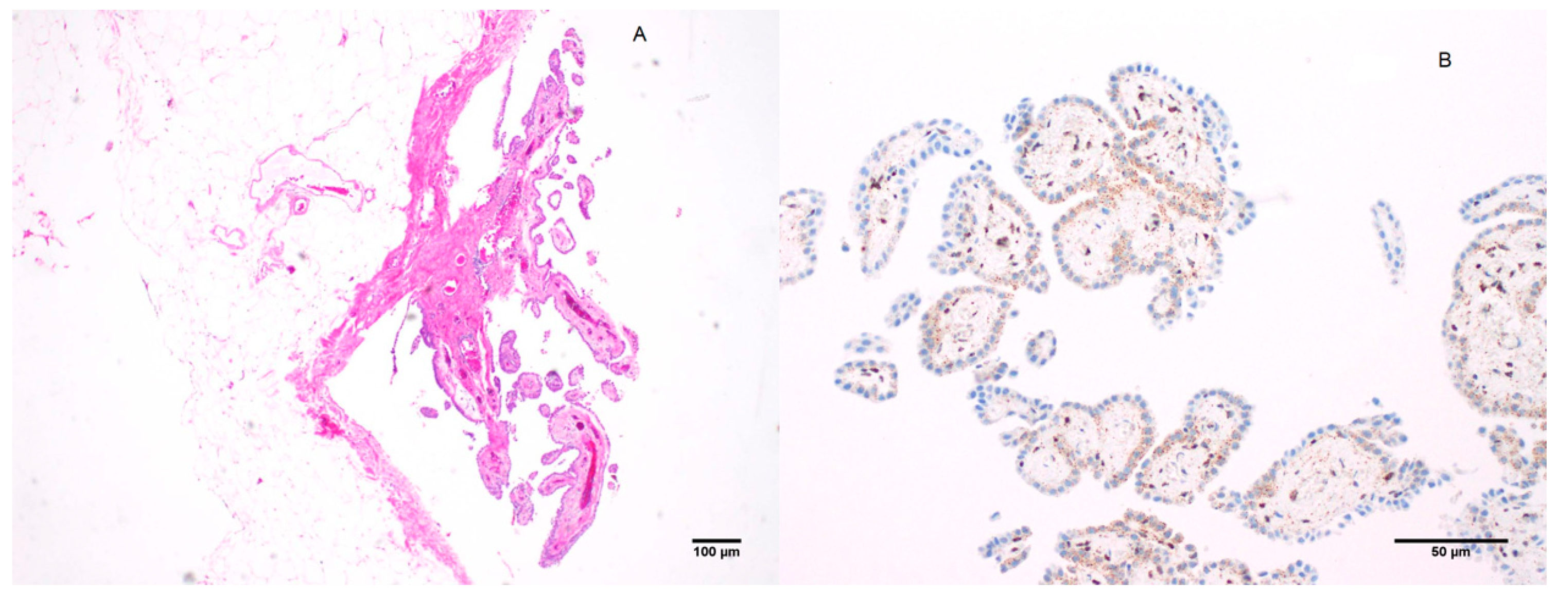
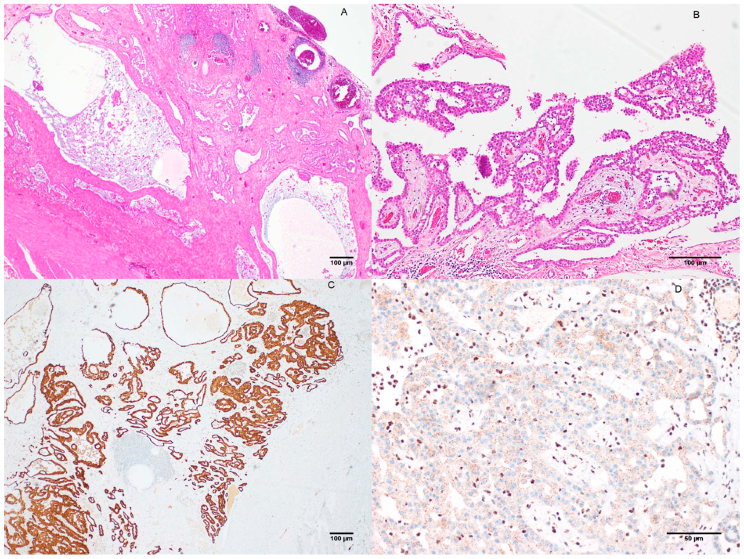
2. Case Presentation

The patient was a 68-year-old woman with a known connective tissue disorder (EDS), along with a history of asthma, hypertension, dyslipidaemia, and glaucoma. She had been exposed to asbestos during extensive home renovations nearly 35 years ago.
She presented with abdominal pain and bloating, with an ultrasound revealing fluid in the pelvis and diffuse intraperitoneal nodules. A laparoscopic biopsy revealed WDPMT described as a papillary lesion with papillary fronds lined by a single layer of bland and attenuated mesothelial cells with broad-based, club-shaped papillae and no invasion. Immunohistochemical labelling (IHC) for calretinin, WT1 and D2-40 (Figure 1) was positive. BAP1 IHC was not routinely available at that time, but we retrospectively demonstrated a loss of nuclear labelling for BAP1.
One year and seven months later, a CT scan revealed disease progression and the accumulation of ascitic fluid. An umbilical hernia repair and a diagnostic laparoscopy were performed, but an obtained biopsy was not interpretable due to poor tissue preservation and minimal material retention.
One month later, the patient was again symptomatic and laparoscopy with debulking was performed. Pathology now revealed widely invasive epithelioid malignant mesothelioma with omental deposits and infiltrating serosal surfaces of the gallbladder, uterus, large bowel and appendix. The tumour cells infiltrated in cords and gland-like tubular structures, but areas of complex surface papillary proliferation were also present. Nuclear expression for BAP1 was lost in the tumour cells (Figure 2).
The decision at the time for the patient was not to perform hyperthermic intraperitoneal chemotherapy (HIPEC). Thirteen months after the debulking procedure, the patient presented with right-sided pleural effusion, and a right VATS procedure with talc pleurodesis was performed, but no biopsy was available. Another ten months later, there was progressive disease with ascites and peritoneal nodularity on CT. One month later, she underwent six cycles of chemotherapy (Cisplatin and Pemetrexed), two years and two months after tumour debulking. Disease progression in the chest occurred 15 months after completion of her chemotherapy, and she was treated with immunotherapy (Nivolumab and Ipilimumab). The patient remains relatively well with CT showing improvement in the disease. This suggests a more indolent course of nearly six years.

3. Discussion

WDPMT has undergone changes in its definition from an indolent, non-invasive tumour found incidentally to symptomatic lesions that included those with ‘limited’ or superficial invasion [7,8,9] and more guarded prognoses. The current WHO classification introduces the term well-differentiated papillary mesothelial tumour, distinguishing it from (malignant by definition) mesothelioma. The detection of retained BAP1 in these tumours is regarded by the WHO as desirable but not an essential criterium for the diagnosis of WDPMT. The change in nomenclature reflects the difficulties in predicting a prognosis, with some lesions pursuing an indolent course whereas others behave in a malignant fashion. The WHO guidelines acknowledge the potential difficulty in differentiating WDPMT and mesothelioma in situ MIS. Loss of BAP is sufficient to diagnose MIS in cytologically bland mesothelial cells, given that BAP1 inactivation is considered an early molecular event in the development of mesothelioma in these tumours [10]. We suggest that WDPMT-like lesions that show a loss of BAP1 may be better categorised along with MIS. Ribeiro et al. presented a report on two siblings with BAP1 germline mutations and WDPMT, one with both pleural mesothelioma and peritoneal WDPMT who survived 12 years, and the other with a peritoneal WDPMT who was in remission in the follow-up period of the study [13]. Lee et al. [14] reported three cases with BAP1 loss in WDPMT, one with peritoneal WDPMT who developed mesothelioma after 9 years, the second case with pleural WDPMT who developed mesothelioma after 20 months and the third case with both pleural and peritoneal WDPMT, who developed mesothelioma at both sites after 7 months. Single-nucleotide polymorphism genomic microarray demonstrated a similar genetic profile in the WDPM and MM components, supporting a clonal relationship between them [14]. Only one of these three patients had exposure to asbestos. The functional significance of BAP1 loss in WDPMT in the development of malignant mesothelioma has not been definitively established, but given that it is regarded as an early event in mesothelioma development, caution is advised [15,16,17,18].
Here, we report a case of WDPMT with BAP1 loss at the time of diagnosis that progressed to malignant mesothelioma within two years. The presence of symptoms attributable to the lesion at the time of presentation was a finding that could have been regarded as worrisome and inconsistent with the diagnosis according to some definitions.
A single mesothelioma arising in the background of EDS has been reported, but the significance of EDS as a potential predisposing factor has not been established [19]. The EDS contributed to recurrent hernias and symptoms may have been attributed to hernias, rather than an intraabdominal tumour [20]. The relationship of conservatively diagnosed WDPMT with asbestos is not definitively established, and whilst past history of exposure to asbestos in patients with WDPMT has been noted in some but not all cases, once invasive mesothelioma is diagnosed, this becomes a moot point [7,8,12].
Our case emphasises the need to prospectively identify those lesions that are (more) likely to progress. There may be at least two subsets of papillary mesothelial tumours that must be distinguished, one representing conservatively defined ‘classical’ WDPMT with a relatively indolent course, and lesions that present with symptoms, show a loss of BAP 1 (and/or possibly MTAP) that potentially pursue a more aggressive clinical course. The morphology of a bland arborizing papillary lesion is in keeping with WDPMT, but the subset of WDPMT with a loss of BAP1 may represent an early form of malignant mesothelioma, akin to and indistinguishable from MIS.
Two recent studies suggest that BAP1 alterations do not occur in WDPMT and that these lesions are genetically distinct from malignant mesothelioma [21,22]. Sun et al. also reported that 94% of peritoneal WDPMT was labelled with PAX-8 on IHC [3]. Only limited molecular data is available, and further studies are required to clarify if these lesions (WDPMT and MIS) show distinct mutation profiles or molecular profiles aligning with conventional mesothelioma.
We suggest that the presence of symptoms attributable to the lesion, or a loss of nuclear expression of BAP1 (or MTAP) in a tumour morphologically indistinguishable from WDMT should raise concern, and that BAP1-deleted lesions may best be designated as ‘atypical papillary mesothelial proliferation with a loss of BAP1, close clinical follow up suggested if clinically indicated’ or similar. This is to facilitate early intervention, given the potential for tumour progression. The morphological pattern of WDPM likely represents part of a spectrum of papillary mesothelial lesions, whose behaviour depends on molecular profiles, rather than morphology alone. Larger series to study the significance of these factors are therefore urgently needed.

Author Contributions

Conceptualisation, S.K., K.J.O. and S.P.; ethics, S.P. and S.K.; Writing—review and editing, S.P., S.K., K.J.O. and M.H.; Methodology, M.H. and S.P. All authors have read and agreed to the published version of the manuscript.

Funding

This research was funded by the Professor Douglas Henderson AO bequest fund of Flinders University.

Institutional Review Board Statement

This work was approved by The Central Adelaide Local Health Network Human Research Ethics Committee (CALHN HREC) (approval number R20190415).

Informed Consent Statement

Written informed consent was obtained from the patient. A copy of the written consent and ethics approval for publication is available on request.

Conflicts of Interest

Professor Sonja Klebe prepares medicolegal reports for the courts of Australia on the diagnosis and causation of occupational lung disease outside of the submitted work. The authors report no other conflicts of interest in this work.

References

  1. Malpica, A.; Sant’Ambrogio, S.; Deavers, M.T.; Silva, E.G. Well-differentiated papillary mesothelioma of the female peritoneum: A clinicopathologic study of 26 cases. Am. J. Surg. Pathol. 2012, 36, 117–127. [Google Scholar] [CrossRef] [PubMed]
  2. Daya, D.; Elliott McCaughey, W. Well-differentiated papillary mesothelioma of the peritoneum. A clinicopathologic study of 22 cases. Cancer 1990, 65, 292–296. [Google Scholar] [CrossRef]
  3. Sun, M.; Zhao, L.; Lao, I.W.; Yu, L.; Wang, J. Well-differentiated papillary mesothelioma: A 17-year single institution experience with a series of 75 cases. Ann. Diagn. Pathol. 2019, 38, 43–50. [Google Scholar] [CrossRef] [PubMed]
  4. Bürrig, K.-F.; Pfitzer, P.; Hort, W. Well-differentiated papillary mesothelioma of the peritoneum: A borderline mesothelioma. Virchows Arch. A 1990, 417, 443–447. [Google Scholar] [CrossRef] [PubMed]
  5. Kim, M.; Kim, H.-S. Clinicopathological characteristics of well-differentiated papillary mesothelioma of the peritoneum: A single-institutional experience of 12 cases. In Vivo 2019, 33, 633–642. [Google Scholar] [CrossRef] [PubMed] [Green Version]
  6. World Health Organization Classification of Tumours. Pathology & Genetics. Tumours of the Lung, Pleura, Thymus and Heart; Travis, W.D., Brambilla, E., Müller-Hermelink, H.K., Harris, C.C., Eds.; IARC Press: Lyon, France, 2004; pp. 135–136. [Google Scholar]
  7. Butnor, K.J.; Sporn, T.A.; Hammar, S.P.; Roggli, V.L. Well-differentiated papillary mesothelioma. Am. J. Surg. Pathol. 2001, 25, 1304–1309. [Google Scholar] [CrossRef] [PubMed]
  8. Galateau-Sallé, F.; Vignaud, J.M.; Burke, L.; Gibbs, A.; Brambilla, E.; Attanoos, R.; Goldberg, M.; Launoy, G. Well-differentiated papillary mesothelioma of the pleura: A series of 24 cases. Am. J. Surg. Pathol. 2004, 28, 534–540. [Google Scholar] [CrossRef] [PubMed]
  9. Churg, A.; Allen, T.; Borczuk, A.C.; Cagle, P.T.; Galateau-Sallé, F.; Hwang, H.; Murer, B.; Murty, V.V.; Ordonez, N.; Tazelaar, H.D. Well-differentiated papillary mesothelioma with invasive foci. Am. J. Surg. Pathol. 2014, 38, 990–998. [Google Scholar] [CrossRef] [PubMed]
  10. World Health Organization. Thoracic Tumours, 5th ed.; International Agency for Research on Cancer: Lyon, France, 2021; Volume 5. [Google Scholar]
  11. Hoekman, K.; Tognon, G.; Risse, E.; Bloemsma, C.; Vermorken, J. Well-differentiated papillary mesothelioma of the peritoneum: A separate entity. Eur. J. Cancer 1996, 32, 255–258. [Google Scholar] [CrossRef]
  12. Jones, M.A.; Young, R.H.; Scully, R.E. Malignant mesothelioma of the tunica vaginalis. A clinicopathologic analysis of 11 cases with review of the literature. Am. J. Surg. Pathol. 1995, 19, 815–825. [Google Scholar] [CrossRef] [PubMed]
  13. Ribeiro, C.; Campelos, S.; Moura, C.; Machado, J.; Justino, A.; Parente, B. Well-differentiated papillary mesothelioma: Clustering in a Portuguese family with a germline BAP1 mutation. Ann. Oncol. 2013, 24, 2147–2150. [Google Scholar] [CrossRef] [PubMed]
  14. Lee, H.E.; Molina, J.R.; Sukov, W.R.; Roden, A.C.; Eunhee, S.Y. BAP1 loss is unusual in well-differentiated papillary mesothelioma and may predict development of malignant mesothelioma. Hum. Pathol. 2018, 79, 168–176. [Google Scholar] [CrossRef] [PubMed]
  15. Churg, A.; Galateau-Salle, F.; Roden, A.C.; Attanoos, R.; von der Thusen, J.H.; Tsao, M.S.; Chang, N.; De Perrot, M.; Dacic, S. Malignant mesothelioma in situ: Morphologic features and clinical outcome. Mod. Pathol. 2020, 33, 297–302. [Google Scholar] [CrossRef] [PubMed]
  16. Dacic, S.; Roy, S.; Lyons, M.A.; von der Thusen, J.H.; Galateau-Salle, F.; Churg, A. Whole exome sequencing reveals BAP1 somatic abnormalities in mesothelioma in situ. Lung Cancer 2020, 149, 1–4. [Google Scholar] [CrossRef] [PubMed]
  17. Pulford, E.; Henderson, D.W.; Klebe, S. Malignant mesothelioma in situ: Diagnostic and clinical considerations. Pathology 2020, 52, 635–642. [Google Scholar] [CrossRef] [PubMed]
  18. Zauderer, M.G.; Bott, M.; McMillan, R.; Sima, C.S.; Rusch, V.; Krug, L.M.; Ladanyi, M. Clinical characteristics of patients with malignant pleural mesothelioma harboring somatic BAP1 mutations. J. Thorac. Oncol. 2013, 8, 1430–1433. [Google Scholar] [CrossRef] [PubMed] [Green Version]
  19. Bisconti, M.; Bisetti, A.; Bidoli, P. Malignant mesothelioma in subjects with Marfan’s syndrome and Ehlers-Danlos syndrome: Only an apparent association? Respiration 2000, 67, 223–228. [Google Scholar] [CrossRef] [PubMed]
  20. Fikree, A.; Chelimsky, G.; Collins, H.; Kovacic, K.; Aziz, Q. Gastrointestinal involvement in the Ehlers–Danlos syndromes. Am. J. Med. Genet. Part C Semin. Med. Genet. 2017, 175C, 181–187. [Google Scholar] [CrossRef] [PubMed] [Green Version]
  21. Shrestha, R.; Nabavi, N.; Volik, S.; Anderson, S.; Haegert, A.; McConeghy, B.; Sar, F.; Brahmbhatt, S.; Bell, R.; Le Bihan, S. Well-Differentiated Papillary Mesothelioma of the Peritoneum is Genetically Distinct from Malignant Mesothelioma. Cancers 2020, 12, 1568. [Google Scholar] [CrossRef] [PubMed]
  22. Stevers, M.; Rabban, J.T.; Garg, K.; Van Ziffle, J.; Onodera, C.; Grenert, J.P.; Yeh, I.; Bastian, B.C.; Zaloudek, C.; Solomon, D.A. Well-differentiated papillary mesothelioma of the peritoneum is genetically defined by mutually exclusive mutations in TRAF7 and CDC42. Mod. Pathol. 2019, 32, 88–99. [Google Scholar] [CrossRef] [PubMed]
Figure 1. Well-differentiated papillary mesothelial tumour. WDPMT characterised by a papillary lesion with broad papillae and bland surface mesothelium. (A) H&E (Hematoxylin-Eosin stain) 4× magnification; (B) BAP1 IHC 20× magnification (retrospectively performed).
Jmp 02 00026 g001
Figure 2. Invasive mesothelioma of epithelioid type forming gland-like structures. (A) H&E (Hematoxylin-Eosin stain) 4× magnification. (B) Atypical mesothelial surface proliferation in close vicinity to invasive tumour 10× magnification. (C) Positive labelling for calretinin in invasive mesothelioma 4× magnification. (D) Loss of nuclear labelling for BAP1 in invasive mesothelioma, with retained nuclear labelling of inflammatory cells (internal control) 20× magnification.
Jmp 02 00026 g002
Publisher’s Note: MDPI stays neutral with regard to jurisdictional claims in published maps and institutional affiliations.

