1. Introduction
Being able to determine and predict the condition of the drinking water distribution network is crucial for optimal asset management. Pipe failure results from an interplay of, on the one hand, external and internal loads, and on the other hand, intrinsic material properties that determine the degradation- and failure mechanisms. In the Netherlands, asbestos cement (AC) still is an important pipe material: roughly 25% (around 30,000 km) of the Dutch drinking water distribution network consists of this material. This pipe material has a relatively high priority to be renewed and therefore received quite some attention when it comes to the development of condition assessments that help water utility experts to prioritize which of these pipes to replace first.
Key in assessing the condition of AC pipes is the development of non-destructive, in-line techniques to measure the remaining wall thickness: the part of the pipe wall that has not yet been degraded by processes such as leaching and chemical attack. Several promising techniques based on wave reflection have been piloted in The Netherlands since the early 2000s, such as ground penetrating radar [
1] and non-collinear wave mixing [
2]. The validation of these techniques has so far been an arduous task, however, dependent on cost-intensive destructive experiments such as phenolphthalein staining on lab-scale samples. As a result, detailed validation based on field samples of representative size has been out of reach.
X-ray computed tomography (CT) can be used to visualize spatial material differences in objects. While the development of this technique has been mainly driven by its invaluable use in medical diagnostics, it is also a useful tool for the non-destructive inspection of building materials. In particular, it has been shown that the technique is suitable for identifying the different components of cement-based materials [
3,
4] and that the technique can be used to visualize the material changes associated with cement degradation through leaching [
5]. CT should therefore prove to be a valuable tool to provide a more detailed view of AC pipe degradation.
The work presented here explores the potential of X-ray computed tomography to provide the detailed description of AC pipe degradation. Access to such information may lead to a better understanding of the degradation processes of asbestos cement pipes. Moreover, such information could be used to guide and validate the development of inspection techniques suitable for field use.
2. Materials and Methods
2.1. Asbestos Cement Pipes
In total, 28 uncoated asbestos cement pipe sections with diameters of 100 mm and with a length of approximately 1 m were collected from the drinking water distribution network by the water utilities Brabant Water, Dunea and WML. All pipe segments were scanned with CT.
2.2. X-ray Computed Tomography
CT images of the asbestos cement pipes were obtained with a medical scanner (Somatom Definition, Siemens, Malvern, USA). The electron beam was set to 250 mA and 140 kVp. Furthermore, the scanner was set to a pitch of 0.6 mm, a rotation time of 0.5 mm and a B50f kernel. With these settings, a cross-section was measured over every 1.5 mm of pipe length with a field of view of 150 mm × 150 mm and a resolution of 512 pixels in each direction (0.29 mm per pixel).
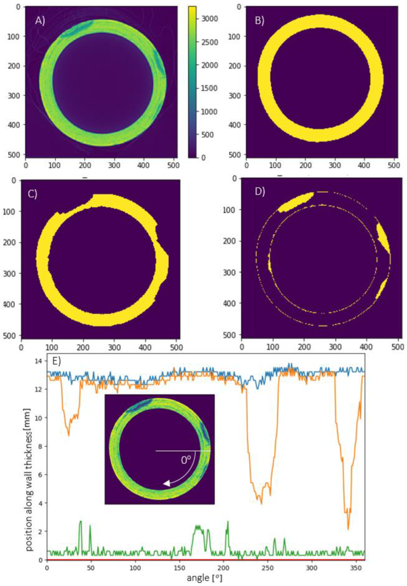
The resulting CT data took the form of a collection of slice images, each image depicting the material composition of a different cross section of the pipe. A custom image processing algorithm was written to translate the images of CT slices to profiles of degraded and undegraded material in terms of angles and millimeters. The algorithm was written in Python, using the publicly available libraries numpy, scikit-image and scipy. The basic approach of the algorithm is to use the grey values in the CT images to identify and distinguish the degraded and undegraded material. An illustration of an original CT slice, the most important steps of the image processing algorithm and the resulting profile data, can be seen in
Figure 1:
(a) The algorithm starts with the original slice, shown in
Figure 1A, with the color scale from the blue to yellow indicating low to high pixel grey values, corresponding to low and high radio densities, respectively;
(b) The silhouette of the pipe material is collected, from the pixels with a value above 1500 (
Figure 1B);
(c) The silhouette of the undegraded pipe material was collected by finding pixels with a value between 1500 and 2400 (
Figure 1C);
(d) The silhouette of the degraded material is constructed, obtained by subtracting c from b (
Figure 1D);
(e) Profiles of the original wall thickness are determined from b (
Figure 1E, blue) and the interfaces between the degraded and undegraded material are determined from d for internal degradation (
Figure 1E, green) and external degradation (
Figure 1E, orange). The arrow illustrates the relation between the angles on the x axis and the position in the original slice image.
3. Results
Degradation Patterns of AC
Once the CT slices of a field sample are processed and converted to the remaining wall thickness data as illustrated in
Figure 1, an overall impression of the pipe degradation can be constructed.
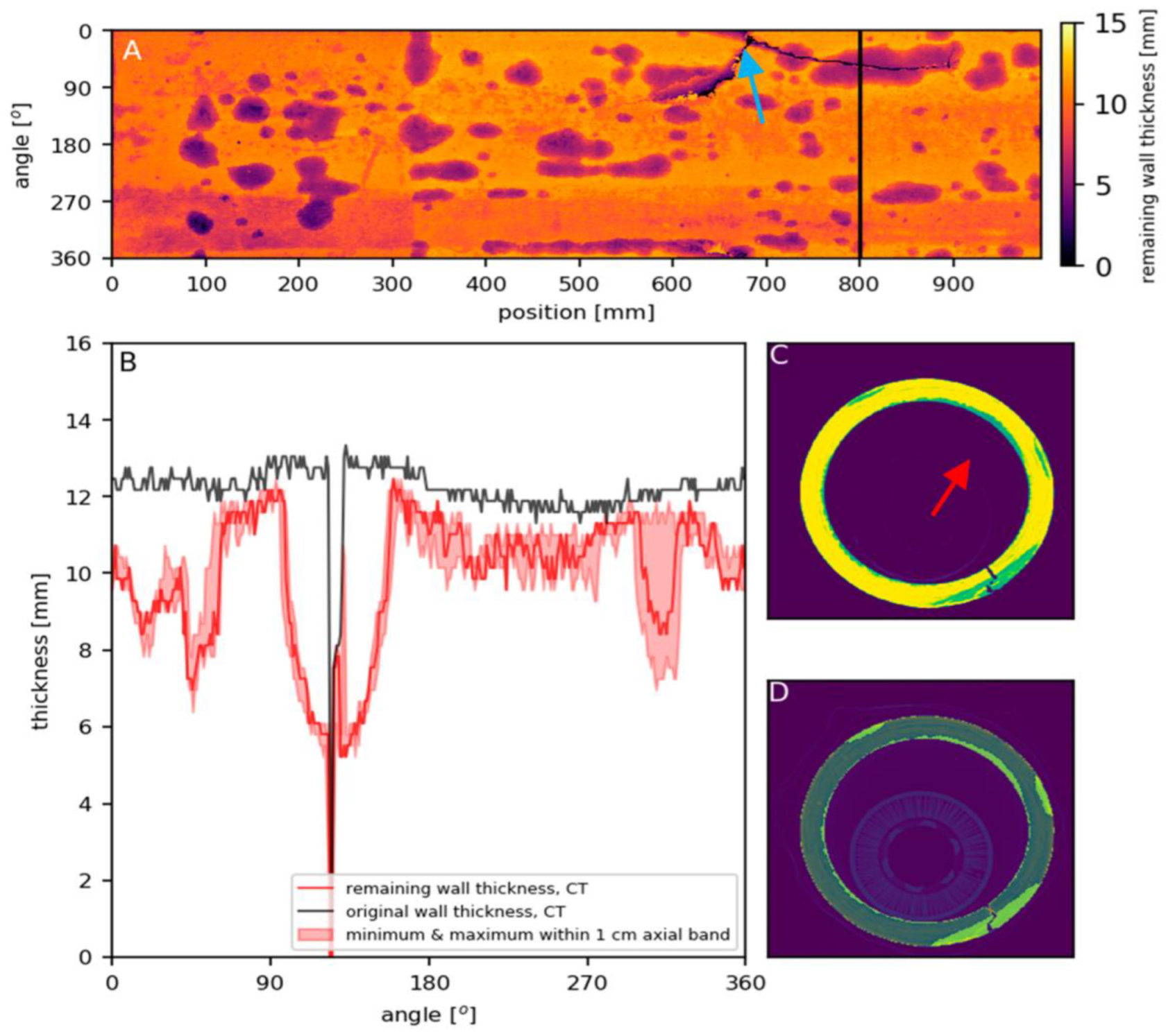
Figure 2 and
Figure 3 give two examples of the analyses made to summarize the information present in a complete CT scan.
Figure 2A and
Figure 3A display height maps that summarize the distribution of degradation across the pipe surface. Color represents the remaining wall thickness at a given position in the pipe as marked by the axis in terms of angle and distance along the length of the pipe segment. The lines of extremely low-value pixels clearly identify cracks in the pipe (blue arrow in
Figure 2A).
The black vertical lines in
Figure 2A and
Figure 3A mark the locations of specific slices in the respective CT scans that are shown in
Figure 2C and
Figure 3C (color scale from blue to yellow indicating low to high radio densities). In
Figure 2D and
Figure 3D, the highlighted areas mark the degraded material visible in the slice, as determined by the image-processing algorithm. The graphs show the profiles obtained from these slices for the wall thickness (
Figure 2B and
Figure 3B, black) and the remaining wall thickness (
Figure 2B and
Figure 3B, red).
Clear but distinct patterns of locations with reduced remaining wall thickness are recognizable for both pipes. The remaining wall thickness of the pipe in
Figure 2 displays a spotty pattern across the pipe surface, which corresponds to many localized areas of external degradation (also recognizable in the CT slices displayed in
Figure 1A and
Figure 2C). A band of reduced wall thickness is also visible across the full length of the pipe between 270° and 360°, which corresponds to an area of internal degradation (marked by the red arrow in
Figure 2C). In
Figure 3, the external degradation is drawn out in a thin band across the surface (the darker area marked by the green bracket in
Figure 3A). The internal degradation is present far more homogeneously across the surface than in
Figure 2, but again a pattern of bands of different levels of degradation across the pipe length is visible.
Presumably, the localized nature of the external degradation in
Figure 2 and
Figure 3 is related to the corresponding distributions of aggressive soil components around the pipe when the pipes were in use. The inhomogeneous nature of the internal degradation is more remarkable, however, since the leaching of calcium hydroxide from the AC pipes into the drinking water is typically understood to be a diffusion-controlled process that occurs homogeneously across the pipe surface [
6]. Nonetheless, the patterns described above were typical for the 32 pipes that were scanned in total. Thirteen pipes displayed inhomogeneous internal degradation, while only eight of the pipes displayed the expected pattern of homogeneous internal degradation. Nineteen of the pipes displayed inhomogeneous external degradation.
4. Conclusions
The CT scans provide the Dutch utilities with a new view on the degradation of asbestos cement pipes with an unprecedented level of detail and completeness. As a technique, CT is well understood, and when specifically applied to AC, the differences between the degraded and undegraded materials are clear enough to be identified and quantified with relatively basic image processing steps. This makes the CT measurements well suited for comparison with the data of less known inspection techniques that are still under development.
The observed degradation patterns show that inhomogeneous degradation is more common than previously thought for AC drinking water pipes. This raises the question of whether a rough impression of the general level of degradation of AC mains is sufficient for a correct estimation of the risk of failure, or that detailed knowledge of the presence of potential ‘weak links’ is required. Answering this question will have implications for the use of current condition models, which typically assume homogeneous degradation (e.g., [
7]) and may therefore currently overestimate the condition of AC pipes. Moreover, the answer to this question may determine the level of detail—in terms of resolution and accuracy—that water utilities should demand from inspection techniques in the field.
At the moment of writing, research efforts are directed towards the validation of current in-line inspection techniques for the Dutch drinking water utilities and towards supporting their further development and tuning to the context of drinking water pipes in the field. Future research should also focus on finding out how the inhomogeneous internal degradation of AC mains can come to pass.
Author Contributions
Conceptualization, K.v.L.; analysis, K.v.L.; writing, K.v.L; review and revision, C.Q. All authors have read and agree to the published version of the manuscript.
Funding
This study was partly funded within the context of the BTO collective research program of the Dutch drinking water sector, project numbers 400544-192 and 402045-092. The CT measurements were funded by the drinking water companies Brabant Water, Dunea, PWN, WMD and WML.
Acknowledgments
The authors are grateful to the Dutch drinking water companies Brabant Water, Dunea and WML for providing the pipe field samples on which the reported measurements were performed.
Conflicts of Interest
The authors declare no conflict of interest. The funders had no role in the design of the study; in the collection, analyses, or interpretation of data; in the writing of the manuscript, or in the decision to publish the results.
References
- Smolders, S.; Verhoest, L.; De Gueldre, G.; Van De Steene, B. Inspection of deteriorating asbestos cement force mains with georadar technique. Water Sci. Technol. 2009, 60, 995–1001. [Google Scholar] [CrossRef][Green Version]
- Hernandez Delgadillo, H.; Kern, B.; Loendersloot, R.; Yntema, D.; Akkerman, R. A methodology based on pulse-velocity measurements to quantify the chemical degradation levels in thin mortar specimens. J. Nondestruct. Eval. 2018, 37, 79–92. [Google Scholar] [CrossRef]
- Aruntas, H.Y.; Tekin, I.; Birgul, R. Determining Hounsfield unit values of mortar constituents by computerized tomography. Measurement 2010, 43, 410–414. [Google Scholar] [CrossRef]
- Wan, K.; Chen, L.Y.; Xu, Q. Calibration of grayscale values of cement constituents using industrial X-ray tomography. Sci. China Technol. Sci. 2015, 58, 485–492. [Google Scholar] [CrossRef]
- Wan, K.; Li, Y.; Sun, W. Application of tomography for solid calcium distributions in calcium leaching cement paste. Constr. Build. Mater. 2012, 36, 913–917. [Google Scholar] [CrossRef]
- Leroy, P.; Shock, M.R.; Wagner, I.; Holtschulte, H. Cement-based materials. In Internal Corrosion of Water Distribution Systems, 2nd ed.; AWWA: Denver, CO, USA, 1996. [Google Scholar]
- Wols, B.; Moerman, A.; Horst, P.; van Laarhoven, K. Prediction of pipe failure in drinking water distribution networks by Comsima. In Proceedings of the 3rd EWaS International Conference on “Insights on the Water-Energy-Food Nexus”, Lefkada Island, Greece, 27–30 June 2018. [Google Scholar]
| Publisher’s Note: MDPI stays neutral with regard to jurisdictional claims in published maps and institutional affiliations. |
© 2022 by the authors. Licensee MDPI, Basel, Switzerland. This article is an open access article distributed under the terms and conditions of the Creative Commons Attribution (CC BY) license (https://creativecommons.org/licenses/by/4.0/).


