Proceeding Paper

Peripheral Venous Simulator Development for Medical Training †

by
Pedro Escudero-Villa
1,*,
Jéssica Núñez-Sánchez
2 and
Jenny Paredes-Fierro
1
1
Facultad de Ingeniería, Universidad Nacional de Chimborazo, Riobamba 060108, Ecuador
2
Facultad de Ciencias Físicas, Universidad Complutense de Madrid, 28040 Madrid, Spain
*
Author to whom correspondence should be addressed.
Presented at the 5th International Electronic Conference on Applied Sciences, 4–6 December 2024; https://sciforum.net/event/ASEC2024.
Eng. Proc. 2025, 87(1), 2; https://doi.org/10.3390/engproc2025087002
Published: 6 February 2025
(This article belongs to the Proceedings of The 5th International Electronic Conference on Applied Sciences)

Abstract

The necessity to develop skills in medical training, from simple procedures such as sutures, venipunctures, and peripheral venous cannulations to complex surgeries, has driven innovation in the fabrication of medical simulators throughout history. These simulators are crafted using materials that mimic the physical and mechanical characteristics of human body parts, providing realistic training experiences. However, the costs associated with developing these simulators pose a significant challenge, especially for low-income areas. This work explores practical options for creating cost-effective and useful simulators by fabricating pieces that represent the forearm, a common site for venipunctures and peripheral venous cannulations. The fabrication process involved combining three types of materials: polydimethylsiloxane (PDMS), food-grade silicone, and Artesil Shore 20 silicone, along with a Foley catheter to simulate the arm veins. The compatibility of these materials was thoroughly evaluated to produce valid prototypes, ensuring that the stress ratios closely matched the properties of human tissue. Preliminary evaluations showed a good acceptability rating from users. Medical students who tested the simulators found them effective for explaining the behavior of fluids in the body during venoclysis simulations and recommended elaborating on the replication of more complex structures.

1. Introduction

The development of medical simulators has given rise to many innovations in the field of healthcare education, providing an interactive approach to training future professionals, as well as maintaining competencies over the years. These tools replicate real-life scenarios, mimicking human body systems and parts, creating a safe and controlled environment to improve skills. The area of application covers basic procedures to complex surgeries where simulators offer a hands-on learning experience to link the theoretical knowledge to practical procedures [1]. The precision and realism of medical simulators have improved, making them indispensable in current medical training programs by reducing the risk of errors in real patients. Regarding clinical training, there is an increasing necessity to limit traditional instruction methods such as patients’ direct physical contact, animal laboratories, and cadaveric dissection to avoid the ethical aspects of these procedures, prevent possible complications, and contain costs [2].
The common materials used to fabricate medical simulators are selected for their similarity to the human body’s look, feel, and function. Silicon-rubber-based materials offer an acceptable balance of durability, flexibility, and realism; moreover, these materials allow ease of fabrication [3,4]. The simulators are considered low- and high-fidelity devices, depending of the functions that each simulator develops; low-fidelity simulators cover basic procedures, such as venous cannulation, venipuncture, etc. [5,6,7]. High-fidelity simulators involve technological developments such as medical software, touch screens that show an anatomical 3D description of the human body, and mannequins that simulate a hospital environment [8,9,10]. These mannequins are capable of exhibiting symptoms of illnesses, such as sweating, convulsions, and bleeding, which can be treated with medical tools. The importance of this medical equipment has a great impact on the medical community, with new developments in the field of simulators emerging every year [11].
Low-cost simulators represent a development trend, where systems are simplified with portable characteristics and designed for single-use applications. Homemade reusable devices were proposed to be used in medical teaching through simulations of clamshell thoracotomy with 89.47% acceptability as a substitute tool for teaching [12], a laparoscopy training box with which it has been possible to improve the confidence of students when faced with surgery [13]. Many possibilities arise from using fruits for teaching procedures such as suturing, cannulation, and injection, as the structure of fruits imitates the soft characteristics of skin [14]. However, human skin is composed of tissue layers, which gives it a unique characteristic that is difficult to replicate directly with fruits or mannequins. For instance, bananas can simulate the feel of human skin for suturing practice [15,16], while oranges can replicate the structure of a breast for practicing fine-needle aspiration biopsies [17]. Grapes can simulate small tumors or cysts for practicing palpation and fine-needle aspiration techniques due to their size and consistency [18]. Apples can be used to simulate the human eye for practicing incision and drainage procedures, as well as suturing delicate tissues [19]. Tomatoes can simulate soft tissue masses for practicing excision and biopsy techniques due to their firm skin and soft interior [19,20]. Watermelons can be used to simulate the abdominal cavity and organs for practicing laparoscopic techniques, as their thick rind and soft interior offer realistic resistance and texture, also helping to model a caesarean section [21,22].
The characteristics of human tissue can also be approximated using raw chicken breast to practice suturing and incision techniques, as its texture closely resembles that of human muscle and skin [23,24]. Hot dogs can be used for practicing endotracheal intubation, as they mimic the resistance and feel of inserting a tube into the trachea [25]. Pork ribs can be used to simulate the human rib cage for practicing thoracic procedures such as chest tube insertion and clamshell thoracotomy [26]. The effectiveness of models used depends on the material characteristics, but most of the cited models are used for a unique or few essays due to the damage by material fatigue or degradation. Silicone-based materials are attractive to fabricate simulators due to flexibility and resistance to tearing by repetitive injection, and they are favorable for suturing in training scenarios.
Motivated to provide a practical solution for medical simulator training, this study aims to fabricate a peripheral venous cannulation simulator with low-cost materials and medical supplies used on a daily basis, aiming enhance confidence performing such procedures with patients. In this work, we detail all the steps to obtain the simulator piece, from assembly to covering and drying of the piece. As the main material of the piece, we used different types of silicone to verify the compatibility of the materials for the piece’s covering with those in the frame. The reason for this was that, in some cases, material incompatibility (inhibition) could occur. The final simulator piece was assessed by medicine students, and we measured their opinions about the usage, characteristics, and appearance of the piece by conducting a survey.

2. Materials and Methods

For the fabrication of a peripheral venous cannulation simulator of the arm, we searched for materials that matched the original in shape, size, and function, considering body anatomy. We fabricated an inexpensive piece for peripheral venous cannulation based on an original forearm simulator. We tested the compatibility of various materials used for the frame and the covering. Figure 1 shows a flowchart describing the entire process for elaborating the peripheral venous simulator piece.

2.1. Materials and Compatibility

We used low-cost materials, combining Do-It-Yourself (DIY) materials, medical supplies, and elastomers, most of which were utilized on a daily basis. For each material employed in the procedure, it was necessary to consider its chemical properties and characteristics. The coating of the piece needed to resemble real skin, being flexible, soft, and allowing easy needle penetration. Based on these requirements, three materials (polydimethylsiloxane (PDMS), alimentary silicone, and Artesyl 20 silicone) were used, each with distinct properties. Additionally, forearm veins were represented by Foley catheters of various sizes (12, 14, 16, and 18), as their diameters (2 mm to 4 mm) closely matched real veins [27,28].
To evaluate the compatibility, we tested the casting of polymers on the Foley catheter surfaces, choosing the ones that completed the cast correctly. Table 1 shows the combination of materials used to test the compatibility.

2.2. Fabrication of the Simulator Piece

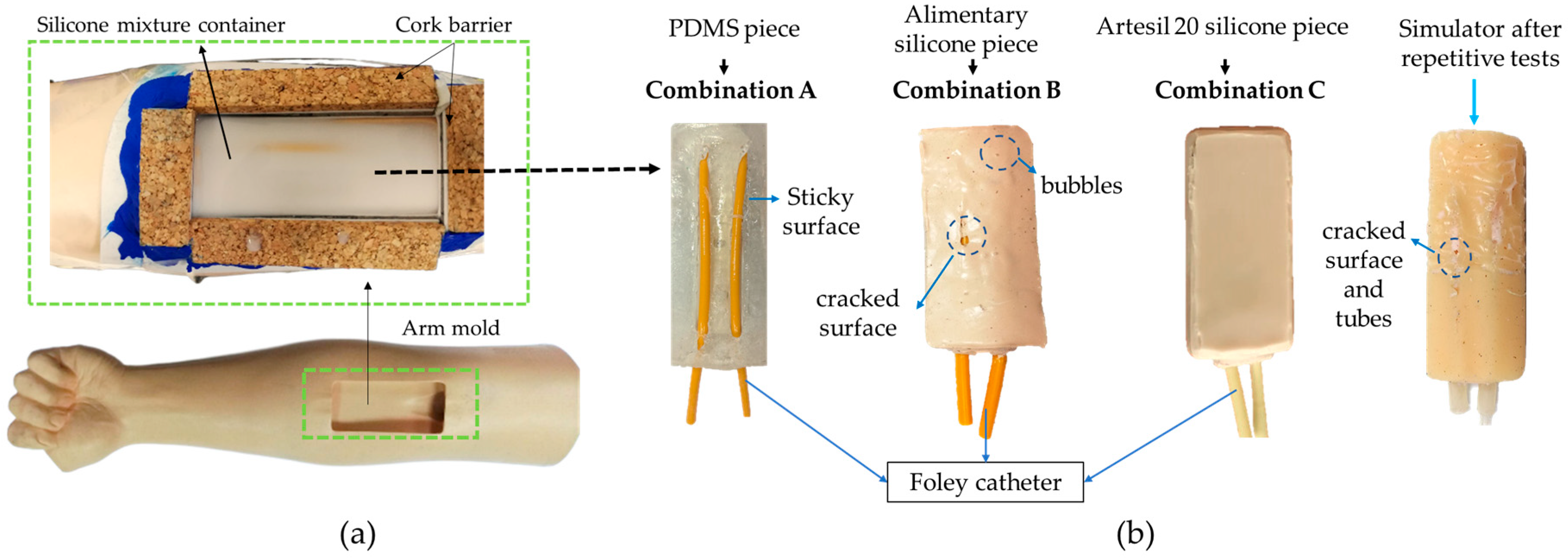
To carry out the fabrication of the simulator piece, the elements were cleaned with 95% ethanol and dried using nitrogen. The Foley catheter was securely fixed to a uniform flat base to prevent movement when the polymers were poured over it. Following Table 1, the combined materials were prepared and poured into three different setups, as shown in Figure 2a.
PDMS was prepared using a standard recipe with a 10:1 ratio of PDMS to crosslinker. After removing bubbles, the mixture was cured in an oven at 80 °C for 2 h.
Alimentary silicone was prepared by weighing 80 g of component A and 80 g of component B. Both components were poured into a vessel and mixed thoroughly. The mixture was cured at room temperature (~18 °C) for 8 h. To achieve a skin-like coloration, pigments were added: 2 g of rose pigment, 8 g of brown pigment, and 10 g of white pigment.
Artesyl 20 silicone was prepared by weighing 200 g of the silicone and pouring it into a vessel. Then, 3 g of brown pigment for silicone and 80 drops of catalyst were added. The mixture was stirred until uniform and left at room temperature for 4 h to complete curing.

2.3. Testing the Simulator Piece

To test the prototype, we used an arm mold and an intravenous infusion set. We performed standard tests that included injection, cannulation, drug delivery, and flow control. We also analyzed the similarity of the material’s texture and tested the hardness of the materials, comparing it with the values of human skin, especially regarding the force used to inject and manipulate the intravenous set. Additionally, we analyzed the material’s damage due to repetitive use.

2.4. Evaluating the Use of the Peripheral Simulator

To assess the acceptability of the simulator, we conducted a survey with a population of 116 students and 5 professors from the medical field, as they frequently use this type of simulator in their daily practice. The goal was to gather feedback on their understanding of the puncture and peripheral venous cannulation process, particularly in identifying the anatomy, shape, and characteristics of the components that make up the system, as well as the steps involved in performing the procedure. The survey aimed to collect responses and recommendations that could help refine and improve the simulator model.

3. Results and Discussion

3.1. Compatibility of Materials with Silicone

After comparing the different simulator pieces, we observed incompatibilities between the Foley catheter and the PDMS (Combination A) and alimentary silicone (Combination B) coverings. In Combination A, the surface remained sticky and humid, resulting in incomplete coverage and hard, inflexible areas. This made PDMS unsuitable for venipuncture simulators. Combination B, despite being considered ideal for simulating skin, also failed to dry properly, with bubbles on the surface and sticky areas near the veins. The presence of inhibition was evident in both combinations, caused by the materials in the Foley catheter. However, Combination C, using Artesyl 20 silicone, demonstrated excellent drying, complete coverage, and compatibility with all simulator components. This combination proved to be the most effective for creating a functional venipuncture simulator. Figure 2b shows the various pieces, each with unique characteristics such as bubbles and cracks. Incompatibility between materials, such as in the case of amines, chemical substances, and materials that contain sulfur, all of which are considered strong inhibitors, produces curing failures.

3.2. Testing the Simulator

With the simulator from Combination C (Artesil 20 silicone piece), we conducted tests to ensure its proper functionality for practical procedures. The simulator clearly distinguishes the flexible, soft covering (epidermis) from the Foley catheter representing veins. The covering withstands punctures from surgical needles, and catheters ranging from 18 G to 24 G effectively pierced both the silicone and Foley catheter. A key consideration was ensuring the catheter needle entered the silicone easily, enabling realistic medical practice. The texture of the simulator piece was approximately similar to human skin when injecting with a green needle due to the similar Young’s modulus. The simulator exhibited small liquid dripping after 120 tests/punctures, demonstrating reasonable resistance of the silicone to repetitive use.

3.3. Measuring Opinions About the Simulator

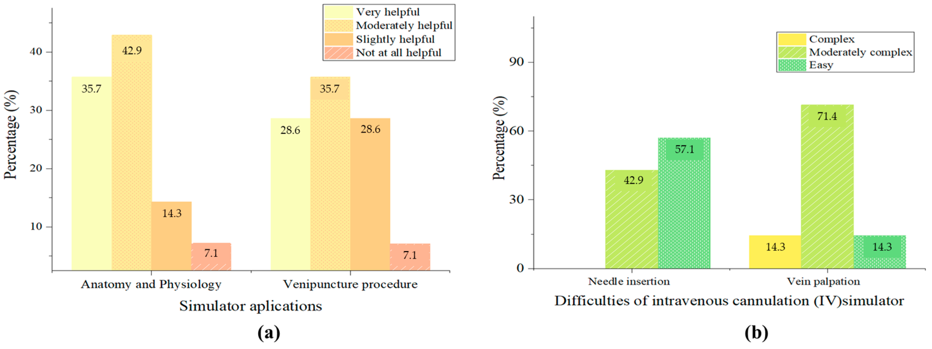
We fabricated five prototypes and evaluated them through a survey completed by medical students during their regular training. The survey revealed that all students were familiar with simulators, with 50% using them monthly. Figure 3 illustrates the frequency of simulator use in medical practices. Most students agreed that simulators provide an excellent way to develop medical skills, reflecting their positive experiences and perceived benefits in training. This feedback highlights the importance of simulators in enhancing practical abilities and ensuring effective preparation for medical procedures.
The vein palpation was considered moderately complex due to the level of difficulty in identifying the vein in the simulator. The medical teacher viewed this characteristic as essential for helping students train to identify the features of veins through palpation.

3.4. Low-Cost Materials and Quality

The quality of the raw materials is crucial when fabricating the simulator, as durability and aesthetic appearance are key characteristics. These devices should have a long lifespan without quickly deteriorating. The term “low-cost” should not be confused with “bad quality”. In this study, the pieces are considered low-cost because they were produced in a basic laboratory with wholesale materials, making the costs affordable. The materials used are versatile and commonly used in daily applications, not limited to medical simulators. However, PDMS, a more expensive material, was found to be incompatible with catheter materials and silicones.

3.5. Helpfulness of the Simulator Piece at Medical Practices

Medical practice for the training of medical professionals. Therefore, using medical simulators is vital for building students’ confidence in performing peripheral venous cannulation and other complex procedures. These simulators also help students understand internal body processes. The simulator we created has had a positive impact on students and medical professionals, as most survey respondents considered it an effective training tool. However, students noted that the simulator could be more flexible to ease needle insertion, although it is already flexible. Alimentary silicone, with a Young’s modulus of 4.2 MPa, is more elastic than Artesyl silicone, which has a modulus of 3.724 MPa. However, latex covering the Foley catheter hose does not accept alimentary silicone, but it does accommodate Artesyl silicone. This suggests the importance of selecting materials with a Young’s modulus similar to that of human skin (4.6 MPa) for better flexibility. The choice of peripheral venous catheter depends on vein size, procedure type (e.g., IV isotonic therapies, surgeries, transfusions), and flow volume. Most students used a 22 G catheter, the most common size for fluid therapy and IV medication. The simulator is designed to accommodate various catheter sizes for comprehensive training.

4. Conclusions

This study was focused on creating a peripheral venous simulator using easily accessible materials like silicone and other elastomers. It demonstrated that material compatibility is key for producing effective medical training tools. Artesyl 20 silicone combined with a drying catalyst was an ideal choice due to its ability to blend well with latex and cork without causing inhibition. The simulator successfully supported venous practices like venoclysis and cannulation. The survey results suggest that materials with a similar Young’s modulus to skin could improve flexibility. This cost-effective method offers a viable solution for medical training, and future research could explore using silicone paste and alginates. The simulator facilitates training for various procedures, including sutures, incisions, and needle insertion.

Author Contributions

Conceptualization, P.E.-V.; methodology, P.E.-V.; software, J.N.-S.; validation, P.E.-V. and J.P.-F.; formal analysis, P.E.-V.; investigation, P.E.-V. and J.N.-S.; resources, P.E.-V.; data curation, J.N.-S. and J.P.-F.; writing—original draft preparation, J.N.-S. and P.E.-V.; writing—review and editing, P.E.-V.; visualization, J.P.-F.; supervision, P.E.-V. and J.P.-F.; project administration, P.E.-V.; funding acquisition, P.E.-V. All authors have read and agreed to the published version of the manuscript.

Funding

This work was partially supported by funding from Vicerrectorado de Investigación, Universidad Nacional de Chimborazo (VIVP Project), Ecuador; and Universidad Indoamérica, Ecuador (CICHE Project).

Institutional Review Board Statement

Not applicable.

Informed Consent Statement

Not applicable.

Data Availability Statement

Data are contained within the article.

Conflicts of Interest

The authors declare no conflicts of interest.

References

  1. Sullivan, N.; Swoboda, S.M.; Breymier, T.; Lucas, L.; Sarasnick, J.; Rutherford-Hemming, T.; Budhathoki, C.; Kardong-Edgren, S.S. Emerging Evidence Toward a 2:1 Clinical to Simulation Ratio: A Study Comparing the Traditional Clinical and Simulation Settings. Clin. Simul. Nurs. 2019, 30, 34–41. [Google Scholar] [CrossRef]
  2. Jones, F.; Passos-Neto, C.; Melro Braghiroli, O. Simulation in Medical Education: Brief History and Methodology. PPCRJ 2015, 1, 56–63. [Google Scholar] [CrossRef]
  3. Izdihar, K.; Abdul Razak, H.R.; Supion, N.; Karim, M.K.A.; Osman, N.H.; Norkhairunnisa, M. Structural, Mechanical, and Dielectric Properties of Polydimethylsiloxane and Silicone Elastomer for the Fabrication of Clinical-Grade Kidney Phantom. Appl. Sci. 2021, 11, 1172. [Google Scholar] [CrossRef]
  4. Duran, M.M.; Moro, G.; Zhang, Y.; Islam, A. 3D Printing of Silicone and Polyurethane Elastomers for Medical Device Application: A Review. Adv. Ind. Manuf. Eng. 2023, 7, 100125. [Google Scholar] [CrossRef]
  5. Cerra, C.L.; Dante, A.; Caponnetto, V.; Franconi, I.; Gaxhja, E.; Petrucci, C.; Alfes, C.M.; Lancia, L. Effects of High-Fidelity Simulation Based on Life-Threatening Clinical Condition Scenarios on Learning Outcomes of Undergraduate and Postgraduate Nursing Students: A Systematic Review and Meta-Analysis. BMJ Open 2019, 9, e025306. [Google Scholar] [CrossRef]
  6. Corvetto, M.; Bravo, M.P.; Montaña, R.; Utili, F.; Escudero, E.; Boza, C.; Varas, J.; Dagnino, J. Simulación en educación médica: Una sinopsis. Rev. Méd. Chile 2013, 141, 70–79. [Google Scholar] [CrossRef] [PubMed]
  7. Motola, I.; Devine, L.A.; Chung, H.S.; Sullivan, J.E.; Issenberg, S.B. Simulation in Healthcare Education: A Best Evidence Practical Guide. AMEE Guide No. 82. Med. Teach. 2013, 35, e1511–e1530. [Google Scholar] [CrossRef]
  8. Zhou, G.; Nagle, A.; Takahashi, G.; Hornbeck, T.; Loomis, A.; Smith, B.; Duerstock, B.; Yu, D. Bringing Patient Mannequins to Life: 3D Projection Enhances Nursing Simulation. In Proceedings of the 2022 CHI Conference on Human Factors in Computing Systems, New Orleans, LA, USA, 29 April–5 May 2022; Association for Computing Machinery: New York, NY, USA, 2022; pp. 1–15. [Google Scholar]
  9. Martin, C.; Boissy, P.; Hamel, M.; Lebel, K. Instrumented Pre-Hospital Care Simulation Mannequin for Use in Spinal Motion Restrictions Scenarios: Validation of Cervical and Lumbar Motion Assessment. Sensors 2024, 24, 1055. [Google Scholar] [CrossRef]
  10. Rash, I.; Shi, K.; Sussel, R.; Smith, R.; Thompson, S.; Cai, E.; Nguyen, T.D.; Cheng, J.; Gong, C.; Farahmand, S.; et al. Virtual 3D Simulation Technology for Interprofessional Team Training. 2024. Available online: https://journals.sagepub.com/doi/full/10.1177/10468781231222969 (accessed on 23 May 2024).
  11. Larraga-García, B.; Quintana-Díaz, M.; Gutiérrez, Á. Simulation-Based Education in Trauma Management: A Scoping Review. Int. J. Environ. Res. Public Health 2022, 19, 13546. [Google Scholar] [CrossRef] [PubMed]
  12. Clifford, E.; Stourton, F.; Willers, J.; Colucci, G. Development of a Low-Cost, High-Fidelity, Reusable Model to Simulate Clamshell Thoracotomy. Surg. Innov. 2023, 30, 739–744. [Google Scholar] [CrossRef]
  13. Seltzer, H.; Swayze, E.; Thottathil, L.; Dewey, J.; Jabara, J.; Mehta, A.; Frederick, J.; Yousif, P.; Parikh, S.; Tsuei, A.; et al. The Impact of Homemade Laparoscopic Box Trainers on Medical Student Surgical Skills: A Randomized Control Pilot Study. Surg. Innov. 2023, 30, 84–93. [Google Scholar] [CrossRef]
  14. Sorribes Del Castillo, J.; Fernández-Gallego, V.; Sinisterra Aquilino, J.A. Un modelo nuevo, sencillo, económico y reutilizable para el aprendizaje y práctica de la canalización ecoguiada de vías centrales. Educ. Médica 2016, 17, 74–79. [Google Scholar] [CrossRef]
  15. Ohn, M.H.; Ohn, K.M.; Souza, U.D.; Yusof, S.; Ariffin, Z. Banana Peel: Is It Useful for Surgical Suturing Training? J. Phys. Conf. Ser. 2019, 1358, 012018. [Google Scholar] [CrossRef]
  16. Wong, K.; Bhama, P.K.; d’Amour Mazimpaka, J.; Dusabimana, R.; Lee, L.N.; Shaye, D.A. Banana Fruit: An “Appealing” Alternative for Practicing Suture Techniques in Resource-Limited Settings. Am. J. Otolaryngol. 2018, 39, 582–584. [Google Scholar] [CrossRef]
  17. Alcaraz-Mateos, E.; Exposito-Afonso, I.J.; Labiano-Miravalles, T.; Pijuan, L.; Temprana-Salvador, J.; Zhao, Q.; Jiang, X. “Sara” How Do Cytopathologists Learn Fine Needle Aspiration Techniques? An International Survey. Cytopathology 2022, 121, 127–131. [Google Scholar] [CrossRef]
  18. Schwartz, C.M.; Ivancic, R.J.; McDermott, S.M.; Bahner, D.P. Designing a Low-Cost Thyroid Ultrasound Phantom for Medical Student Education. Ultrasound Med. Biol. 2020, 46, 1545–1550. [Google Scholar] [CrossRef]
  19. Zhang, Z.; Li, S.; Sun, L.; Yan, W.; Huang, L.; Lu, J.; Wang, Q.; Li, M.; Zheng, D.; Liu, Y.; et al. Skills Assessment after a Grape-Based Microsurgical Course for Ophthalmology Residents: Randomised Controlled Trial. Br. J. Ophthalmol. 2023, 107, 1395–1402. [Google Scholar] [CrossRef]
  20. AL-Bayaty, F.H.; Hidayat, M.F.; Ariffin, F.; Noor, E.; Masud, M.; Ariffin, M.H.B.Z.; Zahari, H.I.M.; Adam, F.A. Fruits and Vegetables: A Cost-Effective Practical Solution in Periodontal Pre-Clinical Surgical Training for Postgraduate Students. J. Int. Dent. Med. Res. 2021, 14, 1498–1502. [Google Scholar]
  21. Valencia, P.; Turner-Lawrence, D. The Gravid Watermelon: An Inexpensive Perimortem Caesarean Section Model. J. Educ. Teach. Emerg. Med. 2019, 4. [Google Scholar] [CrossRef]
  22. Watson, G.; Niang, L.; Chandresekhar, S.; Natchagande, G.; Payne, S.R. The Feasibility of Endourological Surgery in Low-Resource Settings. BJU Int. 2022, 130, 18–25. [Google Scholar] [CrossRef]
  23. Denadai, R.; Saad-Hossne, R.; Martinhão Souto, L.R. Simulation-Based Cutaneous Surgical-Skill Training on a Chicken-Skin Bench Model in a Medical Undergraduate Program. Indian. J. Dermatol. 2013, 58, 200–207. [Google Scholar] [CrossRef] [PubMed]
  24. Zyluk, A.; Szlosser, Z.; Puchalski, P. Undergraduate Microsurgical Training: A Preliminary Experience. Handchir. Mikrochir. Plast. Chir. 2019, 51, 477–483. [Google Scholar] [CrossRef] [PubMed]
  25. Rathbun, K.M.; Brader, W.T.; Norbury, J.W. A Simple, Realistic, Inexpensive Nerve Phantom. J. Ultrasound Med. 2019, 38, 2203–2207. [Google Scholar] [CrossRef] [PubMed]
  26. Fortes, H.M.S.; Barros, P.P.; e Silva, B.M.d.C.; de Lyra Costa, J.M.; de Mendonza, A.J.X.; Feitosa, Y.K.D.S.; Filho, K.M.S.; Teixeira, E.G.; dos Santos França, V. Chest Tube Simulation: Experience Report and Brief Review. Res. Soc. Dev. 2022, 11, e173111032664. [Google Scholar] [CrossRef]
  27. Varela, D.R. Elías Venas Superficiales Del Antebrazo Estudio Anatomoquirúrgico Orientado a Los Angioaccesos Para Hemodiálisis Crónica. Cir. Urug. 1992, 40–44. [Google Scholar]
  28. Corzo-Gómez, E.G.; Gómez-Díaz, O.L.; Serrano-Gómez, S.; Saavedra-Martínez, M.; Rojas-Arenas, L.C. Elementos Morfológicos Implicados en la Enseñanza de las Competencias para Lograr una Adecuada Venopunción Periférica. Int. J. Morphol. 2018, 36, 159–163. [Google Scholar] [CrossRef]
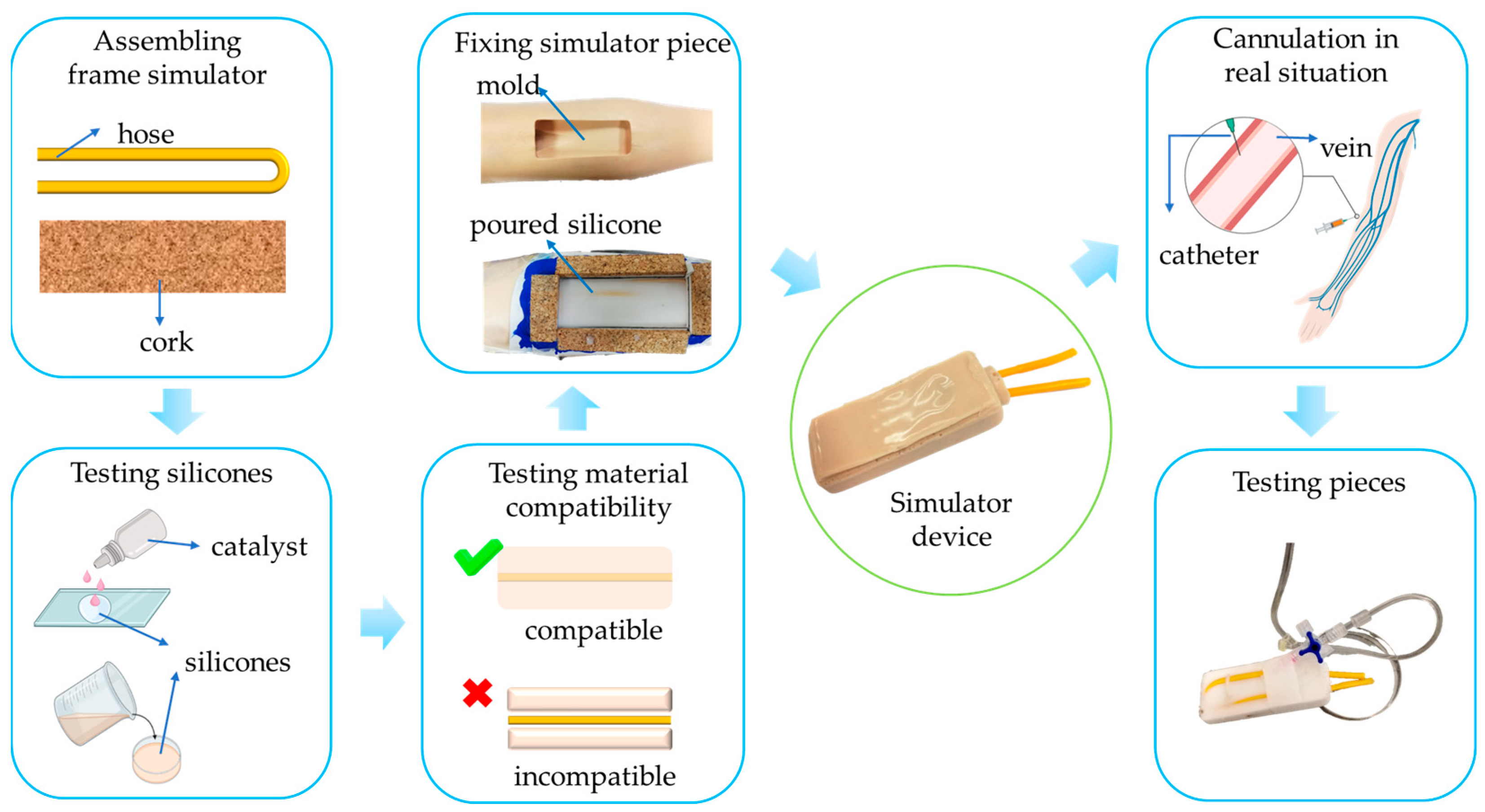
Figure 1. Flowchart to fabricate and evaluate the piece of a peripheral venous cannulation simulator.
Engproc 87 00002 g001
Figure 2. (a) Arm mold for the peripheral simulator fabrication and (b) curing failures produced due to the incompatibility of materials in all three cases compared.
Engproc 87 00002 g002
Figure 3. Testing opinions about simulations with the simulator piece. (a) Percentage of constitution and venipuncture procedure with the simulator piece. (b) Difficulties in needle insertion and vein palpation.
Engproc 87 00002 g003
Table 1. Material combinations.
IdentifierCombinations
A(PDMS + Foley catheter)
B(Alimentary silicone + Foley catheter)
C(Artesyl 20 silicone + Foley catheter)
Disclaimer/Publisher’s Note: The statements, opinions and data contained in all publications are solely those of the individual author(s) and contributor(s) and not of MDPI and/or the editor(s). MDPI and/or the editor(s) disclaim responsibility for any injury to people or property resulting from any ideas, methods, instructions or products referred to in the content.