Abstract
Electrophysiological studies reveal significant organizational and functional differences in the cortex of blind individuals compared to sighted individuals. These differences result from the nervous system’s reorganization to adapt to new sensory modalities used in daily life. Cortical visual prostheses offer a means to restore visual sensations in blind individuals by generating phosphenes, luminous perceptions that provide information about their surroundings. This study investigates the cortical changes associated with the use of a visual neuroprosthesis, focusing on how the brain adapts to the restored visual input. Our findings aim to contribute to understanding neuroplasticity in sensory restoration processes.
1. Introduction
Human evolution has prioritized the integrated processing of multisensory information, facilitated by dynamic neural networks that are continuously modulated by experience and neurobiological development. This multisensory integration enhances the accuracy and relevance of perception, providing redundancy in sensory inputs for a more comprehensive understanding of the environment [1,2,3]. Moreover, this integrative organization underlies compensatory behaviors observed after sensory loss. For instance, blind individuals exhibit neurocognitive adaptations, reorganizing brain areas to optimize other sensory modalities and improve interaction with their surroundings [4,5].
Evidence from human and animal studies indicates that these adaptations occur at multiple levels of the brain, affecting regions associated with the remaining senses and those traditionally linked to the lost modality [6]. In blind individuals, neurophysiological changes include modifications in somatosensory and auditory processing, as well as the functional recruitment of the occipital cortex for non-visual tasks [7,8,9]. These adaptations often enhance the efficiency of information processing, highlighting the brain’s remarkable plasticity [10].
Visual neuroprostheses are emerging technologies designed to restore visual processing in individuals with vision loss by electrically stimulating intact visual structures in the brain [11]. Cortical visual neuroprostheses can evoke the perception of phosphenes—luminous sensations that convey visual information [11]. Despite significant technological advances, these devices require further research to understand their interaction with the brain’s adaptive mechanisms [12]. Prolonged visual deprivation in blind individuals fosters optimized multimodal sensory processing. Consequently, the reintroduction of visual information through neuroprostheses often results in initially limited meaningful perception [13]. However, psychophysical tests, training therapies, and repeated cortical stimulation can progressively enhance the brain’s ability to process artificial visual inputs [11].
The success of functional vision restoration depends on the neuroprosthetic technology and the brain’s capacity for readaptation. Research suggests that intermodal brain reorganization in blind individuals can be partially reversed even after extended periods of blindness [14]. However, neuroplasticity is a gradual process, and the quality of recovered perception varies widely.
This study explores cortical connectivity changes in users of cortical visual neuroprostheses. Using resting-state electroencephalography over a six-month period, we analyzed connectivity patterns to investigate how the brain adapts to restored visual inputs. Resting-state activity provides insights into neural organization, cognitive functions, and sensory processing [15,16]. We hypothesize that brain readaptation to the restored sensory modality involves gradual changes in resting-state connectivity, reflecting neuroplasticity. This longitudinal analysis lays the foundation for understanding multimodal sensory integration dynamics and fostering the development of therapies to enhance neuroplasticity and optimize sensory restoration mediated by neuroprosthetics.
2. Methods
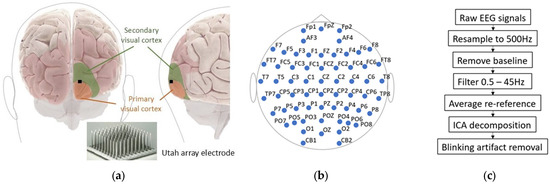
This study was conducted on two blind subjects (males aged 65 and 61 years) who were implanted with an intracortical microelectrode array (Utah Electrode Array, UEA) consisting of 96 electrodes in the visual cortex for a 6-month period. These implants are part of the procedures adopted for the implementation of a cortical visual neuroprosthesis, which is still under research and development within the CORTIVIS project (www.cortivis.org). In both users, the UEA was implanted in the right occipital cortex, near the occipital pole and close to the boundary between V1 and V2 (Figure 1a). All experimental, surgical, and pre-surgical procedures were conducted according to a protocol approved by the Clinical Research Committee of the General University Hospital of Elche and registered in ClinicalTrials.gov (NCT02983370). Furthermore, all relevant ethical guidelines pertaining to clinical trial regulations (EU No. 536/2014, repealing Directive 2001/20/EC), the Declaration of Helsinki, and European Commission Directives (2005/28/EC and 2003/94/EC) were followed. Written informed consent was obtained before any procedures were performed as part of this study.

Figure 1.
Experimental setup. (a) Approximate region where UEA was implanted in both subjects. (b) Location of EEG recording electrodes. Notably, Fp1 position was omitted from analyses due to high levels of noise and artifacts found in signal during recordings. (c) Pipeline used for removing blink artifacts.
UEA implants were performed on different dates, ensuring that the experimental periods for both subjects did not overlap. Over the six months of experimentation, the users underwent various microstimulation protocols at a frequency of 5 times per week, with sessions lasting 4 h each. The electrical microstimulations were administered using a multichannel current stimulation system, Cerestim (Blackrock Microsystems Inc., Salt Lake City, UT, USA). The stimulation primarily consisted of trains lasting 166 ms, with biphasic pulses (−/+) of 0.4 ms per phase, at a frequency of 300 Hz. The amplitudes of the applied stimuli ranged from 5 µA to 160 µA. In some experimental sessions, these parameters varied. Many of the methods and experimental procedures implemented in these two volunteers are described in greater detail in [11].
Electroencephalogram (EEG) signals were recorded in the resting state for 2–5 min before and after daily experimentation. These sessions were conducted at least once every 30 days over the six-month period. EEG recordings were obtained using a 64-channel NeuroScan SynAmps acquisition system (Compumedics, Charlotte, NC, USA) (Figure 1b) with a sampling rate of 1000 Hz. The electrode impedances were maintained below 25 kΩ.
EEG signals were preprocessed using the EEGLAB toolbox on the MATLAB (version R2024a) platform [17]. First, the channels were resampled to 500 Hz, filtered with a FIR band-pass filter of 0.5–45 Hz, and re-referenced to the average. Finally, an Independent Component Analysis (ICA) was performed solely to remove blink artifacts (Figure 1c). This procedure was applied to each of the recordings obtained across all sessions.
Cortical connectivity was studied through spectral coherence determined between all possible pairs of EEG channels. Because electrode pairs spaced close together are subject to significant volume conduction effects [18], first, we focused the analysis on the electrode pairs spaced farther than 10 cm apart from one another. Differences in resting-state connectivity patterns, before the implantation of the stimulation electrode and after its explantation, were analyzed in both users. To perform a longitudinal analysis of connectivity pattern changes, the study focused on assessing local connectivity within distinct cortical regions. This analysis was conducted by calculating spectral coherence between all possible pairs of EEG channels within each cortical region. The temporal evolution of these local coherence values was then evaluated to identify changes over time. The statistical error in coherence estimates depends on both the coherence of the stochastic neural activity and the number of epochs used in the coherence estimate. Here, the coherence estimates are based on more than 120 epochs, well above the minimum of 40 epochs required to obtain reasonable coherence estimates [19].
3. Results
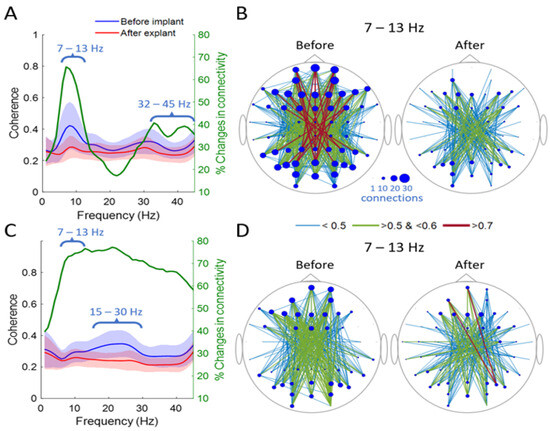
Figure 2 shows the results of the analysis of global connectivity changes in the two users. This analysis is based on the average coherences between cortical positions separated by at least 10 cm. Before implantation, User 1 had an average coherence value of approximately 0.4 ± 0.15 in the 7–13 Hz band (solid blue line, Figure 2A), whereas after UEA explantation, coherence significantly decreased to an average of 0.28 ± 0.07. These values represent significant changes (p < 0.01) in approximately 65% of the analyzed connections (solid green line, Figure 2A), mainly observed in the frontal and occipital regions. Figure 2B shows, in general terms, a reduced number of nodal connections after UEA explantation compared to the reference state (before implantation). This is also observed in terms of the average coherence amplitude.

Figure 2.
Changes in global connectivity in resting state. (A) User 1. Average coherence of all possible comparisons. Solid blue line represents global cortical coherence 1 d after UEA implantation, while solid red line represents global cortical coherence 6 months postimplantation. Shading around means indicates standard deviation. Solid green line represents percentage changes (p < 0.01) between resting states 1 d after implantation vs. 6 months postimplantation. (B) Global connectivity pattern in alpha band (7 to 13 Hz). Node size is related to number of significantly established connections (coherence > 0.5). Color and thickness of connectivity lines are directly related to average coherence value in analyzed band. (C) Same as (A) but for User 2. (D) Same as (B) but for User 2. In all cases, only pairs between cortical locations separated by more than 10 cm were considered.
The percentage change in connectivity exceeded 60% in the 7–45 Hz band for User 2, resulting in significantly lower coherences in the post-explantation UEA condition in the 15–30 Hz band (Figure 2C). In terms of global average coherence, the differences observed in the alpha band are not as pronounced as those in User 1; however, the high rate of change exceeds 70%. This is evident in Figure 2D, where differences are observed both in the number of nodal connections and in the strength of interconnections.
The common factor in both subjects is a generalized decrease in average coherence across all analyzed frequency bands. This connectivity pattern resulted in reductions in both the number of nodal connections and the strength of interconnections following UEA extraction.
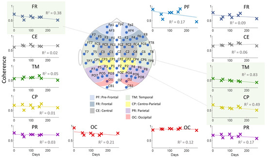
The previously described changes are temporally separated by at least 180 days. A longitudinal analysis of such changes in User 1 reveals linear and decreasing trends in spectral coherence values in the 7–13 Hz band in the left frontal (FR, R2 = 0.32), right temporal (TM, R2 = 0.78), and right parietal central (CP, R2 = 0.43) regions over time (Figure 3). In User 2, the most significant trend in coherence value changes was observed in the left parietal central (CP) region (R2 = 0.48) in the 7–13 Hz band.

Figure 3.
User 1. A longitudinal analysis of coherence in the 7–13 Hz band within predefined cortical regions: PF: Pre-frontal; FR: Frontal; CE: Central; TM: Temporal; CP: Centro-parietal; PR: Parietal; and OC: Occipital. Due to significant interference on some days, the average coherence in the left pre-frontal region (Fp1) is not represented in this figure. Coherence in the occipital region was determined between channels O1 and Oz for the left occipital region and between O2 and Oz for the right occipital region. The dotted lines in each graph represent linear trends obtained from a linear regression fit. R2 is the coefficient of determination resulting from that linear fit.
4. Discussion
Blind individuals implanted with a Utah Electrode Array (UEA) participated in this study aimed at developing a cortical visual neuroprosthesis. Over six months, the subjects underwent cortical electrical stimulation to evoke phosphenes, used as a source of visual sensory information about the environment. Initial psychophysical tests determined stimulation thresholds and assessed parameters such as frequency, intensity, and duration [11,20,21]. Subsequently, functional evaluations included navigation in controlled environments [22], closed-loop stimulation techniques to optimize device performance [21], and an analysis of eye movements related to phosphene perception [23]. These activities aimed to integrate artificial visual information into multimodal processing.
Phosphene perception constitutes a recovered sensory modality that, initially, the brain processes in a fragmented manner. Over time, changes in resting-state cortical connectivity patterns reflect the brain’s neuroplastic adaptation to this new input [13]. In this study, significant changes in connectivity were observed, particularly in the 7–13 Hz frequency band for User 1 and 15–30 Hz for User 2. These alterations involved specific cortical regions, including the right frontal, temporal, and left central–parietal areas, with linearly decreasing coherence trends within and between these regions. Notably, User 2 exhibited less pronounced changes within regions, suggesting connectivity modifications primarily between cortical areas with lower coherence values (< 0.4).
These findings support the hypothesis that resting-state connectivity changes reflect adaptive dynamics associated with the integration of artificial vision into multimodal processing. If multimodal perception were systematic and stable, cortical connectivity in the resting state would remain relatively unchanged (REF). Instead, the observed trends suggest a flexible and adaptive perceptual process required to integrate phosphenes into the sensory framework.
The potential for enhancing neuroplasticity emerges as a critical avenue for improving sensory integration. Factors such as age, blindness duration, and etiology may influence these processes. Tailored therapeutic strategies could accelerate the integration of artificial visual information, with efficacy measurable through resting-state cortical connectivity analysis.
5. Conclusions
This study reveals significant changes in cortical connectivity before UEA implantation and after its explantation. Connectivity trends—both increasing and decreasing—demonstrated adaptive dynamics in response to artificial visual input, with specific frequency bands (7–13 Hz for User 1 and 15–30 Hz for User 2) showing marked differences [13]. These findings highlight the cortical regions involved in integrating phosphenes into multimodal sensory processing and underscore the potential for designing therapies that enhance neuroplasticity, improving outcomes for users of cortical visual neuroprostheses.
Author Contributions
Conceptualization, F.D.F., L.S., and E.F.; methodology, M.d.M.A.A. and F.D.F.; software, M.d.M.A.A. and F.D.F.; validation, F.D.F. and L.S.; formal analysis, F.D.F. and L.S.; investigation, M.d.M.A.A., F.D.F., and L.S.; resources, E.F.; data curation, F.D.F., L.S., and A.L.A.; writing—original draft preparation, M.d.M.A.A. and F.D.F.; writing—review and editing, F.D.F., L.S., A.L.A., and E.F.; visualization, F.D.F.; supervision, E.F.; project administration, E.F.; funding acquisition, F.D.F., L.S., and E.F. All authors have read and agreed to the published version of the manuscript.
Funding
This work was supported by Grants PDC2022-133952-100 and PID2022-1416060B-100 from the Spanish ‘Ministerio de Ciencia, Innovación y Universidades,’ by grant CIPROM/2023/25 from the Generalitat Valenciana, and by the European Union’s Horizon 2020 Research and Innovation Programme under Grant Agreement No. 899287 (NeuraViPeR).
Institutional Review Board Statement
All experimental, surgical, and pre-surgical procedures were conducted according to a protocol approved by the Clinical Research Committee of the General University Hospital of Elche and registered in ClinicalTrials.gov (NCT02983370). Furthermore, all relevant ethical guidelines pertaining to clinical trial regulations (EU No. 536/2014, repealing Directive 2001/20/EC), the Declaration of Helsinki, and European Commission Directives (2005/28/EC and 2003/94/EC) were followed.
Informed Consent Statement
Informed consent was obtained from all subjects involved in this study.
Data Availability Statement
The data presented in this study are available upon request from the corresponding author.
Conflicts of Interest
The authors declare no conflicts of interest.
References
- Calvert, G.A.; Thesen, T. Multisensory integration: Methodological approaches and emerging principles in the human brain. J. Physiol. 2004, 98, 191–205. [Google Scholar] [CrossRef] [PubMed]
- Stein, B.E.; Stanford, T.R. Multisensory integration: Current issues from the perspective of the single neuron. Nat. Rev. Neurosci. 2008, 9, 255–266. [Google Scholar] [CrossRef] [PubMed]
- Driver, J.; Noesselt, T. Multisensory interplay reveals crossmodal influences on ‘sensory-specific’ brain regions, neural responses, and judgments. Neuron. 2008, 57, 11–23. [Google Scholar] [CrossRef]
- Pascual-Leone, A.; Torres, F. Plasticity of the sensorimotor cortex representation of the reading finger in Braille readers. Brain. 1993, 116, 39–52. [Google Scholar] [CrossRef]
- Sterr, A.; Müller, M.M.; Elbert, T.; Rockstroh, B.; Pantev, C.; Taub, E. Perceptual correlates of changes in cortical representation of fingers in blind multifinger Braille readers. J. Neurosci. 1998, 18, 4417–4423. [Google Scholar] [CrossRef]
- Rauschecker, J.P. Compensatory plasticity and sensory substitution in the cerebral cortex. Trends Neurosci. 1995, 18, 36–43. [Google Scholar] [CrossRef]
- Röder, B.; Stock, O.; Bien, S.; Neville, H.; Rösler, F. Speech processing activates visual cortex in congenitally blind humans. Eur. J. Neurosci. 2002, 16, 930–936. [Google Scholar] [CrossRef]
- Burton, H.; Diamond, J.B.; McDermott, K.B. Dissociating cortical regions activated by semantic and phonological tasks: A fMRI study in blind and sighted people. J. Neurophysiol. 2003, 90, 1965–1982. [Google Scholar] [CrossRef]
- Burton, H.; Snyder, A.Z.; Conturo, T.E.; Akbudak, E.; Ollinger, J.M.; Raichle, M.E. Adaptive changes in early and late blind: A fMRI study of Braille reading. J. Neurophysiol. 2002, 87, 589–607. [Google Scholar] [CrossRef]
- Stevens, A.A.; Weaver, K.E. Functional characteristics of auditory cortex in the blind. Behav. Brain Res. 2009, 196, 134–138. [Google Scholar] [CrossRef]
- Fernández, E.; Alfaro, A.; Soto-Sánchez, C.; Gonzalez-Lopez, P.; Lozano, A.M.; Peña, S.; Grima, M.D.; Rodil, A.; Gómez, B.; Chen, X.; et al. Visual percepts evoked with an intracortical 96-channel microelectrode array inserted in human occipital cortex. J. Clin. Investig. 2021, 131, e151331. [Google Scholar] [CrossRef]
- Fernandez, E.; Pelayo, F.; Amermmuller, J.; Ahnelt, P.; Norman, R.A. Cortical visual neuroprostheses for the blind. Restor. Neurol. Neurosci. 2004, 22, 3–14. [Google Scholar]
- Merabet, L.B.; Rizzo, J.F.; Amedi, A.; Somers, D.C.; Pascual-Leone, A. What blindness can tell us about seeing again: Merging neuroplasticity and neuroprostheses. Nat. Rev. Neurosci. 2005, 6, 71–77. [Google Scholar] [CrossRef] [PubMed]
- Castaldi, E.; Lunghi, C.; Morrone, M.C. Neuroplasticity in adult human visual cortex. Neurosci. Biobehav. Rev. 2020, 112, 542–552. [Google Scholar] [CrossRef] [PubMed]
- Uddin, L.Q.; Menon, V. Introduction to Special Topic—Resting-State Brain Activity: Implications for Systems Neuroscience. Front. Syst. Neurosci. 2010, 4, 2190. [Google Scholar] [CrossRef]
- Rogala, J.; Kublik, E.; Krauz, R.; Wróbel, A. Resting-state EEG activity predicts frontoparietal network reconfiguration and improved attentional performance. Sci. Rep. 2020, 10, 5064. [Google Scholar] [CrossRef]
- Delorme, A.; Makeig, S. EEGLAB: An open source toolbox for analysis of single-trial EEG dynamics including independent component analysis. J. Neurosci. Methods. 2004, 134, 9–21. [Google Scholar] [CrossRef]
- Srinivasan, R.; Nunez, P.L.; Silberstein, R.B. Spatial filtering and neocortical dynamics: Estimates of EEG coherence. IEEE Trans. Biomed. Eng. 1998, 45, 814–826. [Google Scholar] [CrossRef]
- Nunez, P.L. Electric Fields of the Brain: The Neurophysics of EEG.; Oxford University Press: Mumbai, India, 2006. [Google Scholar]
- Grani, F.; Soto-Sanchez, C.; Farfan, F.D.; Alfaro, A.; Grima, M.D.; Doblado, A.R.; Fernández, E. Time stability and connectivity analysis with an intracortical 96-channel microelectrode array inserted in human visual cortex. J. Neural Eng. 2022, 19, 45001. [Google Scholar] [CrossRef]
- Grani, F.; Soto-Sánchez, C.; Fimia, A.; Fernández, E. Toward a personalized closed-loop stimulation of the visual cortex: Advances and challenges. Front. Cell. Neurosci. 2022, 16, 1034270. [Google Scholar] [CrossRef]
- Ruiz, R.M.; Soo, L.; Val-Calvo, M.; López-Peco, R.; Grani, F.; Waclawczyk, D.; Doblado, A.R.; Sanchez, C.S.; Fernández, E. A new environment for assessing orientation and mobility in persons with severe visual impairments. IBRO Neurosci. Rep. 2023, 15, S950–S951. [Google Scholar] [CrossRef]
- Waclawczyk, D.; Caspi, A.; Soo, L.; Grani, F.; Peco, R.L.; Doblado, A.R.; Ruiz, R.M.; Sanchez, C.S.; Fernandez, E. The importance of eye movement in visual cortical prosthesis. IBRO Neurosci. Rep. 2023, 15, S725–S726. [Google Scholar] [CrossRef]
Disclaimer/Publisher’s Note: The statements, opinions and data contained in all publications are solely those of the individual author(s) and contributor(s) and not of MDPI and/or the editor(s). MDPI and/or the editor(s) disclaim responsibility for any injury to people or property resulting from any ideas, methods, instructions or products referred to in the content. |
© 2025 by the authors. Licensee MDPI, Basel, Switzerland. This article is an open access article distributed under the terms and conditions of the Creative Commons Attribution (CC BY) license (https://creativecommons.org/licenses/by/4.0/).