Abstract
Hamstring muscle tears are among the most prevalent sports injuries. The occurrence of muscle injuries has been demonstrated to result in alterations to the movement control system, thus prompting the implementation of compensatory strategies. The potential for in-depth study of these adaptive or compensatory strategies for injuries is made possible by the use of electrophysiological biomarkers. The aim of this study was to evaluate the current evidence concerning the analysis of electrophysiological biomarkers in research conducted on subjects with a history of hamstring injury (HSI) on sprinting tasks. A comprehensive literature review was conducted, and five articles were selected based on a rigorous set of selection criteria. The heterogeneity of the results precludes the establishment of broad generalisations regarding hamstring muscle activity during running or sprinting. Notwithstanding these discrepancies, individuals with a history of hamstring injury have been shown to consistently exhibit altered EMG patterns. In order to enhance our comprehension of the neural strategies underpinning movement, it is imperative to employ methodologies that transcend the limitations of EMG amplitude measures.
1. Introduction
Hamstring tears injuries (HSI) represent a significant challenge in the domain of sports and athletic activities, being one of the most common injuries and accounting for approximately 39% of all sports-related injuries. With a recurrence rate of 12% to 63%, these injuries manifest as acute pain in the posterior thigh. According to epidemiological studies, the biceps femoris and, to a lesser extent, the semimembranosus are often affected. Depending on severity, HSI can prevent physical activity for durations ranging from one to seven weeks [1].
A deeper understanding of the mechanisms underpinning these injuries highlights the critical role of high-speed efforts, such as sprinting, hurdling, or kicking, in their occurrence. Specifically, during sprinting—particularly in the late swing and late stance phases—the hamstrings are subjected to significant mechanical demands. This is especially evident when athletes run at speeds exceeding 25 km/h or above 80% of their maximum velocity [1,2,3]. These findings underscore the biomechanical challenges imposed by sprinting, positioning it as a pivotal component of many sports.
Furthermore, the variability in sprinting demands across sports emphasises the relevance of this activity. For example, while Australian football players may accumulate 194 m of sprinting accelerations during a match, rugby union players typically cover approximately 94 m in similar high-intensity efforts [4]. These differences not only illustrate sport-specific demands but also highlight the central role of sprinting in performance optimisation and injury risk management.
Moreover, the complex motor control required for high-speed actions is not confined to biomechanics. Indeed, it is evident that the integration of cortical and subcortical structures is also a prerequisite. Of particular importance are lower motor neurons, which innervate effector muscles and serve as the final common pathway for motor execution [5]. Notably, injuries to the musculoskeletal system, such as HSI, disrupt muscle function and can alter motor control strategies, even after apparent recovery and return to activity [6].
Given these disruptions, a neurophysiological approach becomes essential for understanding the adaptations and compensatory mechanisms associated with muscle injuries. In this context, surface electromyography (EMG) emerges as a powerful tool, providing high temporal resolution to capture detailed electrophysiological data on muscle activity during motor tasks [7]. Recent studies have adopted more integrative approaches, encompassing both bivariate (e.g., intermuscular and corticomuscular functional connectivity) and multivariate (e.g., network analysis and muscle synergies) methods, to facilitate a more profound exploration of complex processes such as low back pain or other musculoskeletal disorders [8,9,10]. These approaches have the potential to offer more comprehensive insights into the multifactorial phenomenon of muscle injuries.
Building on this foundation, it is crucial to examine which parameters of neuromuscular function have been employed as biomarkers in the investigation of compensatory and adaptive strategies associated with HSI during sprint activities. Accordingly, the aim of this study was to review the current evidence on the analysis of electrophysiological biomarkers in studies performed on subjects with a history of HSI on sprinting tasks.
2. Methods
A search strategy was conducted with the objective of identifying all relevant published papers. Specifically, a systematic search was conducted in the PubMed-Medline and Google Scholar databases without imposing restrictions on the year of publication. In order to ensure comprehensive coverage, the search strategy employed key terms based on the research aim (Table 1). The ultimate objective was to retrieve all relevant publications, thereby providing a comprehensive understanding of the subject matter.

Table 1.
Keywords used during database searches.
In order to refine the selection, the lead author evaluated journal articles from peer-reviewed sources. These were subject to a meticulous review of their titles, abstracts, and full texts. The removal of duplicate articles was facilized by utilisation of the bibliographic manager Zotero (v. 7.0.15) (Corporation for Digital Scholarship, Vienna, VA, USA). Furthermore, reference lists from the retrieved papers and systematic reviews were examined to identify further relevant studies that may not have been initially captured in the database search. This supplementary step served to enhance the robustness of the search process.
Finally, a full-text review was conducted to determine eligibility. Prior to the initiation of the search, a set of selection criteria was established. To be eligible for this study, articles had to have the following characteristics:
- The evaluation of neuromuscular function is to be conducted through the utilisation of electromyography and/or electroencephalography.
- The articles include studies that recruited an athletic cohort, defined as individuals participating in either individual or team sports, or those who demonstrated a high level of physical conditioning.
- The articles include participants without pre-existing neurological pathologies.
- The articles include sprinting or running tasks in the analysis.
- The articles include participants who had suffered a hamstring tear and had returned to normal activity at the time of participation.
- The articles have comparative data from a control group or the healthy contralateral limb.
- It must not include other types of injuries, such as ACL tears, sacroiliac joint, or lumbar spine dysfunction.
- It must not be a review article, the full article must be available in English, and it must have been published in a peer-reviewed journal.
Participant demographic data, injury-specific data, and data on neuromuscular function were extracted. Extraction of data was also conducted from the descriptive and inferential statistical analysis of each article.
3. Results
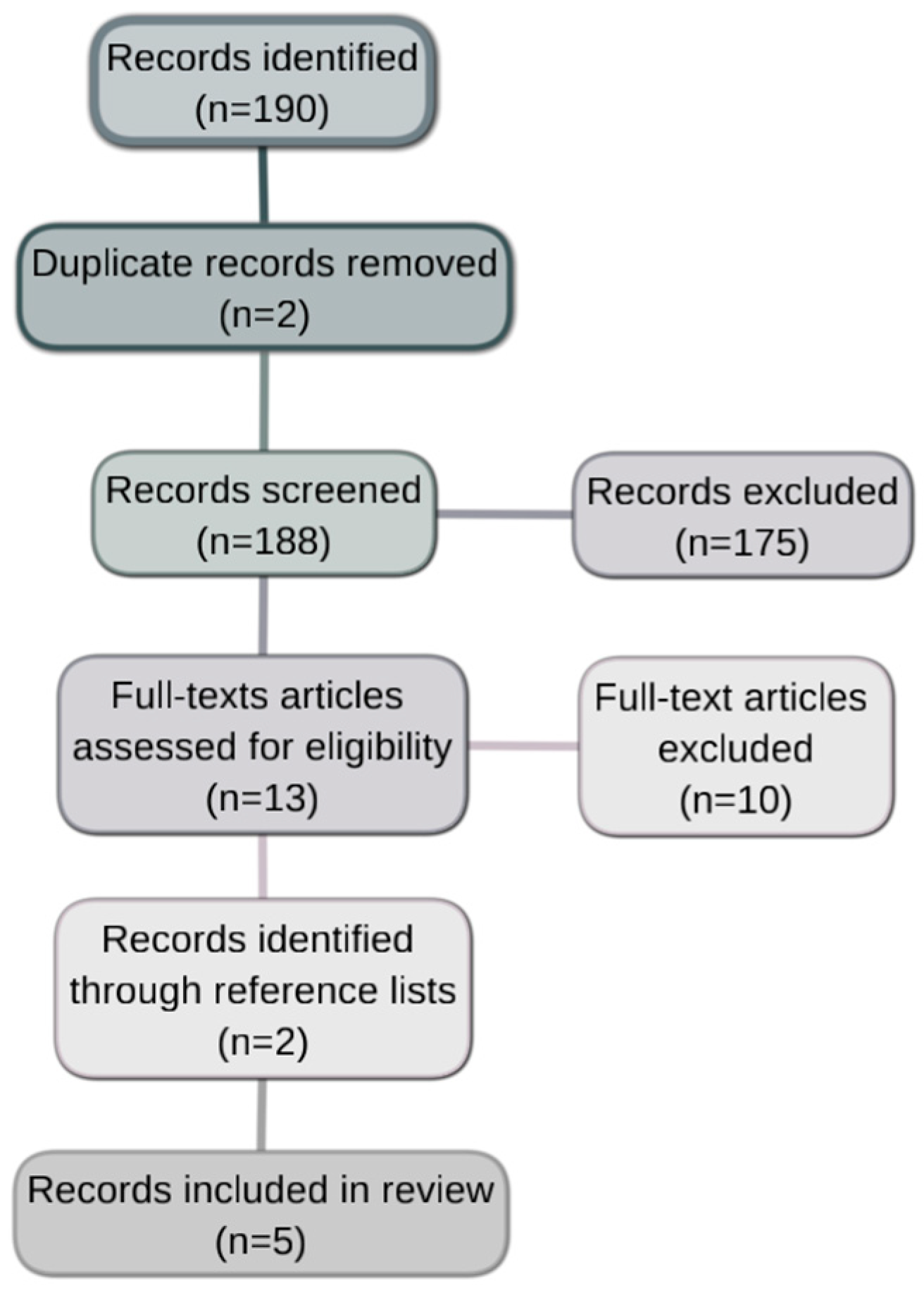
The initial search yielded 190 articles. Following the removal of duplicates and the review of titles and abstracts, a total of 13 full-text articles were assessed for eligibility. A subsequent review of the reference list yielded two additional articles. The application of the eligibility criteria resulted in the inclusion of five articles in the review (Figure 1).
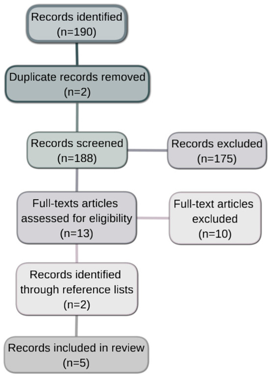
Figure 1.
A schematic representation of the study selection process.
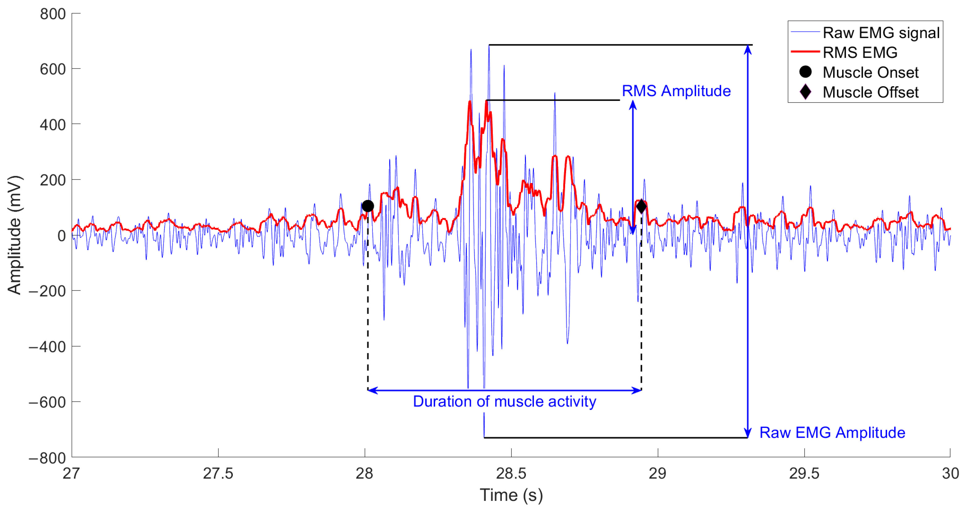
Across the studies, neurophysiological functions were examined using a variety of measures (Figure 2). Furthermore, disparities in signal normalisation methods and sprint or running tasks were observed among studies. Table 2 provides a comprehensive overview of the demographic characteristics under investigation.
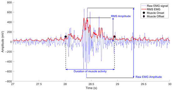
Figure 2.
A schematic representation of an EMG signal highlighting the most common features.
Table 2.
Comparison of principal features of articles.
In one study, Ohtsubo et al. [15] investigated mean EMG activity during each phase of a 30-metre maximum-speed sprint, comparing both limbs in the hamstring strain injured athlete cohort and a control group. A repeated-measures two-way ANOVA was conducted for multiple comparisons, with statical significance setting at p < 0.05. When significant differences were found, effect size (ES) analysis was conducted utilising Hedge’s g (g ≥ 0.20 small ES, g ≥ 0.50 medium ES, and g ≥ 0.80 large ES). The results of the study demonstrated that the level of muscle activity in semitendinosus (ST) during the middle swing phase in HSI, was significantly higher, a similar pattern also was observed in the healthy limb of the HSI group when compared to controls. Subsequent analysis revealed a marked elevation in gluteus medius (Gmed) activity in both lower limbs of the HSI group during the early stance phase. In addition, an evaluation of the external oblique (ObExt) revealed diminished activity in the injured limb during the early stance phase. Notably, similar reductions were observed in the gluteus maximus (Gmax) during the late swing phase. Additionally, the peak RMS of the Gmed was delayed in both limbs of the HSI group compared to controls. In contrast, no statistically significant differences were identified in measurements of the long head of the biceps femoris (BFlh).
Similarly, the study by Higashihara et al. [13], which involved a 40 m sprint and paired t-test to ascertain the inter-limb differences (p < 0.05). The findings revealed diminished BFlh activity in the HSI limb during the late swing phase when compared to the contralateral healthy limb. However, the Gmax exhibited no substantial disparities between the HSI and healthy limbs.
Further investigations by Silder et al. [11], who compared EMG muscle activity during treadmill running at speeds ranging from 60% to 100% of maximal effort, utilised two-way repeated measures ANOVAs to perform multiple comparisons between muscles, limbs, and gait cycle phases and across speeds. The statical significance was set at p < 0.05 for all tests, with bilateral differences reported relative to the uninjured limb as either percent or absolute. The analysis, which included the rectus femoris (RF), vastus lateralis (VL), biceps femoris (BF), and medial hamstring (MH), revealed no significant differences in muscle onset and offset, nor in the duration of muscle activity within the gait cycle. However, a comparison of the magnitude of the normalised EMG (RMS) revealed that BF activity during the late swing phase exhibited an average increase of 67%, while MH activity demonstrated a 37% increase, both in the HSI limb. Conversely, during the stance phase, BF activity exhibited a 34% increase, while MH activity demonstrated a 66% increase in comparison to the non-injured limb. A similar finding regarding the temporal parameters of EMG was reported in a related study [14], in which a 100-metre sprint protocol was employed. In this context, the onset and offset times of muscle activity, as well as the duration of muscle activity, did not differ significantly between the HSI limb of the HSI group and the dominant limb of the control group. Notwithstanding the divergent statistical analysis that was employed, the authors opted for the Mann–Whitney U test in light of the data’s non-normal distribution. The magnitude of the differences between the groups was determined by calculating the effect size.
Finally, Daly et al. [12] conducted a treadmill study in which athletes ran at a constant speed of 20 km/h. Using surface EMG, the authors evaluated the activity of the BFlh, Gmax, RF, ObExt, and erector spinae (ER), with a particular emphasis on ipsilateral and contralateral muscle activity ratios. Subsequent intergroup comparisons were made of muscle activation ratios and kinematics. The results indicated a significant decrease in the BFlh muscle activation ratios in the HSI limb in comparison with the ipsilateral Gmax (maximum difference −12.5%), ipsilateral ER (maximum difference −12.5%), ipsilateral ObExt (maximum difference −23 %), and contralateral RF (maximum difference −22%) during the late swing phase.
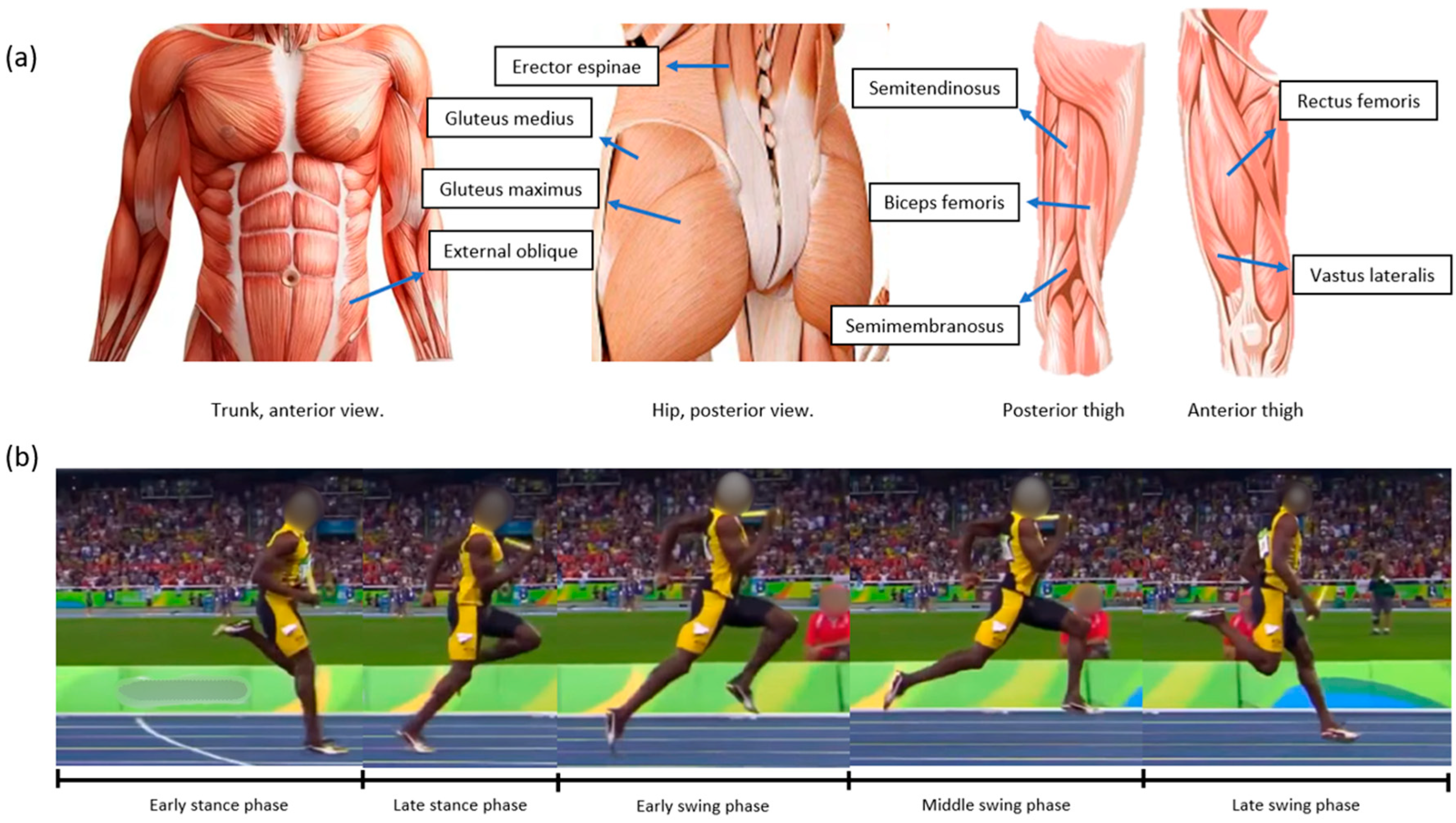
In Figure 3, the most reported muscles in the reviewed studies can be observed along with the specific running phases employed to analyse electromyographic activity.
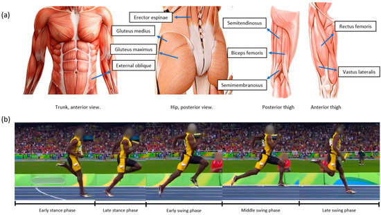
Figure 3.
(a) The anatomical representation of the reported muscles. (b) Specific running phases analysed.
4. Discussion
The aim of this study has been to review the current evidence on the analysis of electrophysiological biomarkers in studies performed on subjects with a history of HSI on sprinting tasks. Despite the limited number of articles analysed, it is necessary to discuss certain relevant aspects that contribute to a more comprehensive approach to the objective of this investigation.
Firstly, there is considerable heterogeneity in the electrophysiological parameters employed to assess neuromuscular function. The utilisation of EMG amplitude as a biomarker has been demonstrated to account for variations related to neuromotor demand in terms of force production [16]. Temporal metrics, such as the onset, offset, and duration of muscle activity, have been shown to reflect coordination-related strategies and the sequencing of different muscle groups during movement [17]. Furthermore, the comparison of muscle activation ratios has the potential to indicate the functional predominance of certain muscles over others, as reported by Daly et al. [12]. These diverse biomarkers capture distinct aspects of neuromuscular function, and such variability across studies poses a significant challenge for direct comparisons and integrative interpretation.
Evidence of the diversity of methodologies can also be observed in the normalisation of EMG signals. For instance, certain studies [12,15] have employed the normalisation of muscle activity to the mean value across the entire trial, incorporating methodologies based on the mean value per stride. An alternative approach was to normalise to the maximum value observed during the running cycle [13,14]. Concurrently, Silder et al. [11] utilised speed-based normalisation, employing the mean signal across a complete cycle at maximum sprint speed. These methodological discrepancies underscore the imperative for prudence when drawing comparisons between results, as each normalisation strategy possesses its own unique characteristics. For instance, standardisation to a mean value may result in an underestimation of peak intensities. Conversely, standardisation to maximum values has the potential to lead to an overestimation of differences and relative changes. On the other hand, task-based normalisation, such as normalisation by speed, is associated with contextual factors, thereby complicating the interpretation of comparisons, even within the same study at varying speeds [11].
It is important to note that the studies reviewed yielded contradictory findings. The main point of divergence pertains to the activity of the BF: while some authors have reported an increase in its activity [11], others have observed a decrease [12,13], and yet others found no significant differences [15] when comparing injured and non-injured individuals or the injured limb versus the contralateral limb, specifically during the same running phase—the late swing phase. As indicated by the citations provided, analogous inconsistencies have been reported in studies that have evaluated tasks related to force production [18,19,20,21]. Consequently, within the context of hamstring strain injury, the activity of the BF appears to be altered, albeit without the presence of a clear or consistent pattern when compared to a control group or limb. In the case of studies by Silder et al. [9] and Crow et al. [10], who analysed the onset, offset, and duration of muscle activity, no differences were found between the injured and control groups for the muscles examined. The incorporation of additional agonist/synergist muscles into the analysis may prove beneficial in facilitating a comparative analysis of temporal variables and an exploration of coordination and sequencing strategies. It is important to consider that variations in protocols, signal processing, and the methods used to determine temporal parameters likely contributed to these discrepancies.
A recurrent theme among the majority of studies is the reliance on EMG amplitude as a biomarker to quantify and compare motor activity in healthy and injured individuals. This univariate approach has yielded significant insights into muscle function in a range of contexts. Nevertheless, when attempting to infer underlying motor control strategies, this approach is subject to significant limitations. In order to gain insight into the neural strategies that govern movement, particularly how they adapt following muscle injury or in the presence of musculoskeletal pain, approaches that extend beyond EMG amplitude measurements are required.
Alternative methodologies have emerged to address the limitations of univariate analysis. For instance, corticomuscular coherence (CMC) provides insights into the coupling between the motor cortex and lower motor units, offering a deeper understanding of cortical control during movement [22]. A body of research has been conducted on the applications of CMC in the domain of strength training adaptations [23] and in the context of brain–muscle interactions during dynamic and isometric tasks [24,25,26,27].
Similarly, intermuscular coherence (IMC), which is defined as the degree of synchronisation in the time–frequency domain between two muscle signals [28], has been associated with motor demands and muscle synergy. IMC has been studied in a variety of contexts. These include strength training, postural challenges, fatigue, and musculoskeletal pain [8,28,29,30].
By quantifying neural synchrony, coherence can offer a more nuanced perspective on the central neural strategies that govern movement, adaptation, and motor recovery. The ability to distinguish neural drive across frequency bands enables the investigation of contributions from distinct cortical, subcortical, and spinal pathways. This approach promises a more detailed and physiologically grounded characterisation of motor control in both clinical and research settings.
Despite the relative novelty of these approaches, their potential to elucidate novel aspects of human motor control renders further investigation imperative. It is evident that both CMC and IMC provide significant insights into the composition of descending neural drive. Moreover, both of these methods benefit from non-invasive recording techniques, thus rendering them suitable for clinical applications. In the context of compensatory or adaptive mechanisms associated with muscle injury, adopting a more comprehensive approach to analysing muscle activity may provide a more accurate estimate of the changes in descending neural drive [31].
5. Conclusions
There is considerable variability in the electrophysiological biomarkers used to assess neuromuscular function in individuals with a history of hamstring strain injuries (HSI), with a predominant emphasis on EMG signal amplitude. The wide range of methodological approaches and the heterogeneity of findings hinder the formulation of definitive conclusions regarding hamstring muscle activity during running or sprinting. However, despite these inconsistencies, the literature consistently demonstrates that EMG activity is altered in individuals with a prior history of muscle injury.
To advance our understanding of the adaptive and compensatory mechanisms that underlie neuromuscular alterations post-injury—particularly regarding intermuscular coordination and corticomuscular connectivity—it is imperative to move beyond the isolated analysis of EMG amplitude. Incorporating more integrative and multidimensional electrophysiological approaches may offer deeper insights into the reorganisation of motor control strategies following injury. This paradigm shift is not only necessary—it is critical and indispensable for developing evidence-based rehabilitation protocols that are truly effective in restoring function, enhancing motor recovery, and preventing recurrence.
Author Contributions
Conceptualisation, methodology, investigation, resources, data curation, and writing—original draft preparation, G.D.G.; writing—review and editing, visualisation, and supervision, L.A.C.; project administration and funding acquisition, M.P.V. and F.D.F. All authors have read and agreed to the published version of the manuscript.
Funding
This study was partially funded by the Grant RESOL-2022-2324-APN-DIR#CONICET from the Argentinian Consejo Nacional de Investigaciones Científicas y Técnicas (CONICET) and Instituto Superior de Investigaciones Biológicas (INSIBIO) and also by PIUNT T705 from Universidad Nacional de Tucuman (UNT).
Institutional Review Board Statement
The study was conducted in accordance with the Declaration of Helsinki and approved by the ethics committee of the Universidad Nacional de Tucumán (Project PIUNT T705 Resol. N° 0356/23).
Informed Consent Statement
Not applicable.
Data Availability Statement
The data presented in this study are available upon request from the corresponding author.
Acknowledgments
The authors would like to thank the Consejo Nacional de Investigaciones Científicas y Técnicas (CONICET) and the Universidad Nacional de Tucumán (UNT), institutions that partially funded the research and made it possible to carry out this study. The authors would also like to thank the Laboratory of Research in Neurosciences and Applied Technologies (LINTEC) of the Department of Bioengineering of the Faculty of Exact Sciences and Technology of the UNT for providing the necessary working spaces for the development of scientific activities.
Conflicts of Interest
The authors declare no conflicts of interest. The funders had no role in the design of the study; in the collection, analyses, or interpretation of data; in the writing of the manuscript; or in the decision to publish the results.
References
- Silvers-Granelli, H.J.; Cohen, M.; Espregueira-Mendes, J.; Mandelbaum, B. Hamstring Muscle Injury in the Athlete: State of the Art. J. ISAKOS 2021, 6, 170–181. [Google Scholar] [CrossRef] [PubMed]
- Bramah, C.; Mendiguchia, J.; Dos’Santos, T.; Morin, J.-B. Exploring the Role of Sprint Biomechanics in Hamstring Strain Injuries: A Current Opinion on Existing Concepts and Evidence. Sports Med. 2024, 54, 783–793. [Google Scholar] [CrossRef] [PubMed]
- Gómez-Piqueras, P.; Alcaraz, P.E. If You Want to Prevent Hamstring Injuries in Soccer, Run Fast: A Narrative Review about Practical Considerations of Sprint Training. Sports 2024, 12, 134. [Google Scholar] [CrossRef] [PubMed]
- Harper, D.J.; Carling, C.; Kiely, J. High-Intensity Acceleration and Deceleration Demands in Elite Team Sports Competitive Match Play: A Systematic Review and Meta-Analysis of Observational Studies. Sports Med. 2019, 49, 1923–1947. [Google Scholar] [CrossRef]
- Sherrington, C.S. The Integrative Action of the Nervous System; Yale University Press: New Haven, CT, USA, 1911. [Google Scholar]
- Areia, C.; Barreira, P.; Montanha, T.; Oliveira, J.; Ribeiro, F. Neuromuscular Changes in Football Players with Previous Hamstring Injury. Clin. Biomech. 2019, 69, 115–119. [Google Scholar] [CrossRef]
- Al-Ayyad, M.; Owida, H.A.; De Fazio, R.; Al-Naami, B.; Visconti, P. Electromyography Monitoring Systems in Rehabilitation: A Review of Clinical Applications, Wearable Devices and Signal Acquisition Methodologies. Electronics 2023, 12, 1520. [Google Scholar] [CrossRef]
- Ghazi, S.; Hadian, M.R.; Shadmehr, A.; Talebian, S.; Olyaei, G.; Hajouj, E. Test-Retest Reliability of EMG β-Band Intermuscular Coherence of Non-Specific Chronic Low Back Pain During Flexion-Extension Task. J. Mod. Rehabil. 2021, 15, 2. [Google Scholar] [CrossRef]
- Murphy, M.C.; Latella, C.; Rio, E.K.; Taylor, J.L.; Martino, S.; Sylvester, C.; Hale, W.; Mosler, A.B. Does Lower-Limb Osteoarthritis Alter Motor Cortex Descending Drive and Voluntary Activation? A Systematic Review and Meta-Analysis. EFORT Open Rev. 2023, 8, 883–894. [Google Scholar] [CrossRef]
- Hubley-Kozey, C.L.; Vezina, M.J. Differentiating Temporal Electromyographic Waveforms between Those with Chronic Low Back Pain and Healthy Controls. Clin. Biomech. 2002, 17, 621–629. [Google Scholar] [CrossRef]
- Silder, A.; Thelen, D.G.; Heiderscheit, B.C. Effects of Prior Hamstring Strain Injury on Strength, Flexibility, and Running Mechanics. Clin. Biomech. 2010, 25, 681–686. [Google Scholar] [CrossRef]
- Daly, C.; McCarthy Persson, U.; Twycross-Lewis, R.; Woledge, R.C.; Morrissey, D. The Biomechanics of Running in Athletes with Previous Hamstring Injury: A Case-control Study. Scand. Med. Sci. Sports 2016, 26, 413–420. [Google Scholar] [CrossRef] [PubMed]
- Higashihara, A.; Ono, T.; Tokutake, G.; Kuramochi, R.; Kunita, Y.; Nagano, Y.; Hirose, N. Hamstring Muscles’ Function Deficit during Overground Sprinting in Track and Field Athletes with a History of Strain Injury. J. Sports Sci. 2019, 37, 2744–2750. [Google Scholar] [CrossRef]
- Crow, J.; Semciw, A.; Couch, J.; Pizzari, T. Does a Recent Hamstring Muscle Injury Affect the Timing of Muscle Activation during High Speed Overground Running in Professional Australian Football Players? Phys. Ther. Sport 2020, 43, 188–194. [Google Scholar] [CrossRef] [PubMed]
- Ohtsubo, R.; Saito, H.; Hirose, N. Characterizing Muscle Activity in Soccer Players with a History of Hamstring Strain Injuries during Accelerated Sprinting. J. Sports Sci. Med. 2024, 23, 656–662. [Google Scholar] [CrossRef] [PubMed]
- Vigotsky, A.D.; Halperin, I.; Lehman, G.J.; Trajano, G.S.; Vieira, T.M. Interpreting Signal Amplitudes in Surface Electromyography Studies in Sport and Rehabilitation Sciences. Front. Physiol. 2018, 8, 985. [Google Scholar] [CrossRef]
- Disselhorst-Klug, C.; Williams, S. Surface Electromyography Meets Biomechanics: Correct Interpretation of sEMG-Signals in Neuro-Rehabilitation Needs Biomechanical Input. Front. Neurol. 2020, 11, 603550. [Google Scholar] [CrossRef]
- Opar, D.A.; Williams, M.D.; Timmins, R.G.; Dear, N.M.; Shield, A.J. Knee Flexor Strength and Bicep Femoris Electromyographical Activity Is Lower in Previously Strained Hamstrings. J. Electromyogr. Kinesiol. 2013, 23, 696–703. [Google Scholar] [CrossRef]
- Buhmann, R.; Trajano, G.S.; Kerr, G.; Shield, A. Voluntary Activation and Reflex Responses after Hamstring Strain Injury. Med. Sci. Sports Exerc. 2020, 52, 1862–1869. [Google Scholar] [CrossRef]
- Avrillon, S.; Hug, F.; Guilhem, G. Bilateral Differences in Hamstring Coordination in Previously Injured Elite Athletes. J. Appl. Physiol. 2020, 128, 688–697. [Google Scholar] [CrossRef]
- Blandford, L.; Theis, N.; Charvet, I.; Mahaffey, R. Is Neuromuscular Inhibition Detectable in Elite Footballers during the Nordic Hamstring Exercise? Clin. Biomech. 2018, 58, 39–43. [Google Scholar] [CrossRef]
- Mima, T.; Hallett, M. Corticomuscular Coherence: A Review. J. Clin. Neurophysiol. 1999, 16, 501. [Google Scholar] [CrossRef] [PubMed]
- Elie, D.; Barbier, F.; Ido, G.; Cremoux, S. Corticomuscular Coherence and Motor Control Adaptations after Isometric Maximal Strength Training. Brain Sci. 2021, 11, 254. [Google Scholar] [CrossRef] [PubMed]
- Kenville, R.; Maudrich, T.; Vidaurre, C.; Maudrich, D.; Villringer, A.; Nikulin, V.V.; Ragert, P. Corticomuscular Interactions during Different Movement Periods in a Multi-Joint Compound Movement. Sci. Rep. 2020, 10, 5021. [Google Scholar] [CrossRef]
- Desmyttere, G.; Mathieu, E.; Begon, M.; Simoneau-Buessinger, E.; Cremoux, S. Effect of the Phase of Force Production on Corticomuscular Coherence with Agonist and Antagonist Muscles. Eur. J. Neurosci. 2018, 48, 3288–3298. [Google Scholar] [CrossRef]
- Dal Maso, F.; Longcamp, M.; Cremoux, S.; Amarantini, D. Effect of Training Status on Beta-Range Corticomuscular Coherence in Agonist vs. Antagonist Muscles during Isometric Knee Contractions. Exp. Brain Res. 2017, 235, 3023–3031. [Google Scholar] [CrossRef]
- Glories, D.; Soulhol, M.; Amarantini, D.; Duclay, J. Specific Modulation of Corticomuscular Coherence during Submaximal Voluntary Isometric, Shortening and Lengthening Contractions. Sci. Rep. 2021, 11, 6322. [Google Scholar] [CrossRef]
- Boonstra, T.W.; Danna-Dos-Santos, A.; Xie, H.-B.; Roerdink, M.; Stins, J.F.; Breakspear, M. Muscle Networks: Connectivity Analysis of EMG Activity during Postural Control. Sci. Rep. 2015, 5, 17830. [Google Scholar] [CrossRef]
- Semmler, J.G.; Ebert, S.A.; Amarasena, J. Eccentric Muscle Damage Increases Intermuscular Coherence during a Fatiguing Isometric Contraction. Acta Physiol. 2013, 208, 362–375. [Google Scholar] [CrossRef]
- Charissou, C.; Vigouroux, L.; Berton, E.; Amarantini, D. Fatigue- and Training-Related Changes in ‘Beta’ Intermuscular Interactions between Agonist Muscles. J. Electromyogr. Kinesiol. 2016, 27, 52–59. [Google Scholar] [CrossRef]
- Boonstra, T.W. The Potential of Corticomuscular and Intermuscular Coherence for Research on Human Motor Control. Front. Hum. Neurosci. 2013, 7, 855. [Google Scholar] [CrossRef]
Disclaimer/Publisher’s Note: The statements, opinions and data contained in all publications are solely those of the individual author(s) and contributor(s) and not of MDPI and/or the editor(s). MDPI and/or the editor(s) disclaim responsibility for any injury to people or property resulting from any ideas, methods, instructions or products referred to in the content. |
© 2025 by the authors. Licensee MDPI, Basel, Switzerland. This article is an open access article distributed under the terms and conditions of the Creative Commons Attribution (CC BY) license (https://creativecommons.org/licenses/by/4.0/).