Abstract
The analysis of the retinal vasculature represents a crucial stage in the diagnosis of several diseases. An exhaustive analysis involves segmenting the retinal vessels and classifying them into veins and arteries. In this work, we present an accurate approach, based on deep neural networks, for the joint segmentation and classification of the retinal veins and arteries from color fundus images. The presented approach decomposes this joint task into three related subtasks: the segmentation of arteries, veins and the whole vascular tree. The experiments performed show that our method achieves competitive results in the discrimination of arteries and veins, while clearly enhancing the segmentation of the different structures. Moreover, unlike other approaches, our method allows for the straightforward detection of vessel crossings, and preserves the continuity of the arterial and venous vascular trees at these locations.
1. Introduction
The analysis of the retinal vasculature represents a crucial stage in the diagnosis of several diseases, such as diabetes, age-related macular degeneration (AMD) and glaucoma [1]. This is due to the presence of these diseases causing changes in the retinal vessels. An exhaustive analysis of the retinal vasculature involves segmenting the vascular tree and classifying their vessels into veins and arteries. Despite its utility, this type of analysis is rarely applied in clinical practice, as performing it manually is arduous, and often leads to partly subjective results. For this reason, several automatic methods have been proposed. Early methods addressed these tasks into two sequential steps [2]. However, this approach causes the classification results to be highly conditioned by the segmentation results. To overcome this issue, the current state of the art (SOTA) addresses both tasks as a single multi-class semantic segmentation problem [3,4,5,6].
In this work, we present an accurate approach, based on deep neural networks, for the joint segmentation and classification of the retinal arteries and veins (JSCAV) from color fundus images. This approach, differently to SOTA, decomposes the joint task into three subtasks: the segmentation of arteries, veins and the whole vascular tree. In the following sections, we discuss this approach and its associated advantages.
2. Materials and Methods
The current SOTA formulates the JSCAV task as a single multi-class semantic segmentation problem. However, this approach leads to incomplete segmentation maps for veins and arteries, and does not directly provide vasculature segmentation maps.
As an alternative, we present an approach that decomposes the joint task into three segmentation subtasks [7]. Each of these subtasks addresses the segmentation of one of three classes of interest: arteries, veins and the whole vascular tree. To implement this multi-segmentation (MS) approach, a deep neural network is trained end-to-end using a novel loss function: BCE3. This loss function computes the loss as the sum of the individual segmentation losses of the aforementioned classes. Each individual loss is computed as the binary cross-entropy (BCE) between the predicted probability map and the manually annotated segmentation map. This setting allows for the intuitive handling of vessel crossings, and directly provides precise and complete segmentation maps of the various vascular trees. It also allows for the direct detection of vessel crossings through the element-wise product of the predicted artery and vein maps.
To train and evaluate the networks in the JSCAV task, we employed the publicly available RITE dataset [8], which is composed of 40 color fundus images and their corresponding arteries, veins and vasculature segmentation masks. To facilitate training of the networks, we used the image preprocessing technique specified in [3], as well as online data augmentation. To validate our method, a U-Net network [9] was trained, using both the traditional and the MS approaches.
3. Results and Conclusions
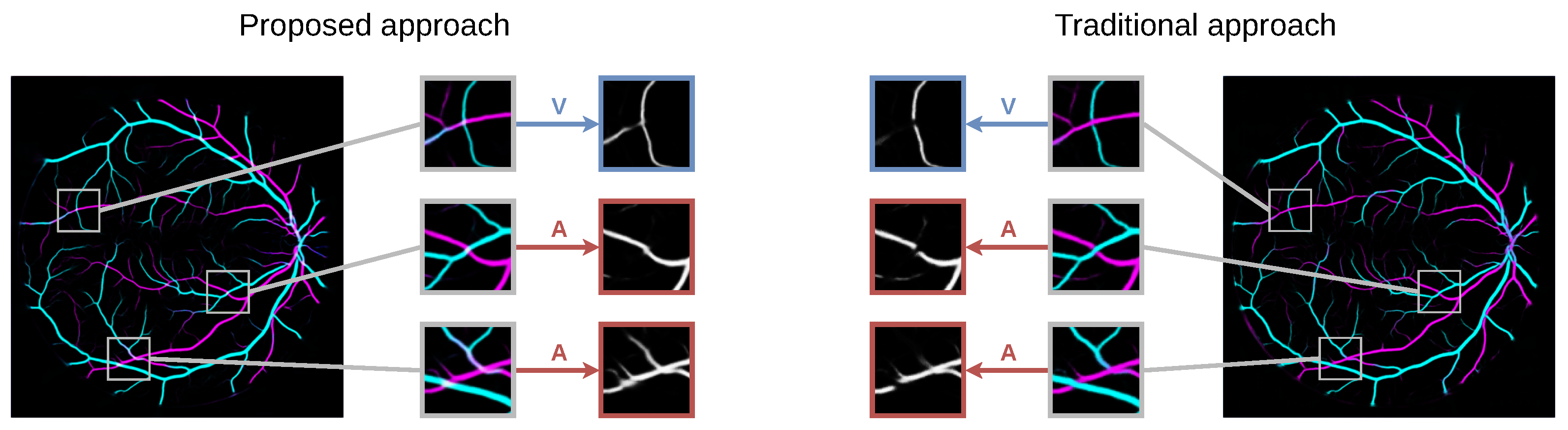
Figure 1 shows an example of an RITE retinography and its arteries, veins, vessels and crossings segmentation maps predicted by a model trained using the MS approach. Figure 2 shows the details of the arteries, veins and vessels segmentation maps of the same retinography predicted by a model trained using the MS and the traditional approaches.
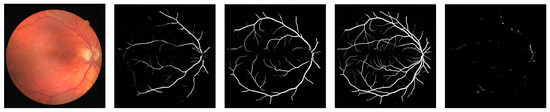
Figure 1.
Example segmentation maps predicted by a model trained using the MS approach. From left to right: arteries, veins, vessels and crossings.
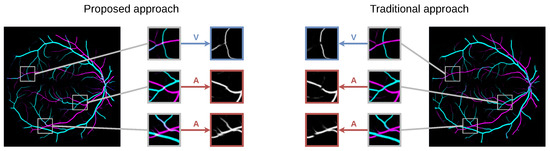
Figure 2.
Examples of arteries, veins and vessels probability maps (in RGB) predicted by the models trained using the MS and the traditional approaches.
The ablation study performed in the RITE dataset shows that our method provides an adequate performance, especially in the segmentation of the different structures. Notably, the MS approach achieves a mean accuracy of in the classification of arteries and veins, and an AUC-ROC of in the segmentation of vessels; for its part, the traditional approach achieves and , respectively.
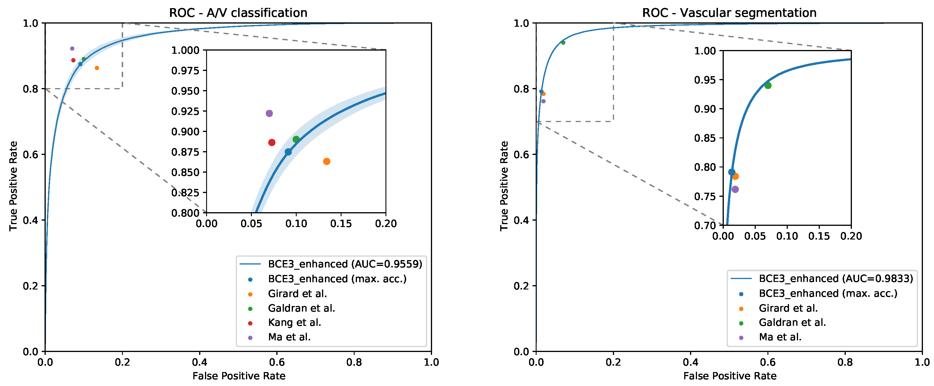
In addition, the comparison with the SOTA works in the same dataset, depicted in Figure 3, clearly demonstrates that the presented method achieves competitive results in the discrimination of arteries and veins, while significantly enhancing the vascular segmentation.
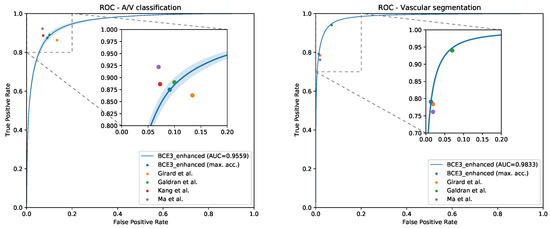
Figure 3.
ROC curves in the RITE dataset for the MS approach along with the point representations of the SOTA approaches for artery/vein classification (left) and vascular segmentation (right).
Therefore, the presented deep multi-segmentation method allows for the detection of more vessels and to better segment the different structures, while achieving competitive classification results. Furthermore, unlike previous approaches, the method allowsfor the straightforward detection of vessel crossings, as well as preserving the continuity of the arterial and venous vascular trees at these locations (see Figure 2).
Author Contributions
Conceptualization, Á.S.H., J.N. and J.R.; methodology, Á.S.H. and J.M.; software, Á.S.H. and J.M.; validation, Á.S.H. and J.M.; formal analysis, Á.S.H. and J.M.; investigation, J.M.; resources, J.N. and J.R.; data curation, Á.S.H. and J.M.; writing—original draft preparation, J.M.; writing—review and editing, J.M. and J.N.; visualization, J.M.; supervision, J.N. and J.R.; project administration, J.N. and J.R.; funding acquisition, J.N. and J.R. All authors have read and agreed to the published version of the manuscript.
Funding
This work was funded by Instituto de Salud Carlos III, Government of Spain, and the European Regional Development Fund (ERDF) of the European Union (EU) through the DTS18/00136 research project; Ministerio de Ciencia e Innovación, Government of Spain, through the RTI2018-095894-B-I00 and PID2019-108435RB-I00 research projects; Axencia Galega de Innovación (GAIN), Xunta de Galicia, ref. IN845D 2020/38; Xunta de Galicia and European Social Fund (ESF) of the EU through the predoctoral grant contracts ED481A-2017/328 and ED481A 2021/140; Consellería de Cultura, Educación e Universidade, Xunta de Galicia, through Grupos de Referencia Competitiva, grant ref. ED431C 2020/24; CITIC, Centro de Investigación de Galicia ref. ED431G 2019/01, is funded by Consellería de Educación, Universidade e Formación Profesional, Xunta de Galicia, through the ERDF (80%) and Secretaría Xeral de Universidades (20%).
Conflicts of Interest
The authors declare no conflict of interest.
References
- Kanski, J.J.; Bowling, B. Clinical Ophthalmology: A Systematic Approach, 7th ed.; Elsevier Health Sciences: New York, NY, USA, 2011. [Google Scholar]
- Staal, J.; Abràmoff, M.D.; Niemeijer, M.; Viergever, M.A.; van Ginneken, B. Ridge-based vessel segmentation in color images of the retina. IEEE Trans. Med. Imaging 2004, 23, 501–509. [Google Scholar] [CrossRef] [PubMed]
- Girard, F.; Kavalec, C.; Cheriet, F. Joint segmentation and classification of retinal arteries/veins from fundus images. Artif. Intell. Med. 2019, 94, 96–109. [Google Scholar] [CrossRef] [PubMed] [Green Version]
- Galdran, A.; Meyer, M.; Costa, P.; MendonÇa; Campilho, A. Uncertainty-Aware Artery/Vein Classification on Retinal Images. In Proceedings of the 2019 IEEE 16th International Symposium on Biomedical Imaging (ISBI 2019), Venice, Italy, 8–11 April 2019; pp. 556–560. [Google Scholar] [CrossRef]
- Ma, W.; Yu, S.; Ma, K.; Wang, J.; Ding, X.; Zheng, Y. Multi-task Neural Networks with Spatial Activation for Retinal Vessel Segmentation and Artery/Vein Classification. In Medical Image Computing and Computer Assisted Intervention—MICCAI 2019; Shen, D., Liu, T., Peters, T.M., Staib, L.H., Essert, C., Zhou, S., Yap, P.T., Khan, A., Eds.; Springer International Publishing: Cham, Switzerland, 2019; pp. 769–778. [Google Scholar] [CrossRef]
- Kang, H.; Gao, Y.; Guo, S.; Xu, X.; Li, T.; Wang, K. AVNet: A retinal artery/vein classification network with category-attention weighted fusion. Comput. Methods Programs Biomed. 2020, 195, 105629. [Google Scholar] [CrossRef] [PubMed]
- Morano, J.; Hervella, Á.S.; Novo, J.; Rouco, J. Simultaneous segmentation and classification of the retinal arteries and veins from color fundus images. Artif. Intell. Med. 2021, 118, 102116. [Google Scholar] [CrossRef] [PubMed]
- Hu, Q.; Abràmoff, M.D.; Garvin, M.K. Automated Separation of Binary Overlapping Trees in Low-Contrast Color Retinal Images. In Medical Image Computing and Computer-Assisted Intervention—MICCAI 2013; Mori, K., Sakuma, I., Sato, Y., Barillot, C., Navab, N., Eds.; Springer: Berlin/Heidelberg, Germany, 2013; pp. 436–443. [Google Scholar] [CrossRef]
- Ronneberger, O.; Fischer, P.; Brox, T. U-Net: Convolutional Networks for Biomedical Image Segmentation. In Medical Image Computing and Computer-Assisted Intervention—MICCAI 2015; Navab, N., Hornegger, J., Wells, W.M., Frangi, A.F., Eds.; Springer International Publishing: Cham, Switzerland, 2015; pp. 234–241. [Google Scholar] [CrossRef] [Green Version]

Publisher’s Note: MDPI stays neutral with regard to jurisdictional claims in published maps and institutional affiliations. |
© 2021 by the authors. Licensee MDPI, Basel, Switzerland. This article is an open access article distributed under the terms and conditions of the Creative Commons Attribution (CC BY) license (https://creativecommons.org/licenses/by/4.0/).