Proceeding Paper

Combined Application of Honokiol and 2-Deoxyglucose against MCF7 Breast Cancer Cells Under Hypoxia †

by
Alexander M. Scherbakov
1,2,*,
Ekaterina Igorevna Mikhaevich
1,
Alexandra L. Mikhaylova
1 and
Danila V. Sorokin
1,2
1
N.N. Blokhin National Medical Research Center of Oncology, The Ministry of Health of Russia, Moscow 115522, Russia
2
Molecular Genetics Laboratory, Institute of Clinical Medicine, National Research Lobachevsky State University of Nizhny Novgorod, Nizhny Novgorod 603022, Russia
*
Author to whom correspondence should be addressed.
Presented at the 4th International Electronic Conference on Applied Sciences, 27 October–10 November 2023; Available online: https://asec2023.sciforum.net/.
Eng. Proc. 2023, 56(1), 168; https://doi.org/10.3390/ASEC2023-16376
Published: 29 November 2023
(This article belongs to the Proceedings of The 4th International Electronic Conference on Applied Sciences)

Abstract

:
Breast cancer is the most common cancer among women. Epidemiologists estimate that over 2.3 million new cases of breast cancer are diagnosed worldwide each year. Natural compounds represent promising molecules for the development of antitumor drugs; among them, lignans show significant antiproliferative effects against breast cancer cells. The goal of the study was to analyse the antiproliferative effects of lignan honokiol on MCF7 breast cancer cells, find synergistic combinations of honokiol with 2-deoxyglucose, and evaluate the effects of the combinations found on cells in hypoxia. The antiproliferative effects of the compounds were evaluated by the MTT test, and protein expression analysis was performed by immunoblotting. Honokiol inhibited MCF7 cell growth with an IC50 value of 19.7 μM. Synergistic combinations of honokiol with the glycolysis inhibitor 2-deoxyglucose were detected; the compounds at low doses caused significant suppression of MCF7 cell growth. The established combinations of compounds inhibited HIF-1α expression and were effective in hypoxia, considered as the leading factor of chemotherapeutic resistance. Oestrogen receptor alpha (ERα) is the main growth driver of hormone-dependent breast tumours. Honokiol combined with 2-deoxyglucose reduced ERα expression in MCF7 cells, and expression of the hormone-dependent protein GREB1 was also downregulated. Honokiol at a concentration of 15 μM in combination with 6 mM 2-deoxyglucose induced the cleavage of PARP (a marker of apoptosis) in MCF7 cells after 48 h of incubation. The cells treated with the combination of honokiol and 2-deoxyglucose demonstrated a decrease in the expression of cyclin D1. Thus, honokiol represents a promising basis for the development of antitumor agents; the combination of this natural compound with glycolysis inhibitors can be used to reduce the applied doses.

1. Introduction

The majority (up to 70%) of breast tumours are oestrogen-dependent. 4-Hydroxytamoxifen (selective ERα modulator) and later fulvestrant (selective ERα degrader) were the standard therapeutics for ERα(+) breast cancer [1,2]. However, tamoxifen also has estrogen agonist activity, which may contribute to treatment failure [3]. The resistance to both drugs develops over time through the dysregulation of the PI3K/AKT/mTOR pathway that cross-talks with ERα-mediated signalling [4,5]. At the moment, there are different approaches to the treatment of breast cancer, and the choice of method depends on the genotype of a particular tumour: therapy with PARP inhibitors in the presence of BRCA1/2 and BRCAness TNBC mutations, anthracyclines, taxanes, and platinum-based therapy and immunotherapy with monoclonal antibodies that block CTLA4, PD1 or PDL1 [6]. Natural compounds represent promising molecules for the development of antitumor drugs; among them, lignans show significant antiproliferative effects against breast cancer cells. Such phytocomponents and their metabolites act on metabolic pathways, which affect proliferation, migration, and apoptosis in cancer cells [7].
Honokiol is a pleiotropic compound which has been isolated from Magnolia species such as Magnolia grandiflora and Magnolia obovata [8]. According to the data obtained, honokiol exhibits a wide range of anticancer activity in various types of tumour cells, including breast cancer. Honokiol blocks the cell cycle, inhibits epithelial–mesenchymal transition through the suppression of mesenchymal markers and activation of epithelial markers and suppression of tumour cell migration. Induction of anti-angiogenesis activity (by suppression of vascular endothelial growth factor (VEGFR) and vascular endothelial growth factor (VEGF)) has been noted. The combined use with paclitaxel showed the synergism of these substances in the induction of apoptosis [9]. Additionally, honokiol participates in many metabolic processes, while having many positive effects, such as neuroprotective, antispasmodic, antithrombotic, and other properties [8].
Increased glucose uptake may represent an important regulatory point in maintaining the growth and synthetic activity of malignant cells and suppressing apoptosis [10]. 2-Deoxy-D-glucose (2-deoxyglucose, 2-DG, 2-DOG), a glucose analogue, targets glucose metabolism, resulting in energy depletion in tumour cells [11]. Thus, breast cancer cells, after 2-DG treatment, express higher levels of the Glut1 transporter due to inhibition of glucose metabolism and accumulation of oxidative stress [10]. As stress intensifies, N-linked glycosylation is suppressed, autophagy and apoptosis processes (induction of caspase 3 activity and poly(ADP-ribose) polymerase cleavage) are triggered, and cell proliferation is inhibited [10,11]. All this speaks of effects of 2-DG on cancer cells and the possibility of using it in combination therapy, along with other drugs, to prevent spontaneous resistance to 2-DG [12]. The goal of the study was to analyse the antiproliferative effects of lignan honokiol on MCF7 breast cancer cells, find synergistic combinations of honokiol with 2-deoxyglucose, and evaluate the effects of the combinations found on cells in hypoxia.

2. Materials and Methods

2.1. Reagents and Cells

2-Deoxyglucose and honokiol (Figure 1a) were purchased from Cayman Chemical Company (Ann Arbor, MI, USA); honokiol was dissolved in DMSO (AppliChem, Darmstadt, Germany) and then the solutions were stored at −70 °C, while 2-DG was solubilised in water from a milli-Q water system immediately before the experiments. MCF7 breast cancer cell line (HTB-22) was obtained from the ATCC (Manassas, VA, USA). MCF7 cells were maintained in DMEM medium (PanEco, Moscow, Russia) with 10% fetal bovine serum (HyClone, Logan, UT, USA) at 37 °C, 5% CO2, and 80–85% humidity (NuAire CO2 incubator, Plymouth, MN, USA). A two-gas incubator system (Binder, Tuttlingen, Germany) was used to model hypoxia (atmosphere containing 1% oxygen).

2.2. Evaluation of Cell Viability

The antiproliferative activity of honokiol, 2-DG, and their combination was assessed by the MTT test [13], as described earlier in [14]. Absorbance was measured at 571 nm with Multiskan™ FC Microplate Photometer (Thermo Fisher Scientific, Waltham, MA, USA). The half-maximal inhibitory concentrations (IC50) were determined with GraphPad Prism 9.0 (GraphPad Software, Boston, MA, USA).

2.3. Immunoblotting

Immunoblotting with modifications was held as described earlier in [15]. HIF-1α, CA IX, PCNA, ERα, GREB1, cyclin D1, and cleaved PARP expression were evaluated using Cell Signaling Technology (Danvers, MA, USA) antibodies. To analyse HIF-1α, CA IX, and PCNA expression, the samples were sonicated as indicated in [16]. GAPDH antibodies (Cell Signaling Technology, Danvers, MA, USA) were used as a loading control. The detection was performed using secondary antibodies to rabbit Ig conjugated with horseradish peroxidase (Jackson ImmunoResearch Laboratories, West Grove, PA, USA) and ImageQuant LAS 4000 imager (GE HealthCare Technologies, Chicago, IL, USA), as described in Mruk and Cheng’s protocol [17].

2.4. Statistical Analysis

Data are presented as mean values and standard deviation (mean ± std. deviation). Student’s t-test (GraphPad Prism 9.0, Boston, MA, USA) at p < 0.05 was considered to indicate a statistically significant result.

3. Results and Discussion

MCF7 breast cancer cells were treated with honokiol and 2-DG for 72 h, and then cell survival was evaluated by the MTT assay. As shown in Figure 1b, honokiol at a concentration of 6.2 μM caused a slight inhibition of MCF7 cell growth. Increasing the concentration of honokiol caused significant inhibition of MCF7 cell growth. Less than 10% of cells were detected as viable after treatment with honokiol at a concentration of 50 μM; the IC50 value was 19.7 μM for this lignan. 2-DG showed activity at the millimolar concentration range; the growth of MCF7 cells was inhibited by this treatment at a concentration of 1.6 mM and above; the IC50 value was 6.2 mM (Figure 1c). The subsequent experiment was performed to analyse the effect exerted by the compounds in combination. As presented in Figure 1d, a significant synergism of honokiol and 2-DG was found.
When tumour growth is rapid, oxygen and nutrient deficiency occurs [18]. Hypoxia is characteristic of most solid neoplasms [19]. The following experiments were performed in an atmosphere containing 1% oxygen; this is consistent with the hypoxia that occurs in solid tumours. In hypoxic cells, activation of various signalling pathways that support adaptation to this stress is observed. HIF-1α is the main factor activated by hypoxia in cells. As shown in Figure 2a, there was a significant accumulation of HIF-1α protein in MCF7 cells under hypoxia. 2-DG caused a decrease in the expression level of HIF-1α. The greatest suppression of HIF-1α expression was detected when MCF7 cells were treated with 2-DG in combination with honokiol. CA IX belongs to the proteins whose expression increases significantly upon HIF-1α activation. CA IX expression was found to be unchanged in MCF7 cells in hypoxic conditions. We found no increase in CA IX expression; the combination of 2-DG and honokiol did not affect the expression of this protein. Interestingly, a greater activation of CA IX in hypoxia is seen in triple negative cancers; this may be due to their high aggressiveness. Güttler et al. showed that, contrary to triple negative MDA-MB-231 cells, a hypoxia-induced increase in CA IX activity was lacking in MCF7 luminal breast cancer cells [20].
Proliferating Cell Nuclear Antigen (PCNA) is a nuclear nonhistone protein that is necessary for DNA synthesis and is an accessory protein for DNA polymerase α [21]. PCNA belongs to the group of proliferation markers. Short-term incubation of cells with the combination of honokiol and 2-DG did not alter PCNA expression. Thus, in this series of experiments, we found selective inhibition of HIF-1α expression by honokiol combined with 2-DG.
Subsequent experiments were performed to analyse the effect of the combination of honokiol and 2-DG on the expression of cell cycle regulators and apoptosis markers. MCF7 cells in hypoxia were treated with honokiol in combination with 2-DG for 48 h, and then protein expression levels were assessed by immunoblotting. Oestrogen receptor α is considered a major driver of the growth of hormone-dependent tumour cells. Oestrogens activate ERα and thus affect the expression of a number of proliferation regulator genes [22]. As shown in Figure 2b, the combination of honokiol and 2-DG decreased ERα expression. The active oestrogen receptor α penetrates into the cell nucleus, where it regulates gene transcription. The expression level of such oestrogen-dependent genes can be used to assess the ability of a compound to inhibit ERα. In particular, such genes include GREB1 (Growth regulation by estrogen in breast cancer 1). Laviolette et al. identified GREB1 as a highly oestrogen-upregulated gene in tumours from an oestrogen-responsive mouse model of ovarian cancer [23]. GREB1 expression correlates with ERα positivity in breast cancer cell lines and primary breast tumours, and GREB1 is induced by ERα binding to estrogen response elements (EREs) upstream of the GREB1 promoter. A significant level of GREB1 expression was detected in MCF7 cells, which indirectly indicates high ERα activity. Combination of honokiol with 2-DG blocked GREB1 expression; interestingly, honokiol even at submicromolar concentration reduced the expression of this protein. In MCF7 cells treated with a combination of 15 μM honokiol and 6 mM 2-DG, GREB1 expression was almost undetectable. This suggests a high anti-estrogenic potency of the drug combination tested. The effect of the combination of honokiol and 2-DG on the cell cycle regulator protein cyclin D1 was also analysed. A dose-dependent suppression of the expression of this marker in MCF7 cells was revealed. Induction of apoptosis leads to activation of various enzymes; increased expression of cleaved PARP is considered a proven marker of this type of cell death. As shown in Figure 2b, the combination of honokiol with 2-DG caused a significant accumulation of cleaved PARP. Thus, here we have described the high antiproliferative potency of honokiol combined with 2-deoxyglucose. The ability of the selected combination to inhibit key signalling pathways in MCF7 breast cancer cells was observed. The combination of honokiol with 2-deoxyglucose is of great interest for development; in particular, such approaches will be of concern for solid neoplasms with large hypoxic regions.

Author Contributions

Conceptualization, A.M.S.; methodology, A.M.S.; formal analysis, A.M.S. and E.I.M.; investigation, A.M.S., E.I.M. and D.V.S.; resources, E.I.M.; data curation, A.M.S. and D.V.S.; writing—original draft preparation, A.M.S. and A.L.M.; writing—review and editing, A.L.M. and E.I.M.; visualization, A.M.S.; supervision, A.M.S. and D.V.S.; project administration, E.I.M.; funding acquisition, E.I.M. All authors have read and agreed to the published version of the manuscript.

Funding

This research was funded by the Russian Science Foundation, grant number 22-25-00628, investigation of the biological effects of lignans and their derivatives in breast cancer cells of various molecular subtypes: search for effective combinations to overcome chemoresistance, https://rscf.ru/project/22-25-00628/, accessed on 20 August 2023).

Institutional Review Board Statement

Not applicable.

Informed Consent Statement

Not applicable.

Data Availability Statement

Data available from the authors.

Acknowledgments

The authors would like to thank Mikhail Alexandrovich Krasil’nikov for the helpful suggestions regarding the manuscript. Figure 1a was kindly created by Aglaya Alexandrovna Shcherbakova. The authors are grateful to Olga Victorovna Stasevich, who supported the evolution of lignans research in Belarus and Russia. The study was carried out using the unique scientific facility Transgenebank.

Conflicts of Interest

The authors declare no conflicts of interest. The funders had no role in the design of the study; in the collection, analyses, or interpretation of data; in the writing of the manuscript; or in the decision to publish the results.

References

  1. Wakeling, A.E.; Dukes, M.; Bowler, J. A potent specific pure antiestrogen with clinical potential. Cancer Res. 1991, 51, 3867–3873. [Google Scholar] [PubMed]
  2. Nathan, M.R.; Schmid, P. A Review of Fulvestrant in Breast Cancer. Oncol. Ther. 2017, 5, 17–29. [Google Scholar] [CrossRef] [PubMed]
  3. Jaiyesimi, I.A.; Buzdar, A.U.; Decker, D.A.; Hortobagyi, G.N. Use of tamoxifen for breast cancer: Twenty-eight years later. J. Clin. Oncol. 1995, 13, 513–529. [Google Scholar] [CrossRef] [PubMed]
  4. Scherbakov, A.M.; Basharina, A.A.; Sorokin, D.V.; Mikhaevich, E.I.; Mizaeva, I.E.; Mikhaylova, A.L.; Bogush, T.A.; Krasil’nikov, M.A. Targeting hormone-resistant breast cancer cells with docetaxel: A look inside the resistance. Cancer Drug Resist. 2023, 6, 103–115. [Google Scholar] [CrossRef]
  5. Dong, C.; Wu, J.; Chen, Y.; Nie, J.; Chen, C. Activation of PI3K/AKT/mTOR Pathway Causes Drug Resistance in Breast Cancer. Front. Pharmacol. 2021, 12, 628690. [Google Scholar] [CrossRef]
  6. Collignon, J.; Lousberg, L.; Schroeder, H.; Jerusalem, G. Triple-negative breast cancer: Treatment challenges and solutions. Breast Cancer 2016, 8, 93–107. [Google Scholar]
  7. Ali Abdalla, Y.O.; Subramaniam, B.; Nyamathulla, S.; Shamsuddin, N.; Arshad, N.M.; Mun, K.S.; Awang, K.; Nagoor, N.H. Natural Products for Cancer Therapy: A Review of Their Mechanism of Actions and Toxicity in the Past Decade. J. Trop. Med. 2022, 2022, 5794350. [Google Scholar] [CrossRef]
  8. Rauf, A.; Olatunde, A.; Imran, M.; Alhumaydhi, F.A.; Aljohani, A.S.M.; Khan, S.A.; Uddin, M.S.; Mitra, S.; Emran, T.B.; Khayrullin, M.; et al. Honokiol: A review of its pharmacological potential and therapeutic insights. Phytomedicine 2021, 90, 153647. [Google Scholar] [CrossRef]
  9. Ong, C.P.; Lee, W.L.; Tang, Y.Q.; Yap, W.H. Honokiol: A Review of Its Anticancer Potential and Mechanisms. Cancers 2019, 12, 48. [Google Scholar] [CrossRef]
  10. Aft, R.L.; Zhang, F.W.; Gius, D. Evaluation of 2-deoxy-D-glucose as a chemotherapeutic agent: Mechanism of cell death. Br. J. Cancer 2002, 87, 805–812. [Google Scholar] [CrossRef]
  11. Zhang, D.; Li, J.; Wang, F.; Hu, J.; Wang, S.; Sun, Y. 2-Deoxy-D-glucose targeting of glucose metabolism in cancer cells as a potential therapy. Cancer Lett. 2014, 355, 176–183. [Google Scholar] [CrossRef] [PubMed]
  12. Schmidt, M.C.; O’Donnell, A.F. ‘Sugarcoating’ 2-deoxyglucose: Mechanisms that suppress its toxic effects. Curr. Genet. 2021, 67, 107–114. [Google Scholar] [CrossRef] [PubMed]
  13. Iselt, M.; Holtei, W.; Hilgard, P. The tetrazolium dye assay for rapid in vitro assessment of cytotoxicity. Arzneimittelforschung 1989, 39, 747–749. [Google Scholar] [PubMed]
  14. Volkova, Y.A.; Antonov, Y.S.; Komkov, A.V.; Scherbakov, A.M.; Shashkov, A.S.; Menchikov, L.G.; Chernoburova, E.I.; Zavarzin, I.V. Access to steroidal pyridazines via modified thiohydrazides. RSC Adv. 2016, 6, 42863–42868. [Google Scholar] [CrossRef]
  15. Scherbakov, A.M.; Komkov, A.V.; Komendantova, A.S.; Yastrebova, M.A.; Andreeva, O.E.; Shirinian, V.Z.; Hajra, A.; Zavarzin, I.V.; Volkova, Y.A. Steroidal Pyrimidines and Dihydrotriazines as Novel Classes of Anticancer Agents against Hormone-Dependent Breast Cancer Cells. Front. Pharmacol. 2017, 8, 979. [Google Scholar] [CrossRef] [PubMed]
  16. Scherbakov, A.M.; Kuznetsov, Y.V.; Yastrebova, M.A.; Khamidullina, A.I.; Sorokin, D.V.; Tserfas, M.O.; Levina, I.S. Antiproliferative effects of 13α/β-steroids on triple-negative MDA-MB-231 breast cancer cells: Unraveling intracellular signaling without ERα. Braz. J. Pharm. Sci. 2023, 59, e22540. [Google Scholar] [CrossRef]
  17. Mruk, D.D.; Cheng, C.Y. Enhanced chemiluminescence (ECL) for routine immunoblotting: An inexpensive alternative to commercially available kits. Spermatogenesis 2011, 1, 121–122. [Google Scholar] [CrossRef] [PubMed]
  18. Ghasemali, S.; Farajnia, S.; Barzegar, A.; Rahmati-Yamchi, M.; Baghban, R.; Rahbarnia, L.; Nodeh, H.R.Y. New Developments in Anti-Angiogenic Therapy of Cancer, Review and Update. Anti-Cancer Agents Med. Chem. 2021, 21, 3–19. [Google Scholar] [CrossRef]
  19. Muz, B.; de la Puente, P.; Azab, F.; Azab, A.K. The role of hypoxia in cancer progression, angiogenesis, metastasis, and resistance to therapy. Hypoxia 2015, 3, 83–92. [Google Scholar] [CrossRef]
  20. Güttler, A.; Theuerkorn, K.; Riemann, A.; Wichmann, H.; Kessler, J.; Thews, O.; Bache, M.; Vordermark, D. Cellular and radiobiological effects of carbonic anhydrase IX in human breast cancer cells. Oncol. Rep. 2019, 41, 2585–2594. [Google Scholar]
  21. Bologna-Molina, R.; Mosqueda-Taylor, A.; Molina-Frechero, N.; Mori-Estevez, A.D.; Sánchez-Acuña, G. Comparison of the value of PCNA and Ki-67 as markers of cell proliferation in ameloblastic tumors. Med. Oral Patol. Oral Y Cir. Bucal 2013, 18, e174–e179. [Google Scholar] [CrossRef] [PubMed]
  22. Clusan, L.; Ferrière, F.; Flouriot, G.; Pakdel, F. A Basic Review on Estrogen Receptor Signaling Pathways in Breast Cancer. Int. J. Mol. Sci. 2023, 24, 6834. [Google Scholar] [CrossRef] [PubMed]
  23. Laviolette, L.A.; Hodgkinson, K.M.; Minhas, N.; Perez-Iratxeta, C.; Vanderhyden, B.C. 17β-estradiol upregulates GREB1 and accelerates ovarian tumor progression in vivo. Int. J. Cancer 2014, 135, 1072–1084. [Google Scholar] [CrossRef] [PubMed]
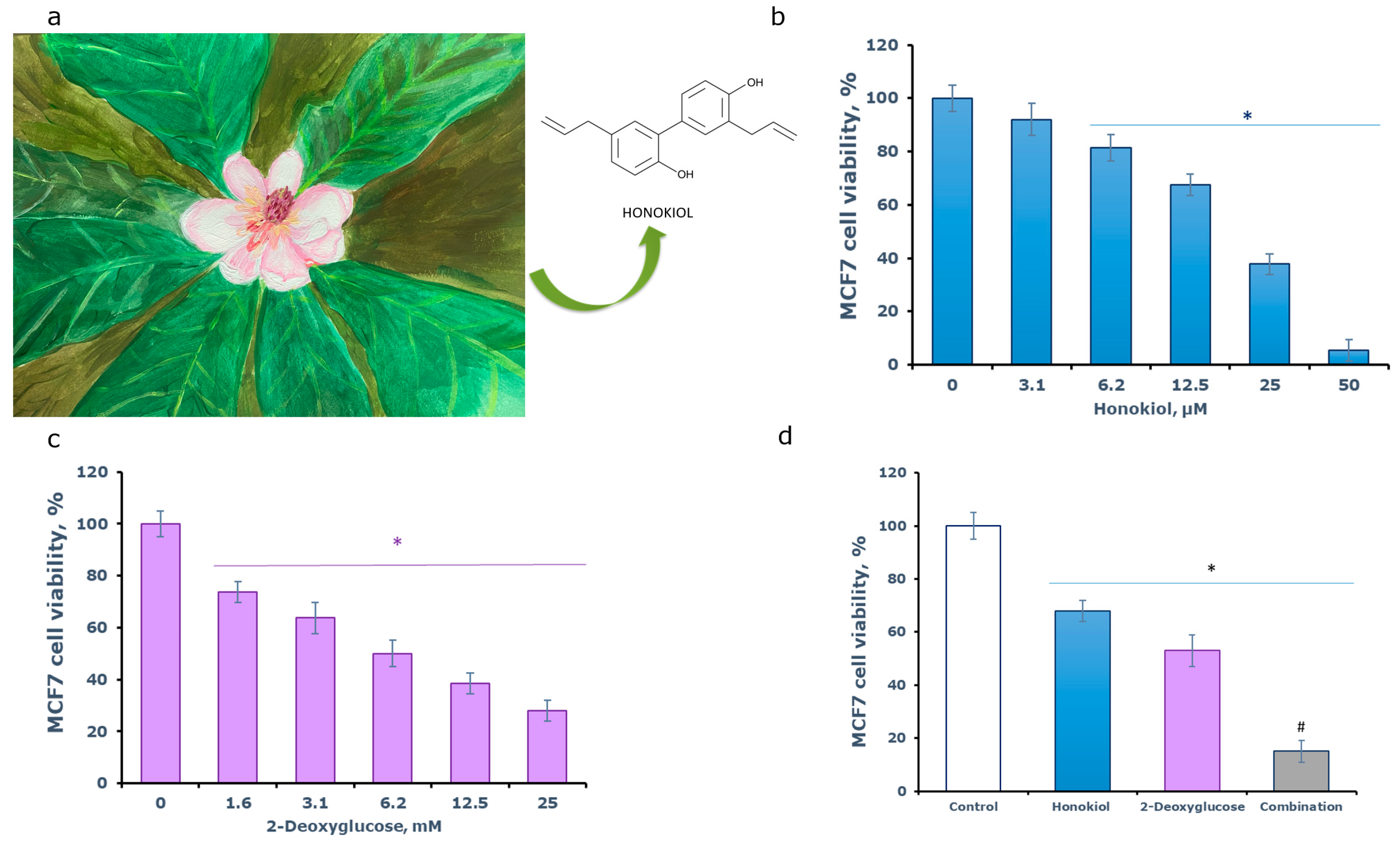
Figure 1. (a) Magnolia obovata (Magnolia hypoleuca) as a source of honokiol. (b) Antiproliferative activity of honokiol and (c) 2-deoxyglucose. (d) Drug combination study of honokiol and 2-deoxyglucose. The MCF7 cells were treated with 12 µM honokiol, 6 mM 2-deoxyglucose, or their combination. The cell viability was assessed by the MTT assay. *—p < 0.05 versus control, #—p < 0.05 versus single-agent treatment.
Engproc 56 00168 g001
Figure 2. Immunoblotting analysis of signalling pathways in MCF7 cells treated with honokiol combined with 2-deoxyglucose (2-DG). The MCF7 were treated with the compounds and then subjected to hypoxia for the indicated periods, (a)—7 h and (b)—48 h, norm—normoxia (21% O2).
Engproc 56 00168 g002
Disclaimer/Publisher’s Note: The statements, opinions and data contained in all publications are solely those of the individual author(s) and contributor(s) and not of MDPI and/or the editor(s). MDPI and/or the editor(s) disclaim responsibility for any injury to people or property resulting from any ideas, methods, instructions or products referred to in the content.