
Image Enhancement CNN Approach to COVID-19 Detection Using Chest X-ray Images †

 ,
, 
Abstract
:1. Introduction
2. Methodology
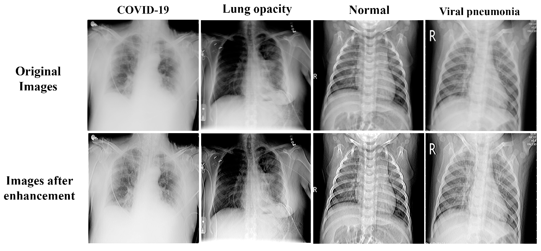
2.1. Dataset
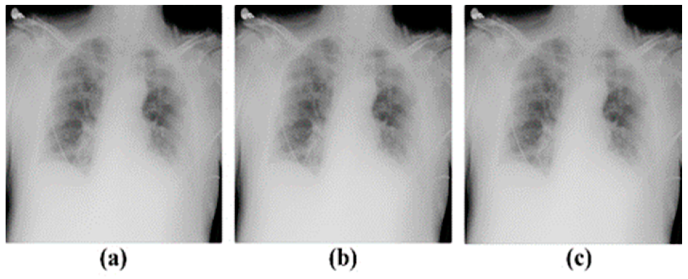
2.2. Pre-Processing
2.3. Proposed Method
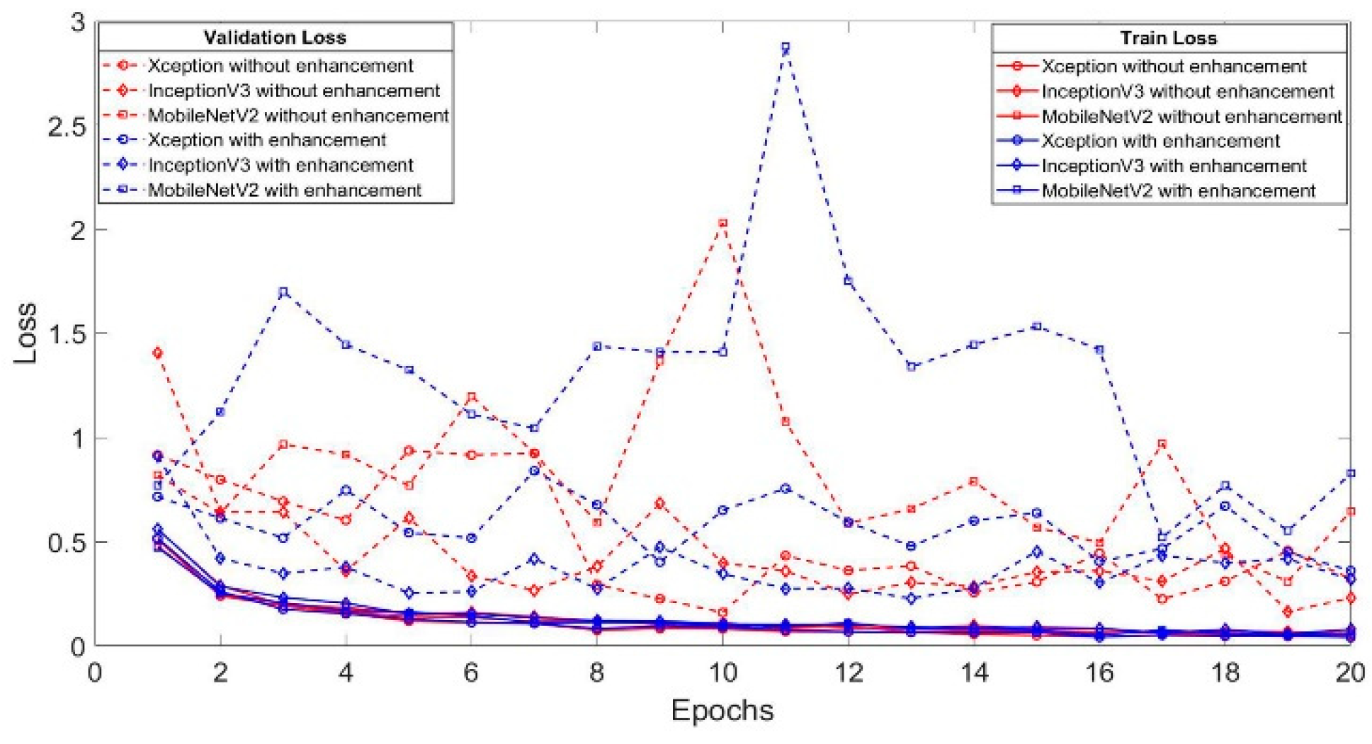
2.4. Specification of Model Training
3. Result and Discussion
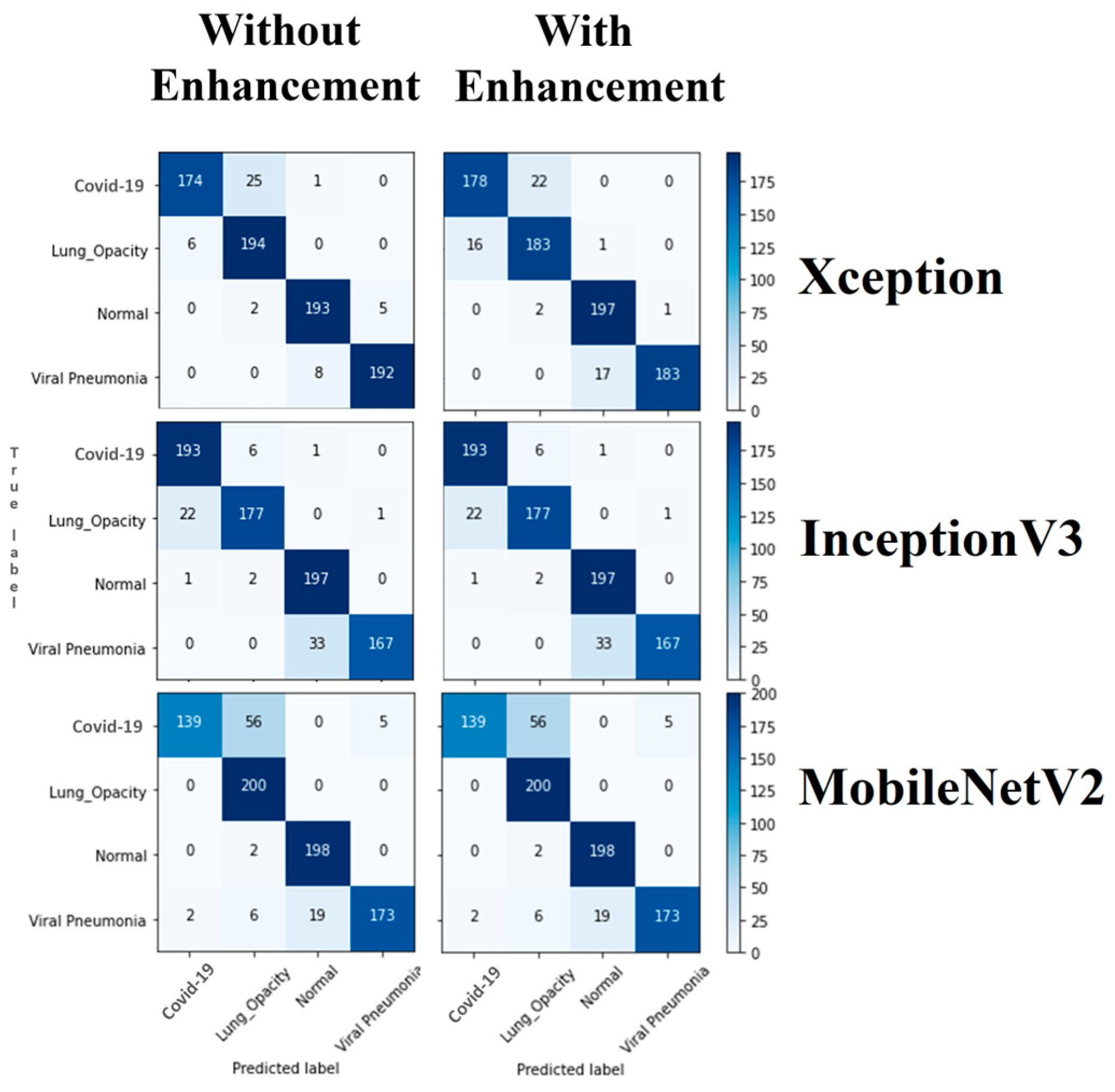
3.1. Without the Enhancement Technique
3.2. With the Enhancement Technique
4. Conclusions
Author Contributions
Funding
Institutional Review Board Statement
Informed Consent Statement
Data Availability Statement
Conflicts of Interest
References
- COVID Live Update: 222,276,536 Cases and 4,593,898 Deaths from the Coronavirus—Worldometer. Available online: https://www.worldometers.info/coronavirus/?utmcampaign=homeAdvegas1? (accessed on 8 September 2021).
- Kaggle. COVID-19 Radiography Dataset. Available online: https://www.kaggle.com/tawsifurrahman/covid19-radiography-database/activity (accessed on 25 May 2021).
- Kadir, M.A.; Mahbub, Z.B.; Islam, K.R.; Khan, M.S.; Can, A.I. Help in Screening Viral and COVID-19 Pneumonia? IEEE Access 2020, 8, 132665–132676. [Google Scholar] [CrossRef]
- Narin, A.; Kaya, C.; Pamuk, Z. Automatic detection of coronavirus disease (COVID-19) using X-ray images and deep convolutional neural networks. Pattern Anal. Appl. 2021, 24, 1207–1220. [Google Scholar] [CrossRef] [PubMed]
- Jain, R.; Gupta, M.; Taneja, S.; Hemanth, D.J. Deep learning based detection and analysis of COVID-19 on chest X-ray images. Appl. Intell. 2021, 51, 1690–1700. [Google Scholar] [CrossRef] [PubMed]
- Rahman, T.; Khandakar, A.; Qiblawey, Y.; Tahir, A.; Kiranyaz, S.; Kashem, S.B.A.; Islam, M.T.; Al Maadeed, S.; Zughaier, S.M.; Khan, M.S.; et al. Exploring the effect of image enhancement techniques on COVID-19 detection using chest X-ray images. Comput. Biol. Med. 2021, 132, 104319. [Google Scholar] [CrossRef] [PubMed]
- Toğaçar, M.; Ergen, B.; Cömert, Z. COVID-19 detection using deep learning models to exploit Social Mimic Optimization and structured chest X-ray images using fuzzy color and stacking approaches. Comput. Biol. Med. 2020, 121, 103805. [Google Scholar] [CrossRef] [PubMed]
- Ozturk, T.; Talo, M.; Yildirim, E.A.; Baloglu, U.B.; Yildirim, O.; Acharya, U.R. Automated detection of COVID-19 cases using deep neural networks with X-ray images. Comput. Biol. Med. 2020, 121, 103792. [Google Scholar] [CrossRef] [PubMed]
- Wang, L.; Lin, Z.Q.; Wong, A. COVID-Net: A tailored deep convolutional neural network design for detection of COVID-19 cases from chest X-ray images. Sci. Rep. 2020, 10, 19549. [Google Scholar] [CrossRef] [PubMed]
- Khan, A.I.; Shah, J.L.; Bhat, M.M. CoroNet: A deep neural network for detection and diagnosis of COVID-19 from chest x-ray images. Comput. Methods Programs Biomed. 2020, 196, 105581. [Google Scholar] [CrossRef] [PubMed]
- Muralidharan, N.; Gupta, S.; Prusty, M.R.; Tripathy, R.K. Detection of COVID19 from X-ray images using multiscale Deep Convolutional Neural Network. Appl. Soft Comput. 2022, 119, 108610. [Google Scholar] [CrossRef] [PubMed]
- Asghar, U.; Arif, M.; Ejaz, K.; Vicoveanu, D.; Izdrui, D.; Geman, O. An Improved COVID-19 Detection using GAN-Based Data Augmentation and Novel QuNet-Based Classification. BioMed Res. Int. 2022, 2022, 8925930. [Google Scholar] [CrossRef] [PubMed]
- Sanida, T.; Sideris, A.; Tsiktsiris, D.; Dasygenis, M. Lightweight Neural Network for COVID-19 Detection from Chest X-ray Images Implemented on an Embedded System. Technologies 2022, 10, 37. [Google Scholar] [CrossRef]
- SIIM-ISIC Melanoma Classification. Available online: https://kaggle.com/c/siim-isic-melanoma-classification (accessed on 15 March 2022).
- Results—dupeGuru 4.0.3 Documentation. Available online: https://dupeguru.voltaicideas.net/help/en/results.html (accessed on 26 March 2022).
- Shah, A. (Exploring Neurons) Through the Eyes of Gabor Filter. Medium, 17 June 2018. Available online: https://medium.com/@anuj-shah/through-the-eyes-of-gabor-filter-17d1fdb3ac97 (accessed on 21 March 2022).
- Team, K. Keras Documentation: Keras Applications. Available online: https://keras.io/api/applications/ (accessed on 21 March 2022).
- Marcelino, P. Transfer Learning from Pre-Trained Models. Medium, 23 October 2018. Available online: https://towardsdatascience.com/transfer-learning-from-pre-trained-models-f2393f124751 (accessed on 15 September 2021).
- Team, K. Keras Documentation: Transfer Learning & Fine-Tuning. Available online: https://keras.io/guides/transfer-learning/ (accessed on 26 March 2022).
- Google Colaboratory. Available online: https://colab.research.google.com/?utm-source=scs-index (accessed on 26 March 2022).


| Research Paper. | Dataset | Number of Classes | Best Model Name | Best Accuracy (%) | |
|---|---|---|---|---|---|
| CXR Images | COVID-19 | ||||
| [3] | 3487 | 423 | 2 (COVID-19, Normal) | DenseNet201 | 99.70 |
| 3 (COVID-19, Viral Pneumonia, Normal) | 97.94 | ||||
| [4] | 7406 | 341 | 2 (COVID-19, Normal) | ResNet50 | 96.10 |
| 2 (COVID-19, Viral Pneumonia) | ResNet101 | 99.50 | |||
| 2 (COVID-19, Bacterial Pneumonia) | ResNet50 | 99.70 | |||
| [5] | 6432 | 576 | 3 (COVID-19, Pneumonia, Normal) | Xception | 97.97 |
| [6] | 18,479 | 3616 | 3 (COVID-19, Lung opacity, Normal) | ChexNet | 96.29 |
| [7] | 458 | 295 | 3 (COVID-19, Pneumonia, Normal) | SqueezeNet | 99.27 |
| [8] | 1125 | 125 | 3 (COVID-19, Pneumonia, No Findings) | DarkCovidNet | 98.08 |
| 2 (COVID-19, No Findings) | 87.02 | ||||
| [9] | 13,975 | 358 | 3 (COVID-19, Pneumonia, Normal) | COVID-Net | 93.30 |
| [10] | 1251 | 284 | 4 (COVID-19, Viral Pneumonia, Bacterial Pneumonia, Normal) | CoroNet | 89.60 |
| 3 (COVID-19, Pneumonia, Normal) | 95.00 | ||||
| 2 (COVID-19, Normal) | 99.00 | ||||
| [11] | 1125 | 125 | 3 (COVID-19, Pneumonia, No Findings) | Multiscale deep CNN | 96.00 |
| 2 (COVID-19, No Findings) | 100.00 | ||||
| 9000 | 3000 | 3 (COVID-19, Pneumonia, No Findings) | 97.17 | ||
| 2 (COVID-19, No Findings) | 96.06 | ||||
| [12] | 450 | 150 | 3 (COVID-19, Pneumonia, Normal) | QuNet | 92.92 |
| [13] | 21,165 | 3616 | 4 (COVID-19, Viral Pneumonia, Normal, Lung Opacity) | Modified MobileNetV2 | 95.80 |
| Medical Case | Number of CXR Images | Used Images | ||
|---|---|---|---|---|
| Training | Validation | Testing | ||
| COVID-19 (positive) | 3615 | 1000 | 145 | 200 |
| Normal (healthy) | 10,192 | 1000 | 145 | 200 |
| Lung opacity | 6012 | 1000 | 145 | 200 |
| Viral pneumonia | 1345 | 1000 | 145 | 200 |
| Model Name | Enhancement Techniques Used | Accuracy (%) | Time Spent (min) | ||
|---|---|---|---|---|---|
| Training | Validation | Testing | |||
| MobileNetV2 | Not used | 83.67 | 83.28 | 88.75 | 50 |
| Gabor filter | 93.05 | 87.76 | 89.25 | 43 | |
| InceptionV3 | Not used | 97.85 | 91.38 | 91.75 | 84 |
| Gabor filter | 96.63 | 89.48 | 92.00 | 76 | |
| Xception | Not used | 99.10 | 91.21 | 94.13 | 111 |
| Gabor filter | 98.68 | 89.83 | 92.62 | 125 | |
| Class | Enhancement Techniques | Xception | InceptionV3 | MobileNetV2 | Support | ||||||
|---|---|---|---|---|---|---|---|---|---|---|---|
| Precision | Recall | F1-Score | Precision | Recall | F1-Score | Precision | Recall | F1-Score | |||
| COVID-19 | Not used | 0.967 | 0.870 | 0.916 | 0.894 | 0.965 | 0.928 | 0.986 | 0.695 | 0.815 | 200 |
| Gabor Filter | 0.918 | 0.890 | 0.904 | 0.865 | 0.960 | 0.910 | 0.855 | 0.945 | 0.898 | 200 | |
| Lung opacity | Not used | 0.878 | 0.970 | 0.922 | 0.957 | 0.885 | 0.919 | 0.758 | 1.000 | 0.862 | 200 |
| Gabor Filter | 0.884 | 0.915 | 0.899 | 0.994 | 0.865 | 0.925 | 0.983 | 0.845 | 0.909 | 200 | |
| Normal | Not used | 0.955 | 0.965 | 0.960 | 0.853 | 0.985 | 0.914 | 0.912 | 0.990 | 0.950 | 200 |
| Gabor Filter | 0.916 | 0.985 | 0.949 | 0.857 | 0.990 | 0.919 | 0.988 | 0.790 | 0.878 | 200 | |
| Viral pneumonia | Not used | 0.975 | 0.960 | 0.967 | 0.994 | 0.835 | 0.908 | 0.972 | 0.865 | 0.915 | 200 |
| Gabor Filter | 0.995 | 0.915 | 0.953 | 1.000 | 0.865 | 0.928 | 0.802 | 0.990 | 0.886 | 200 | |
| Accuracy | Not used | 0.941 | 0.917 | 0.887 | 800 | ||||||
| Gabor Filter | 0.926 | 0.920 | 0.892 | 800 | |||||||
| Macro avg | Not used | 0.944 | 0.941 | 0.941 | 0.924 | 0.917 | 0.917 | 0.907 | 0.887 | 0.886 | 800 |
| Gabor Filter | 0.928 | 0.926 | 0.926 | 0.929 | 0.920 | 0.920 | 0.907 | 0.893 | 0.893 | 800 | |
| Weighted avg | Not used | 0.944 | 0.941 | 0.941 | 0.924 | 0.917 | 0.917 | 0.907 | 0.887 | 0.886 | 800 |
| Gabor Filter | 0.928 | 0.926 | 0.926 | 0.929 | 0.920 | 0.920 | 0.907 | 0.892 | 0.893 | 800 | |
Disclaimer/Publisher’s Note: The statements, opinions and data contained in all publications are solely those of the individual author(s) and contributor(s) and not of MDPI and/or the editor(s). MDPI and/or the editor(s) disclaim responsibility for any injury to people or property resulting from any ideas, methods, instructions or products referred to in the content. |
© 2023 by the authors. Licensee MDPI, Basel, Switzerland. This article is an open access article distributed under the terms and conditions of the Creative Commons Attribution (CC BY) license (https://creativecommons.org/licenses/by/4.0/).
Share and Cite
Kumara, C.T.; Pushpakumari, S.C.; Udhyani, A.J.; Aashiq, M.; Rajendran, H.; Kumara, C.W. Image Enhancement CNN Approach to COVID-19 Detection Using Chest X-ray Images. Eng. Proc. 2023, 55, 45. https://doi.org/10.3390/engproc2023055045
Kumara CT, Pushpakumari SC, Udhyani AJ, Aashiq M, Rajendran H, Kumara CW. Image Enhancement CNN Approach to COVID-19 Detection Using Chest X-ray Images. Engineering Proceedings. 2023; 55(1):45. https://doi.org/10.3390/engproc2023055045
Chicago/Turabian StyleKumara, Chamoda Tharindu, Sandunika Charuni Pushpakumari, Ashmini Jeewa Udhyani, Mohamed Aashiq, Hirshan Rajendran, and Chinthaka Wasantha Kumara. 2023. "Image Enhancement CNN Approach to COVID-19 Detection Using Chest X-ray Images" Engineering Proceedings 55, no. 1: 45. https://doi.org/10.3390/engproc2023055045
APA StyleKumara, C. T., Pushpakumari, S. C., Udhyani, A. J., Aashiq, M., Rajendran, H., & Kumara, C. W. (2023). Image Enhancement CNN Approach to COVID-19 Detection Using Chest X-ray Images. Engineering Proceedings, 55(1), 45. https://doi.org/10.3390/engproc2023055045