Proceeding Paper

A Sulfo-Cyanine Dye as a Colorimetric Chemosensor for Metal Cation Recognition †

by
Cátia D. F. Martins
,
M. Manuela M. Raposo
and
Susana P. G. Costa
*
Centre of Chemistry, University of Minho, Campus de Gualtar, 4710-057 Braga, Portugal
*
Author to whom correspondence should be addressed.
Presented at the 9th International Electronic Conference on Sensors and Applications, 1–15 November 2022; Available online: https://ecsa-9.sciforum.net/.
Eng. Proc. 2022, 27(1), 12; https://doi.org/10.3390/ecsa-9-13219
Published: 1 November 2022

Abstract

:
Metal cations play important roles in several industrial and biochemical processes. However, high levels of some cations are toxic and consequently cause serious health and environmental problems. Because of these features, the search for organic molecules capable of coordinating these analytes is an increasingly studied topic in the scientific community, especially those with optical responses (optical chemosensors). Following the research group’s interest in heterocyclic optical chemosensors for various ions, a sulfo-cyanine, which absorbs and emits in the NIR region, was studied as a chemosensor for the recognition of metal cations with biological and environmental relevance. Chemosensing studies showed that this sulfo-cyanine displayed a highly sensitive colorimetric response, from blue to colorless, for Cu2+ and Fe3+ in acetonitrile solution.

1. Introduction

The development of small organic molecules as optical chemosensors for the recognition and detection of environmentally and biologically important metal ions is an essential research topic in supramolecular chemistry. In particular, chemosensors based on colorimetric changes have attracted considerable attention due to their advantages, namely cost-effectiveness, high sensitivity, and naked eye detection without the need for expensive equipment [1,2,3].
To date, various classic dyes have been applied as chemosensory signaling units, such as coumarin [4], pyrene [5], 1,8-naphthalimide [6], xanthenes [7], boron dipyrromethene difluoride (BODIPY) [8], and cyanine [9] derivatives. Among them, cyanine-based platforms have gained significant interest because of their remarkable spectral properties, including narrow absorption bands, large molar absorptivity, high sensitivity, and particularly an intense π-π* absorption which can be easily tuned from the visible to the near-infrared (NIR) region by structural modifications at the chromophore moiety [10,11,12]. For example, classic cyanine-based Cy3 dyes with three methine groups emit visible light, while pentamethine cyanine dyes Cy5 are fluorescent in the NIR region [9].
Furthermore, metal ion–cyanine interactions can produce a color change associated with the delocalization of π electrons, allowing for the easy detection of the intended analyte in solution, making them ideal candidates as colorimetric sensors for metal recognition. Therefore, there is great interest in exploring colorimetric chemosensors based on the cyanine moiety through the insertion of suitable functional groups [13,14].
Bearing these facts in mind, and considering the research group’s experience on the design, synthesis, and characterization of heterocyclic optical chemosensors [8,15,16,17], we report herein the evaluation of a sulfonated pentamethine cyanine (sulfo-Cy5) dye as a potential colorimetric chemosensor for metal cation recognition. Therefore, interaction studies of this dye in the presence of several cations were performed through a preliminary chemosensory study, followed by spectrophotometric titrations.

2. Experimental Section

2.1. Methods and Materials

The synthesis and structural characterization of sulfo-Cy5 carboxylic acid 1 has been reported by us elsewhere [18]. UV–vis absorption spectra were obtained using a Shimadzu UV/2501PC spectrophotometer (Shimadzu Europa GmbH, Duisburg, Germany) and fluorescence spectra were collected using a FluoroMax-4 spectrofluorometer (HORIBA Europe GmbH, Darmstadt, Germany) in standard quartz cuvettes.

2.2. Photophysical Characterization

The photophysical characterization of sulfo-Cy5 carboxylic acid 1 was carried out by UV–vis absorption and fluorescence spectroscopy of a 5 × 10⁻⁶ M solution in UV-grade acetonitrile. The fluorescence spectrum of 1 was obtained by excitation at the wavelength of maximum absorption. The relative fluorescence quantum yield was calculated using a solution of Nile blue in methanol as the standard (ΦF = 0.27) [19].

2.3. Preliminary Chemosensing Studies and Spectrophotometric Titrations

An evaluation of sulfo-Cy5 carboxylic acid 1 as a colorimetric chemosensor was undertaken in the presence of several cations (Ag+, K+, Li+, Na+, Cu+, TBT+, Hg2+, Ca2+, Co2+, Pb2+, Mn2+, Fe2+, Zn2+, Ni2+, Cd2+, Cu2+, Pd2+, Cs2+, Sn2+, Fe3+, and Al3+). Solutions of compound 1 (1 × 10⁻⁵ M) and solutions of cations under study (ca. 1 × 10⁻² to 1 × 10⁻³ M) were prepared in UV-grade acetonitrile.
A preliminary study was carried out by addition of 50 equivalents of each cation to the solution (1 mL) of compound 1. Considering this preliminary result, spectrophotometric titrations of compound 1 with selected cations were performed by the sequential addition of the cation stock solution to the solution of compound 1, with the collection of absorption spectra.

3. Results and Discussion

3.1. Photophysical Characterization

The photophysical properties of the pentamethine sulfo-cyanine 1 (Figure 1), previously synthesized [18], were evaluated by UV–vis absorption and fluorescence spectroscopy in acetonitrile solution. The compound’s solution displayed a vibrant blue color and the UV–vis absorption and fluorescence data for sulfo-Cy5 carboxylic acid 1 (maximum absorption wavelength, λmax; molar extinction coefficient, ε; maximum emission wavelength, λem; relative fluorescence quantum yield, ΦF; and Stokes’ shift, ∆λ) are compiled in Table 1.
The sulfo-Cy5 carboxylic acid 1 displayed a narrow absorption band (Figure 2) and high molar absorptivity (log ε = 4.99) at 651 nm. Upon excitation at the wavelength of maximum absorption, compound 1 exhibited a sharp emission band with a wavelength of maximum fluorescence in the NIR region (λem = 672 nm). The narrow bands and the small Stokes’ shift are typical features of this class of compounds [20].

3.2. Preliminary Chemosensing Studies and Spectrophotometric Titrations

An evaluation of sulfo-Cy5 carboxylic acid 1 as a colorimetric chemosensor was carried out in the presence of several cations with biological and environmental relevance, through preliminary chemosensory studies, followed by spectrophotometric titrations.
Firstly, the colorimetric behavior of compound 1 was studied by the addition of 50 equivalents of each cation in acetonitrile (Figure 3). The chromogenic response of compound 1 was remarkably visible to the naked eye, with a color change from blue to colorless in the presence of Cu2+ and Fe3+ and from blue to darker blue upon interaction with Hg2+, Ca2+, and Pb2+.
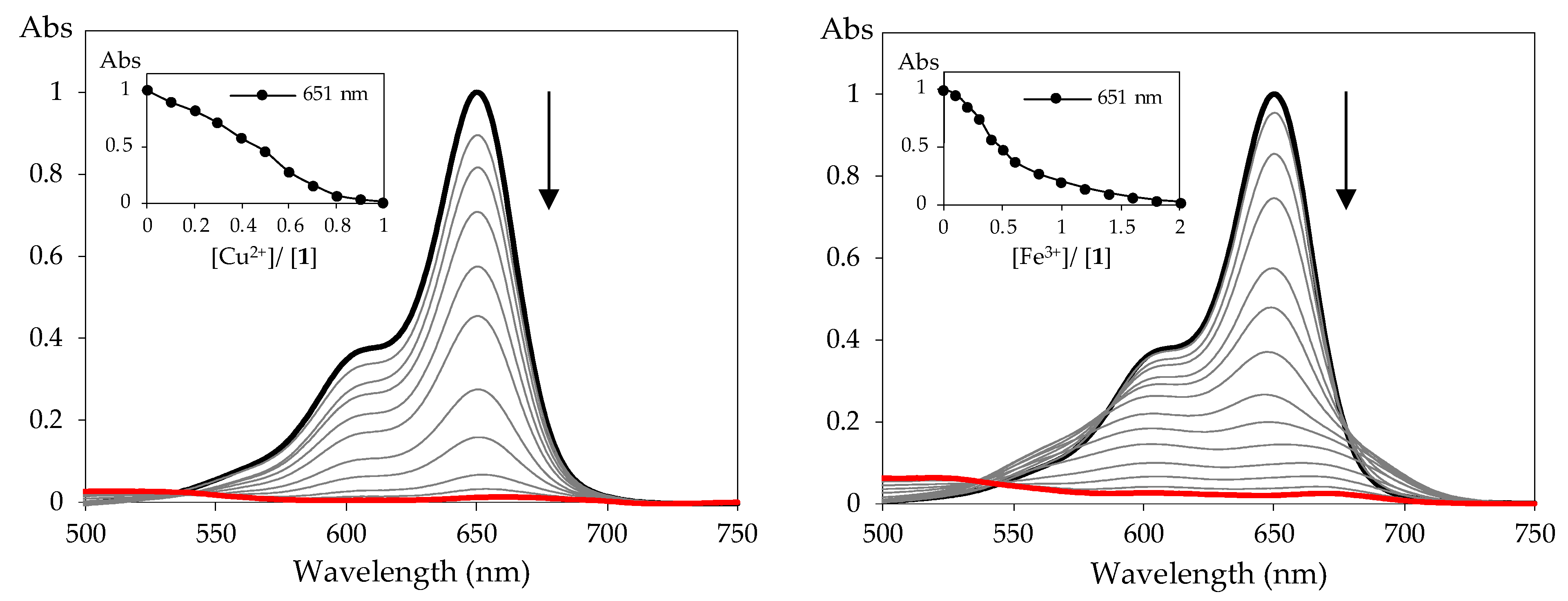
Given the preliminary colorimetric sensing results and the most dramatic changes, spectrophotometric titrations were performed for compound 1 with Cu2+ and Fe3+ in acetonitrile solutions (Figure 4). The UV–visible spectra revealed a trend: the intensity of the longest wavelength absorption band at 651 nm decreased upon the addition of the metal cation. Moreover, the sensitivity of compound 1 towards Cu2+ and Fe3+ was evident, since the addition of only 1 and 2 equivalents, respectively, was necessary to achieve a plateau.
The very low number of Cu2+ and Fe3+ equivalents necessary for a noticeable color change is a good indicator of the sensitivity of the tested sulfo-Cy5 1 towards these cations. Among transition metal ions, copper and iron are essential trace elements in the human body, playing important roles in many fundamental physiological processes, but excessive levels of these cations can result in serious disorders such as Huntington, Parkinson’s, Alzheimer’s, Menke’s, and Wilson’s diseases [21,22]. Therefore, finding highly sensitive receptors that can detect Cu2+ and Fe3+ is of great importance.

4. Conclusions

In summary, the photophysical properties of a sulfo-cyanine containing two sulfonic acid groups were studied by UV–vis absorption and fluorescence spectroscopy in acetonitrile. Furthermore, this compound was evaluated as a colorimetric chemosensor for several cations with biological and environmental relevance. A preliminary chemosensing study showed that cyanine 1 displayed a highly colorimetric response, from blue to colorless, for Cu2+ and Fe3+ in acetonitrile solution. As a result, spectrophotometric titrations were performed for compound 1 with the selected cations, confirming the sensitivity of this compound in the presence of these analytes. These results clearly indicate that sulfo-Cy5 carboxylic acid 1 can be used to detect Cu2+ and Fe3+ with remarkable sensitivity.

Author Contributions

Conceptualization, S.P.G.C.; methodology, C.D.F.M. and S.P.G.C.; validation, S.P.G.C. and M.M.M.R.; formal analysis, C.D.F.M.; M.M.M.R. and S.P.G.C.; investigation, C.D.F.M.; resources, S.P.G.C. and M.M.M.R.; writing—original draft preparation, C.D.F.M.; writing—review and editing, C.D.F.M.; M.M.M.R. and S.P.G.C.; supervision, S.P.G.C. and M.M.M.R.; project administration, S.P.G.C.; funding acquisition, M.M.M.R. and S.P.G.C. All authors have read and agreed to the published version of the manuscript.

Funding

This research was funded by the Foundation for Science and Technology (FCT) for financial support to CQ/UM (UID/QUI/00686/2020) and project PTDC/QUI-COL/28052/2017 and a PhD grant to C. D. F. Martins (SFRH/BD/05277/2020). The NMR spectrometer Bruker Avance III 400 is part of the National NMR Network and was purchased within the framework of the National Program for Scientific Re-Equipment (contract REDE/1517/RMN/2005), with funds from POCI 2010 (FEDER) and FCT.

Institutional Review Board Statement

Not applicable.

Informed Consent Statement

Not applicable.

Data Availability Statement

Not applicable.

Conflicts of Interest

The authors declare no conflict of interest.

References

  1. You, L.; Zha, D.; Anslyn, E.V. Recent advances in supramolecular analytical chemistry using optical sensing. Chem. Rev. 2015, 115, 7840–7892. [Google Scholar] [CrossRef]
  2. Kaur, B.; Kaur, N.; Kumar, S. Colorimetric metal ion sensors—A comprehensive review of the years 2011–2016. Coord. Chem. Rev. 2018, 358, 13–69. [Google Scholar] [CrossRef]
  3. Mohammadi, A.; Yaghoubi, S. A new dual colorimetric chemosensor based on quinazolinone for CN, AcO and Cu2+ ions. Sens. Actuators B Chem. 2017, 241, 1069–1075. [Google Scholar] [CrossRef]
  4. Zhou, Y.; Liu, K.; Li, J.-Y.; Fang, Y.; Zhao, T.-C.; Yao, C. Visualization of nitroxyl in living cells by a chelated copper(II) coumarin complex. Org. Lett. 2011, 13, 1290–1293. [Google Scholar] [CrossRef] [PubMed]
  5. Gai, L.; Chen, H.; Zou, B.; Lu, H.; Lai, G.; Li, Z.; Shen, Z. Ratiometric fluorescence chemodosimeters for fluoride anion based on pyrene excimer/monomer transformation. Chem. Commun. 2012, 48, 10721–10723. [Google Scholar] [CrossRef]
  6. Banerjee, S.; Veale, E.B.; Phelan, C.M.; Murphy, S.A.; Tocci, G.M.; Gillespie, L.J.; Frimannsson, D.O.; Kelly, J.M.; Gunnlaugsson, T. Recent advances in the development of 1,8-naphthalimide based DNA targeting binders, anticancer and fluorescent cellular imaging agents. Chem. Soc. Rev. 2013, 42, 1601–1618. [Google Scholar] [CrossRef] [Green Version]
  7. Chen, X.; Pradhan, T.; Wang, F.; Kim, J.S.; Yoon, J. Fluorescent chemosensors based on spiro ring-opening of xanthenes and related derivatives. Chem. Rev. 2012, 112, 1910–1956. [Google Scholar] [CrossRef]
  8. Presti, M.L.; Martínez-Máñez, R.; Ros-Lis, J.V.; Batista, R.M.F.; Costa, S.P.G.; Raposo, M.M.M.; Sancenón, F. A dual channel sulphur-containing macrocycle functionalised BODIPY probe for the detection of Hg(II) in mixed aqueous solution. New J. Chem. 2018, 42, 7863–7868. [Google Scholar] [CrossRef] [Green Version]
  9. Sun, W.; Guo, S.; Hu, C.; Fan, J.; Peng, X. Recent development of chemosensors based on cyanine platforms. Chem. Rev. 2016, 116, 7768–7817. [Google Scholar] [CrossRef]
  10. Wolf, N.; Kersting, L.; Herok, C.; Mihm, C.; Seibel, J. High-yielding water-soluble asymmetric cyanine dyes for labeling applications. J. Org. Chem. 2020, 85, 9751–9760. [Google Scholar] [CrossRef]
  11. Owens, E.A.; Bruschi, N.; Tawney, J.G.; Henary, M. A microwave-assisted and environmentally benign approach to the synthesis of near-infrared fluorescent pentamethine cyanine dyes. Dyes Pigm. 2015, 113, 27–37. [Google Scholar] [CrossRef]
  12. Yi, X.; Wang, F.; Qin, W.; Yang, X.; Yuan, J. Near-infrared fluorescent probes in cancer imaging and therapy: An emerging field. Int. J. Nanomed. 2014, 9, 1347–1365. [Google Scholar] [CrossRef] [PubMed] [Green Version]
  13. Hu, Y.; Yin, J.; Yoon, J. A multi-responsive cyanine-based colorimetric chemosensor containing dipicolylamine moieties for the detection of Zn(II) and Cu(II) ions. Sens. Actuators B Chem. 2016, 230, 40–45. [Google Scholar] [CrossRef]
  14. Shi, Y.; Wang, R.; Yuan, W.; Liu, Q.; Shi, M.; Feng, W.; Wu, Z.; Hu, K.; Li, F. An easy-to-use colorimetric cyanine probe for the detection of Cu2+ in Wilson’s Disease. ACS Appl. Mater. Interfaces 2018, 10, 20377–20386. [Google Scholar] [CrossRef]
  15. Martins, C.D.F.; Batista, P.M.R.; Raposo, M.M.M.; Costa, S.P.G. Crown ether benzoxazolyl-alanines as fluorimetric chemosensors for the detection of palladium in aqueous environment. Chem. Proc. 2021, 3, 5. [Google Scholar]
  16. Esteves, C.I.C.; Ferreira, R.C.M.; Raposo, M.M.M.; Costa, S.P.G. New fluoroionophores for metal cations based on benzo[d]oxazol-5-yl-alanine bearing pyrrole and imidazole. Dyes Pigm. 2018, 151, 211–218. [Google Scholar] [CrossRef]
  17. Okda, H.E.; El Sayed, S.; Ferreira, R.C.M.; Gonçalves, R.C.R.; Costa, S.P.G.; Raposo, M.M.M.; Martínez-Máñez, R.; Sancenón, F. N,N-diphenylanilino-heterocyclic aldehyde-based chemosensors for UV-vis/NIR and fluorescence Cu(II) detection. New J. Chem. 2019, 43, 7393–7402. [Google Scholar] [CrossRef] [Green Version]
  18. Martins, C.D.F.; Raposo, M.M.M.; Costa, S.P.G. Synthesis and characterization of a water-soluble pentamethine indocyanine dye for peptide labeling. Chem. Proc. 2022, 8, 91. [Google Scholar]
  19. Jose, J.; Ueno, Y.; Burgess, K. Water-soluble Nile Blue derivatives: Syntheses and photophysical properties. Chem. Eur. J. 2009, 15, 418–423. [Google Scholar] [CrossRef]
  20. Wang, L.; Fan, J.; Qiao, X.; Peng, X.; Dai, B.; Wang, B.; Sun, S.; Zhang, L.; Zhang, Y. Novel asymmetric Cy5 dyes: Synthesis, photostabilities and high sensitivity in protein fluorescence labeling. J. Photochem. Photobiol. A Chem. 2010, 210, 168–172. [Google Scholar] [CrossRef]
  21. Wu, D.; Chen, L.; Lee, W.; Ko, G.; Yin, J.; Yoon, J. Recent progress in the development of organic dye based near-infrared fluorescence probes for metal ions. Coord. Chem. Rev. 2018, 354, 74–97. [Google Scholar] [CrossRef]
  22. Li, S.; Zhang, D.; Xie, X.; Ma, S.; Liu, Y.; Xu, Z.; Gao, Y.; Ye, Y. A novel solvent-dependently bifunctional NIR absorptive and fluorescent ratiometric probe for detecting Fe3+/Cu2+ and its application in bioimaging. Sens. Actuators B Chem. 2016, 224, 661–667. [Google Scholar] [CrossRef]
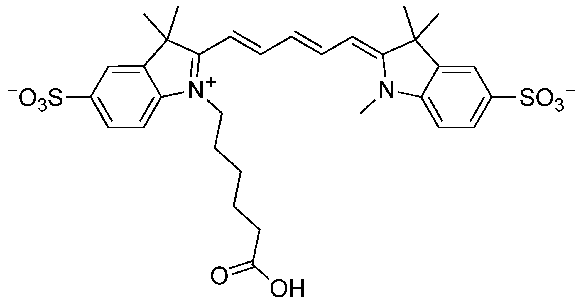
Figure 1. Structure of sulfo-Cy5 carboxylic acid 1.
Engproc 27 00012 g001
Figure 2. Normalized absorption and fluorescence spectra of sulfo-Cy5 carboxylic acid 1 (5 × 10⁻⁶ M) in acetonitrile solution.
Engproc 27 00012 g002
Figure 3. Color changes observed for sulfo-Cy5 carboxylic acid 1 after the addition of 50 equivalents of several cations in acetonitrile (1 × 10⁻⁵ M).
Engproc 27 00012 g003
Figure 4. Spectrophotometric titration of sulfo-Cy5 carboxylic acid 1 with the addition of increasing amounts of Cu2+ and Fe3+ in acetonitrile. The inset represents the normalized absorption at 651 nm ([1] = 1 × 10⁻⁵ M).
Engproc 27 00012 g004
Table 1. UV–vis absorption and fluorescence data of sulfo-Cy5 carboxylic acid 1 (5 × 10⁻⁶ M) in acetonitrile solution.
CompoundUV–Vis AbsorptionFluorescence
λmax (nm)log ελem (nm)ΦF∆λ (nm)
16514.996720.3021
Publisher’s Note: MDPI stays neutral with regard to jurisdictional claims in published maps and institutional affiliations.