Proceeding Paper

Influence of the Type and Amount of Plasticizer on the Sensory Properties of Microspheres Sensitive to Lipophilic Ions †

by
Aleksandra Kalinowska
,
Patrycja Matusiak
,
Sandra Skorupska
,
Ilona Grabowska-Jadach
and
Patrycja Ciosek-Skibińska
*
Chair of Medical Biotechnology, Faculty of Chemistry, Warsaw University of Technology, Noakowskiego 3, 00-664 Warsaw, Poland
*
Author to whom correspondence should be addressed.
Presented at the 1st International Electronic Conference on Chemical Sensors and Analytical Chemistry, 1–15 July 2021; Available online: https://csac2021.sciforum.net/.
Chem. Proc. 2021, 5(1), 90; https://doi.org/10.3390/CSAC2021-10487
Published: 30 June 2021

Abstract

:
Working parameters of chemical sensors, such as selectivity and sensitivity, can be adjusted by optimizing components of chemosensitive layers, including type and amount of plasticizer in the case of PVC membranes in optodes. Plasticizers are also used in the process of creating micro/nanospheres that are incorporated with chemical indicators to form micro/nano-scale optodes. This study investigated the influence of the type of plasticizer (polar o-NPOE and non-polar DOS) on the optical response of microspheres that are sensitive to lipophilic ions. Moreover, the amount of plasticizer was also adjusted in order to obtain satisfactory sensitivity in the widest linear range. The chemosensory response of the developed microspheres was studied with the use of spectrophotometry and spectrofluorimetry, while size of the optodes was estimated by confocal microscopy.

Published: 30 June 2021

1. Introduction

For many years, ion-selective electrodes (ISE) have been considered important analytical chemistry tools. They are widely used both in environmental analysis and in medical diagnostics [1]. Chemosensitive membranes of the ISEs whose signal is potentiometric can be easily modified by the incorporation of chromoionophores, which gives the possibility for optical transduction. On the other hand, polymer scaffolds of such optode membrane can be replaced by surfactants, which leads to the fabrication of suspension of micro- or nano-sized optodes.
Various achievements in the field of micellization of dyes can be applied for the fabrication of micro- and nano-optodes. Micelles are micro- or nanometer-sized particles that exist solution in equilibrium with the particles or ions. They can be incorporated with additional lipophilic components forming nano/microspheres, aggregates of surfactant molecules with a hydrophilic part being in contact with outer water solution, and a hydrophobic part creating inner lipophilic microenvironment [2]. Micelles are widely used in medicine for drug delivery and increase water solubility and pharmaceuticals’ bioavailability. In recent years, the huge potential of micellar systems for various applications in analytical chemistry has been noticed [3].
Working parameters of chemical sensors, such as selectivity and sensitivity, can be adjusted by optimizing components of chemosensitive membranes, including type and amount of plasticizer [4]. A plasticizer is a substance allowing for increasing polymer flexibility, as well as susceptibility to its further processing. In membranes of ISEs and optodes, it forms a lipophilic environment in which receptors and additional components responsible for sensing are suspended. A plasticizer in the simplest sense is considered an organic solvent. There are many types of such compounds, such as animal fats, petroleum fractions, and all kinds of plant extracts. Plasticizers can be used in the process of creating micro/nanospheres incorporated with chemical indicators and stabilized by surfactants. As environmental conditions change, such systems may change their optical properties, becoming optodes in the micro/nanoscale [3,5].
This study aimed to develop two microsphere types sensitive towards lipophilic ions: anion-sensitive (AS) and cation-sensitive (CS). Each of these systems includes a chromoionophore, an ion exchanger, a surfactant, and a plasticizer. In this work we were focused on selecting an appropriate plasticizer, as well as adjusting its quantity. Changes in chemosensory properties were observed in absorbance and fluorescence mode, while confocal microscopy observations were applied to verify the microspheres’ size.

2. Experimental

2.1. Chemicals

Sodium phosphate monohydrate, disodium phosphate dodecahydrate, Tris-HCl, Pluronic F127 were supplied by Sigma-Merck (Poznań, Poland). Milli-Q water was used for preparation of all aqueous solutions, including phosphate buffer pH 7.4 and Tris-HCl buffer pH 9.0. Plasticizers (2-Nitrophenyl octyl ether, o-NPOE, Bis(2-ethylhexyl) sebacate, DOS), lipophilic salts (Tridodecylmethylammonium chloride, TDMAC, Potassium tetrakis [3,5-bis(trifluoromethyl)phenyl]borate, KTFPB), chromoionophores I and XI were obtained from Fluka (Selectophore). Freshly distilled tetrahydrofuran, THF (Fluka) was used as a solvent for the microspheres’ components. All chemicals were used as received.

2.2. Preparation of Microspheres Suspensions

Two types of optode microspheres: with generic anion-sensitivity (AS) and with generic cation-sensitivity (CS), were prepared (Figure 1). The composition of the membranes and proposed mechanism of optical signal generation are based on the literature data on optodes [6,7,8,9,10]. AS microspheres contained Chromoionophore XI, TDMAC, o-NPOE or DOS, and Pluronic (F-127), whereas CS microspheres contained Chromoionophore I, KTFPB, o-NPOE or DOS, and Pluronic (F-127). Each of the components included in the given microsphere was weighed and dissolved in 1.5 mL of THF. To ensure that all ingredients dissolve well, the vial was placed in an ultrasonic bath for 5 min. 0.5 mL portions of THF solutions were pipetted to 4.5 mL of deionized water on a vortex. The last step was to remove the solvent by passing compressed air through the solution (the process was carried out for 1 h). Clear particle suspensions were obtained that were applied for further measurements using microtiter plates. For this purpose, 100 µL of the prepared microsphere suspension were pipetted to each well and 100 µL of appropriate analyte solution was added. We examined the chemosensory response in the presence of 0.1 M NaOH, 0.1 M HCl, and calibration solutions appropriate for each type of microspheres (1 µM-0.1 M NaClO4 and 1 µM-0.1 M NH4NO3 for AS and CS optodes, respectively). When protonation degree in AS was determined, microspheres suspensions were two times diluted (50 µL of optode cocktail + 50 µL of deionized water) to avoid recording signals out of range by microtiter plate reader.

2.3. Examination of the Optical Properties of Microspheres

Both spectrophotometric and spectrofluorimetric measurements were applied to study the chemosensory properties of the obtained microspheres. They were tested using Synergy 2 Multi-Mode Reader (BioTek Instruments, Inc., Winooski, VT, USA). In order to examine the sizes of the obtained microspheres, a confocal microscope Fluoview FV10i (Olympus, Japanese) was used. The following parameters were used to observe the samples: λex = 473 nm, λem = 490–590 nm for AS microspheres and λex = 635 nm, λem = 660–760 nm for CS microspheres. Measurements of microspheres were made using the FV10i-SW software. Samples were observed using CellviewTM Cell Culture Dish (Greiner Bio One, Germany) with a glass bottom and four-compartments. Three. Hundred microliters of suspension of microspheres were placed in each compartment.

3. Results and Discussion

3.1. Spectrophotometric Measurements of Chemosensory Properties

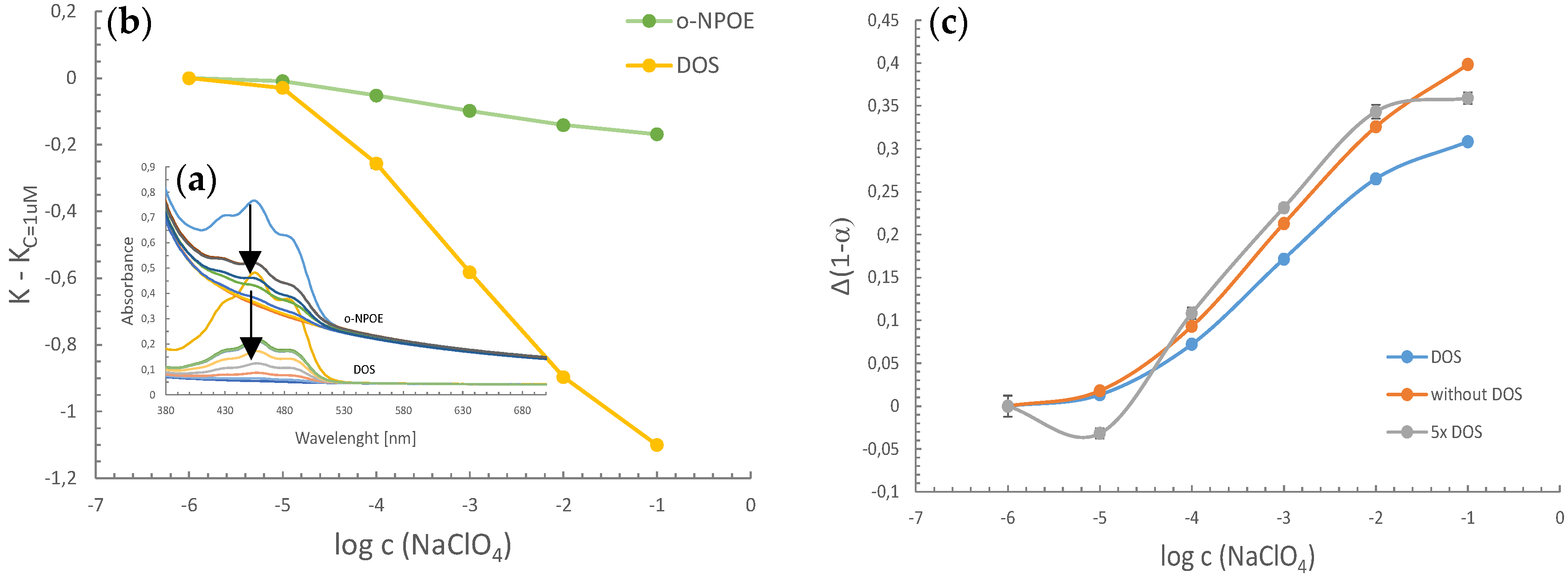
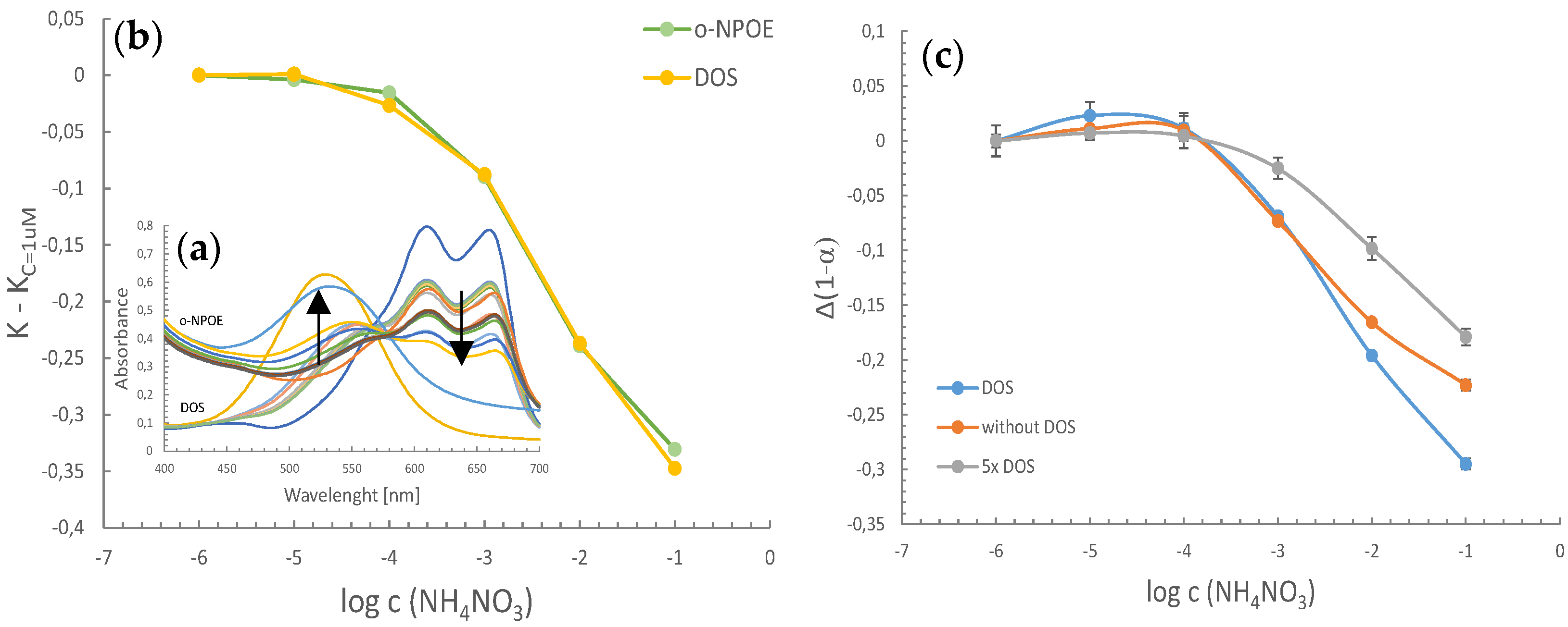
The microspheres prepared for this work, both AS and CS, contain an ion exchanger in their composition, whose task is to exchange ions between the aqueous and organic phases according to equations given in Figure 1 (exchange of lipophilic cations to protons in the case of CS microspheres, coextraction of protons with lipophilic anions in the case of AS microspheres). This processes ensure the change in the protonation degree of the chromoionophores and thus cause a change in the optical properties of the produced system. The sensory response of four independent replicates of microspheres suspensions was tested by recording UV-Vis spectra in the presence of model lipophilic ions—perchlorate ions in the case of AS system, and ammonium ions in the case of CS system (Figure 2 and Figure 3, respectively). The tested solutions were buffered to make the change of the chromoionophore spectrum independent of pH, in order to ensure, that the spectral change was influenced only by the change of the target ion concentration. The spectra were gradually changing following change in ion concentration and thus protonation degree of chromoionophore (represented by arrows in Figure 2a and Figure 3a). Calibration curves were determined for the obtained UV-Vis spectra Figure 2b and Figure 3b). First, signal based on baseline correction was calculated (ratio of absorbances obtained for 454 nm and 380 nm, K = A454/A380, and then only change related to this signal in smallest concentration was presented (K − KC=1μM) for more precise comparison of the two systems using various plasticizers (Figure 2b). The obtained results clearly show that in the AS optode system, the non-polar plasticizer DOS allowed for obtaining responses with a greater degree of sensitivity and enables the determination of anions in almost the entire concentration range.
In the case of CS microspheres, baseline correction was calculated using absorbances at 610 nm and 570 nm, and resulting signal was the change of K = A454/A380 value (Figure 3b). For CS microspheres based on DOS and o-NPOE, there was no significant difference in calibration curves—they were comparable. However, in the case of DOS-based optodes, a slightly more comprehensive linear range was observed.
Thus, a non-polar plasticizer DOS was chosen for further experiments, as the better for both microsphere types. The next phase of the research involved the study of the influence of plasticizer amount on chemosensory properties. Three formulations for AS optodes were prepared that was based on various plasticizer content. Again, UV-Vis spectra were recorded as previously, and calibration curves were determined (Figure 2c). Signals were presented as change in protonation degree, 1-α, of chromoionophore (change of 1-α related to this value obtained for the smallest concentration of the analyte, for clarity of presentation). The observation of these calibration curves revealed that the different amount of plasticizer did not significantly affect the sensory response of the system. The microspheres without plasticizers and with its five-fold amount exhibited slightly better sensitivity compared to the system with the standard amount of plasticizer. Additionally, without the addition of DOS, slightly better quantification range was noticed (10 µM–0.1 M). The narrowest linear range was observed for optode system with the highest amount of DOS. The same procedure was repeated for CS microsphere optodes. Respective calibration curves for three studied systems with various amounts of plasticizer are presented in Figure 3c. The best results in terms of sensitivity and quantification range were obtained for the standard amount of the plasticizer.

3.2. Spectrofluorimetric Measurements of Chemosensory Properties

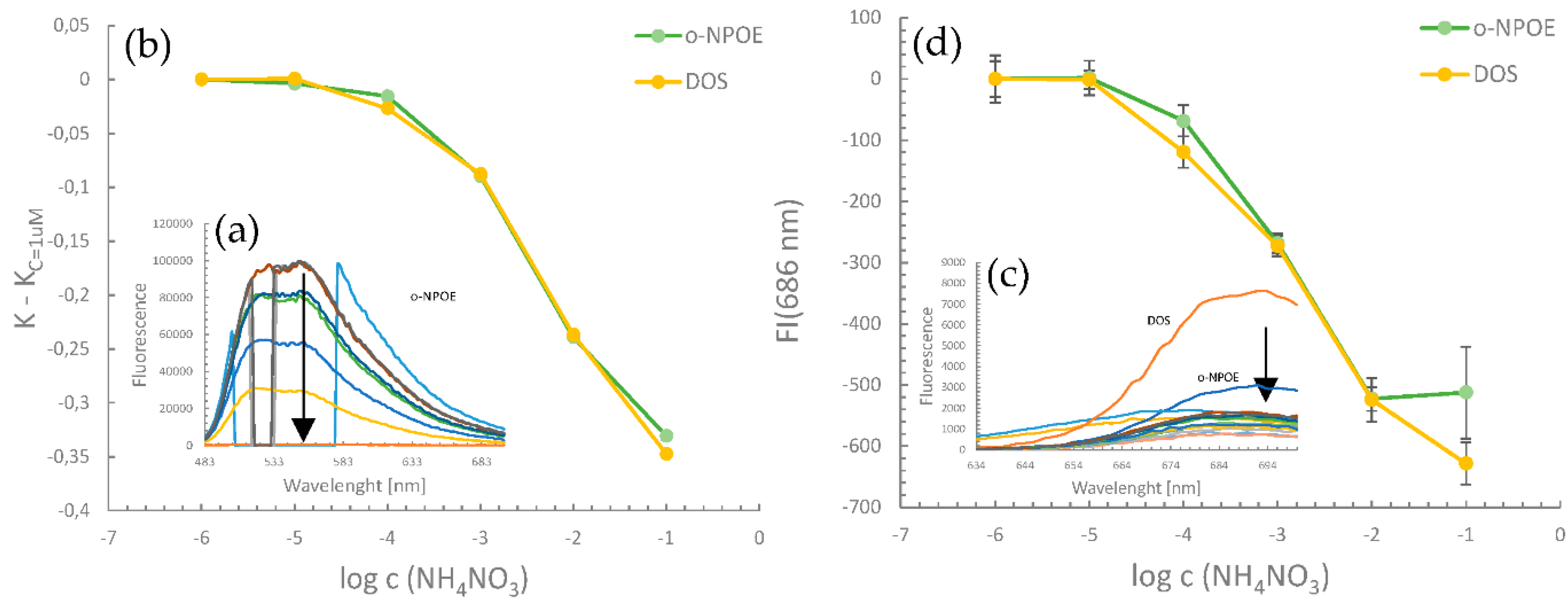
In addition to measuring the absorbance, chemosensory properties were also tested in the fluorescence mode. Similarly, as in the case of absorbance, a gradual change in the spectrum of AS and CS microspheres, influenced by the growing concentration of target ions was observed (respective emission spectra and calibration curves presented in Figure 4). In the case of both AS and CS microspheres, DOS was recognized as the better plasticizer, which enables the determination of analytes in a wider concentration range with high sensitivity. Observation of protonation degree (data not shown) revealed that the widest linear range was obtained when the standard amount of DOS was applied, while its five-fold amount caused increase in the sensitivity.

3.3. Confocal Microscope

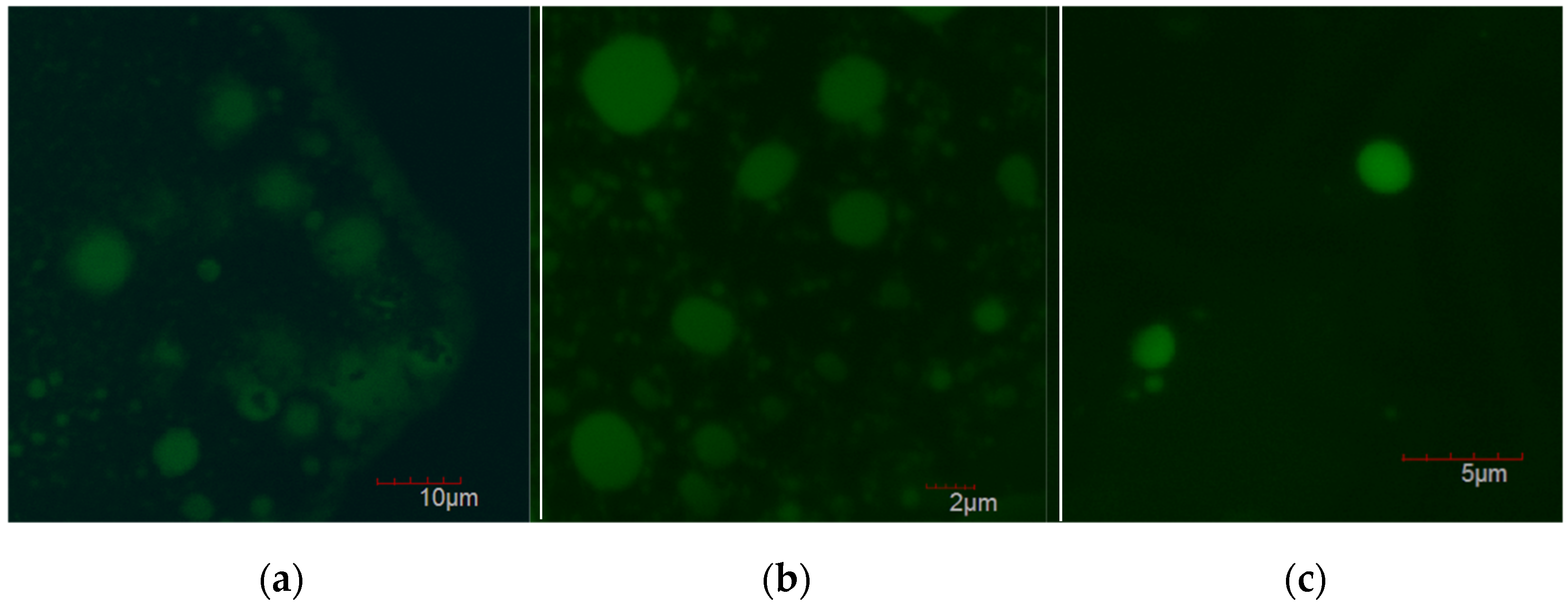
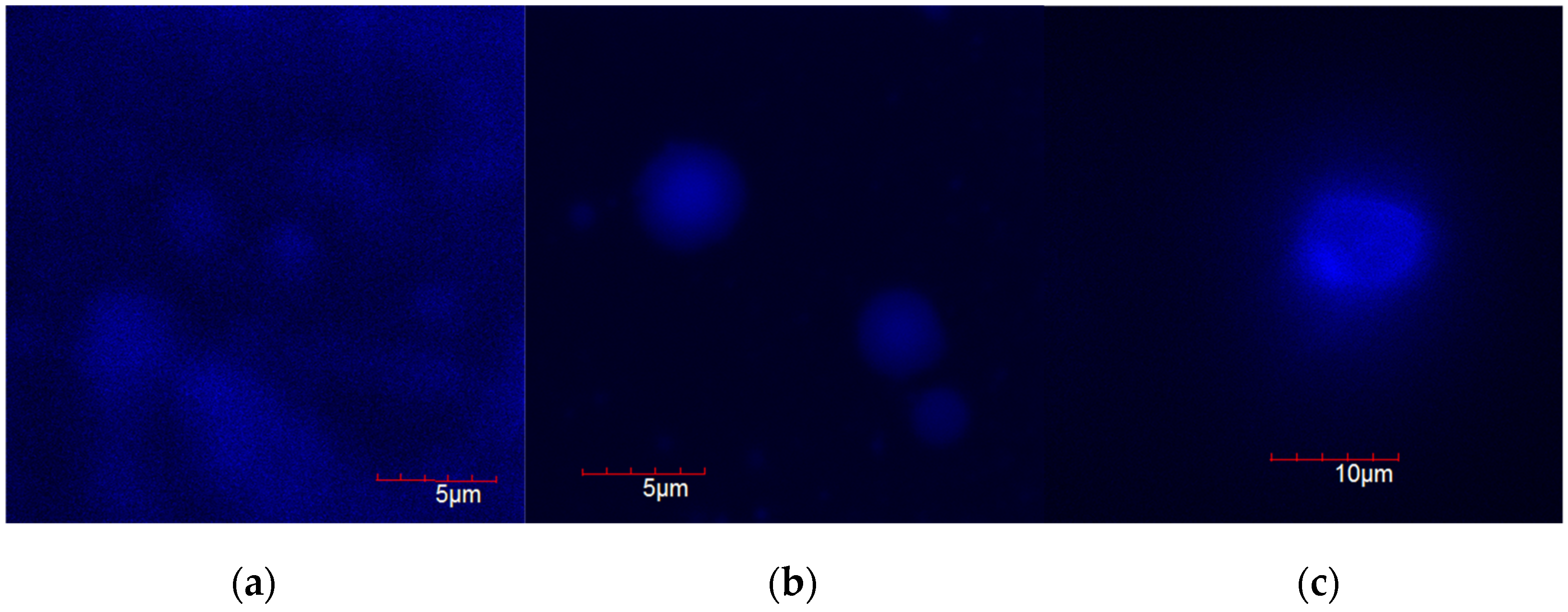
Figure 5 and Figure 6 show pictures of microspheres taken with the use of a confocal microscope. The analyzes confirmed the formation of spherical microspheres in all experimental samples regardless of the amount of plasticizer added. A slight spread of the size of the microspheres was observed. Their diameters oscillated between 2–10 µm in the case of both types of microspheres.
Based on the observation of the fluorescence intensity of the population of microspheres, it was found that they are homogeneous in terms of fluorescence intensity, which proves the incorporation of the chromoionophore. Moreover, the chromoionophores are evenly distributed in both AS and CS microspheres.

4. Conclusions

The presented results show that in the case of both AS and CS microspheres used in this work, the type and amount of plasticizer change chemosensory properties of the micro-optodes. For both types of microspheres, non-polar plasticizer DOS allowed for obtaining better results compared to the polar plasticizer in the composition. Thanks to DOS, it was possible to determine perchlorate and ammonium ions in a wide concentration range and the obtained sensitivity was higher compared to microspheres with o-NPOE content. The plasticizer amount should also be carefully adjusted to obtain satisfactory chemosensory characteristics. Generally, in the case of microspheres studied in this work, the standard amount led to the best results. All these observations were confirmed both in spectrophotometric and spectrofluorimetric measurements. The obtained results from the confocal microscope confirm the formation of spherical microspheres in size range of 2–10 μm. No influence of the amount of added plasticizer on the shape and size of the microspheres was observed. In summary, the use of an appropriate amount of a non-polar plasticizer allows for obtaining chemosensory microspheres sensitive to lipophilic ions in a broad quantification range from 10 µM to 0.1 M in both absorbance and fluorescence intensity mode.

Supplementary Materials

The following are available online at https://www.mdpi.com/article/10.3390/CSAC2021-10487/s1.

Author Contributions

Conceptualization, P.C.-S. and I.G.-J.; methodology, design of experiments, A.K. and S.S; investigation, P.M., A.K. and S.S.; Original draft preparation, A.K. and S.S.; resources, supervision, project administration, funding acquisition, review and editing of the manuscript were completed by P.C.-S. All authors have read and agreed to the published version of the manuscript.

Funding

National Science Centre (Poland): UMO-2018/30/E/ST4/00481.

Institutional Review Board Statement

Not applicable.

Informed Consent Statement

Not applicable.

Data Availability Statement

Not applicable.

Acknowledgments

This work was financially supported by National Science Centre (Poland) within the framework of the SONATA BIS project No. UMO-2018/30/E/ST4/00481. Aleksandra Kalinowska acknowledges financial support from IDUB project (Scholarship Plus program).

Conflicts of Interest

The authors declare no conflict of interest.

References

  1. Wygladacz, K.; Radu, A.; Xu, C.; Qin, Y.; Bakker, E. Fiber-Optic Microsensor Array Based on Fluorescent Bulk Optode Microspheres for the Trace Analysis of Silver Ions. Anal. Chem. 2005, 77, 4706–4712. [Google Scholar] [CrossRef] [PubMed]
  2. Xie, X.; Bakker, E. Ion selective optodes: From the bulk to the nanoscale. Anal. Chem. 2015, 407, 3899–3910. [Google Scholar] [CrossRef] [PubMed]
  3. Wang, C.X.; Wu, B.; Zhou, W.; Wang, Q.; Yu, H.; Deng, K.; Li, J.M.; Zhuo, R.X.; Huang, S.W. Turn-on fluorescent probe-encapsulated micelle as colloidally stable nanochemosensor for highly selective detection of Al3+ in aqueous solution and living cell imaging. Sens. Actuators B. Chem. 2018, 271, 225–238. [Google Scholar] [CrossRef]
  4. Bakker, E.; Bühlmann, P.; Pretsch, E. Carrier-Based Ion-Selective Electrodes and Bulk Optodes. 1. General Characteristics. Chem. Rev. 1997, 97, 3083–3132. [Google Scholar] [CrossRef] [PubMed]
  5. Wypych, G. Handbook of Plasticizers, 3rd ed.; ChemTech Publishing: Toronto, ON, Canada, 2017. [Google Scholar]
  6. Zeng, H.H.; Wang, K.M.; Li, D.; Yu, R.Q. Development of an alcohol optode membrane based on fluorescence enhancement of fluorescein derivatives. Talanta 1994, 41, 969–975. [Google Scholar] [CrossRef]
  7. Ortuño, J.A.; Albero, M.I.; García, M.S.; Sánchez-Pedreño, C.; García, M.I.; Expósito, R. Flow-through bulk optode for the fluorimetric determination of perchlorate. Talanta 2003, 60, 563–569. [Google Scholar] [CrossRef]
  8. Chan, W.H.; Lee, A.W.M.; Wang, K. Design of a primary amine-selective optode membrane based on a lipophilic hexaester of calix[6]arene. Analyst 1994, 119, 2809–2812. [Google Scholar] [CrossRef]
  9. Chan, W.H.; Lee, A.W.M.; Lee, C.M.; Yau, K.W.; Wang, K. Design and characterization of sodium-selective optode membranes based on the lipophilic tetraester of calix[4]arene. Analyst 1995, 120, 1963–1967. [Google Scholar] [CrossRef]
  10. Du, X.; Zhu, C.; Xie, X. Thermochromic Ion-Exchange Micelles Containing H+ Chromoionophores. Langmuir 2017, 33, 5910–5914. [Google Scholar] [CrossRef] [PubMed]
Figure 1. Schematic representation of the prepared microspheres, their composition, and the mechanisms of target analyte recognition with optical signal generation. The mechanisms by which the spheres operate are based on the extraction of the analyte from the aqueous medium (aq subscript) into the lipophilic interior of the microsphere (m subscript). Each type of microsphere solution is based on a plasticizer emulsion stabilized with a Pluronic non-ionic surfactant having hydrophobic and hydrophilic domains (PPO and PEO, respectively). The plasticizer droplets contain all components needed to extract target analytes (lipophilic salt) and generate a signal (IND) [6,7,8,9,10].
Chemproc 05 00090 g001
Figure 2. Sensory properties of AS microspheres: (a) Absorbance spectrum of microspheres with various plasticizers obtained in the presence of 0.1 M NaOH (deprotonated form of chromoionophore), in 0.1 M HCl (protonated form of chromoionophore), in 0.01 M PBS pH = 7.4 (control sample) and when NaClO4 was added (calibration solutions in concentration range 1 µM–0.1 M); (b) calibration curves with signal based on change of baseline correction for K = A454/A380; (c) signals stated as change in the protonation degree of chromoionophore for microspheres with various amount of DOS. All calibration solutions were buffered with 0.01 M phosphate buffer pH 7.4. Points of calibration curves were determined as mean ± SD; n = 4.
Chemproc 05 00090 g002
Figure 3. Spectrophotometric sensory properties of CS microspheres: (a) Absorbance spectrum of microspheres with various plasticizers obtained in the presence of 0.1 M NaOH (deprotonated form of chromoionophore), in 0.1 M HCl (protonated form of chromoionophore), in 0.01M Tris-HCl pH = 9 (control sample) and when NH4NO3 was added (calibration solutions in concentration range 1 µM–0.1 M); (b) Calibration curves with signal based on change of baseline correction for K = A610/A570; (c) signals stated as change in the protonation degree of chromoionophore for microspheres with various amount of DOS. All calibration solutions were buffered with 0.01 M Tris-HCl buffer pH 9.0. Points of calibration curves were determined as mean ± SD; n = 4.
Chemproc 05 00090 g003
Figure 4. Spectrofluorimetric sensory properties of AS (a, b) and CS (c,d) microspheres with various plasticizers. Calibration curves (b,d) obtained on the basis of fluorescence emission spectra (a,c). All calibration solutions were buffered (0.01 M phosphate buffer pH 7.4 for AS microspheres, 0.01 M Tris-HCl buffer pH 9.0 for CS microspheres). Points of calibration curves were determined as means; n = 4.
Chemproc 05 00090 g004
Figure 5. Confocal microscope pictures of AS microspheres: (a) without plasticizer; (b) with a standard amount of plasticizer; (c) with its five-fold amount.
Chemproc 05 00090 g005
Figure 6. Confocal microscope pictures of CS microspheres: (a) without plasticizer; (b) with a standard amount of plasticizer; (c) with its five-fold amount.
Chemproc 05 00090 g006
Publisher’s Note: MDPI stays neutral with regard to jurisdictional claims in published maps and institutional affiliations.