Proceeding Paper

Sucralose Disrupts LuxR-Type Quorum Sensing: Implications for Anti-Cariogenic Activity †

Department of Medical Biochemistry, Faculty of Medicine, Near East University, 99138 Lefkosa, Mersin, Turkey
Presented at the 29th International Electronic Conference on Synthetic Organic Chemistry, 14–28 November 2025; Available online: https://sciforum.net/event/ecsoc-29.
Chem. Proc. 2025, 18(1), 4; https://doi.org/10.3390/ecsoc-29-26692
Published: 11 November 2025

Abstract

Unlike sucrose, sucralose is a non-cariogenic artificial sweetener, commonly included in dental care products such as chewing gums, toothpastes, and mouth rinses to enhance palatability for consumers. While its non-cariogenic action is well established, there is limited evidence regarding the potential anti-cariogenic mechanisms of sucralose. This study investigated whether sucralose interferes with QS involved in oral bacterial biofilm formation. A representative LuxR-type QS regulator, LasR, was expressed in the presence of sucralose and/or its native ligand, N-acyl homoserine lactone (AHL). The expressed protein was purified using nickel-affinity chromatography and quantified by the Bradford assay. The findings reveal that sucralose significantly inhibits AHL-dependent signaling, presumably by disrupting receptor–ligand interactions. These results provide insights into a possible molecular mechanism underlying the anti-cariogenic action of sucralose, highlighting its potential as a functional additive in oral health formulations.

1. Introduction

Out of the six artificial sweeteners approved by the Food and Drug Administration of the United States (FDA), sucralose is one of the most widely consumed artificial sweeteners today [1] and the only one synthesized from natural sugar, sucrose (Figure 1). Sucralose is approximately 600 times sweeter than sucrose [2]. Three hydroxyl groups in sucrose at positions 4, 1′, and 6′ were replaced with chlorines to produce sucralose, known scientifically as 4,1′,6′-trichloro-4,1′,6′-trideoxy-galacto-sucrose or 1,6-dichloro-1, 6-dideoxy-β-d-fructofuranosyl 4-chloro-4-deoxy-α-d-galacto-pyranoside [3]. Despite its discovery about 50 years ago (in 1976), it was not until 1998 that sucralose was approved for use in the US [4].
Today, sucralose is used in a wide range of products in the market, including food, pharmaceutical, and dental care products. In contrast with other artificial sweeteners, sucralose remains stable when heated and thus is utilized in both fried and baked foods [5]. Sucralose is stable in a wide range of temperatures and pH, with an achievable solubility of 280 g L−1 in water at 20 °C [3]. Like in foods, the use of sucralose in dental products has increased because of its perceived benefits to dental health. Unlike sucrose, sucralose is non-cariogenic [6]. Some of the commercially available dental products that contain sucralose are chewing gum, toothpastes, and mouthrinses [6,7]. In addition to the fact that sucralose is non-cariogenic, it is also commonly used in oral health products to mask the taste of other ingredients and make them more palatable for consumers [6,8]. Most people perceive sucralose to have a flavour profile that is most similar to sucrose, with an evident and readily perceptible sweetness [9].

2. Methods

2.1. Bacterial Cultivation

The E. coli BL21-pETM-11 strain was cultivated for 24 h at 37 °C in an incubator (Binder, Camarillo, CA, USA) on LB-agar plates (15 g L−1 agar; 10 g L−1 tryptone; 10 g L−1 NaCl; 5 g L−1 yeast extract) supplemented with 50 µg mL−1 kanamycin. A colony from the agar plate was inoculated into 5 mL of LB broth supplemented with 50 µg mL−1 kanamycin (the tube cap was half-opened and stabilized with autoclave tape)—this was the starter culture. The culture was then incubated for 18 h (15:00–9:00) at 37 °C while being shaken at 140 rpm on a rotary thermoshaker (Gerhardt, Germany). For future usage, the LB-agar plates containing the BL21-pETM-11 strain were kept at 4 °C for no longer than a month. Double-distilled water (DDW) was used to prepare the kanamycin stock (50 mg mL−1), which was then kept at −20 °C.

2.2. Protein Expression

The protein expression system used was Escherichia coli BL21 carrying a pETM-11 vector that codes for the LasR-LBD (His6-tagged LasR construct) [10]. An overnight starter culture of E. coli BL21-pETM-11 with an approximate OD600 of 1 was diluted a hundred times using LB broth. Also, 300 µL kanamycin (50 mg mL−1) was included in the diluted starter culture. Before adding the sucralose, the solution was incubated at 37 °C for 2 h with 180 rpm of agitation until the OD600 reached 0.4. After the sucralose was added, 450 μL of 3-oxo-C12-HSL (5 mM) was added right away. While 450 μL of dimethyl sulfoxide (DMSO) (final concentration: 0.15%, v/v) was used in the negative control, 450 μL 3-oxo-C12-HSL was added in the positive control. After 30 min of agitation (150 rpm) at 20 °C, protein expression was induced in both the tests and controls cultures by adding 120 μL IPTG (1M). With agitation (150 rpm), the cultures were maintained at 20 °C overnight (12:00–9:00).

2.3. Protein Purification and Determination

Following protein expression, the cells’ pellets were separated using centrifugation for 15 min at 4 °C and 6000 rpm and reconstituted in lysis buffer pH 8 (10 mM imidazole, 300 mM NaCl, 50 mM Na2HPO4), which contained 1 mM phenylmethylsulfonyl fluoride (PMSF) and 1 mg mL−1 lysozyme. About 3 mL of the lysis buffer was then added to each 1g of pellets, and the mixture underwent peptization to create a colloidal solution, which was transferred into 15 mL sterile Eppendorf tubes and placed on ice for 30 min. Next, the colloidal solution was sonicated for 40 s at 4 s intervals and 3 s pulses off at 30% amplitude. The resultant lysate underwent a 15 min centrifugation at 13,000 rpm and 4 °C. The supernatant was collected for purification.
A Ni-NTA slurry portion was prepared by centrifuging 1.5 mL of beads with 10 mL DDW for 5 min at 1500 rpm. Following centrifugation, the concentrated nickel beads were collected at the tube’s bottom, while the DDW on top was carefully removed. The protein fraction was added to the nickel beads and shaken at 30 rpm on a rotary shaker for 60 min at 4 °C. Each 1 mL of Ni-NTA required 4 mL of lysate. Chromatography columns (properly labelled) were filled with the protein-Ni-NTA mixture and left until the blue beads settled at the bottom. Next, the columns’ bottom caps were removed and the flow-through was collected. The columns were washed twice each with 4 mL of wash buffer pH 8 (20 mM imidazole, 300 mM NaCl, 50 mM Na2HPO4). To elute the protein from the columns, 0.5 mL of elution buffer pH 8 (250 mM imidazole, 300 mM NaCl, 50 mM Na2HPO4) was added to each of the columns. The proteins were quantified using the Bradford assay [11].

3. Result

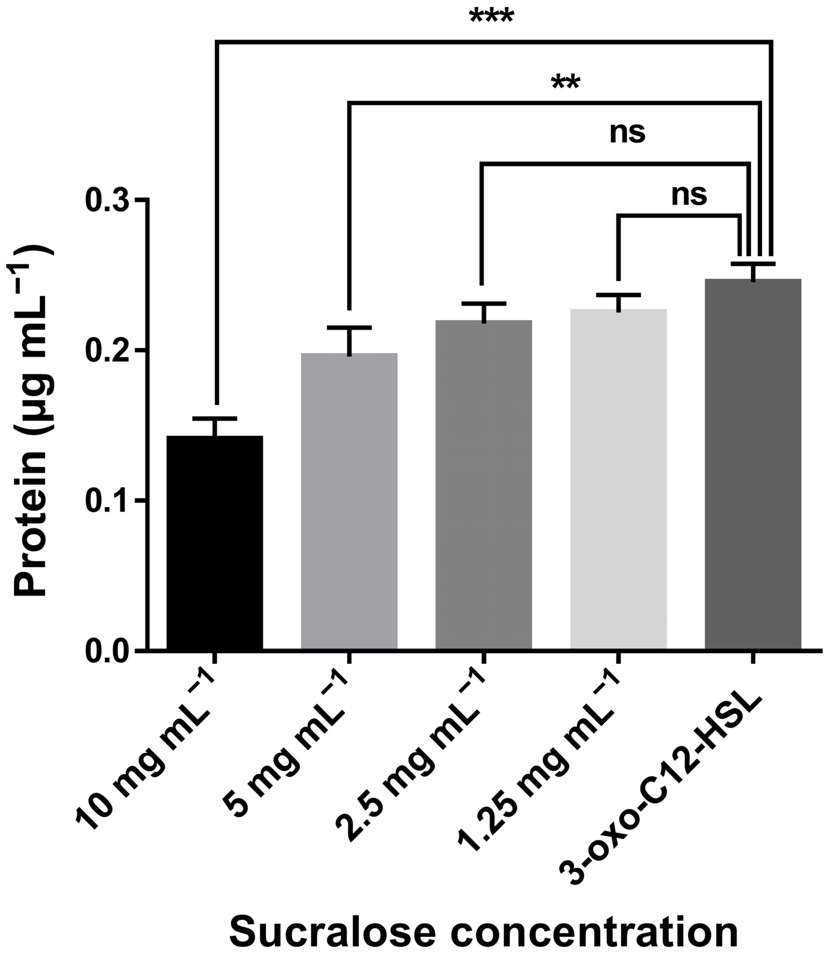
In E. coli BL21, the LasR-LBD protein was expressed in the presence of 3-oxo-C12-HSL and/or sucralose. After the protein expression, nickel-affinity chromatography was used to purify it. Nickel attached to agarose beads via nitriloacetic acid (NTA) selectively binds the His6-tag on the LasR-LBD construct. Phosphate buffer containing a low concentration of imidazole was used to remove non-specifically bound proteins. The LasR-LBD was eluted by employing a phosphate buffer with high imidazole concentration. The Bradford assay was used to quantify the pure protein, as shown in Figure 2. There was no LasR protein in the supernatant of the negative control, which contained only 0.15% (v/v) DMSO. The positive control, which included 7.5 μM 3-oxo-C12-HSL, showed that the protein was significantly expressed. While there was no significant reduction in the protein level at lower concentrations of sucralose, there was a significant decrease in protein levels at higher concentrations of sucralose (5 and 10 mg mL−1) compared with the control.

4. Discussion

The findings of this study show that sucralose inhibits the expression of the LasR protein in E. coli BL21. Sucralose interfered with the protein expression, as seen by the poor protein expression, whereas 3-oxo-C12-HSL significantly increased protein expression. LasR:3-oxo-C12-HSL binding has been proposed to be extremely strong [12] and crucial for the folding, stability, and solubility of the protein [13]. The LasR:3-oxo-C12-HSL binding may have been disrupted by sucralose, whose action would have reduced the protein’s solubility.
Several oral diseases, such as oropharyngeal candidiasis and dental caries, are biofilm-based [14]. Therefore, it means that the pathophysiology of various oral diseases depends critically on the production of biofilms. Notably, QS, a sophisticated bacterial communication system, controls the production of biofilms [15]. Thus, blocking QS may prevent the production of biofilms [16] and hence tackle biofilm-based oral diseases. That suggests sucralose may have anti-cariogenic properties in addition to being non-cariogenic. The biofilm production of Streptococcus sanguinis, Streptococcus mutans [17], and Porphyromonas gingivalis [18] was reported to be inhibited by sucralose. Remarkably, at the concentration that inhibited the formation of biofilms, sucralose did not exhibit cytotoxic effects in human cell lines [18].

5. Conclusions

According to this study, sucralose inhibits the expression of LasR, a QS regulator. There was a considerable drop in protein levels at higher doses of sucralose (5 and 10 mg mL−1) in comparison to the control, but no substantial drop at lower concentrations of sucralose. Sucralose suppression of AHL-dependent QS signalling was most likely by interfering with receptor–ligand interactions. Since QS controls biofilm and several oral diseases are biofilm based, sucralose may have anticariogenic properties in addition to being noncariogenic. These findings emphasise sucralose’s potential as a useful ingredient in formulations for oral health and shed light on a putative molecular mechanism behind its anti-cariogenic activity.

Funding

This research received no external funding.

Institutional Review Board Statement

Not applicable.

Informed Consent Statement

Not applicable.

Data Availability Statement

The raw data supporting the conclusions of this article will be made available by the author on request.

Conflicts of Interest

The author declares no conflicts of interest.

Abbreviations

The following abbreviations are used in this manuscript:
QSQuorum sensing
AHLN-acyl homoserine lactone
NTANitriloacetic acid
DMSODimethyl sulfoxide
FDAThe Food and Drug Administration of the United States
DDWDouble-distilled water
PMSFPhenylmethylsulfonyl fluoride

References

  1. Debras, C.; Chazelas, E.; Sellem, L.; Porcher, R.; Druesne-Pecollo, N.; Esseddik, Y.; de Edelenyi, F.S.; Agaësse, C.; De Sa, A.; Lutchia, R.; et al. Artificial sweeteners and risk of cardiovascular diseases: Results from the prospective NutriNet-Santé cohort. BMJ 2022, 378, e071204. [Google Scholar] [CrossRef] [PubMed]
  2. Schiffman, S.S.; Rother, K.I. Sucralose, A Synthetic Organochlorine Sweetener: Overview Of Biological Issues. J. Toxicol. Environ. Heal Part B 2013, 16, 399–451. [Google Scholar] [CrossRef] [PubMed]
  3. Glória, M.B.A. SWEETENERS|Others. In Encyclopedia of Food Sciences and Nutrition; Elsevier: Amsterdam, The Netherlands, 2003; pp. 5695–5702. ISBN 9780122270550. [Google Scholar]
  4. Aguayo-Guerrero, J.A.; Méndez-García, L.A.; Solleiro-Villavicencio, H.; Viurcos-Sanabria, R.; Escobedo, G. Sucralose: From Sweet Success to Metabolic Controversies—Unraveling the Global Health Implications of a Pervasive Non-Caloric Artificial Sweetener. Life 2024, 14, 323. [Google Scholar] [CrossRef] [PubMed]
  5. AlDeeb, O.A.A.; Mahgoub, H.; Foda, N.H. Sucralose. In Profiles of Drug Substances, Excipients and Related Methodology; Brittain, H.G., Ed.; Elsevier: Amsterdam, The Netherlands, 2013; Volume 38, pp. 423–462. ISBN 9780124076914. [Google Scholar]
  6. Gardner, E. Alternative sugars: Sucralose. Br. Dent. J. 2018, 224, 5. [Google Scholar] [CrossRef] [PubMed]
  7. Herrera, M.N.R.; Román, C.L.; Altamirano, E.M.; Pamatz, F.J.G. Effect of Toothpaste Sweeteners on Glucose Homeostasis. Asian J. Res. Rep. Endocrinol. 2025, 8, 1–9. [Google Scholar] [CrossRef]
  8. Panel, E.; Nda, A. Scientific Opinion on the substantiation of health claims related to the sugar replacers xylitol, sorbitol, mannitol, maltitol, lactitol, isomalt, erythritol, D-tagatose, isomaltulose, sucralose and polydextrose and maintenance of tooth mineralisation by. EFSA J. 2011, 9, 2076. [Google Scholar] [CrossRef]
  9. BeMiller, J.N. Carbohydrate and Noncarbohydrate Sweeteners. In Carbohydrate Chemistry for Food Scientists; Elsevier: Amsterdam, The Netherlands, 2019; pp. 371–399. [Google Scholar]
  10. Dümmler, A.; Lawrence, A.M.; de Marco, A. Simplified screening for the detection of soluble fusion constructs expressed in E. coli using a modular set of vectors. Microb. Cell Factories 2005, 4, 1–10. [Google Scholar] [CrossRef] [PubMed]
  11. Kielkopf, C.L.; Bauer, W.; Urbatsch, I.L. Bradford Assay for Determining Protein Concentration. Cold Spring Harb. Protoc. 2020, 2020, pdb.prot102269. [Google Scholar] [CrossRef] [PubMed]
  12. Schuster, M.; Urbanowski, M.L.; Greenberg, E.P. Promoter specificity in Pseudomonas aeruginosa quorum sensing revealed by DNA binding of purified LasR. Proc. Natl. Acad. Sci. USA 2004, 101, 15833–15839. [Google Scholar] [CrossRef] [PubMed]
  13. Bottomley, M.J.; Muraglia, E.; Bazzo, R.; Carfì, A. Molecular Insights into Quorum Sensing in the Human Pathogen Pseudomonas aeruginosa from the Structure of the Virulence Regulator LasR Bound to Its Autoinducer. J. Biol. Chem. 2007, 282, 13592–13600. [Google Scholar] [CrossRef] [PubMed]
  14. Bertolini, M.; Costa, R.; Barão, V.; Villar, C.C.; Retamal-Valdes, B.; Feres, M.; Silva Souza, J. Oral Microorganisms and Biofilms: New Insights to Defeat the Main Etiologic Factor of Oral Diseases. Microorganisms 2022, 10, 2413. [Google Scholar] [CrossRef] [PubMed]
  15. Kilian, M.; Chapple, I.L.C.; Hannig, M.; Marsh, P.D.; Meuric, V.; Pedersen, A.M.L.; Tonetti, M.S.; Wade, W.G.; Zaura, E. The oral microbiome—An update for oral healthcare professionals. Br. Dent. J. 2016, 221, 657–666. [Google Scholar] [CrossRef] [PubMed]
  16. Paluch, E.; Rewak-Soroczyńska, J.; Jędrusik, I.; Mazurkiewicz, E.; Jermakow, K. Prevention of biofilm formation by quorum quenching. Appl. Microbiol. Biotechnol. 2020, 104, 1871–1881. [Google Scholar] [CrossRef] [PubMed]
  17. Zhu, J.; Liu, J.; Li, Z.; Xi, R.; Li, Y.; Peng, X.; Xu, X.; Zheng, X.; Zhou, X. The Effects of Nonnutritive Sweeteners on the Cariogenic Potential of Oral Microbiome. BioMed Res. Int. 2021, 2021, 9967035. [Google Scholar] [CrossRef] [PubMed]
  18. Shinohara, M.; Maetani, M.; Kitada, C.; Nishigami, Y.; Yazawa, A.; Kamitani, S. Analysis of the Effects of Food Additives on Porphyromonas gingivalis. Pathogens 2022, 11, 65. [Google Scholar] [CrossRef] [PubMed]
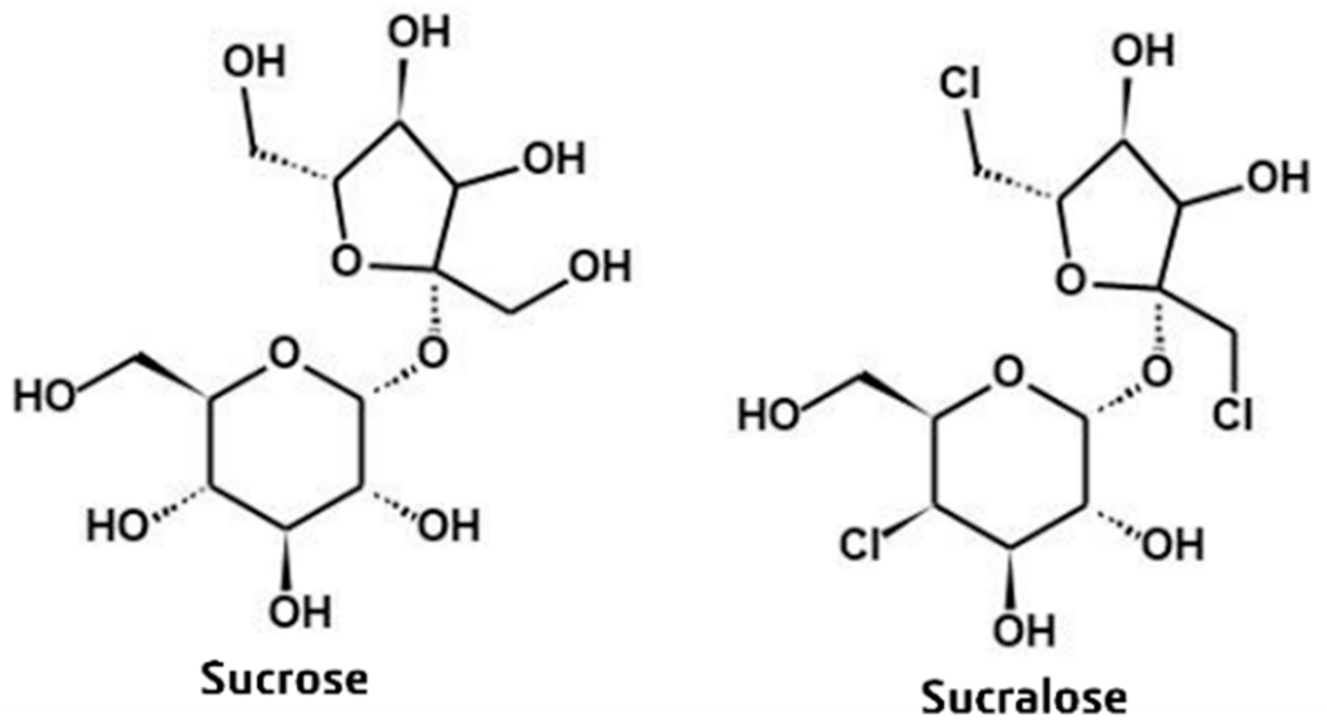
Figure 1. Chemical structures of sucrose and sucralose. Three chlorine atoms are substituted for three hydroxyl groups in sucrose at positions 4, 1′, and 6′ to synthesize sucralose. Structures were drawn using MedChem Designer, Version 5.5 (Simulations Plus Inc., Lancaster, CA, USA).
Chemproc 18 00004 g001
Figure 2. Protein quantification of LasR-LBD protein expressed in E. coli BL21 in the presence of 3-oxo-C12-HSL and/or sucralose. Every concentration that is displayed is the final concentration. In the negative control (containing just 0.15% (v/v) DMSO), there was no LasR protein expressed. The presence of 7.5 μM 3-oxo-C12-HSL in the positive control indicated that the protein was highly expressed. The statistical technique employed was Student’s t-test; the tests were compared with the positive control. ** p < 0.01; *** p < 0.001; and ns, not significant. The values show the mean ± SD for n = 3.
Chemproc 18 00004 g002
Disclaimer/Publisher’s Note: The statements, opinions and data contained in all publications are solely those of the individual author(s) and contributor(s) and not of MDPI and/or the editor(s). MDPI and/or the editor(s) disclaim responsibility for any injury to people or property resulting from any ideas, methods, instructions or products referred to in the content.