Proceeding Paper

Evaluation of the Photostability of Ivermectin †

1
Laboratorio de Fotoquímica, Instituto Venezolano de Investigaciones Científicas, Caracas 1020-A, Venezuela
2
Laboratorio de Fisicoquímica Orgánica, Instituto Venezolano de Investigaciones Científicas, Caracas 1020-A, Venezuela
*
Author to whom correspondence should be addressed.
Presented at the 28th International Electronic Conference on Synthetic Organic Chemistry (ECSOC-28), 15–30 November 2024; Available online: https://sciforum.net/event/ecsoc-28.
Chem. Proc. 2024, 16(1), 78; https://doi.org/10.3390/ecsoc-28-20182
Published: 14 November 2024

Abstract

:
Ivermectin (IVM) is a pharmaceutical antiparasitic agent with a broad range of medicinal properties that are comparable in impact to those of penicillin and aspirin. The molecule’s structural composition includes functional groups that indicate the potential for photoreactivity. However, there is a paucity of information regarding its photostability, particularly in tropical regions where parasitic diseases and intense solar radiation intersect. It would be beneficial to investigate the chemical transformation of this compound in a variety of natural aqueous environments under different irradiation sources. This knowledge gap motivated this study. Therefore, the chemical alterations of IVM were investigated in various natural aqueous media when exposed to solar radiation (UVA-Vis). In particular, an evaluation of its photostability was conducted at wavelengths of 350 and 254.5 nm. It is noteworthy that photodegradation occurred primarily at 350 nm. Additionally, IVM demonstrated photohemolytic effects on human erythrocytes, indicating phototoxicity. This suggests the presence of photoinduced mechanisms by this drug for the generation of free radicals, including singlet oxygen (1O2, type II mechanism), superoxide anion, and hydroxyl radical (.O2 and .OH, type I mechanism). The latter would also entail the interaction of the IVM molecule with the membrane of human red blood cells, which would signify a considerable biological impact. Furthermore, through computational calculations, potential photoproducts formed during IVM irradiation were deduced, simulating experimental conditions. Our findings contribute to an enhanced comprehension of IVM’s behavior under solar exposure, particularly in tropical contexts. Additional research is imperative to address its emerging biological activity status and potential implications for biomedical applications.

1. Introduction

Ivermectin (IVM) is an anti-parasitic pharmaceutical product with antiviral, antibacterial, and antitumor properties, which earned its discoverers, Omura and Campbell, the 2015 Nobel Prize in Medicine. Its origin dates back to 1974 in Japan, where Satoshi Ōmura isolated organisms with biocidal properties from soils, extracting avermectins, which were later modified to obtain ivermectin [1,2].
The applications of IVM were initially veterinary, but over time, it proved to be useful in human medicine, combating numerous diseases, such as onchocerciasis, intestinal parasitosis, and lice, among others, which afflict millions of inhabitants in rural and densely populated areas [3,4,5].
Recent studies show encouraging results in the use of IVM for the treatment of diseases, such as malaria, Chagas, leishmaniasis, trichinosis, allergic asthma, and neurological disorders. In addition, during the pandemic, it was approved to treat COVID-19, with doses of 150–200 µg/kg twice daily. Although its effectiveness has not been proven, toxicological evaluations concluded that it is a safe drug with moderate side effects [6,7].
IVM has a high degree of lipophilicity and insolubility in water. However, it is soluble in methanol and 95% ethanol. The excipients employed in its preparation include microcrystalline cellulose, corn starch, butylated hydroxyanisole, and others. The maximum dose of IVM administered to humans does not exceed 200 µg/kg, although it varies according to the patient’s individual characteristics. Following administration, the compound is rapidly absorbed and distributed throughout the body. Elimination occurs via the excretory system, with a half-life of approximately 24 to 60 h [8].
IVM (Figure 1a) consists of an 80:20 ratio of the avermectins B1a (C48H74O14) and B1b (C47H72O14), both derived from the lactone produced by the actinobacterium Streptomyces avermitilis [7,8]. The presence of a carbonyl group, forming an ester, as well as conjugated double bonds, alcohols, and ethers, points to the possible photoreactivity of IVM. However, information on its photostability is scarce, although some reports indicate that it is not stable when exposed to light during storage [9,10].
In this study, the photostability of IVM was evaluated by UV-Vis spectrophotometry. UVA and UVC lamps were used as irradiation sources. Furthermore, the photodegradation of IVM in ordinary seawater and a sample from the Orinoco River (Venezuela) was also compared. Computational calculations were also performed with the aim of proposing a mechanistic insight for the photodegradation of this drug. Additionally, the phototoxic activity of IVM in human erythrocytes was assessed.

2. Materials and Methods

2.1. Materials and Equipment

Commercial Ivermectin (IVM) Remeny®, Perkin Elmer Lambda 35 UV-VIS Spectrophotometer, Luzchem L2C-4V photoreactor, ethanol, PBS buffer, UV-C, and UV-A lamps: These materials and equipment were obtained from Sigma Aldrich (St. Louis, MO, USA) and Perkin Elmer (Waltham, MA, USA).

2.2. Methods

2.2.1. Sample Preparation

IVM was extracted from two 6 mg tablets of the active ingredient, Remeny®, by dissolution in 25 mL of 95% ethanol of IVM and subsequent separation of the excipients by filtration of the suspended solids.

2.2.2. Evaluation of Photostability by UV-Visible Spectrometry

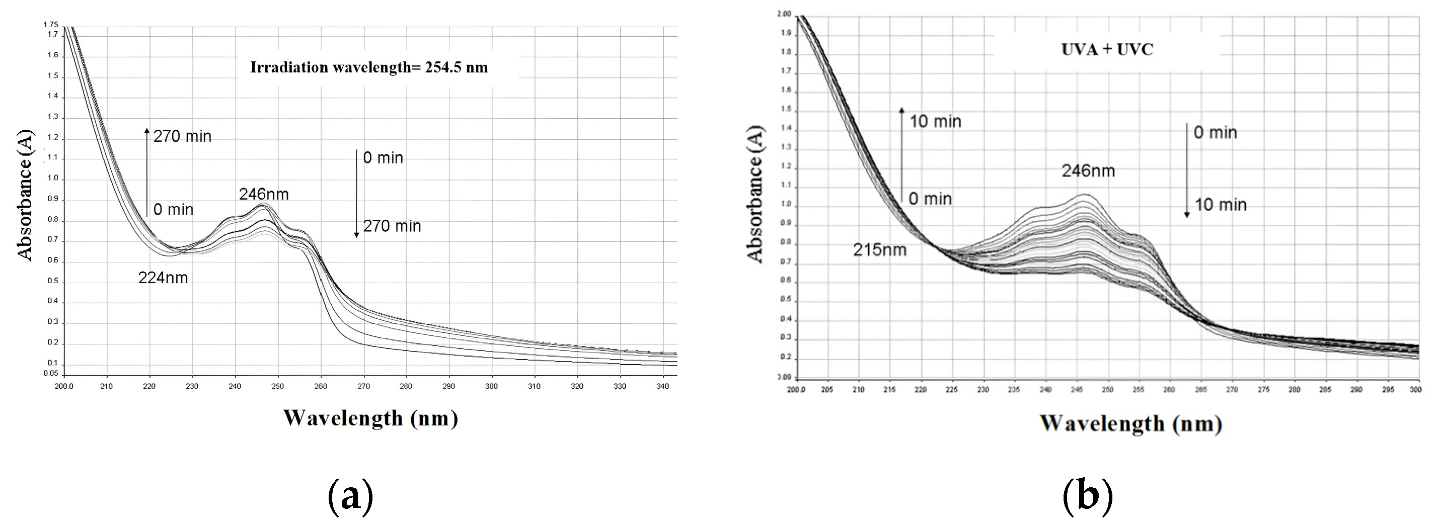
An IVM solution was prepared at a concentration of approximately 6.85 × 10⁻³ mM in ultrapure water. The solution was subjected to ultraviolet (UV) radiation in two separate experiments. In the initial experiment, the solution was subjected to ultraviolet (UV) radiation for a period of 270 min utilizing a set of Hitachi FL8BL-B lamps. Secondly, the solution was irradiated with a combination of UVA and UVC light for a period of 10 min. The irradiation power and wavelength of the lamps differed between the two experiments. The absorbance spectrum of the solution was measured using a Perkin Elmer Lambda 35 UV-Visible spectrophotometer.

2.2.3. Phototoxicity on Human Erythrocytes

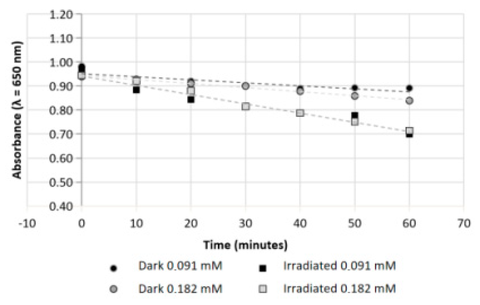
Solutions of the isolated human erythrocytes were prepared in PBS buffer (pH 7.4), and amounts of the IVM sample (0.091 and 0.182 mM) were added. Mixtures of human erythrocytes with IVM were irradiated at 350 nm and compared with non-irradiated mixtures to compare the level of photoinduced damage. Photohemolysis was followed by UV-VIS spectrophotometry, tracking the decrease in absorbance at 650 nm for 1 h.

2.2.4. Computational Calculations

DFT calculations were conducted for the IVMB1a molecule. B3LYP methods, designed by [11], were employed in conjunction with the 6-311+G(d,p) basis set [12], utilizing the Gaussian 09 software packages [13]. The optimized structure was validated through a frequency analysis. Additionally, Natural Bond Orbital (NBO) charge and Wiberg bond index (WBI) analysis were performed. The effect of water as a solvent was taken into consideration in the calculations, using the Polarized Continuum Model, in particular, the IEFPCM variant [14].

3. Discussion and Results

3.1. Photostability of IVM

During the UVA irradiation of IVM in distilled water (Figure 2a), the inflection of the absorption curve at 224 nm in addition to the decrease in the absorbance value for the peak of maximum (246 nm) is seen, suggesting the alteration of avermectins.
Figure 2b evidences further alteration with additional inflection at 265 nm, especially for the most altered samples, corresponding to the longest irradiation time (10 min), which may be due to the modification of specific bonds or the formation of degradation products. The inflection in the absorbance at 224 nm suggests changes in the conformation of the avermectins. Diminished absorbance at 246 nm could indicate the breaking of bonds or the formation of new functional groups [15].
It should be noted that UVA and UVC irradiation could have induced changes in the avermectins, generating molecular fragments, as well as free radicals, which may interact with the structure of the avermectins. It is also necessary to consider that the functional groups may have changed position due to radiation-induced isomerization. These changes in molecular structure could affect the pharmacological activity of avermectins [9,15].
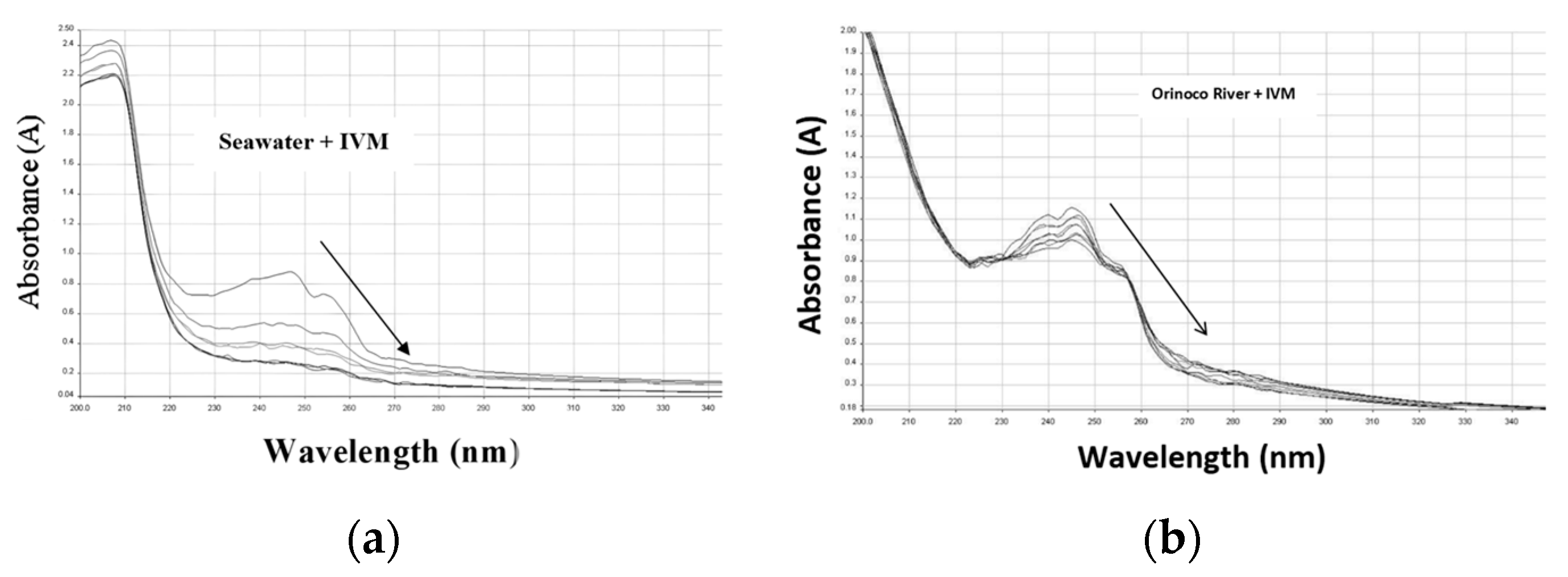
Figure 3a,b show the effect of irradiation on IVM in seawater and Orinoco River water, respectively. A greater effect of irradiation on the IVM dissolved in common seawater is highlighted. This implies that the photodegradation of IVM is affected by the species present in the water, being favored in seawater, although it is inhibited in Orinoco water. Therefore, the composition of the water must be considered in its degradation.
In this regard, it is important to consider that seawater contains a high concentration of salts, which may act as sensitizing agents, increasing the efficiency of photodegradation. The ions present can interact with IVM and facilitate bond breaking. In addition, the presence of trace metals in seawater (e.g., copper, iron) may catalyze photochemical reactions. These metals could accelerate the degradation of IVM [16].
On the other hand, Orinoco River water may contain dissolved organic matter, such as humic and fulvic acids. These compounds may compete with IVM for light absorption, reducing the efficiency of photodegradation. Likewise, the presence of organic matter can also absorb some of the UV radiation, decreasing the amount of light available to trigger IVM photodegradation [17,18]. Plus, the turbidity of the Orinoco River water could generate “shadow zones” where the IVM is less exposed to direct sunlight, which could inhibit its degradation, which could be a determining factor in the differences shown in the results of this study [16].

3.2. Photohemolytic Effects of IVM

Figure 4 shows the photoinduced effect of IVM on human blood cells and its comparison with non-irradiated samples. The concentration of erythrocytes decreases for the sample irradiated at 350 nm compared to the one kept in the dark. These results suggest the presence of free radical photogeneration from IVM, such as singlet oxygen (1O2, type II mechanism), superoxide anion, and hydroxyl radical (.O2 and .OH, type I mechanism) [18,19].

3.3. Computational Calculations

The NBO analysis of IVMB1a shows that the bonds α, β, and γ are polarized, while the carbon of the C=O group of the lactone has a large positive charge (+0.860). The WBIs indicate that the bonds α, β, and γ have a bond order less than 0.90 (see Supplementary Material Figures S2 and S3). The polarization of the α and β bonds makes them susceptible to photoinduced dissociation and attack by -OH free radicals, which corresponds to the bonds that are broken in the degradation pathways of ivermectin [20]. Moreover, the WBIs of these bonds indicate that they are labile.
On the other hand, the γ bond obtained the highest polarization and the lowest WBI, indicating a higher lability and propensity to photoinduced cleavage. This also provides an explanation for the monohydroxylated fragments derived from lactone oxidation [20]. Therefore, it can be assumed that the photodegradation of ivermectin is attributable to the lability of the bonds depicted in Figure 1b.

4. Conclusions

The study on the photostability of ivermectin has revealed that exposure to UVA and UVC radiation causes significant alterations in its chemical structure, as evidenced by changes in absorption spectra. The results demonstrate that ivermectin is susceptible to degradation in different aquatic environments, with seawater proving a particularly challenging medium. This suggests that the composition of the medium in which it is used influences its stability. Furthermore, the computational results provide additional insight by suggesting potential photodegradation products, which helps to elucidate the degradation mechanisms. These findings are crucial for understanding the efficacy and safety of ivermectin use in treatments, especially in tropical regions where sun exposure is intense.

Supplementary Materials

The following supporting information can be downloaded at: https://www.mdpi.com/article/10.3390/ecsoc-28-20182/s1, Figure S1: Optimized structure of IVMB1a; Figure S2: NBO charges of the atoms forming α, β and γ bonds; Figure S3: Wiberg bond order indices for α, β and γ bonds; Date S1: Binding lengths in Å for labile bonds of IVMB1a.

Author Contributions

Conceptualization, F.V. and M.L.; methodology, F.V. and B.A.; software, A.M. and J.G.; validation, F.V. and Á.Á.; formal analysis, F.V., M.L., and J.G.; investigation, M.L. and J.G.; resources, Á.Á.; writing—original draft preparation, M.L.; writing—review and editing, M.L. and A.M.; supervision, F.V.; funding acquisition, F.V. All authors have read and agreed to the published version of the manuscript.

Funding

This research was funded by the Instituto Venezolano de Investigaciones Científicas (IVIC).

Institutional Review Board Statement

Not applicable.

Informed Consent Statement

Not applicable.

Data Availability Statement

All the data are contained within the article.

Acknowledgments

We thank Álvaro Álvarez for his permission and assistance with the computational calculations.

Conflicts of Interest

The authors declare no conflicts of interest.

References

  1. Crump, A.; Ōmura, S. Ivermectin, ‘wonder drug’ from Japan: The human use perspective. Proc. Jpn. Acad. Ser. B Phys. Biol. Sci. 2021, 87, 13–28. [Google Scholar] [CrossRef] [PubMed]
  2. Crump, A. Ivermectin: Enigmatic multifaceted ‘wonder’ drug continues to surprise and exceed expectations. J. Antibiot. 2017, 70, 495–505. [Google Scholar] [CrossRef] [PubMed]
  3. Geary, T. Ivermectin 20 years on: Maturation of a wonder drug. Trends Parasitol. 2005, 21, 530–532. [Google Scholar] [CrossRef] [PubMed]
  4. González, P.; González, F.A.; Ueno, K. Ivermectin in human medicine, an overview of the current status of its clinical applications. Curr. Pharm. Biotechnol. 2012, 13, 1103–1109. [Google Scholar] [CrossRef]
  5. Shirazi, F.; Mirzaei, R.; Nakhaee, S.; Nejatian, A.; Ghafari, S.; Mehrpour, O. Repurposing the drug, ivermectin, in COVID-19: Toxicological points of view. Eur. J. Med. Res. 2022, 27, 21. [Google Scholar] [CrossRef] [PubMed]
  6. González, C.; Sahagún, P.; Diez, L.; Fernández, N.; Sierra, M.; García, J. The pharmacokinetics and interactions of ivermectin in humans—A mini-review. AAPS J. 2008, 10, 42–46. [Google Scholar] [CrossRef] [PubMed]
  7. Õmura, S.; Crump, A. The life and times of ivermectin—A success story. Nat. Rev. Microbiol. 2004, 2, 984–989. [Google Scholar] [CrossRef]
  8. Roz, L.; Victoria, G.; Eileen, D. Ivermectin–Old Drug, New Tricks? Trends Parasitol. 2017, 33, 463–472. [Google Scholar] [CrossRef]
  9. David, W. Fink, Ivermectin. In Analytical Profiles of Drug Substances; Florey, K., Ed.; Academic Press: Cambridge, MA, USA, 1988; Volume 17, pp. 155–184. [Google Scholar] [CrossRef]
  10. Mirta, Č.; Davor, L.; Lidija, Ć.; Irena, Š.; Sandra, B. Kinetics and degradation pathways of photolytic and photocatalytic oxidation of the anthelmintic drug praziquantel. J. Hazard. Mater. 2017, 323, 500–512. [Google Scholar] [CrossRef]
  11. Becke, A.D. Density-functional thermochemistry. I. The effect of the exchange-only gradient correction. J. Chem. Phys. 1993, 98, 5648–5652. [Google Scholar] [CrossRef]
  12. Krishnan, R.; Binkley, J.S.; Seeger, R.; Pople, J.A. Self-consistent molecular orbital methods. XX. A basis set for correlated wave functions. J. Chern. Phys. 1980, 72, 650–654. [Google Scholar] [CrossRef]
  13. Frisch, M.J.; Trucks, G.W.; Schlegel, H.B.; Scuseria, G.E.; Robb, M.A.; Cheeseman, J.R.; Scalmani, G.; Barone, V.; Mennucci, B.; Petersson, G.A.; et al. Gaussian 09, Revision A.02; Gaussian, Inc.: Wallingford, CT, USA, 2009. [Google Scholar]
  14. Tomasi, J.; Mennucci, B.; Cammi, R. Quantum Mechanical Continuum Solvation Models. Chem. Rev. 2005, 105, 2999–3093. [Google Scholar] [CrossRef] [PubMed]
  15. Gwenzi, W.; Selvasembian, R.; Offiong, N.A.O.; El Din Mahmoud, A.; Sanganyado, E.; Mal, J. COVID-19 drugs in aquatic systems: A review. Environ. Chem. Lett. 2022, 20, 1275–1294. [Google Scholar] [CrossRef]
  16. John, R.E.; John, I.H.; Allan, H.D.; Jefrey, E.R. Dissolved humic substances of the Amazon River system. Limnol. Oceanogr. 1986, 31, 139–154. [Google Scholar] [CrossRef]
  17. Peiris, R.; Budman, H.; Moresoli, C.; Legge, R. Identification of humic acid-like and fulvic acid-like natural organic matter in river water using fluorescence spectroscopy. Water Sci. Technol. 2011, 63, 2427–2433. [Google Scholar] [CrossRef]
  18. Kahn, G.; Fleischaker, B.I. Red Blood Cell Hemolysis by Photosensitizing Compounds. J. Investig. Dermatol. 1971, 56, 85–90. [Google Scholar] [CrossRef]
  19. Costanzo, L.; De Guidi, G.; Condorelli, G.; Cambria, A.; Fama, M. Molecular mechanism of drug photosensitization–II. Photohemolysis sensitized by ketoprofen. Photochem. Photobiol. 1989, 50, 359–365. [Google Scholar] [CrossRef] [PubMed]
  20. Havlíkova, L.; Satínský, D.; Solich, P. Aspects of decontamination of ivermectin and praziquantel from environmental waters using advanced oxidation technology. Chemosphere 2016, 144, 21–28. [Google Scholar] [CrossRef] [PubMed]
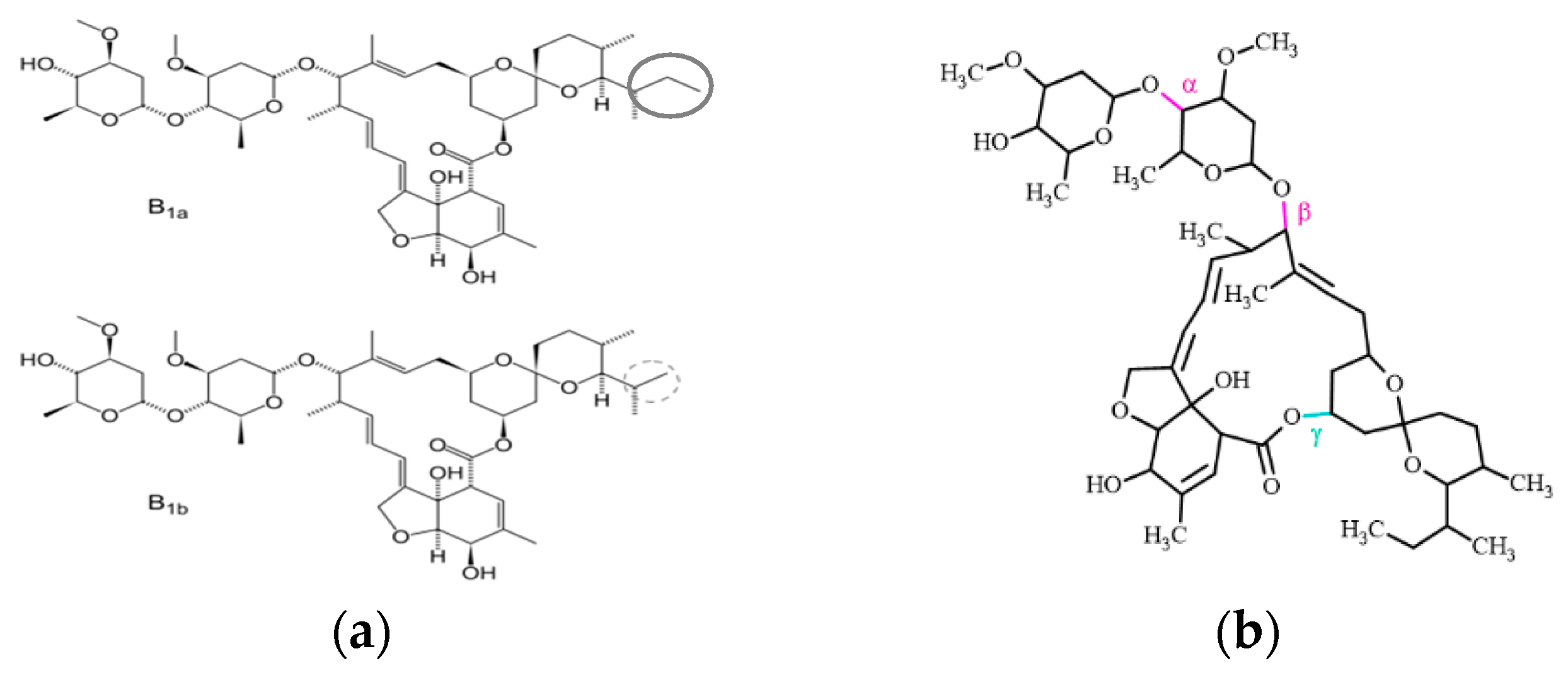
Figure 1. (a) Molecular structure of the avermectins (80% B1a/20% B1b); (b) optimized IVMB1a molecule.
Chemproc 16 00078 g001
Figure 2. (a) Alteration of IVM in distilled water, exposed to UVA radiation (λ = 350 ± 50); (b) alteration of IVM in distilled water, exposed to UVA + UVC radiation λ = 350 + 254.5).
Chemproc 16 00078 g002
Figure 3. (a) Irradiation of IVM in seawater.; (b) MVI irradiation in Orinoco River water.
Chemproc 16 00078 g003
Figure 4. Phototoxicity of IVM on human erythrocytes.
Chemproc 16 00078 g004
Disclaimer/Publisher’s Note: The statements, opinions and data contained in all publications are solely those of the individual author(s) and contributor(s) and not of MDPI and/or the editor(s). MDPI and/or the editor(s) disclaim responsibility for any injury to people or property resulting from any ideas, methods, instructions or products referred to in the content.