Proceeding Paper

Synthesis, Self-Assembling and Photophysical Property Exploration of Water Self-Dispersible, Grafted Poly(p-Phenylene Vinylene)s with Nonionic, Hydrophilic and Biocompatible Side Chains †

1
Centre of Advanced Research in Bionanoconjugates and Biopolymers, “Petru Poni” Institute of Macromolecular Chemistry, 41 A, Grigore-Ghica Voda Alley, 700487 Iasi, Romania
2
Department of Chemistry, Faculty of Science and Letters, Istanbul Technical University, 34469 Maslak, Istanbul, Turkey
3
Department of Physics of Polymers and Polymeric Materials, “Petru Poni” Institute of Macromolecular Chemistry, 41 A, Grigore-Ghica Voda Alley, 700487 Iasi, Romania
*
Authors to whom correspondence should be addressed.
Presented at the 28th International Electronic Conference on Synthetic Organic Chemistry (ECSOC-28), 15–30 November 2024; Available online: https://sciforum.net/event/ecsoc-28.
Chem. Proc. 2024, 16(1), 73; https://doi.org/10.3390/ecsoc-28-20198
Published: 14 November 2024

Abstract

:
Conjugated polymers (CPs), in particular poly(p-phenylene vinylene)s (PPVs), are recognized as “smart” materials with potential applications ranging from optoelectronic devices to emergent technologies and to precision medicine. The present communication reports on the synthesis and structural characterization of new dibrominated macromonomers and their derived PPVs, of rod–graft–coil architecture, whose grafted, biocompatible and hydrophilic side chains are either PEG-2000 or poly(2-methyl-2-oxazoline) or poly(2-ethyl-2-oxazoline). The Suzuki–Heck cascade reaction was used for PPVs’ obtainment. After PPVs’ structural characterization using specific techniques (such as 1H-NMR; GPC), the micellar, fluorescent nanoparticles formed by spontaneous self-assembling during simple direct dissolution in water were evaluated using dynamic light scattering for their size, complementarily combined with Atom Force Microscopy (AFM) for their shape assessing. The PPV micelles’ photophysical properties were revealed using UV-vis spectroscopy and fluorescence measurements.

1. Introduction

Photonic nanomedicine promotes the progress of early detection and diagnosis of diseases, and new modalities of light-guided and light-activated therapies, providing opportunities to advance healthcare technology with unprecedented precision and safety [1]. As such, there is an increasing demand to develop and optimize a materials platform for biophotonic applications. Particularly, the design of biocompatible and biodegradable materials with desired optical properties, improved biocompatibility and biological functionalities is required [2]. Originally designed for use in electronic and optoelectronic devices, conjugated polymers (CPs) have emerged as one of the most appropriate agents for biophotonics [3]. Typical examples are PPVs, recognized as representatives of electroluminescent polymers, used for the construction of the first polymer light-emitting diode [4]. Later on, it turned out that they could be the materials of choice for various types of bioapplications. Taking advantage of their light-harvesting, light-emitting and photosensitizing capabilities, PPVs found applications for in vivo bioimaging [5], photodynamic- [6], gene- [7] and immunotherapy [8], drug delivery [9], interesting biodegradable afterglow imaging agents [10] or for afterglow imaging-guided photothermal therapy [11]. It has previously been found that when the polymer side chains attached to the conjugated backbone are hydrophilic and biocompatible, better interaction with biological entities occurs [12]. The resulting amphiphilic rod–graft–coil topology mediated enhanced physiological stability for their derived micellar nanoparticles, while preserving the CPs’ optical properties; this can be an optimized alternative to CPs’ nanoparticle encapsulation in an amphiphilic biocompatible matrix with nanoprecipitation [13]. Following a similar strategy, the present communication introduces new amphiphilic, grafted poly(p-phenylene vinylene)s (g-PPVs), having hydrophilic side chains and differing in length, chemical nature and way of attaching. Using the “grafting through” approach [14], the new g-PPVs were synthesized with the Suzuki–Heck cascade polycondensation [15] of macromonomers derived from PEG or poly(2-alkyl-oxazolines) (POXA). The macromonomers and new g-PPVs were structurally confirmed using 1H-NMR spectroscopy, whereas complementary use of DLS and AFM evidenced the size and shape of the micellar nanoparticles formed by spontaneous self-assembling in water. The formed micelles are fluorescent and the values of absorption and emission maxima wavelengths were established using UV-vis and fluorescence measurements. To the best of our knowledge, only a few g-PPVs with hydrophilic polymer side chains, aimed at bioapplications, have been reported [10,11,16], but this is the first one regarding g-PPVs having poly(2-alkyl-2-oxazoline) side chains.

2. Materials and Methods

2.1. Materials

All chemicals were purchased from Sigma-Aldrich and used without further purification. All the solvents were purified and dried with usual methods before use.

2.2. Syntheses

Macromonomer M1 in Scheme 1 was prepared by following a procedure previously described in detail [12]. 1,4-Dibromo-2,5-bis(bromomethyl)benzene (compound 4 in Scheme 1), used as initiator for the synthesis of macromonomers M2 and M3, was synthesized following the steps described in reference [17]. For polymer synthesis, in each case, an equimolar amount of potassium vinyltrifluoroborate and the macromonomer were used, following a slightly modified protocol reported in reference [15]. The crude g-PPVs were obtained by precipitating the reaction mixture in cold diethyl ether. They were purified through reprecipitation from methanol to a cold diethyl ether three times, followed by drying in a vacuum oven at room temperature for 24 h.

2.3. Methods

Nuclear magnetic resonance (NMR) spectroscopy was performed on a Bruker Avance NEO 400 MHz spectrometer (Bruker BioSpin GmbH, Rheinstetten, Germany,) operating at 400.1 MHz for 1H nuclei. All the experiments were recorded at room temperature (24 °C) using CD3OD and CDCl3 as deuterated solvents. The relative molecular weight (Mn) and index of polydispersity (IPD) were determined with gel permeation chromatography (GPC). The measurements were performed by using a WGE SEC-3010 multidetection system, consisting of a pump, two PL gel columns (PL gel 5 micro Mixed C Agilent and PL gel 5 micro Mixed D Agilent), dual detector refractometer/viscometer (RI/VI) WGE SEC-3010 and a flow rate of 1.0 mL/min at 30 °C. The RI/VI detector was calibrated with PS standards (580–467,000 DA) having a narrow molecular weight distribution. UV–vis absorption spectra were measured using a Specord 200 Analytik Jena spectrophotometer. Fluorescence measurements were carried out using a Perkin Elmer LS 55 apparatus. Particle size measurement was carried out through dynamic light scattering (DLS), using a Malvern Zetasizer Nano ZS instrument equipped with a 4.0 mW He–Ne laser operating at 633 nm and a detection angle of 173°. AFM micrographs of the polymer films were taken in air, on a SPM SOLVER Pro-M instrument. An NSG10/Au Silicon tip with a 35 nm radius of curvature and a 255 kHz oscillation mean frequency was used. The apparatus was operated in semi-contact mode. The polymer films were prepared by drop casting the aqueous dispersion of the polymers at a concentration of 0.01mg/mL on mica support.

3. Results and Discussion

3.1. Syntheses of Macromonomers and of g-PPVs Based on Them-Structural Characterization

Macromonomer M1 was obtained by end-group functionalization, through esterification of the hydroxyl function of the PEG2000 monomethylether with 2,5-dibromobenzoic acid, in a similar manner as reported in [12]. By cationic ring-opening polymerization (CROP) of 2-methyl- or 2-ethyl-2-oxazoline, respectively, were synthesized macromonomers M1 and M2, using the initiator 4 (Scheme 1) in ratios to the monomer (I/M) of 10/1 and 15/1, respectively.
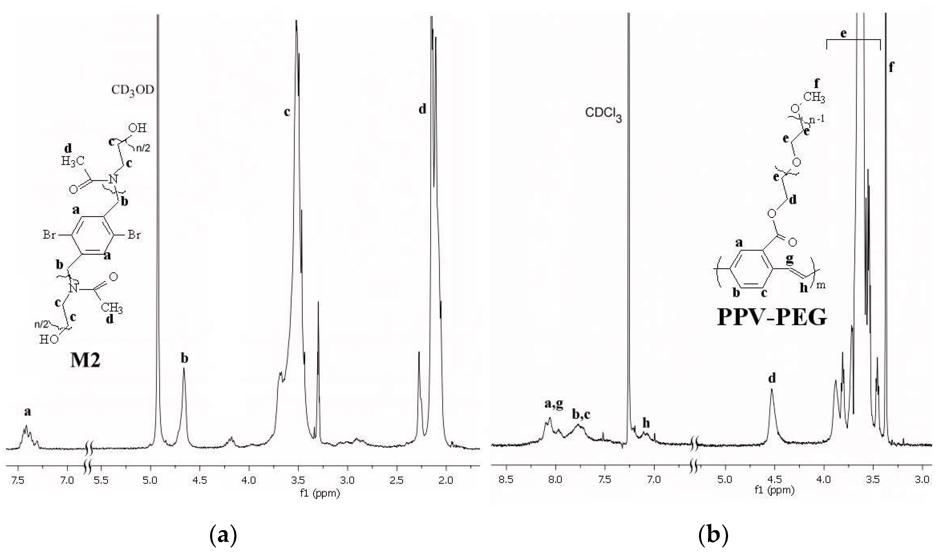
The expected structure of the three macromonomers was confirmed, besides another technique (like FT-IR, DSC), using 1H-NMR spectroscopy. In Figure 1a, the spectrum of M2 shows all the signals belonging to the protons characterizing the structure and exemplified in the enclosed molecular formula. By using the values of the integrals of peaks a or b in conjunction with the ones for protons c or d, the polymerization degree of obtained PMeOx was evaluated as n = 10. In a similar manner to M3, the degree of polymerization n of PEtOx was calculated as 14. For the synthesis of amphiphilic g-PPVs, the versatile protocol of the Suzuki-–eck cascade reaction was chosen, as it claimed that the percentage of structural 1,1-diarylenevinylene defects can be minimized [15]. In Figure 1b, the 1H-NMR spectrum of polymer PPV-PEG is given, which shows, besides the signal characteristic to the starting macromonomer M1, the peaks for protons g and h of the newly formed vinylene linkage.
The apparent molecular weight of the synthesized polymers was measured by GPC and the obtained values are given in Table 1.
The increased number of the electron-withdrawing carbonyl groups in the structures of M2 and M3 could have decreased the reactivity of bromine functionality, resulting in lower values of Mn,GPC for PPV-PMeOx and PPV-PEtOx, respectively.

3.2. Self-Assembling of PPVs in Water by Direct Dissolution

New amphiphilic g-PPVs self-assemble upon simple “dissolution” in water.
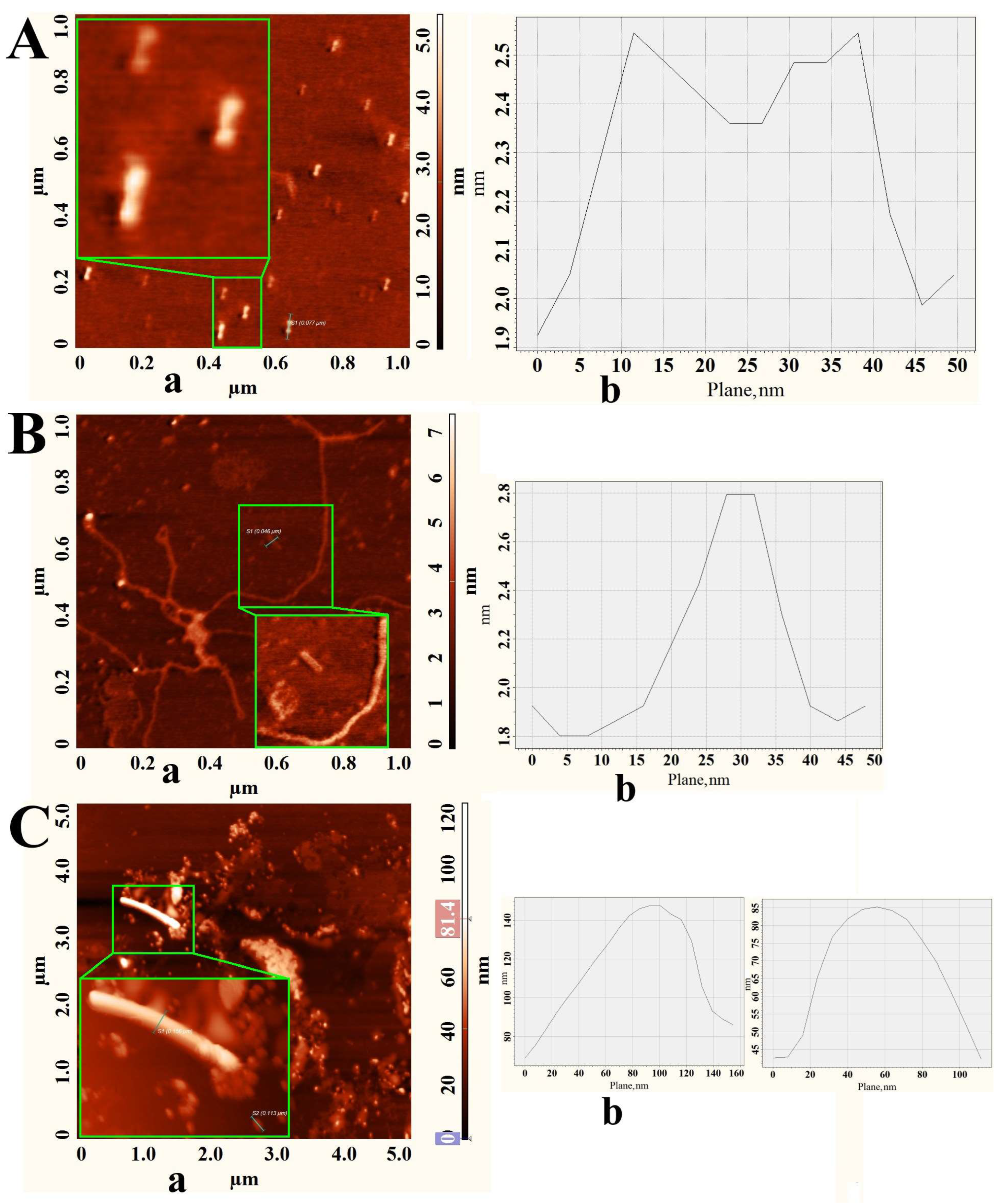
The visual confirmation of the formation of the self-assembled structures was conducted using AFM (Figure 2), which supports the obtained results of DLS (Table 2).
The data from Table 2 suggest a dependence of the formed particles’ size on the concentration of the polymers in water, with higher values both for the lowest concentrations and for the highest ones. PPV-PEG self-assembled in an interesting dumbbell-like shape (Figure 2A), while for PPV-PMeOx and PPV-PEtOx, a heterogeneous, mixed morphology can be seen, dominated by the “rod-like” structures and the sinuous fibers (Figure 2B,C).

3.3. Optical Properties of the PPVs’ Self-Assembled Micellar Nanoparticles

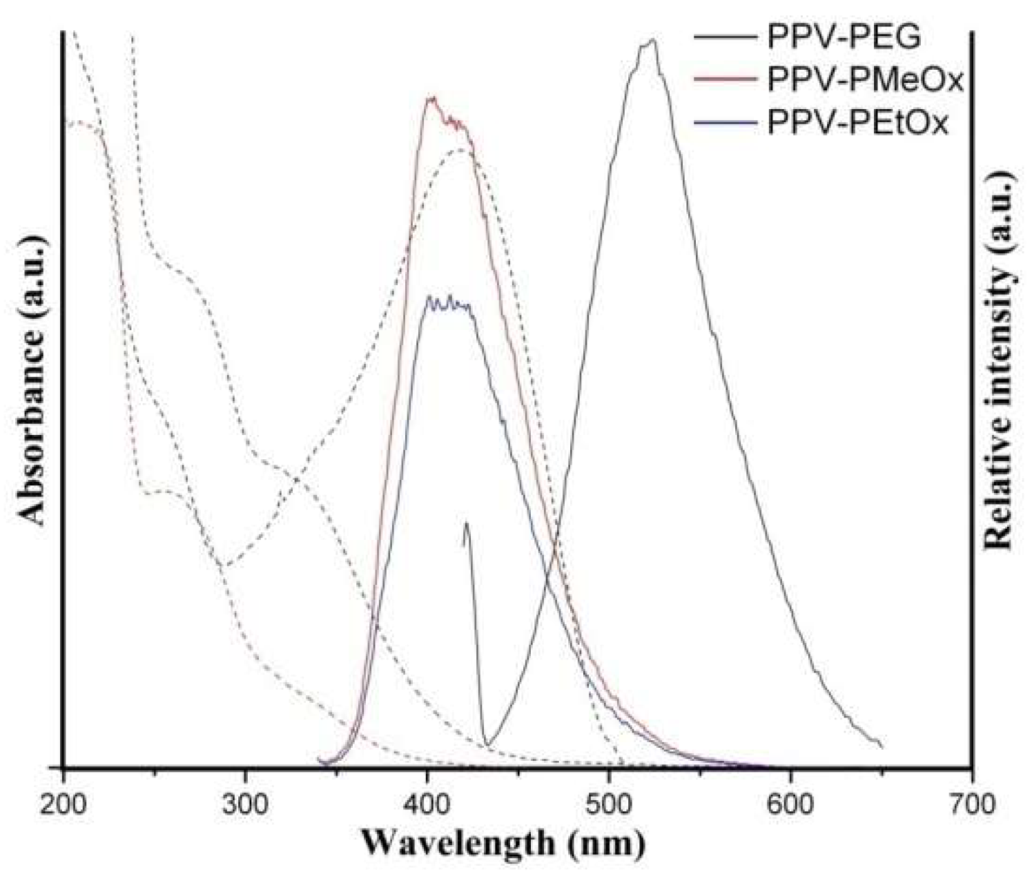
As expected, the formed micellar structures were fluorescent in water, and the side chains preserved the optical properties typical of PPVs but significantly influenced them. All polymers showed large emission bands (200 nm). While PPV-PEG micelles absorb light in the visible region (Table 2) and emit in the green–yellow range, with a maximum centered at around 524 nm, those formed by PPV-PMeOx and PPV-PEtOx absorbed light in the UV range and emitted in the visible range, with the maximum in the blue rage (Table 2 and Figure 3). When compared to the values reported for bare PPV [18], there are practical no differences for PPV-PEG, but the introduction of the POXA side chains produced a relevant hypsochromic effect, the emission maxima being blue-shifted with more than 100 nm. This phenomenon was noticed by us [19,20] and by others [21] for g-PPVs. It could be due to the effect of the synergistic combination of the steric hindrance, imposed on the conjugated chain by the side chains, with the grafts’ electronic peculiarities; in this case, the electron-withdrawing nature of the carbonyl groups in the side chains of POXA.
The dependence of the photophysical properties on the size of the micelles as well as on the presence of some biomolecules (like proteins) will be studied in the future.

Author Contributions

Conceptualization, I.C. and M.P.; methodology, I.C. and L.C.; validation, A.-D.B., D.G.-C. and L.C.; formal analysis, A.-D.B. and D.G.-C.; investigation, I.C., D.G.-C., A.-D.B. and E.-G.H.; data curation, D.G.-C., A.-D.B., L.C. and E.-G.H.; writing—original draft preparation, L.C.; writing—review and editing, L.C., I.C. and M.P.; visualization, I.C.; supervision, I.C. and M.P.; project administration, M.P. All authors have read and agreed to the published version of the manuscript.

Funding

This research received no external funding.

Institutional Review Board Statement

Not applicable.

Informed Consent Statement

Not applicable.

Data Availability Statement

Data are contained within the article.

Conflicts of Interest

The authors declare no conflicts of interest.

References

  1. Shan, D.; Gerhard, E.; Zhang, C.; Tierney, J.W.; Xie, D.; Liu, Z.; Yang, J. Polymeric biomaterials for biophotonic applications. Bioact. Mater. 2018, 3, 434–445. [Google Scholar] [CrossRef] [PubMed]
  2. Rebelo, R.; Caldas, M.; Neves, M.A.D.; Kundu, S.C.; Reis, R.L.; Correlo, V. Natural Polymers for Biophotonic Use. In Polysaccharides of Microbial Origin; Oliveira, J.M., Radhouani, H., Reis, R.L., Eds.; Springer: Cham, Switzerland, 2022. [Google Scholar] [CrossRef]
  3. Jiang, Y.; Pu, K. Multimodal biophotonics of semiconducting polymer nanoparticles. Acc. Chem. Res. 2018, 51, 1840–1849. [Google Scholar] [CrossRef] [PubMed]
  4. Burroughes, J.H.; Bradley, D.D.C.; Brown, A.R.; Marks, R.N.; Mackay, K.; Friend, R.H.; Bums, P.L.; Holmes, A.B. Light-emitting diodes based on conjugated polymers. Nature 1990, 347, 539–541. [Google Scholar] [CrossRef]
  5. Kim, S.; Lim, C.K.; Na, J.; Lee, Y.D.; Kim, K.; Choi, K.; Leary, J.F.; Kwon, I.C. Conjugated polymer nanoparticles for biomedical in vivo imaging. Chem. Commun. 2010, 46, 1617–1619. [Google Scholar] [CrossRef]
  6. Doshi, M.; Copik, A.; Gesquiere, A.J. Development and characterization of conducting polymer nanoparticles for photodynamic therapy in vitro. Photodiagn. Photodyn. Ther. 2015, 12, 476–489. [Google Scholar] [CrossRef] [PubMed]
  7. Li, S.; Yuan, H.; Chen, H.; Wang, X.; Zhang, P.; Lv, F.; Liu, L.; Wang, S. Cationic Poly(p-phenylene vinylene) Materials as a Multifunctional Platform for Light-Enhanced siRNA Delivery. Chem. Asian J. 2016, 11, 2686–2689. [Google Scholar] [CrossRef]
  8. Yang, Z.; Shen, Q.; Xing, L.; Fu, X.; Qiu, Z.; Xiang, H.; Huang, Y.; Lv, F.; Bai, H.; Huo, Y.; et al. A biophotonic device based on a conjugated polymer and a macrophage-laden hydrogel for triggering immunotherapy. Mater. Horiz. 2023, 10, 2226–2236. [Google Scholar] [CrossRef] [PubMed]
  9. Senthilkumar, T.; Zhou, L.; Gu, Q.; Liu, L.; Lv, F.; Wang, S. Conjugated Polymer Nanoparticles with Appended Photo-Responsive Units for Controlled Drug Delivery Release, and Imaging. Angew. Chem. Int. Ed. 2018, 57, 13114–13119. [Google Scholar] [CrossRef] [PubMed]
  10. Xie, C.; Zhen, X.; Miao, Q.; Lyu, Y.; Pu, K. Self-Assembled Semiconducting Polymer Nanoparticles for Ultrasensitive Near-Infrared Afterglow Imaging of Metastatic Tumors. Adv. Mater. 2018, 30, 1801331. [Google Scholar] [CrossRef] [PubMed]
  11. Zhen, X.; Xie, C.; Pu, K. Temperature-Correlated Afterglow of a Semiconducting Polymer Nanococktail for Imaging-Guided Photothermal Therapy. Angew. Chem. Int. Ed. 2018, 57, 3938–3942. [Google Scholar] [CrossRef] [PubMed]
  12. Colak, D.G.; Cianga, I.; Demirkol, D.O.; Kozgus, O.; Medine, E.I.; Sakarya, S.; Unak, P.; Timur, S.; Yagci, Y. The synthesis and targeting of PPP-type copolymersto breast cancer cells: Multifunctional platforms for imaging and diagnosis. J. Mater. Chem. 2012, 22, 9293–9300. [Google Scholar] [CrossRef]
  13. Xie, C.; Zhen, X.; Lei, Q.; Ni, R.; Pu, K. Self-Assembly of Semiconducting Polymer Amphiphiles for In Vivo Photoacoustic Imaging. Adv. Funct. Mater. 2017, 27, 1605397. [Google Scholar] [CrossRef]
  14. Bendrea, A.-D.; Cianga, L.; Ailiesei, G.-L.; Colak, D.G.; Popescu, I.; Cianga, I. Thiophene α-Chain-End-Functionalized Oligo(2-methyl-2-oxazoline) as Precursor Amphiphilic Macromonomer for Grafted Conjugated Oligomers/Polymers and as a Multifunctional Material with Relevant Properties for Biomedical Applications. Int. J. Mol. Sci. 2022, 23, 7495. [Google Scholar] [CrossRef]
  15. Grisorio, R.; Mastrorilli, P.; Nobile, C.F.; Romanazzi, G.; Suranna, G.P. A novel synthetic protocol for poly(fluorenylenevinylene)s: A cascade Suzuki–Heck reaction. Tetrahedron Lett. 2005, 46, 2555–2558. [Google Scholar] [CrossRef]
  16. Damavandi, M.; Pilkington, L.I.; Whitehead, K.A.; Wilson-Nieuwenhuis, J.; McBrearty, J.; Dempsey-Hibbert, N.; Travis-Sejdic, J.; Barker, D. Poly(para-phenyleneethynylene) (PPE)- and poly(para-phenylene vinylene) (PPV)-poly[(2-(methacryloyloxy)ethyl) trimethylammonium chloride] (PMETAC) graft copolymers exhibit selective antimicrobial activity. Eur. Polym. J. 2018, 98, 368–374. [Google Scholar] [CrossRef]
  17. Cianga, I.; Yagci, Y. Synthesis and characterization of comb-like polyphenylenes viaSuzuki coupling of polystyrene macromonomers prepared by atom transfer radical polymerization. Eur. Polym. J. 2002, 38, 695–703. [Google Scholar] [CrossRef]
  18. Wong, W.W.H.; Banal, J.L. Poly(arylene-vinylene)s. In Encyclopedia of Polymeric Nanomaterials; Kobayashi, S., Müllen, K., Eds.; Springer: Berlin/Heidelberg, Germany, 2013. [Google Scholar] [CrossRef]
  19. Colak, D.G.; Egbe, D.A.M.; Birckner, E.; Yurteri, S.; Cianga, I.; Tekin, E.; Schubert, U.S.; Yagci, Y. Photophysical properties of PPP and PPV derivatives bearing polystyrene or polycaprolactone as side groups. Eur. Polym. J. 2009, 45, 940–945. [Google Scholar] [CrossRef]
  20. Colak, D.G.; Cianga, I.; Yagci, Y.; Cirpan, A.; Karasz, F.E. Novel Poly(phenyle vinylenes) with Well-Defined Poly(e-caprolactone) or Polystyrene as Lateral Substituents: Synthesis and Characterization. Macromolecules 2007, 40, 5301–5310. [Google Scholar] [CrossRef]
  21. Kerr-Phillips, T.; Damavandi, M.; Pilkington, L.I.; Whitehead, K.A.; Travas-Sejdic, J.; Barker, D. Effects of Neutral, Anionic and Cationic Polymer Brushes Grafted from Poly(para-phenylene vinylene) and Poly(para-phenylene ethynylene) on the Polymer’s Photoluminescent Properties. Polymers 2022, 14, 2767. [Google Scholar] [CrossRef] [PubMed]
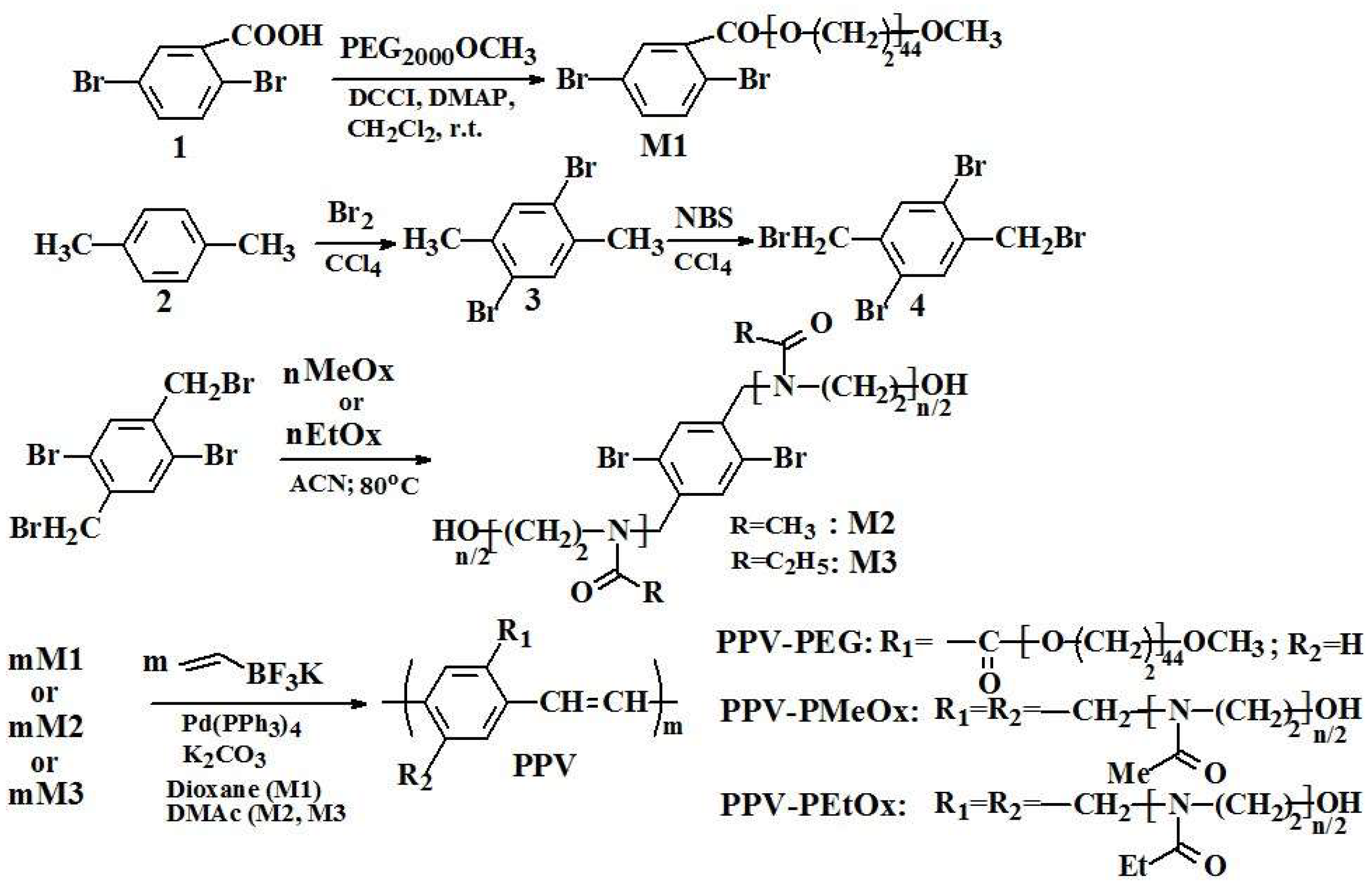
Scheme 1. The route for synthesis of macromonomers (M1, M2 and M3) and their derived g-PPVs.
Chemproc 16 00073 sch001
Figure 1. 1H-NMR spectrum of (a) macromonomer M2 in CD3OD and of (b) PPV-PEG in CDCl3.
Chemproc 16 00073 g001
Figure 2. AFM micrographs of (A)—PPV-PEG; (B)—PPV-PMeOx; (C)—PPV-PEtOx; (a)—height contrast and (b)—cross-sectional traces.
Chemproc 16 00073 g002
Figure 3. UV-vis (left side) and fluorescence (right side) spectra of g-PPVs, in water at 0.01 mg/mL.
Chemproc 16 00073 g003
Table 1. GPC data of PPV grafted polymers.
SampleMn,GPC (IPD) aMn,GPC (IPD) b
PPV-PEG3825 (1.13)44,580 (1.13)
PPV-PMeOx
PPV-PEtOx
276 * (1.02)
705 * (1.07)
8834 (1.07)
10,950 (1.1)
a—GPC measured in THF; b—GPC-measured in DMF; IPD-index of polydispersity; *—unrealistic values.
Table 2. Dependence of particle size on polymers’ concentration in water and some optical data.
SampleConcentration (mg/mL)Size (nm)λabsem (nm) a
PPV-PEG0.001- *nd
0.016.1; 806418/524
0.1- *nd
PPV-PMeOx0.001600; 4830nd
0.01
0.1
1
- *
28
373; 5290
nd
331/403
nd
PPV-PEtOx0.00172.5; 4960nd
0.01
0.1
1
16.5; 4430
1.58; 134
383; 4030
nd
267/403; 420
nd
* Obtained values exceeded the apparatus maximum limit; nd-not determined; a the values of the peak maxima, obtained in water, by UV-vis and fluorescence spectroscopy.
Disclaimer/Publisher’s Note: The statements, opinions and data contained in all publications are solely those of the individual author(s) and contributor(s) and not of MDPI and/or the editor(s). MDPI and/or the editor(s) disclaim responsibility for any injury to people or property resulting from any ideas, methods, instructions or products referred to in the content.