Proceeding Paper

A Refined Model of the CFTR Membrane Transporter as a Tool to Revert Misbehavior †

by
Pedro M. S. Suzano
1,*,
Michael González-Durruthy
2,
Ricardo J. Ferreira
3,
Cátia A. Bonito
4,
Margarida D. Amaral
5 and
Daniel J. V. A. dos Santos
1,*
1
CBIOS—Research Center for Biosciences & Health Technologies, Lusófona University, Campo Grande 376, 1749-024 Lisboa, Portugal
2
LAQV@REQUIMTE/Department of Chemistry and Biochemistry, Faculty of Sciences, University of Porto, 4169-007 Porto, Portugal
3
RG Discovery AB, Medicon Village, Building 403, Scheeletorget 1, 223 81 Lund, Sweden
4
Medicinal Chemistry, Research and Early Development, Cardiovascular, Renal and Metabolism (eCVRM), BioPharmaceuticals R&D, AstraZeneca, Gothenburg, 431 83 Mölndal, Sweden
5
BioISI—Biosystems & Integrative Sciences Institute, Faculty of Sciences, University of Lisboa, Campo Grande, C8, 1749-016 Lisboa, Portugal
*
Authors to whom correspondence should be addressed.
Presented at the 28th International Electronic Conference on Synthetic Organic Chemistry (ECSOC-28), 15–30 November 2024; Available online: https://sciforum.net/event/ecsoc-28.
Chem. Proc. 2024, 16(1), 109; https://doi.org/10.3390/ecsoc-28-20247
Published: 15 November 2024

Abstract

:
ABC proteins are large transmembrane efflux pumps that export substrates against the concentration gradient through ATP hydrolysis. Due to their efflux capabilities, they are crucial in drug metabolism. Point mutations in ABC proteins can lead to many forms of illness by causing protein misfolding and misbehavior. CFTR, or ABCC7, is a member of this transporter family, which, when mutated, can lead to cystic fibrosis, the most common life-shortening rare disease. Here, we report a refined and functional model of CFTR using in silico methods, aiming at further understanding anion permeation and the impact of different mutations on the gating mechanism to shine light on ways to reverse mutations’ effects.

1. Introduction

Cystic fibrosis (CF) is a genetic disorder mostly found in European populations, specifically those from Northern Europe. It is estimated to affect over 100,000 people worldwide, causing progressive damage to the lungs and pancreas [1]. Due to a very low life expectancy associated with this condition, CF has been considered the most common deadly genetic disorder of children in these populations. However, advancements in CF therapeutics from recent decades have enabled those affected to live, on average, close to 50 years [2].
The gene responsible for CF and coding for the cystic fibrosis transmembrane conductance regulator (CFTR) was successfully cloned in 1989 by Riordan and coworkers [3]. Since then, several therapies have been developed to improve the life expectancy and quality of these patients by targeting the genetic defect. This proved to be quite complex, as today, more than 2000 mutations in the CFTR gene have been found to cause CF. Despite this variety, around 80% of CF cases present a deletion of residue F508, making it the most problematic and common mutation [4]. Thus, targeting this variant has been the main goal of CF research in the past few decades.
The discovered CFTR mutations were grouped into six main classes, regarding their effects in cellular phenotypes. Class I is related to a lack of expression, class II is characterized by folding disabilities, and class III is associated with gating defects. Class IV is marked by conductance defects, while class V mutations lead to a significantly lower quantity in CFTR proteins and class VI mutations produce a very low amount of surface proteins. The F508del mutation is a class II mutation, meaning the CFTR protein fails to fold properly and is flagged for proteosomal degradation [4,5,6]. Hence, designing therapies for this phenotype can be achieved by focusing on rescuing the CFTR protein by correcting protein folding and increasing its activity.
To date, the most successful therapy option on the market for this deletion is a combination of three modulators named Trikafta. Trikafta is the FDA-approved therapy combining the effects of two folding correctors (VX-661 or tezacaftor and VX-445 or elexacaftor), necessary to rescue the defective protein, and a gating potentiator (VX-770 or Ivacaftor), which boosts gating function [4,5]. Despite the overall success and applicability of this form of therapy, the full mechanism of action of some of these compounds is not yet fully understood and insights into many of the rare mutant forms are scarce. Hence, a functional model of the CFTR protein would be valuable for understanding structural effects and details of these interaction changes.
CFTR, also known as ABCC7, belongs to the ABC (ATP-Binding Cassette) superfamily of proteins. It differs from most other proteins in this family, as many are known membrane transporter proteins while CFTR is an ion channel, using ATP binding and hydrolysis in anion translocation [7]. Still, CFTR shares structural similarities with many proteins in this family: it features two transmembrane domains (TMDs) that cross the membrane facilitating substrate crossing and two nucleotide-binding domains (NBDs) facing the cytoplasm, which are responsible for ATP recognition, binding and hydrolysis [8]. However, the architecture of the CFTR protein differs from other ABC transporters in the regulatory domain (R domain) [7,9].
Over the years, electrophysiological studies have demonstrated the importance of the R domain in the substrate transport process. This domain needs to undergo phosphorylation by protein kinase A (PKA) to allow the shift between the closed and open channel states [7,9,10]. Despite the importance of this domain, and the increasing number of available crystallographic structures, a resolved structure of the protein including the R domain does not exist, as of today. It is our opinion that a functional and refined model of the CFTR protein would be quite important to fully understand the mechanistic aspects of ion translocation, effects of modulators and correctors, and effects of point mutations in the CFTR protein.
Chen and coworkers postulate that some regions of the CFTR R domain are likely to be helical [9]. However, due to being mostly unstructured and quite long, a full refined model of this domain does not yet exist. To circumvent this, we researched functional R domain deletions. We found that deleting amino acids 708–835 generated a CFTR channel that was constitutively active [11]. Thus, by deleting this portion of the R domain, we generated an active channel that can be useful to study CFTR mechanics and the effects of amino acid mutations and deletions.

2. Methods

The CFTR atomistic model was built starting with the cryo-electron microscopy (cryo-EM) 6O2P deposited in the protein data bank (PDB) [12]. Although this structure describes the interaction with the ivacaftor potentiator, several segments could not be atomistically resolved, although it is, so far, the most complete resolved sequence.
To obtain a complete and functional atomistic model, we followed a protocol already successfully used to build the atomistic structures of Pgp [13] and BCRP [14]. Especially in the case of Pgp, the importance of including missing sequences that are relevant for function and/or structural stability was quite clear [13,15].
The third chain of the PDB structure 6O2P containing co-crystallized molecules/ions like ATP was removed and the protein was saved in a PDB file to be used as the starting point for model building. The second protein chain, containing a short sequence for which the side chains were not resolved, was kept to be used in the modeling. Both ATP and the corresponding magnesium ions were also saved on another file to be later included in the simulations. All other molecules were not used.
The missing segments were built using the Molecular Operating Environment (MOE v2019.01 [16]). Three short missing sequences exist between amino acids 410–436 and 1174–1201 (loops connecting a TMD with the NBD that follows in the sequence), and 890–899 (extracellular loop 4, uniting helices 7 and 8). The R domain (638–844) that needs to be phosphorylated to allow the channel to open is also mostly unmapped. However, for 17 amino acids (half organized as a helix), although their position is known, the side chains are not resolved and, therefore, their relative position in the sequence is unknown.
For small missing sequences, a search was made in MOE using the Loop/Linker Modeler module and the best scored loop was used. However, since the R domain is too large to be correctly modeled and positioned, we used a different approach. Experimentally, it was demonstrated that the R domain could be shortened while keeping the CFTR function [11].
To model the end part of the R domain sequence until Thr844, the “PredictProtein” server was used [17]. The mapped helix starting at Thr845 to Ile853 was included in the sequence uploaded to the server to match this helix sequence with the prediction and to assign the rest of the sequence. MOE’s Protein Builder was used to build this sequence as predicted by the server. For the remaining R domain sequence, the MOE’s Protein Builder was also used by choosing an extended geometry conformation for each added amino acid, always followed by a simple energy minimization using default parameters and linking the last added amino acid to the next known amino acid position.
All the added protein sequences were assembled in a PDB file. To correctly maintain the physiological function of CFTR, the experimentally known amino acids that need to be phosphorylated were phosphorylated using the CHARMM36 force field used throughout. The experimentally reported phosphorylated amino acids kept in the CFTR model were Ser422 (in NBD1), and Ser660, Ser670 and Ser700 in the R domain [18]. When needed, the charmm-gui web server was used to convert MOE’s PDB to the corresponding CHARMM36 atom types. To allow the newly modeled sequences to better adjust to their environment, the assembled CFTR protein was solvated and neutralized to be used in a NVT simulated annealing from 303 to 323 K using 10 points for a total simulation time of 10 ns, while keeping all protein position restrained but the added sequences. Except when otherwise stated, all simulations were executed in GROMACS (v2019.3).
A POPC bilayer containing 512 lipids and 20,480 water molecules was assembled from a previously used smaller POPC patch. This patch in water was equilibrated at NpT (1 bar, 303K), using parameters like those used for the fully assembled CFTR system and described below. The g_membed GROMACS (v4.6.2) program was used to embed the CFTR on the POPC bilayer patch and the position of the membrane relative to CFTR was defined using information obtained from the OPM database [19]. The number of lipids with the CFTR embedded in the bilayer was reduced to 468.
The system containing the CFTR protein embedded in the POPC patch and the ATP and magnesium ions previously saved from the 6O2P PDB structure was solvated. Other ions were also added to neutralize the system and the final system contained 468 lipids, 65,170 water molecules, 24 chlorine ions, 11 sodium ions, 2 ATP and 2 magnesium ions in a box of size about x = 125, y = 131 and z = 166 Å. Energy minimization using the steepest descent algorithm and default converging parameters were used and an NVT simulation was performed for just 50 ps to complete the fast adjustment of atoms and to allow the pressure to stabilize.
A run sampling the NpT ensemble was conducted for 200 ns to equilibrate the system followed by 3 replicates starting with different velocities sampled from the respective Maxwell–Boltzmann velocity distribution in accordance with a temperature of 303 K to collect statistics for production analysis. In all simulations, periodic boundary conditions (PBCs) were applied in all three axes. For NVT, the Nosé–Hoover thermostat [20,21] was used with a coupling constant of 0.2 ps. For NpT simulations, a Parrinello–Rahman barostat [22,23,24] with 5 ps semi-isotropic (x and y) coupling and a system compressibility of 4.5 × 10⁻⁵ bar−1 was used. The time-step was 2fs, and data were collected periodically to trajectory and energy files.
The Particle Mesh Ewald (PME) summation method for the electrostatic interactions used cubic interpolation, with an identical cutoff radius for electrostatic and short-range Lennard–Jones interactions (12 Å), was employed. The SETTLE (for water molecules) and LINCS algorithms were used to constrain bond lengths with hydrogen atoms.

3. Results and Discussion

After the system was built and calibrated, it became imperative to analyze and test the overall system health, guaranteeing no significant alterations resulting from the deletion. In Figure 1, the system’s structural fluctuations during a 500 ns production run can be observed. The root-mean square deviation (RMSD) of alpha carbons belonging to the protein main chain was used to study the system stability. The three modeled missing regions present significantly higher RMSD values than the rest of the protein, suggesting much higher mobility and the underlying reason for the lack of structural information in the experimental structures deposited in the PDB. The R domain, due to having been spliced in our model, tends to stick to the body of the protein and presents significantly lower movement. Our results show that this shorter domain model could probably be determined by cryo-EM, shedding light on a shorter but functional R domain.
The TMDs and NBDs are quite stable along the simulations and the contributions of the added protein sequences have a smaller impact on the global changes in the protein.
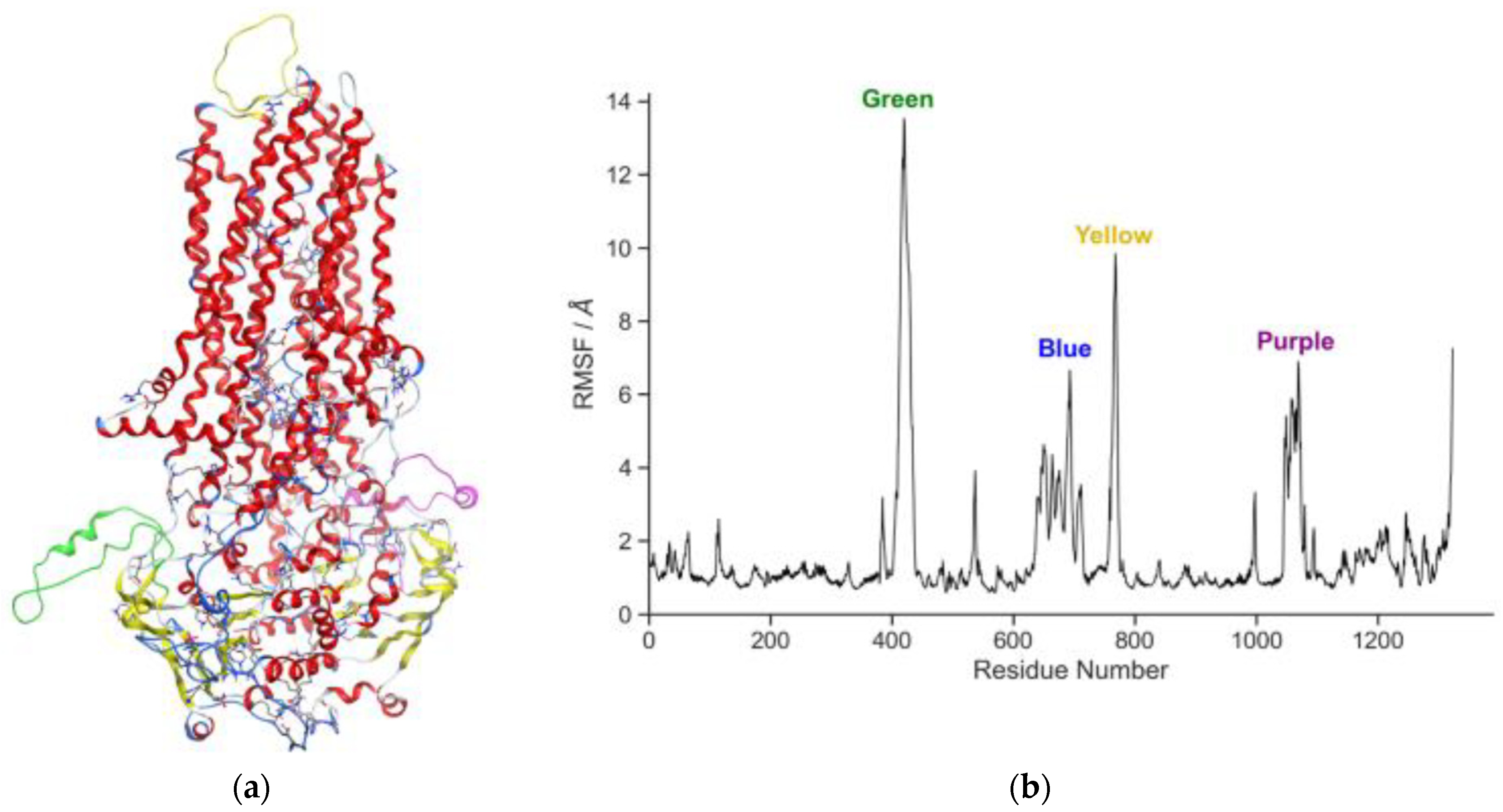
To further understand protein stability, a root-mean square fluctuation (RMSF) analysis was performed on the modeled CFTR, measuring the average deviation of atomic positions from their mean positions over time. In Figure 2, the specific modeled sequences can be compared to the fluctuation of all other residues. Herein, it is possible to verify that the most mobile regions are indeed the four regions missing in the original structure.
As an ion gate, CFTR needs to be open to both ions and water molecules. Hence, to check our system for functionality, we analyzed the pore in the center of the protein. In Figure 3, the transmembrane region of the protein (where most water molecules accumulate, and ions diffuse) is visible. Aside from the standard water flow that was observed, we found that some chlorine ions could move in and out of the protein pore (maximum of two; no sodium ions were ever found inside the channel) but the bilayer could never be transposed to what corresponds to the extracellular space (there is no ion gradient and, due to the PBC, both sides of the bilayer are in indirect contact through the simulation box xy wall). Moreover, water molecules seem to stack outside the main pore, near the proposed binding site for Ivacaftor. This finding is very interesting and requires further investigation, as it could lead to a greater understanding of one of the most powerful CFTR potentiator compounds with specific characteristics to bind in a quite peculiar environment: hydrophobic helices of the protein, matching the hydrophobic slab of the bilayer, although in a pocket that attracts water molecules.

4. Conclusions

Herein, a functional CFTR refined structure was obtained (DeltaR(708-835)-CFTR) that correlates to the behavior of the WT CFTR with one or two chlorine ions moving spontaneously to and inside the channel. In fact, in a following simulation already performed of a mutated model (G85E; not yet published), the chlorine ion is very quickly evicted from the channel and, in opposition to the present sliced WT model, no other chlorine ion ever returns to the channel. This needs further analysis and evaluation, but could be the basis for understanding the mechanism of the cystic fibrosis condition due to this mutation and could be used to further simulate other CFTR mutated structures. Through this work, the chlorine ion channel exit could be determined and the binding site of the potentiators and their mechanism of action could be clarified in the future.
Although several new experimental structures have become available, there are few computational models so far. One model by Tieleman and co-workers used the incomplete structure of the zebrafish to study the structural role of TM8 [25]. Another zebrafish CFTR model by Hegedüs and co-workers [26] used metadynamics simulations to reveal two possible exits on the channel and, more recently, Pomès and co-workers [27] used a strong hyperpolarizing electric field to study the translocation of chlorine ions. However, although these simulations used models corresponding to a phosphorylated state, none used an atomistic description containing phosphorylated residues that can impact the behavior of the models and, like the herein described model, only the latter used the human CFTR sequence.

Author Contributions

M.G.-D. R.J.F., C.A.B., M.D.A. and D.J.V.A.d.S. contributed to the conceptualization of the experiments and methodology. All authors, P.M.S.S., M.G.-D. R.J.F., C.A.B., M.D.A. and D.J.V.A.d.S. participated in writing and/or revising the manuscript; simulations were performed by D.J.V.A.d.S. and M.G.-D.; analysis of the simulations by P.M.S.S. and D.J.V.A.d.S.; resources provided by D.J.V.A.d.S. and M.D.A. All authors have read and agreed to the published version of the manuscript.

Funding

The authors of this work received financial support from the European Union (FEDER funds POCI/01/0145/FEDER/007265) and National Funds (FCT/MEC, Fundação para a Ciência e Tecnologia and Ministério da Educação e Ciência) under the Partnership Agreement PT2020UID/QUI/50006/2013 and H2020 PTDC/MED-QUI/28800/2017. Cátia C. Bonito acknowledges the PhD grant SFRH/BD/130750/2017 from FCT.

Institutional Review Board Statement

Not applicable.

Informed Consent Statement

Not applicable.

Data Availability Statement

A final confirmation of the MD refined CFTR structure and all files needed to perform GROMACS simulations are available for download at our website (http://chemistrybits.com (accessed on 26 September 2024)) or upon request to a corresponding author (D.J.V.A.d.S.).

Conflicts of Interest

The authors declare no conflict of interest.

References

  1. Kim, C.; Higgins, M.; Liu, L.; Volkova, N.; Zolin, A.; Naehrlich, L.; Andreas, P.; Elise, L.; Duška, T.-D.; Pavel, D.; et al. Effectiveness of Lumacaftor/Ivacaftor Initiation in Children with Cystic Fibrosis Aged 2 through 5 Years on Disease Progression: Interim Results from an Ongoing Registry-Based Study. J. Cyst. Fibros. 2024, 23, 436–442. [Google Scholar] [CrossRef]
  2. Scotet, V.; L’Hostis, C.; Férec, C. The Changing Epidemiology of Cystic Fibrosis: Incidence, Survival and Impact of the CFTR Gene Discovery. Genes 2020, 11, 589. [Google Scholar] [CrossRef]
  3. Riordan, J.R.; Rommens, J.M.; Kerem, B.-S.; Alon, N.; Rozmahel, R.; Grzelczak, Z.; Zielenski, J.; Lok, S.; Plavsic, N.; Chou, J.-L.; et al. Identification of the Cystic Fibrosis Gene: Cloning and Characterization of Complementary DNA. Science 1989, 245, 1066–1073. [Google Scholar]
  4. Veit, G.; Roldan, A.; Hancock, M.A.; Da Fonte, D.F.; Xu, H.; Hussein, M.; Frenkiel, S.; Matouk, E.; Velkov, T.; Lukacs, G.L. Allosteric Folding Correction of F508del and Rare CFTR Mutants by Elexacaftor-Tezacaftor-Ivacaftor (Trikafta) Combination. JCI Insight 2020, 5, e139983. [Google Scholar] [CrossRef]
  5. Dastoor, P.; Muiler, C.; Garrison, A.; Egan, M.; Carlos Dos Reis, D.; Santos, A.; Ameen, N.A. Localization and Function of Humanized F508del-CFTR in Mouse Intestine Following Activation of Serum Glucocorticoid Kinase 1 and Trikafta. Eur. J. Pharmacol. 2024, 978, 176771. [Google Scholar] [CrossRef]
  6. Yeh, J.; Yu, Y.; Hwang, T. Structural Mechanisms for Defective CFTR Gating Caused by the Q1412X Mutation, a Severe Class VI Pathogenic Mutation in Cystic Fibrosis. J. Physiol. 2019, 597, 543–560. [Google Scholar] [CrossRef]
  7. Liu, F.; Zhang, Z.; Csanády, L.; Gadsby, D.C.; Chen, J. Molecular Structure of the Human CFTR Ion Channel. Cell 2017, 169, 85–95.e8. [Google Scholar] [CrossRef]
  8. Alam, A.; Locher, K.P. Structure and Mechanism of Human ABC Transporters. Annu. Rev. Biophys. 2023, 52, 275–300. [Google Scholar] [CrossRef]
  9. Zhang, Z.; Liu, F.; Chen, J. Molecular Structure of the ATP-Bound, Phosphorylated Human CFTR. Proc. Natl. Acad. Sci. USA 2018, 115, 12757–12762. [Google Scholar] [CrossRef]
  10. Levring, J.; Terry, D.S.; Kilic, Z.; Fitzgerald, G.; Blanchard, S.C.; Chen, J. CFTR Function, Pathology and Pharmacology at Single-Molecule Resolution. Nature 2023, 616, 606–614. [Google Scholar] [CrossRef]
  11. Ostedgaard, L.S.; Baldursson, O.; Welsh, M.J. Regulation of the Cystic Fibrosis Transmembrane Conductance Regulator Cl− Channel by Its R Domain. J. Biol. Chem. 2001, 276, 7689–7692. [Google Scholar] [CrossRef]
  12. Liu, F.; Zhang, Z.; Levit, A.; Levring, J.; Touhara, K.K.; Shoichet, B.K.; Chen, J. Structural Identification of a Hotspot on CFTR for Potentiation. Science 2019, 364, 1184–1188. [Google Scholar] [CrossRef]
  13. Ferreira, R.J.; Ferreira, M.-J.U.; dos Santos, D.J.V.A. Insights on P-Glycoprotein’s Efflux Mechanism Obtained by Molecular Dynamics Simulations. J. Chem. Theory Comput. 2012, 8, 1853–1864. [Google Scholar] [CrossRef]
  14. Ferreira, R.J.; Bonito, C.A.; Cordeiro, M.N.D.S.; Ferreira, M.-J.U.; dos Santos, D.J.V.A. Structure-Function Relationships in ABCG2: Insights from Molecular Dynamics Simulations and Molecular Docking Studies. Sci Rep 2017, 7, 15534. [Google Scholar] [CrossRef]
  15. Ferreira, R.J.; Ferreira, M.-J.U.; dos Santos, D.J.V.A. Assessing the Stabilization of P-Glycoprotein’s Nucleotide-Binding Domains by the Linker, Using Molecular Dynamics. Mol. Inform. 2013, 32, 529–540. [Google Scholar] [CrossRef]
  16. Molecular Operating Environment (MOE), 2019.01 Chemical Computing Group ULC. 910-1010 Sherbrooke St. W., Montreal, QC H3A 2R7, 2019. Available online: https://www.chemcomp.com/en/index.htm (accessed on 26 September 2024).
  17. Bernhofer, M.; Dallago, C.; Karl, T.; Satagopam, V.; Heinzinger, M.; Littmann, M.; Olenyi, T.; Qiu, J.; Schütze, K.; Yachdav, G.; et al. PredictProtein—Predicting Protein Structure and Function for 29 Years. Nucleic Acids Res. 2021, 49, W535–W540. [Google Scholar] [CrossRef]
  18. Bozoky, Z.; Krzeminski, M.; Chong, P.A.; Forman-Kay, J.D. Structural Changes of CFTR R Region upon Phosphorylation: A Plastic Platform for Intramolecular and Intermolecular Interactions. FEBS J. 2013, 280, 4407–4416. [Google Scholar] [CrossRef]
  19. Lomize, M.A.; Lomize, A.L.; Pogozheva, I.D.; Mosberg, H.I. OPM: Orientations of Proteins in Membranes Database. Bioinformatics 2006, 22, 623–625. [Google Scholar] [CrossRef]
  20. Nosé, S. A Molecular Dynamics Method for Simulations in the Canonical Ensemble. Mol. Phys. 1984, 52, 255–268. [Google Scholar] [CrossRef]
  21. Hoover, W.G. Canonical Dynamics: Equilibrium Phase-Space Distributions. Phys. Rev. A 1985, 31, 1695–1697. [Google Scholar] [CrossRef]
  22. Parrinello, M.; Rahman, A. Crystal Structure and Pair Potentials: A Molecular-Dynamics Study. Phys. Rev. Lett. 1980, 45, 1196–1199. [Google Scholar] [CrossRef]
  23. Parrinello, M.; Rahman, A. Polymorphic Transitions in Single Crystals: A New Molecular Dynamics Method. J. Appl. Phys. 1981, 52, 7182–7190. [Google Scholar] [CrossRef]
  24. Parrinello, M.; Rahman, A. Strain Fluctuations and Elastic Constants. J. Chem. Phys. 1982, 76, 2662–2666. [Google Scholar] [CrossRef]
  25. Corradi, V.; Gu, R.-X.; Vergani, P.; Tieleman, D.P. Structure of Transmembrane Helix 8 and Possible Membrane Defects in CFTR. Biophys. J. 2018, 114, 1751–1754. [Google Scholar] [CrossRef]
  26. Farkas, B.; Tordai, H.; Padányi, R.; Tordai, A.; Gera, J.; Paragi, G.; Hegedűs, T. Discovering the Chloride Pathway in the CFTR Channel. Cell. Mol. Life Sci. 2020, 77, 765–778. [Google Scholar] [CrossRef]
  27. Zeng, Z.W.; Linsdell, P.; Pomès, R. Molecular Dynamics Study of Cl− Permeation through Cystic Fibrosis Transmembrane Conductance Regulator (CFTR). Cell. Mol. Life Sci. 2023, 80, 51. [Google Scholar] [CrossRef]
Figure 1. (a)—Representation of the CFTR model. Ions are represented by light and dark blue spheres. Protein helices are in purple and beta sheets are in yellow. Lipid bilayer is represented by light blue sticks with orange phosphates and, for clarity, water molecules were not depicted. (b)—Root-mean square deviations over a 500 ns production run. Total modeled protein RMSD is shown in black, TMDs in orange and NBDs in red. Modeled regions 1 (amino acids 410–436), 2 (890–899) and 3 (1174–1201) are in green, yellow and purple, respectively. R domain RMSD is depicted in blue.
Chemproc 16 00109 g001
Figure 2. (a)—CFTR represented in cartoon. Modeled sequence 1 (amino acids 410–436) is in green, the R domain is in blue and modeled sequences 2 (890–899) and 3 (1174–1201) are in yellow (loop on the top of the structure) and purple, respectively. (b)—Root-mean square fluctuations per protein residue. Higher peaks mean larger average deviation from the mean atomic positions.
Chemproc 16 00109 g002
Figure 3. Cartoon representation of the CFTR ion channel with water molecules crossing the pore. Water molecules are represented as red and white spheres and protein TMD in purple. Water molecules inside the bilayer and outside the pore can be seen on the left side. A chlorine ion inside the channel is depicted as a large cyan ball.
Chemproc 16 00109 g003
Disclaimer/Publisher’s Note: The statements, opinions and data contained in all publications are solely those of the individual author(s) and contributor(s) and not of MDPI and/or the editor(s). MDPI and/or the editor(s) disclaim responsibility for any injury to people or property resulting from any ideas, methods, instructions or products referred to in the content.