Proceeding Paper

Preparation of a Fluorescent Peptide Substrate to Target Tumor-Associated Macrophages †

by
Ângela B. M. P. Leite
,
Cátia D. F. Martins
,
M. Manuela M. Raposo
and
Susana P. G. Costa
*
Centre of Chemistry, University of Minho, Campus de Gualtar, 4710-057 Braga, Portugal
*
Author to whom correspondence should be addressed.
Presented at the 27th International Electronic Conference on Synthetic Organic Chemistry (ECSOC-27), 15–30 November 2023; Available online: https://ecsoc-27.sciforum.net/.
Chem. Proc. 2023, 14(1), 63; https://doi.org/10.3390/ecsoc-27-16101
Published: 15 November 2023

Abstract

:
One of the significant challenges in targeting M2 macrophages is the need for specific and high-affinity targeting ligands that can distinguish them from phenotype M1 macrophages. In order to investigate the selectivity of the polypeptide M2pep (YEQDPWGVKWWY) towards M2 macrophages using fluorescence-based techniques, we report here the synthesis and characterization of the M2pep substrate labeled with a fluorophore. This peptide comprising a 12-amino acid sequence with a spacer consisting of three additional glycines (GGG) was synthesized and coupled to Rhodamine B at its N-terminal via microwave-assisted solid-phase synthesis. The structure of the labeled peptide was confirmed by NMR, UV-Vis, fluorescence spectroscopy and mass spectrometry.

1. Introduction

Tumor-associated macrophages (TAMs) are immune cells that perform essential roles within the tumor microenvironment. These macrophages can change their functional characteristics in response to local environmental cues, switching between an M1 phenotype (anti-tumor) or an M2 phenotype (tumor-promoting). Unfortunately, TAMs are largely polarized into the M2 phenotype, contributing to tumor growth and immunosuppression [1,2]. Therefore, strategies to modulate and reprogram M2 macrophages toward an M1 phenotype have been explored as potential approaches to boost anti-cancer immune responses and enhance the effectiveness of cancer therapies [3]. Recent studies have reported that the polypeptide M2pep (YEQDPWGVKWWY) preferentially binds to M2 macrophages, making it a promising candidate for targeted drug delivery and imaging of M2 TAMs [3,4,5].
The conjugation of fluorescent probes with peptides and other biomolecules is a widely used strategy for the investigation of cellular processes and the development of innovative methodologies for diseases such as cancer, chronic infections, and neurodegenerative disorders. Fluorescent probes, known for their sensitivity and selectivity, find applications in various fields, including bioimaging, disease diagnosis and drug delivery, allowing for in situ, real-time and non-invasive analysis [6]. Xanthene derivatives, such as Rhodamine B (RhoB), are commonly employed as cost-effective and reliable fluorescent labels for peptides and proteins in biological and medical research [7,8].
RhoB exhibits an equilibrium between two distinct forms, a fluorescent open form A and a non-fluorescent lactone form B, as depicted in Figure 1. Under acidic environments, the pink-colored form A predominates, while the colorless form B dominates in basic conditions [9]. The acidic conditions prevalent within the tumor microenvironment promote the transition of RhoB into its fluorescent form; therefore, rhodamine derivatives have been developed as fluorescence-based imaging reporters for cancer diagnosis and therapy [7,10].
Our recent research is focused on re-programming TAMs toward an M1 phenotype as a means to impact cancer prognosis. Therefore, to investigate the selectivity of M2pep to target M2 macrophages using fluorescence-based techniques, we report here the synthesis and the characterization of M2pep labeled with Rhodamine B.

2. Experimental Section

2.1. General

All the chemicals and solvents were purchased from commercial suppliers and were used as received. Fmoc protected amino acids, Fmoc-Gly-Wang resin (loading, 0.51 mmol/g) and ethyl-2-cyano-2-(hydroxyimino) acetate (Oxyma) were purchased from AAPPTec (Louisville, KY, USA). N,N′-diisopropylcarbodiimide (DIC), piperidine, triisopropylsilane (TIS) and trifluoroacetic acid (TFA) were obtained from Acros Organics (Geel, Belgium). Rhodamine B was purchased from Sigma Aldrich (St. Louis, MO, USA).
1H NMR spectra were recorded on a Bruker Avance III 400 at an operating frequency of 400 MHz, using the solvent peak as the internal reference at 25 °C. The synthesis of M2pep-GGG 1 was performed on an automatic peptide synthesizer (Liberty Blue 2.0, CEM Corp., Matthews, NC, USA), while the conjugation of M2pep-GGG with RhoB was carried out on a manual peptide synthesizer (Discover SPS, CEM Corp., Matthews, NC, USA). High-resolution mass spectra, in positive ESI mode, were acquired using a QqTOF Impact IITM mass spectrometer (Bruker, Billerica, MA, USA). Low-resolution ESI-MS analyses were performed using a Thermo Scientific LxQ linear ion trap equipped with an electrospray ionization source (ThermoFisher Scientific, Waltham, MA, USA). Probe purification was carried out via semi-preparative HPLC on a Shimadzu LC-8A, UV/Vis JASCO 875-UV detector and a Shimadzu C-RGA Chromatopac register (Shimadzu Europa GmbH, Duisburg, Germany) on a Europa Peptide 120 C18 (5 mm) column using ACN/water (1:1, v/v) containing 0.1% TFA (λdet = 540 nm). UV/Vis absorption spectra were recorded on a Shimadzu UV/2501PC (Shimadzu Europa GmbH, Duisburg, Germany) spectrophotometer, and fluorescence spectra were collected using a FluoroMax-4 (HORIBA Europe GmbH, Darmstadt, Germany) spectrofluorometer in standard quartz cuvettes.

2.2. Methods

2.2.1. Synthesis of M2pep 1

M2pep with a triple glycine spacer at the C-terminal (YEQDPWGVKWWYGGG) 1 was synthesized following standard Fmoc solid-phase peptide synthesis on a 0.25 mmol scale using a preloaded Fmoc-Gly-Wang resin. All Fmoc-protected amino acids were coupled using DIC and Oxyma as coupling agents, and Fmoc protecting group removal was performed with 20% piperidine in DMF. A portion of the resin was subjected to cleavage to afford the free peptide (see details below) for characterization via NMR spectroscopy and mass spectrometry.
HRMS: m/z (ESI, positive mode) m/z for C89H112N20O23, calculated 914.4099; found 914.9124 (+2 charge).

2.2.2. Synthesis of RhoB-M2pep 2

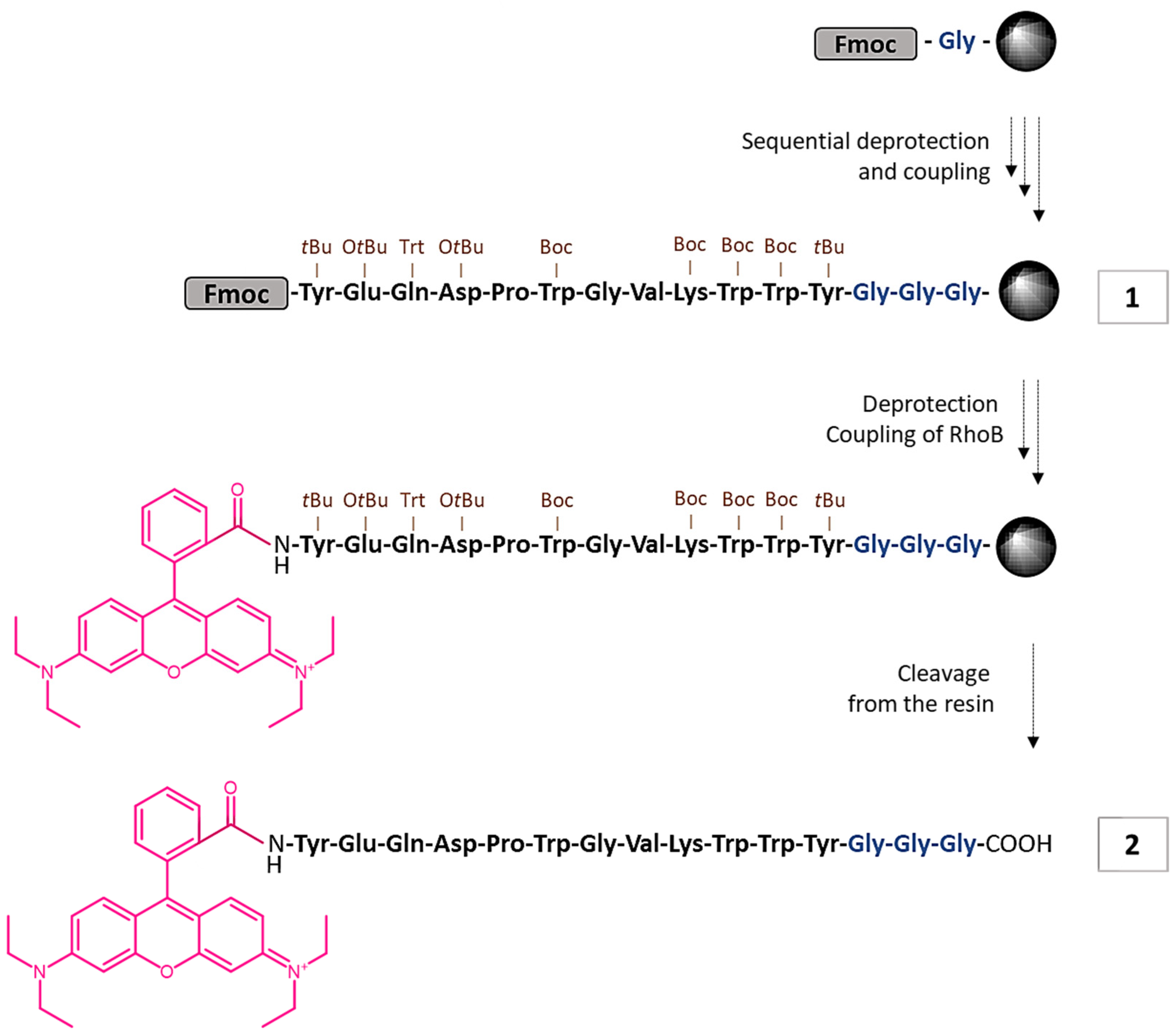
After the last Fmoc group had been removed from the on-resin M2pep-GGG 1, Rhodamine B was coupled at its N-terminal using DIC and Oxyma in DMF. This mixture was added to resin M2pep-GGG 1 and subjected to five microwave irradiation cycles of 5 min (P = 25 W, T = 75 °C). Then, the resin was washed with DMF followed by MeOH several times. The substrate was cleaved from the resin with a mixture of TFA/H2O/TIS (95:2.5:2.5, v/v). After stirring with the cleavage cocktail for 2 h at room temperature, the solution was filtered, and the product was precipitated by the addition of cold diethyl ether to the filtrate and collected via centrifugation. The crude product 2 was purified via semi-preparative HPLC using an ACN/water mixture (1:1) containing 0.1% TFA at a flow rate of 1 mL/min. The desired compound was eluted from the column with a retention time of 29 min. Probe 2 was obtained as a pink solid (0.019 g, yield 6%).
MS (ESI, positive mode): m/z 1133 (100) (corresponds to fragment resulting from loss of the triple Gly spacer and cleavage at the D-P bond).

2.2.3. Photophysical Characterization

The spectroscopic characterization of M2pep-GGG 1 and RhoB-M2pep-GGG 2 was carried out using UV/Vis absorption and fluorescence spectroscopy. Solutions of compounds 1 and 2 (1.0 × 10⁻⁵ M) were prepared in phosphate buffer at pH 5.6. Fluorescence spectra were obtained by excitation at 540 nm, with a 3 nm slit.

3. Results and Discussion

3.1. Synthesis

Based on our research interest in using fluorescence-based techniques to target tumor-associated macrophages for cancer diagnosis and therapy, we prepared and fully characterized a new fluorogenic substrate for M2 TAMs.
Firstly, M2pep-GGG (YEQDPWGVKWWYGGG) 1 was synthesized via microwave-assisted solid-phase peptide synthesis (MW-SPPS), applying the Fmoc strategy on a preloaded glycine Wang resin. The peptide chain was elongated in consecutive cycles of deprotection and coupling of the required amino acids. Then, M2pep-GGG 1 was cleaved from a portion of the resin and its structure was characterized via 1H NMR spectroscopy and mass spectrometry. Although not all proton signals could be assigned unequivocally in the 1H NMR spectrum due to signal overlap, they were consistent with the expected structure, as were the data obtained via mass spectrometry.
Subsequently, the free N-terminal of the on resin M2pep-GGG 1 was conjugated with RhoB by MW-SPPS using DIC/Oxyma in dry DMF. The labeled substrate, RhoB-M2pep-GGG 2, as shown in Scheme 1, was cleaved from the solid support via treatment with TFA/H2O/TIS (95:2.5:2.5, v/v). Finally, the obtained product was purified using semi-preparative HPLC to ensure proper separation from free RhoB, which could interfere with subsequent fluorescence measurements. The desired product, RhoB-M2pep-GGG 2, was obtained in 6% yield and its structure was confirmed via mass spectrometry.

3.2. UV/Vis Absorption and Fluorescence Spectroscopy

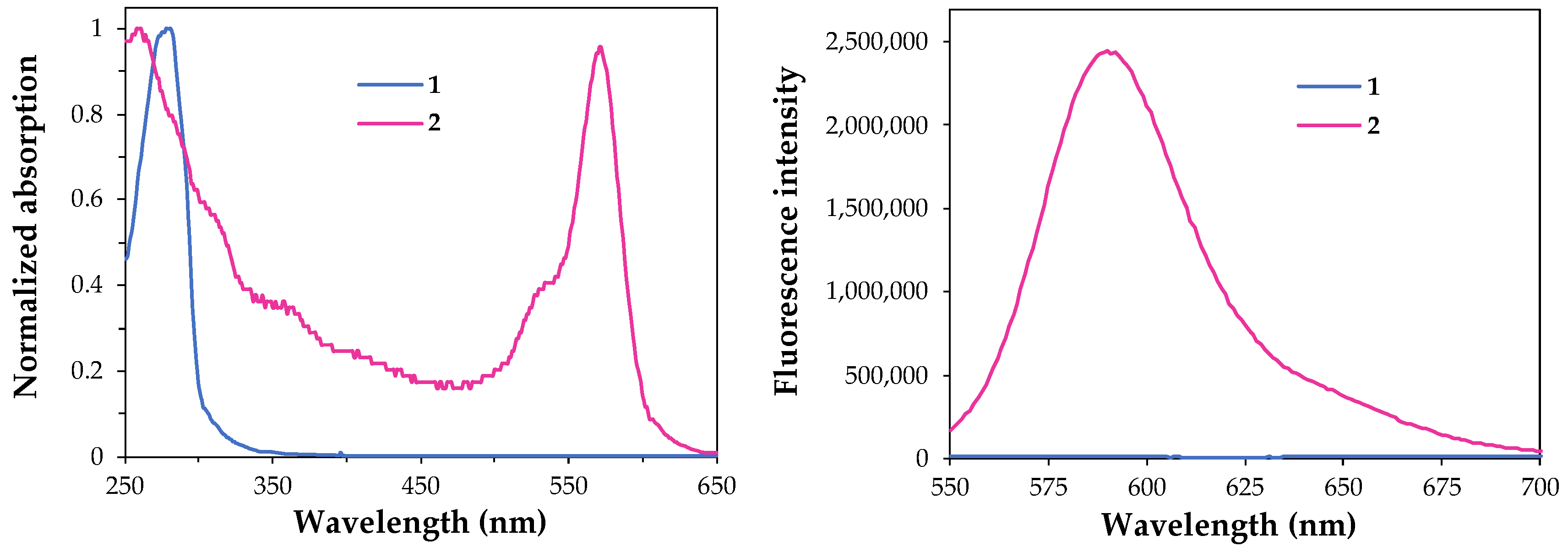
The photophysical properties, namely the absorption and fluorescence spectra of both free peptide 1 and labeled peptide 2, were measured in phosphate buffer at pH 5.6 to evaluate the potential application of probe 2 as a fluorescent reporter in the acidic conditions of the tumor microenvironment.
It was found that the free peptide 1 solution exhibited an intense absorption band at 280 nm, while RhoB-M2pep-GGG 2 showed absorption bands at 260 nm and 572 nm (Figure 2, left a). The latter is responsible for the pink color solution of 2 and is ascribed to Rhodamine B, while the absorption band at 260–280 nm observed for both compounds 1 and 2 can be attributed to the presence of tryptophan and tyrosine residues.
A comparison of the fluorescence spectrum of labeled peptide 2 with that of free peptide 1 (Figure 2, right) clearly showed that the conjugation of Rhodamine B with M2pep-GGG significantly enhanced the fluorescence intensity, showing a wavelength of maximum fluorescence at 590 nm. Therefore, RhoB-M2pep-GGG 2 could be used as an efficient probe to target M2 macrophages in cancer diagnosis.

4. Conclusions

In summary, we have reported the synthesis of a new fluorescent peptide substrate to target M2 macrophages. This peptide comprising a 15-amino acid sequence was successfully prepared and coupled to Rhodamine B via microwave-assisted solid-phase synthesis. However, further work will be carried out to optimize the synthesis and isolation processes to enhance the overall synthesis yield of the labeled substrate.
Furthermore, the photophysical characterization of RhoB-M2pep-GGG 2 showed an intense emission band at 590 nm in phosphate buffer at pH 5.6, and so this fluorescent peptide is currently being used to target M2 macrophages in vitro.

Author Contributions

Conceptualization, Â.B.M.P.L., C.D.F.M. and S.P.G.C.; methodology, Â.B.M.P.L., C.D.F.M. and S.P.G.C.; validation, S.P.G.C. and M.M.M.R.; formal analysis, Â.B.M.P.L., C.D.F.M. and S.P.G.C.; investigation, Â.B.M.P.L. and C.D.F.M.; resources, S.P.G.C. and M.M.M.R.; writing—original draft preparation, Â.B.M.P.L. and C.D.F.M.; writing—review and editing, Â.B.M.P.L., C.D.F.M., M.M.M.R. and S.P.G.C.; supervision, S.P.G.C. and M.M.M.R.; project administration, S.P.G.C. and M.M.M.R.; funding acquisition, S.P.G.C. and M.M.M.R. All authors have read and agreed to the published version of the manuscript.

Funding

This research was funded by the Foundation for Science and Technology (FCT) who provided financial support to CQ/UM (UID/QUI/00686/2020), project (PTDC/QUI-OUT/3143/2021) and a PhD grant to C. D. F. Martins (SFRH/BD/05277/2020). The NMR spectrometer Bruker Avance III 400 is part of the National NMR Network and was purchased within the framework of the National Program for Scientific Re-equipment, contract REDE/1517/RMN/2005, with funds from POCI 2010 (FEDER) and FCT.

Institutional Review Board Statement

Not applicable.

Informed Consent Statement

Not applicable.

Data Availability Statement

The data presented in this study are available in the article.

Conflicts of Interest

The authors declare no conflict of interest.

References

  1. He, W.; Kapate, N.; Shields, C.W.; Mitragotri, S. Drug Delivery to Macrophages: A Review of Targeting Drugs and Drug Carriers to Macrophages for Inflammatory Diseases. Adv. Drug. Deliv. Rev. 2020, 165–166, 15–40. [Google Scholar] [CrossRef] [PubMed]
  2. Obeid, E.; Nanda, R.; Fu, Y.-X.; Olopade, O.I. The Role of Tumor-Associated Macrophages in Breast Cancer Progression (Review). Int. J. Oncol. 2013, 43, 5–12. [Google Scholar] [CrossRef] [PubMed]
  3. Hourani, T.; Holden, J.A.; Li, W.; Lenzo, J.C.; Hadjigol, S.; O’Brien-Simpson, N.M. Tumor Associated Macrophages: Origin, Recruitment, Phenotypic Diversity, and Targeting. Front. Oncol. 2021, 11, 788365. [Google Scholar] [CrossRef] [PubMed]
  4. Cieslewicz, M.; Tang, J.; Yu, J.L.; Cao, H.; Zavaljevski, M.; Motoyama, K.; Lieber, A.; Raines, E.W.; Pun, S.H. Targeted Delivery of Proapoptotic Peptides to Tumor-Associated Macrophages Improves Survival. Proc. Natl. Acad. Sci. USA 2013, 110, 5919–15924. [Google Scholar] [CrossRef] [PubMed]
  5. Ngambenjawong, C.; Cieslewicz, M.; Schellinger, J.G.; Pun, S.H. Synthesis and Evaluation of Multivalent M2pep Peptides for Targeting Alternatively Activated M2 Macrophages. J. Control. Release 2016, 224, 103–111. [Google Scholar] [CrossRef] [PubMed]
  6. Li, X.; Zhao, S.; Li, B.; Yang, K.; Lan, M.; Zeng, L. Advances and Perspectives in Carbon Dot-Based Fluorescent Probes: Mechanism, and Application. Coord. Chem. Rev. 2021, 431, 213686. [Google Scholar] [CrossRef]
  7. Birtalan, E.; Rudat, B.; Kolmel, D.K.; Fritz, D.; Vollrath, S.B.; Schepers, U.; Brase, S. Investigating Rhodamine B-Labeled Peptoids: Scopes and Limitations of Its Applications. Biopolymers 2011, 96, 694–701. [Google Scholar] [CrossRef] [PubMed]
  8. Belko, N.; Maltanava, H.; Lugovski, A.; Ferreira, R.A.S.; Correia, S.F.H.; Shabunya, P.; Fatykhava, S.; Tabolich, A.; Kulahava, T.; Bahdanava, A.; et al. pH-Sensitive Fluorescent Sensor for Fe(III) and Cu(II) Ions Based on Rhodamine B Acylhydrazone: Sensing Mechanism and Bioimaging in Living Cells. Microchem. J. 2023, 191, 108744. [Google Scholar] [CrossRef]
  9. Hoenke, S.; Serbian, I.; Deigner, H.-P.; Csuk, R. Mitocanic Di- and Triterpenoid Rhodamine B Conjugates. Molecules 2020, 25, 5443. [Google Scholar] [CrossRef] [PubMed]
  10. García-Hevia, L.; Casafont, Í.; Oliveira, J.; Terán, N.; Fanarraga, M.L.; Gallo, J.; Bañobre-López, M. Magnetic Lipid Nanovehicles Synergize the Controlled Thermal Release of Chemotherapeutics with Magnetic Ablation While Enabling Non-Invasive Monitoring by MRI for Melanoma Theranostics. Bioact. Mater. 2021, 8, 153–164. [Google Scholar] [CrossRef]
Figure 1. Structure of RhoB in its open form (A) and closed lactone form (B).
Chemproc 14 00063 g001
Scheme 1. Synthesis of RhoB-M2pep-GGG 2.
Chemproc 14 00063 sch001
Figure 2. Normalized UV/Vis absorption (left) and fluorescence (right) spectra of M2pep-GGG 1 and RhoB-M2pep-GGG 2 (1.0 × 10⁻⁵ M) in phosphate buffer at pH 5.6.
Chemproc 14 00063 g002
Disclaimer/Publisher’s Note: The statements, opinions and data contained in all publications are solely those of the individual author(s) and contributor(s) and not of MDPI and/or the editor(s). MDPI and/or the editor(s) disclaim responsibility for any injury to people or property resulting from any ideas, methods, instructions or products referred to in the content.