Proceeding Paper

Molecular Dynamics Simulations on the Mesophilic Enzyme Vibrio Cholerae Endonuclease I: Salt Effect Study †

by
Djamila Benrezkallah
1,2
1
Department of Basic Teachings in Sciences and Technologies (EBST), Faculty of Technology, Djillali Liabes University, Ben M’HIDI BP 89, Sidi Bel Abbes 22000, Algeria
2
LCPM Laboratory, Chemistry Department, Faculty of Exact and Applied Sciences, University Oran 1 Ahmed Ben Bella, El Mnaouer BP 1524, Oran 31000, Algeria
Presented at the 27th International Electronic Conference on Synthetic Organic Chemistry (ECSOC-27), 15–30 November 2023; Available online: https://ecsoc-27.sciforum.net/.
Chem. Proc. 2023, 14(1), 40; https://doi.org/10.3390/ecsoc-27-16165
Published: 15 November 2023

Abstract

:
Some of the most extensively studied marine or estuarine bacteria belong to the genus Vibrio, with Vibrio cholerae being the most notorious species as it is the cause of cholera in humans. V. cholerae is found in tropical and temperate areas and can be classified as a mesophilic bacterium with its growth optimum at around 37 °C. One of the important factors in the activity and stability of each enzyme is its physiological environment. A previous study on the secreted mesophilic enzyme Endonuclease I from the Vibrio cholerae genus (VcEndA) showed that its activity was strongly dependent not only on temperature but also on NaCl concentration. Here, we report a structural study on the mesophilic enzyme (VcEndA) using molecular dynamics simulations at different salt concentrations (NaCl). The analysis of molecular dynamic simulation trajectories reveals that the enzyme is not tolerant and not sensitive to salt since the profile of the rmsf as a function of different concentrations does not show a large difference in the mobility of the enzyme for high values of the NaCl concentration (450 and 650 mM). However, the most flexible regions of the enzyme are recorded under the concentration of 175 mM, which coincides well with the previous experimental work.

1. Introduction

The Gram-negative bacterium Vibrio cholerae has always been, throughout history, a terrible microorganism due to its pathogenic and contagious properties, which have caused cholera pandemics affecting all continents in the world [1]. Vibrio cholerae is also a natural bacterial inhabitant of aquatic environments and is associated with copepod crustaceans and aquatic plants [2]. Bacteria isolated from the majority of environmental samples exhibit non-pathogenic properties due to the lack of the gene for cholera enterotoxin [2]. As the bacterium Vibrio cholerae is linked to water, and pandemics with few exceptions have their origins in the Indian subcontinent and the Ganges delta in Bengal, it is suggested that water acts as a reservoir for the bacteria [1].
The characterization and cloning of Vibrio cholerae Endonuclease I (VcEndA) were first described by Focareta and others [3], but the structural determination of the nuclease was carried out twenty years later by Altermark and others [4]. There are two structures of VcEndA deposited in the Protein Data Bank, both of which are solved by X-ray crystallography. The highest resolution structure (1.95 Å), with PDB entry 2g7f, is crystallized at neutral pH and has optimal catalytic activity at a concentration of NaCl equal to 175 mM at pH 7.5–8.0 and a temperature of 50 °C [5].
Among the orthologs of Endonuclease I of several species of bacteria which are described in the literature [5,6,7,8,9], we were interested in the study of Vibrio cholerae Endonuclease I (VcEndA) because the biochemical characterization of this enzyme has already been reported and compared to that of its counterpart, Vibrio salmonicida Endonuclease I VsEndA [5]. In this article, we will present, by molecular dynamics simulation at T = 300 K, the effect of different NaCl concentration values (Table 1) on the structural flexibility of the VcEndA enzyme.

2. Materials and Methods

All molecular dynamic simulations presented in the present article were performed using the CHARMm27 force field [10,11] implemented in a parallel architecture in the program GROMACS 4.5.3 [12,13]. The initial crystal structure of the Vibrio cholera Endonuclease I (VcEndA) enzyme was downloaded from the protein data bank (PDB entry 2g7f [5]). Hydrogen atoms were added with the pdb2gmx program in the GROMACS code. The starting structure was immersed in a dodecahedron box of TIP3P water molecules [14]. The minimal distance between the protein atoms and the box edges was set to 1 nm. In order to create an electric neutral system, 6 water molecules were replaced by Cl ions. We added also a number of Cl and Na+ ions to ensure the salt concentration values of 50, 175, 425, and 650 mM (Table 1). The system was first submitted to energy minimization using the steepest descent algorithm. Then, several MD simulations of 50 ps each were run at the constant-volume, constant-temperature ensemble (NVT) by increasing temperature gradually from 0 to 300 K. The final MD runs were subjected to the constant-pressure, constant-temperature (NPT) ensemble (P = 1 bar, T = 300 K), for 10ns each at five different salt concentrations (0, 50, 175, 425 and 650 mM). Periodic boundary conditions were used under isothermal and isobaric conditions using the Berendsen coupling algorithm [15] with relaxation times of 0.1 and 1ps, respectively. The LINCS algorithm [16] was used to constrain bond lengths using a time step of 2 fs for all calculations. Electrostatic interactions were calculated using the particle Mmesh Ewald (PME) [17,18] summation scheme. Van der Waals and Coulomb interactions were truncated at 1.2 nm. The non-bonded pair list was updated every 5 steps, and conformations were stored every 2 ps.

3. Results

3.1. Stability of the Model

Figure 1 shows the temporal evolution of the RMSDs of the VcEndA enzyme backbone for the different simulations in which we varied the value of the NaCl concentration (Table 1). According to this figure, the RMSDs reflect fairly stable MD trajectories with RMSDs average values that do not exceed 1.6 A°.

3.2. Structural Flexibility

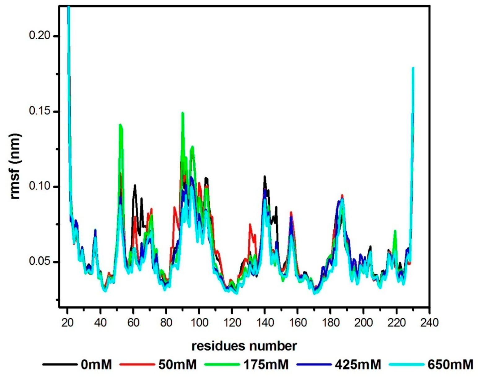
To evaluate the relative mobility of the protein regions during MD simulations, the (RMSF) root mean square fluctuation of Cα atoms in a residue of the average structure obtained from the merged images of the MD trajectories is calculated for the VcEndA enzyme as a function of the number of residues, at different NaCl salt concentrations, at 300 K (Figure 2).

3.3. Radial Distribution Functions

The radial distribution functions of Cl ions around the backbone of the residue with the highest value of the rmsf profile, GLN90, as well as those of water molecules around the most flexible residues of the protein structure, GLY52, LYS53, and GLN90, as a function of different salt concentrations, are presented in Figure 3.

4. Discussion

The rmsf profile for the mesophilic enzyme, under different salt concentrations (Figure 2), is almost the same, with the exception of two regions, where the flexibility of the enzyme is high at C = 175 mM compared to the other concentration values, the region (GLY52-LYS53), and the GLN90 residue.
Located in the loop region, between two β-sheets (β1 and β2, Figure 4), residue GLY52 presents a high rmsf value of 0.1415 nm, and the residue which succeeds it in the primary structure of the enzyme; LYS53, has an rmsf value of 0.1385 nm, for the salt concentration equal to 175 mM.
Glycine is sometimes grouped with hydrophobic amino acids. However, it is unique in having no side chains. This absence of a side chain allows glycine to have bond angles (in the backbone bends) that are much more extreme than other amino acids. It, therefore, plays a vital role in allowing a large number of conformations in proteins without excessive deformation. It is also worth noting that the GLY52 residue is substituted by the polar ASN52 residue in the primary structure of the cold-adapted enzyme, VsEndA, and that the largest fluctuation recorded regarding the structural flexibility of this psychrophilic enzyme was for this polar residue where the rmsf has reached the maximum value of 0.2133 nm, under the salt concentration of 425 mM [20]. Therefore, by homology to the cold-adapted enzyme, the mesophilic enzyme is also characterized by greater structural flexibility of the loop region (52–53) under the optimal salt concentration for catalytic activity; 175 mM [5].
The other equally important region in the VcEndA rmsf profile is residue GLN90. Located near the ARG99 active site, in a loop region between two α helices (α3 and α4, Figure 4 and Figure 5), this polar residue presents the highest value of the rmsf profile of this mesophilic enzyme for the salt concentration of 175 mM; 0.1493 nm. Our structural flexibility results coincide well with subsequent work on the Endonuclease I enzyme pair, where the mesophilic VcEndA enzyme from low salinity brackish water had an optimal salt concentration for activity lower than that of its psychrophilic homolog VsEndA [5].
Although it is weak, we should note the interaction of Cl ions with the backbone (bb) of the polar residue GLN90 at C = 175 mM (Figure 3d). We note a slight accumulation of water molecules at a concentration value of 175 mM, compared to the other values, around two residues: GLY52 and LYS53 (Figure 3a,b).However, the polar residue GLN90 is surrounded by more water molecules at the salt concentration of 650 mM (Figure 3c).

5. Conclusions

It is generally believed that proteins, particularly enzymes, are vulnerable structures and sensitive to environmental changes. The reason they are so sensitive to changes in salt and temperature is that such a change can affect the interactions that hold the protein chain in place.
In this article, we presented the results of the effect of NaCl concentration on the dynamics of the mesophilic enzyme Vibrio cholera (VcEndA). The analysis of the molecular dynamics trajectories reveals that the enzyme is not very tolerant and sensitive to salt since the profile of the root mean square fluctuations (rmsf) as a function of the different concentrations does not show a big difference in the mobility of the enzyme, especially for high values of the NaCl concentration (450 and 650 mM). However, the most flexible regions of the enzyme are recorded under the salt concentration of 175 mM, which coincides well with the previous experimental work [5]. Just like its psychrophilic counterpart Vibrio Salmonicida (VsEndA) [20], the mesophilic enzyme (VcEndA) is characterized by significant flexibility of the loop region (52–53) under the optimal concentration of 175 mM, which is essentially due to the GLY52 residue. However, no correlation was found between flexibility and the Clions positions around this residue since the radial distribution functions reveal rather a slight accumulation of water molecules and not ions accumulation around the glycine GLY52 for the salt concentration of 175 mM.
The same cannot be said of the polar residue GLN90, which is considered, according to the rmsf profile, as the most flexible residue in the structure of VcEndA. Under the salt concentration optimal for the activity of the mesophilic enzyme, 175 mM, the backbone of the residue GLN90 is surrounded by more Cl ions, and this gives it more flexibility under the considered concentration.

Funding

This research received no external funding.

Institutional Review Board Statement

Not applicable.

Informed Consent Statement

Not applicable.

Data Availability Statement

Data are unavailable due to privacy or ethical restrictions.

Acknowledgments

Computations were performed on the Al-Farabi Cluster computer of the Ecole Nationale Polytechnique Oran, Maurice Audin.

Conflicts of Interest

The author declares no conflict of interest.

References

  1. Kaper, J.B.; Morris, J.G., Jr.; Levine, M.L. Cholera. Clin. Microbiol. Rev. 1995, 8, 48–86. [Google Scholar] [CrossRef] [PubMed]
  2. Singh, D.V.; Matte, M.H.; Matte, G.R.; Jiang, S.; Sabeena, F.; Shukla, B.N.; Sanyal, S.C.; Huq, A.; Colwell, R.R. Molecular analysis of Vibrio cholerae O1, O139, non-O1, and non O139 strains: Clonal Relationships between clinical and environmental isolates. Appl. Environ. Microbiol. 2001, 67, 910–921. [Google Scholar] [CrossRef] [PubMed]
  3. Focareta, T.; Manning, P.A. Extracellular proteins of Vibrio cholerae: Molecular cloning, nucleotide sequence and characterization of the deoxyribonuclease (DNase) together with its periplasmic localization on Esterichia coli K-12. Gene 1987, 53, 31–40. [Google Scholar] [CrossRef]
  4. Altermark, B.; Smalås, A.O.; Willassen, N.P.; Helland, R. The structure of Vibrio cholerae extracellular endonuclease I reveals the presence of a buried chloride ion. Acta Crystallogr. D Biol. Crystallogr. 2006, 62, 1387–1391. [Google Scholar] [CrossRef]
  5. Altermark, B.; Willassen, N.P.; Smalås, A.O.; Moe, E. Comparative studies of endonuclease I from cold-adapted Vibrio salmonicida and mesophilic Vibrio cholerae. FEBS J. 2007, 274, 252–263. [Google Scholar] [CrossRef] [PubMed]
  6. Chang, M.C.; Chang, S.Y.; Chen, S.L.; Chuang, S.M. Cloning and expression in Escherichia coli of the gene encoding an extracellular deoxyribonuclease (DNase) from Aeromonas hydrophila. Gene 1992, 122, 175–180. [Google Scholar] [CrossRef] [PubMed]
  7. Wu, S.I.; Lo, S.K.; Shao, C.P.; Tsai, H.W.; Hor, L.I. Cloning and characterization of a periplasmic nuclease of Vibrio vulnificus and its role in preventing uptake of foreign DNA. Appl. Environ. Microbiol. 2001, 67, 82–88. [Google Scholar] [CrossRef]
  8. Salikhova, Z.Z.; Sokolova, R.B.; Ponomareva, A.Z.; Iusupova, D.V. Endonuclease from Proteus mirabilis. Prikl. Biokhim. Mikrobiol. 2001, 37, 43–47. [Google Scholar]
  9. Altermark, B.; Helland, R.; Moe, E.; Willassen, N.P.; Smalas, A.O. Structural adaptation of endonuclease I from the cold-adapted and halophilic bacterium Vibrio salmonicida. Acta Crystallogr. D Biol. Crystallogr. 2008, 64, 368–376. [Google Scholar] [CrossRef]
  10. MacKerell, J.A.D.; Feig, M.; Brooks, C.L., III. Extending the treatment of backbone energetic in protein force fields: Limitations of gas-phase quantum mechanics in reproducing protein conformational distributions in molecular dynamics simulations. J. Comp. Chem. 2004, 25, 1400–1415. [Google Scholar] [CrossRef]
  11. MacKerell, A.D.; Bashford, D.; Bellott, M.; Dunbrack, R.L., Jr.; Evanseck, J.D.; Field, M.J.; Fischer, S.; Gao, J.; Guo, H.; Ha, S.; et al. All-atom empirical potential for molecular modeling and dynamics studies of proteins. J. Phys. Chem. B. 1998, 102, 3586–3616. [Google Scholar] [CrossRef]
  12. Hess, B.; Kutzner, C.; Van der Spoel, D.; Lindahl, E. Gromacs 4: Algorithms for highly efficient, load-balanced, and scalable molecular simulation. J. Chem. Theory Comput. 2008, 4, 435–447. [Google Scholar] [CrossRef]
  13. Van der Spoel, D.; Lindahl, E.; Hess, B.; Groenhof, G.; Mark, A.E.; Berendsen, H.J.C. Gromacs: Fast, flexible, and free. J. Comp. Chem. 2005, 26, 1701–1719. [Google Scholar] [CrossRef]
  14. Jorgensen, W.L.; Chandrasekhar, J.; Madura, J.D.; Impey, R.W.; Klein, M.L. Comparison of simple potential functions for simulating liquid water. J. Chem. Phys. 1983, 79, 926–935. [Google Scholar] [CrossRef]
  15. Berendsen, H.J.C.; Postma, J.P.M.; Dinola, A.; Hakk, J.R. Molecular dynamics with coupling to an external bath. J. Chem. Phys. 1984, 81, 3684–3690. [Google Scholar] [CrossRef]
  16. Hess, B.; Bekker, H.; Berendsen, H.J.C.; Fraaije, J.G.E.M. Lincs: A linear constraint solver for molecular simulations. J. Comput. Chem. 1997, 18, 1463–1472. [Google Scholar] [CrossRef]
  17. Darden, T.; York, D.; Pedersen, L. Particle mesh ewald: An N-log(N) method for ewald sums in large systems. J. Chem. Phys. 1993, 98, 10089–10092. [Google Scholar] [CrossRef]
  18. Essmann, U.; Perera, L.; Berkowitz, M.L.; Darden, T.; Lee, H.; Pederson, L.G. A smooth particle mesh ewald method. J. Chem. Phys. 1995, 103, 8577–8592. [Google Scholar] [CrossRef]
  19. Robert, X.; Gouet, P. Deciphering key features in protein structures with the new ENDscript server. Nucleic Acids Res. 2014, 42, 320–324. [Google Scholar] [CrossRef] [PubMed]
  20. Benrezkallah, D.; Dauchez, M.; Krallafa, A.M. Molecular dynamics of the salt dependence of a cold adapted enzyme: Endonuclease I. J. Biomol. Struct. Dyn. 2015, 33, 2511–2521. [Google Scholar] [CrossRef]
  21. Humphrey, W.; Dalke, A.; Schulten, K. VMD: Visual molecular dynamics. J. Mol. Graph. 1996, 14, 33–38. [Google Scholar] [CrossRef] [PubMed]
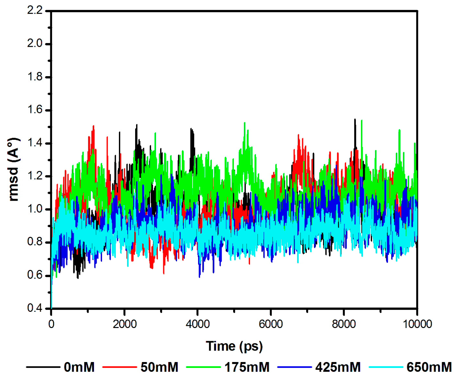
Figure 1. rmsd of the VcEndA enzyme under different values of the NaCl concentrations.
Chemproc 14 00040 g001
Figure 2. rmsf of the VcEndA enzyme under different values of the NaCl concentrations.
Chemproc 14 00040 g002
Figure 3. RDF of water molecules around the most flexible residues of VcEndA: (a) GLY52, (b) LYS53, (c) GLN90, (d) RDF of Cl ions around GLN90.
Chemproc 14 00040 g003
Figure 4. Secondary structure of VcEndA (pdb code; 2g7f [5]), using the program ESPript 3.0 [19]. Asterisks indicate the most flexible residues. Numbers indicate cysteines involved in disulfide bridges.
Chemproc 14 00040 g004
Figure 5. Snapshot of MD simulation of VcEndA_175mM_300K, indicating the protein in Cartoon style and the most flexible residues of the structure; GYL52, LYS53, and GLN90 in CPK style. Chloride and sodium ions are colored in cyan and blue, respectively. Water molecules are represented in a line transparent style. This image is made with VMD 1.8.7 [21].
Chemproc 14 00040 g005
Table 1. Number of Cl and Na+ ions for each simulation.
VcEndA_0 mMVcEndA_50 mMVcEndA_175 mMVcEndA_425 mMVcEndA_650 mM
Cl6153776122
Na+093182116
Disclaimer/Publisher’s Note: The statements, opinions and data contained in all publications are solely those of the individual author(s) and contributor(s) and not of MDPI and/or the editor(s). MDPI and/or the editor(s) disclaim responsibility for any injury to people or property resulting from any ideas, methods, instructions or products referred to in the content.