1. Introduction
The Wnt/β-catenin signaling pathway is crucial in a colorectal carcinoma, and its central component β-catenin is the focus of many targeted anticancer therapies [
1]. In healthy cells, a Wnt signaling pathway is not active, and β-catenin is mainly located in the peripheral part of the cytoplasm. In the cell membrane area, β-catenin is bound to E-cadherin, thus forming a complex with a role in connecting neighboring cells, which reflects its importance in the epithelium tissue. If it is found free in the cytoplasm, it is immediately degraded by the APC (Adenomatous Polyposis Coli) complex [
2]. However, in cancer cells, the Wnt pathway is deregulated and if the APC or β-catenin are mutated or absent, β-catenin is not degraded. It becomes accumulated in the cell cytoplasm and transported into the nucleus, where it acts as a transcription factor regulating cell proliferation, migration, and invasion, and thus the formation of metastases [
1].
Data from the literature report the existence of many natural products as sources of biologically active molecules originating from plants and animals with already proven effects on Wnt signaling. The inhibitory properties of these products have been described and are desired when it comes to the Wnt signal pathway. These features designate significant therapeutic potential and make them candidates for successful targeted therapies against cancer [
3].
One of recognized natural products reported to be useful as an anticancer agent is royal jelly. This special food derived from bees and intended for their consumption showed inhibitory effects on cancer cell proliferation, tumor growth, and cell invasion [
4].
We examined the effect of this natural product, royal jelly (RJ) sampled in Serbia, on β-catenin expression and concentration, since previous studies have shown that RJ has certain suppressive properties on cancer, as well as on its progression in terms of metastasis.
In this study, we examined the effects of RJ on the β-catenin gene and protein expression in colorectal cancer cell line.
2. Methods
β-catenin gene expression was evaluated 24 h after treatment with two RJ concentrations (10 and 100 µg/mL) using the qRT-PCR method, as previously described in detail [
5]. Results are presented as the fold change in mRNA expression in a target sample normalized to a reference gene (
β-actin) and relative to the control sample. Values of relative gene expression were calculated according to 2
−∆∆Ct method.
Meanwhile, protein level and localization of β-catenin fractions in the nucleus and cytoplasm were determined by immunofluorescent assay [
5] 24 h after treatment of cells with two RJ concentrations (10 and 100 µg/mL). Results of protein expression are presented as relative fluorescence intensity (%).
3. Results
3.1. Gene Expression
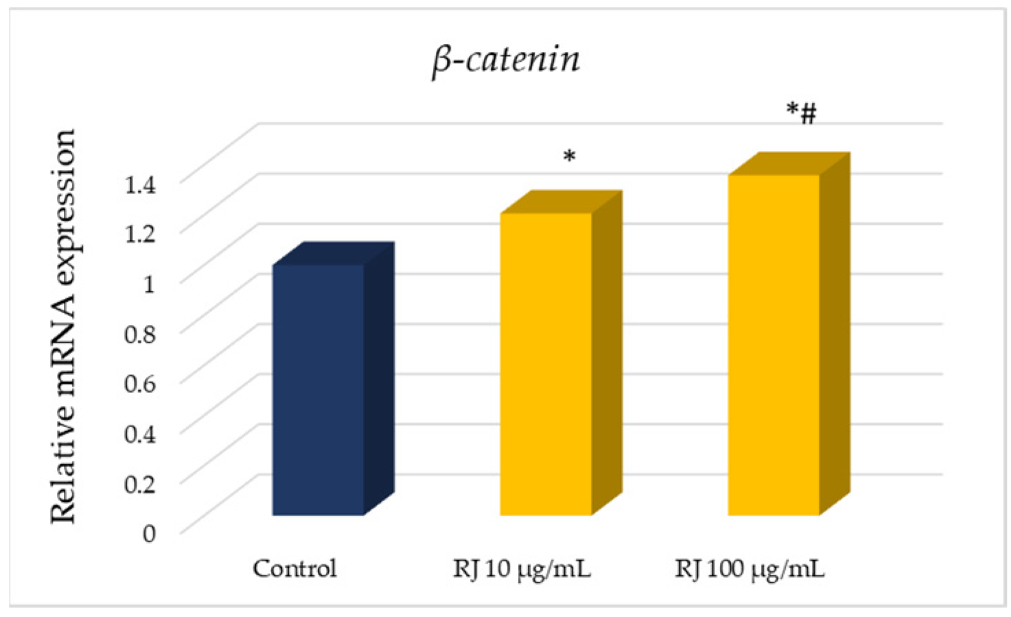
The results showed that this natural treatment caused a significant increase in β-catenin gene expression in both applied concentrations (
Figure 1). A dose-dependent effect of this treatment compared to the level of the control housekeeping gene
β-actin can be observed in
Figure 1.
3.2. Protein Expression and Localization
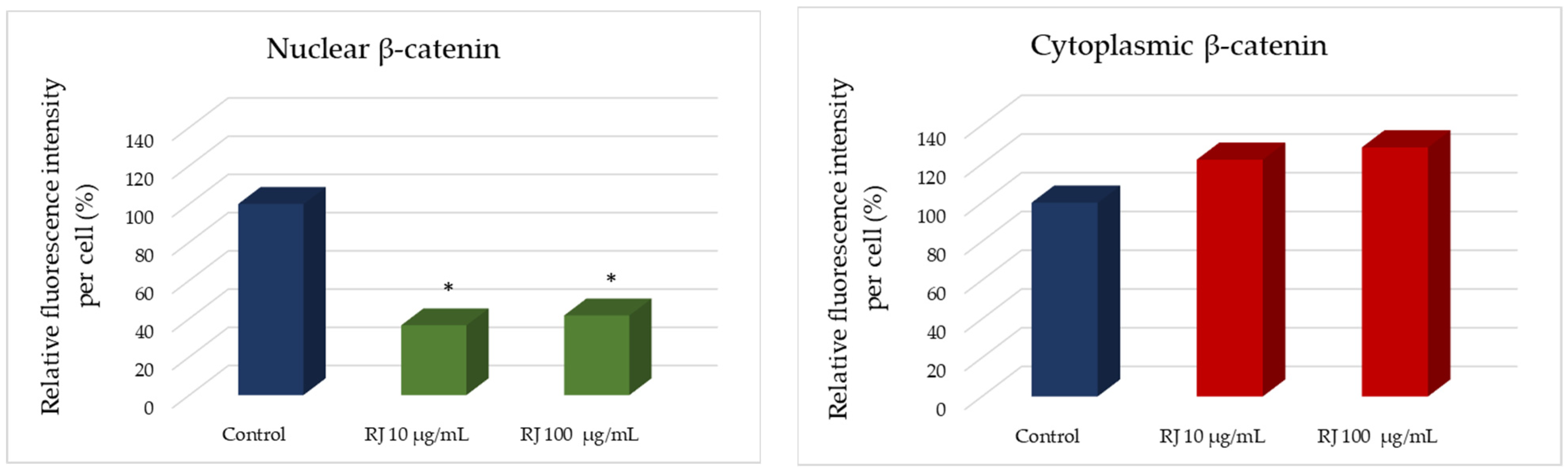
Our results show that RJ treatment had an impact on total β-catenin concentration when compared to the control values. Namely, both applied RJ concentrations (10 and 100 µg/mL) were able to decrease β-catenin, as it can be observed in
Table 1.
We noticed a significant decrease of nuclear β-catenin in HCT-116 cells after treatment with RJ (
Figure 2). Meanwhile, a dose-dependent increase in the cytoplasmic fraction of this protein was evident after 24 h of treatment with RJ when compared to the control values (
Figure 2).
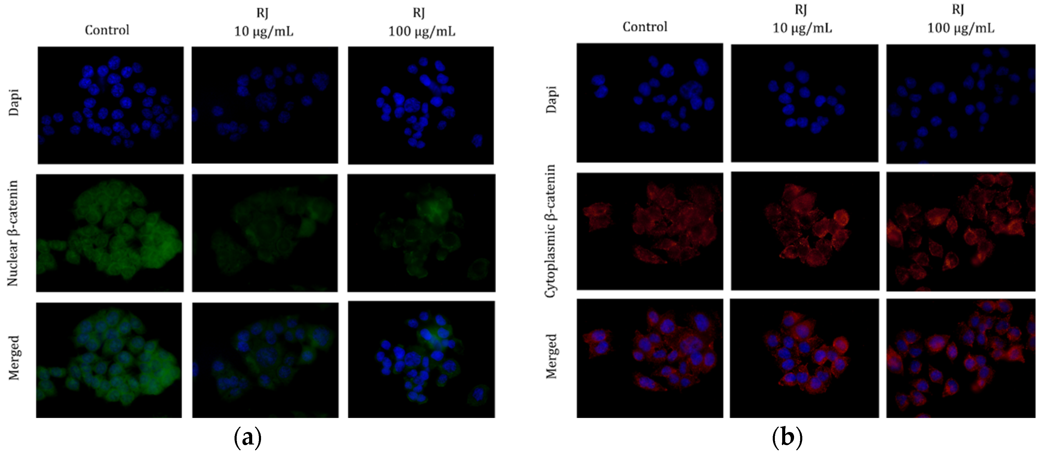
Immunofluorescent technique showed that the localization of nuclear β-catenin was not only limited to the nucleus of control (untreated) HCT-116 cells but was also found in the cell cytoplasm. When these cells were treated with a lower RJ concentration, the localization of this protein was found in both cell compartments again, however, its concentration was lower than in the control cells. Furthermore, a higher RJ concentration induced mostly cytoplasmic localization of this protein (
Figure 3a).
Meanwhile, we observed the localization of cytoplasmic β-catenin in control and HCT-116 cells treated with RJ and it can be concluded that this protein was mostly concentrated on the cell membrane area (
Figure 3b).
4. Discussion
The Wnt/β-catenin signal pathway is conserved signaling and its dysregulation plays a crucial role in cancer development and progression. Hence, agents that are able to target its components are increasingly the focus of worldwide researchers as innovative therapeutic drugs for combating cancer [
6].
Considering that royal jelly, as a natural product with proven beneficial effects on human wellness, has already shown significant anticancer activity [
4], we aimed to test its potential to modulate the Wnt/β-catenin pathway in selected colorectal cancer cell line.
Firstly, the HCT-116 cell line possesses one mutated
β-catenin gene allele, while the
APC gene is a wild type [
1,
2]. Therefore, this cell line represents a good model system for examining the impact of royal jelly on the central component of Wnt signal pathway, β-catenin.
Our results indicate that the treatment caused the obvious decrease in
β-catenin gene expression, indicating royal jelly’s efficacy in targeting nuclear processes, which is a prominent result that should not be neglected. A previous study conducted by Wang et al. [
7] proved that bee products were able to downregulate the
β-catenin mRNA level in cancer cells, which concords with our results.
Considering total β-catenin protein level in these cells, we observed a decrease of this marker in HCT-116 cells induced by royal jelly. Furthermore, a lower concentration of β-catenin was found in the cell nucleus and a slightly increased level of this protein was found in the cytoplasmic area. This could indicate that treatment started APC-independent signaling pathways are responsible for the export of β-catenin from the nucleus, or that the passive export could occur as result of the treatment effect. This could be a consequence of a reduced interaction of β-catenin with transcription factors and the interaction with DNA in the nucleus. It is known that when present in the cell nucleus, this protein regulates the expression of many markers and plays a significant role in cell proliferation, invasion, and migration. Therefore, it is very important that the level of β-catenin is on a minimum when found in cell nuclei, in order to inhibit cancer development and progression [
2].
Furthermore, its relocation from the nucleus to the cytoplasm most probably indicates its migration to the membrane in order to bind to E-cadherin and restore intercellular connections. It is also possible that after its export from the nucleus, royal jelly activated APC protein complexes responsible for the degradation of one portion of β-catenin protein resulted from wild type gene allele that is substrate capable for degradation by APC, therefore its lower concentration in cell cytoplasm.
In conclusion, royal jelly proved to be a powerful agent in affecting
β-catenin gene expression and in the export of this protein from the nucleus to the cytoplasm where it can be degraded. Similar results in HCT-116 cells treated with natural products have been published earlier by Šeklić et al. [
1], confirming the potential that can be found in agents originating from nature regarding targeted activity in cancer cells.
5. Conclusions
Obviously, this natural product was effective in targeting an important component included in a significant signal pathway already deregulated in colorectal cancer cells; however, enormous examinations remain to be completed. We anticipate that these findings will be focus the of increasing attention in both the scientific and clinical fields of research.
Author Contributions
Conceptualization, D.Š. and M.M.J.; methodology, K.V., D.A. and K.P.; software, D.A.; validation, D.Š. and M.M.J.; formal analysis, K.V., D.A. and K.P.; investigation, K.V., D.A. and K.P.; resources, D.Š.; data curation, D.Š.; writing—original draft preparation, K.V., D.A. and K.P.; writing—review and editing, D.Š.; visualization, M.M.J.; supervision, D.Š.; project administration, D.Š.; funding acquisition, D.Š. All authors have read and agreed to the published version of the manuscript.
Funding
This research was funded by the Ministry of Education, Science, and Technological Development of the Republic of Serbia, grant number 451-03-68/2022-14/200124 and 451-03-68/2022-14/200122.
Institutional Review Board Statement
Not applicable.
Informed Consent Statement
Not applicable.
Data Availability Statement
Not applicable.
Conflicts of Interest
The authors declare no conflict of interest.
References
- Šeklić, D.S.; Stanković, M.S.; Milutinović, M.G.; Topuzović, M.D.; Štajn, A.Š.; Marković, S.D. Cytotoxic, antimigratory and pro/antioxidative activities of extracts from medicinal mushrooms on colon cancer cell lines. Arch. Biol. Sci. 2016, 68, 93–105. [Google Scholar] [CrossRef]
- Šeklić, D.S.; Jovanović, M.M.; Virijević, K.D.; Grujić, J.N.; Živanović, M.N.; Marković, S.D. Pseudevernia furfuracea inhibits migration and invasion of colorectal carcinoma cell lines. J. Ethnopharmacol. 2022, 284, 114758. [Google Scholar] [CrossRef] [PubMed]
- Blagodatski, A.; Klimenko, A.; Jia, L.; Katanaev, V.L. Small Molecule Wnt Pathway Modulators from Natural Sources: History, State of the Art and Perspectives. Cells 2020, 9, 589. [Google Scholar] [CrossRef] [PubMed]
- Miyata, Y.; Sakai, H. Anti-cancer and protective effects of royal jelly for therapy-induced toxicities in malignancies. Int. J. Mol. Sci. 2018, 19, 3270. [Google Scholar] [CrossRef] [PubMed]
- Jovanović, M.M.; Šeklić, D.S.; Rakobradović, J.D.; Planojević, N.S.; Vuković, N.L.; Vukić, M.D.; Marković, S.D. Royal jelly and trans-10-hydroxy-2-decenoic acid inhibit migration and invasion of colorectal carcinoma cells. Food Technol. Biotechnol. 2022, 60, 213–224. [Google Scholar] [CrossRef] [PubMed]
- Sferrazza, G.; Corti, M.; Brusotti, G.; Pierimarchi, P.; Temporini, C.; Serafino, A.; Calleri, E. Nature-derived compounds modulating Wnt/β-catenin pathway: A preventive and therapeutic opportunity in neoplastic diseases. Acta Pharm. Sin. B 2020, 10, 1814–1834. [Google Scholar] [CrossRef] [PubMed]
- Wang, X.; Li, H.; Lu, X.; Wen, C.; Huo, Z.; Shi, M.; Tang, X.; Chen, H.; Peng, C.; Fang, Y.; et al. Melittin-induced long non-coding RNA NONHSAT105177 inhibits proliferation and migration of pancreatic ductal adenocarcinoma. Cell Death Dis. 2018, 9, 940. [Google Scholar] [CrossRef] [PubMed]
| Disclaimer/Publisher’s Note: The statements, opinions and data contained in all publications are solely those of the individual author(s) and contributor(s) and not of MDPI and/or the editor(s). MDPI and/or the editor(s) disclaim responsibility for any injury to people or property resulting from any ideas, methods, instructions or products referred to in the content. |
© 2022 by the authors. Licensee MDPI, Basel, Switzerland. This article is an open access article distributed under the terms and conditions of the Creative Commons Attribution (CC BY) license (https://creativecommons.org/licenses/by/4.0/).


 ,
, 


