Article

Novel Insights of Lithium Chloride Therapeutic Approach for Managing Type 2 Diabetic Kidney Disease: Crosslinking Tau Hyperphosphorylation and TGF Beta Signaling

1
Department of Biological Sciences, Faculty of Science, Beirut Arab University, Beirut P.O. Box 11-5020, Lebanon
2
Department of Medical Laboratory Sciences, Faculty of Health Sciences, Beirut Arab University, Beirut P.O. Box 11-5020, Lebanon
3
Molecular Biology Unit, Department of Zoology, Faculty of Science, Alexandria University, Alexandria 21568, Egypt
4
Department of Anatomy, Cell Biology, and Physiological Sciences, Faculty of Medicine, American University of Beirut, Beirut 1107-2020, Lebanon
5
AUB Diabetes, American University of Beirut Medical Center, Beirut 1107-2020, Lebanon
*
Authors to whom correspondence should be addressed.
Diabetology 2025, 6(4), 26; https://doi.org/10.3390/diabetology6040026
Submission received: 18 December 2024 / Revised: 1 February 2025 / Accepted: 25 February 2025 / Published: 2 April 2025

Abstract

:
Background: Diabetic kidney disease (DKD) represents a chronic microvascular complication with diabetes, affecting around one-third of diabetic individuals. Despite current therapies, progression to end-stage kidney disease remains a challenge. Abnormal hyperphosphorylation of the Tau protein is implicated in various age-related diseases. This study aimed to explore the link between renal Tau protein hyperphosphorylation and kidney damage in type 2 diabetes mellitus (T2DM). Methods: Sprague Dawley rats were administered lithium chloride (LiCl), an inhibitor of a glycogen synthase kinase-3 (GSK3) inhibitor known to reduce Tau hyperphosphorylation. LiCl was administered either daily or every other day at a dosage of 1 mmol/kg. The effects of LiCl on kidney function were assessed through proteinuria, the kidney-to-bodyweight ratio, inflammation, fibrosis, and TGF-β1 expression levels. Results: Diabetic rats exhibited increased proteinuria, renal hypertrophy, inflammation, fibrosis, and elevated TGF-β1 expression. Lithium chloride treatment reduced kidney hypertrophy, inflammation, and fibrosis, indicating that Tau hyperphosphorylation contributes to the pathogenesis of DKD. LiCl also regulated TGF-β1 expression, which was associated with improved renal outcomes. Conclusions: The inhibition of Tau hyperphosphorylation by lithium chloride offers a potential therapeutic strategy for mitigating kidney damage in diabetic kidney disease. This study proposes LiCl as a novel treatment approach to attenuate DKD progression.

1. Introduction

Diabetes mellitus is a chronic metabolic condition defined by persistently elevated blood glucose levels, causing increased morbidity and mortality among 8% to 10% of the world’s population [1]. Diabetic kidney disease (DKD) is a significant microvascular complication of diabetes, impacting one-third of people with the condition and often progressing to end-stage kidney disease [2]. It has been shown that exceeding the renal threshold for glucose re-absorption (180 mg/dL) could cause symptoms of glycosuria, polyuria, and polydipsia [3]. Structural and functional changes characterize this major health issue. Structurally, it is characterized by glomerular basement membrane (GBM) thickening, mesangial expansion, and the development of glomerulosclerosis. However, functionally, it is mainly characterized by glomerular hyperfiltration and increased proteinuria, causing renal dysfunction [4,5].
The pathophysiological events of DKD are complex and multifactorial, involving but not limited to metabolic and hemodynamic alterations and chronic inflammation that triggers fibrosis [6]. Renal fibrosis development is predominantly driven by hyperglycemia and the activation of intracellular signaling pathways mediated by Smad and non-Smad-dependent transforming growth factor-β1 (TGF-β1) [7]. TGF-β1 facilitates myofibroblast activation and promotes the excessive accumulation of extracellular matrix (ECM) proteins including collagen and fibronectin, thus disrupting the homeostatic balance between synthesis, deposition, and proteolytic clearance [8]. Subsequently, the quantification and characterization of the ECM correlate with the assessment of the progression of glomerular disease, the deterioration of renal function, and thus the efficacy of therapeutic interventions [9].
The Tau protein is highly soluble and is generated through alternative splicing of the MAPT gene, which encodes microtubule-associated proteins (MAPs) [10]. Primarily, Tau is located in the axons of mature neurons before it is shown to be expressed in various tissues, including the kidneys [11,12]. The Tau protein primarily operates through phosphorylation mediated by protein kinases, such as glycogen synthase kinase-3β (GSK3β) [13]. As a proline-directed serine/threonine protein kinase, GSK3β specifically phosphorylates Tau at two residues, namely S214 and S404, respectively [14]. It is important to note that several sites, including Ser214, require the prephosphorylation of Tau by cAMP-dependent protein kinase A (PKA), which is then subsequently phosphorylated by GSK3β in the rat brain, thus improving the overall amount of GSK-3 phosphorylation [15,16]. Other proteins, such as Ser 404, are phosphorylated directly by GSK3β [17]. Specific phosphorylation sites play different roles in Tau, including in cell process development [18], axonal transport [19], and signal transduction [20]. Excess intracellular glucose activates Tau protein kinases and induces hyperphosphorylation [21]. On the other hand, Tau can initiate kidney damage through its ability to trigger inflammation and tissue remodeling, consequently dysregulating the local metabolic environment [2]. Recently, the protective effects of lithium chloride (LiCl) against Tau hyperphosphorylation have been reported in diabetic neuropathy [13]. Despite the lack of investigation into the relationship between renal Tau protein levels and the pathogenesis of renal damage in diabetic patients, the established role of TGF-β1 in promoting renal fibrosis and its potential interaction with Tau highlights the crucial need for such studies to enhance our understanding of DKD progression.
Our study aimed to elucidate the relationship between the hyperphosphorylation of Tau, TGF-β activation, and the progression of DKD using an experimental rat model of T2DM. While several studies have identified Tau as a potential mediator of renal fibrosis through TGF-β signaling [7,8], our findings uniquely demonstrate that lithium chloride (LiCl) not only inhibits Tau hyperphosphorylation [10,11] but also significantly downregulates TGF-β1 expression in the kidneys of diabetic rats [7,8]. This dual action suggests a novel therapeutic approach that targets both Tau pathology and TGF-β signaling, potentially mitigating the progression of DKD [2,6]. By clarifying these relationships, our research advances the understanding of DKD pathogenesis and highlights LiCl as a promising protective agent against renal damage in individuals with T2DM [1,2].

2. Materials and Methods

2.1. Power Analysis and Sample Size Justification

Before conducting the study, a power analysis was performed using G*Power software (Version 3.1.9.4) to determine the appropriate sample size required to detect significant differences between groups. Based on preliminary data, an effect size of 0.95 was anticipated, with an alpha level set to 0.05 and a desired power of 0.80. This analysis indicated that a minimum of five animals per group would be necessary to achieve statistically significant results. Consequently, we assigned five male Sprague Dawley rats to each of the four experimental groups (control, diabetic, LiCl daily treatment, and LiCl every other day treatment) to ensure adequate power for detecting differences in kidney function and pathology.

2.2. Animals and Treatments

Male rats of the Sprague Dawley strain, aged one month and weighing 100–200 g, were randomly assigned to four groups, each consisting of five animals. The control group received an injection of sodium citrate buffer (0.1 M, pH 4.5) and was provided with standard chow alone; Group 1. The other groups (Groups 2, 3, and 4) received an in-house prepared HFD containing 60% kcal from fat for a duration of 8 weeks and intravenously injected through the tail with 30 mg/kg body weight streptozotocin (STZ) prepared in (0.01 M, pH 4.5) sodium citrate buffer to induce diabetes. Subsequently, Group 2 was assigned as a diabetic group, while diabetic rats in Groups 3 and 4 received intraperitoneal injections of LiCl at a dosage of 1 mmol/kg, either daily or every other day for a duration of 14 weeks, respectively. Blood glucose levels were measured 24 h later and weekly until the end of the study using AccuCheck Performa, Roche [22]. The study was carried out following the protocols authorized by the Institutional Animal Care and Use Committee (IACUC) at the American University of Beirut. The control rats in Group 1 were provided with unrestricted access to food and water. All other groups of rats had unrestricted access to a high-fat diet (60% kcal fat) [22]. Urine samples were collected over a 24 h period using metabolic cages. Proteinuria levels were quantified using the Lowry method and reported as micrograms of albumin per 24 h. At week 14, both kidneys of the euthanized rats were removed, weighed, and washed in PBS. Slices of both the right and left kidney cortices at the poles of each were cut, either embedded in paraffin or rapidly frozen in liquid nitrogen and then stored at a temperature of −80 °C for subsequent histological and biochemical analyses.

2.3. Kidney Sieving

The kidneys were cut in half lengthwise (midsagittal section), and the medullas were removed and discarded. The cortical slices of each rat kidney were then used to isolate the glomeruli and tubules using the differential sieving method. After wetting the slices with PBS with 5% pen/strep, they were minced with a blade and then pressed through a stainless-steel mesh of 180 µm pore size using a spatula to sieve them into the ice bottom beaker. Successive periodic small washings with PBS were performed to collect the whole sample in a beaker, avoiding sample dilution. The collected sample was reused, pressed into another 75 µm pore size mesh, and suspended in PBS. While the glomeruli were retained on top of the 75 µm sieve, the sample was collected in the beaker. Each sample was transferred into two separate 50 mL plastic conical tubes and placed on ice. The conical tubes were subjected to centrifugation at 1500 rpm for 5 min at a temperature of 4 °C to collect the glomeruli and tubule pellets for Western blotting later [23].

2.4. Lithium Chloride Administration

Lithium chloride (1 mmol/kg, Thermo Fisher Scientific, Waltham, MA, USA) was dissolved in normal saline and administered intraperitoneally daily to Group 3 and every other day to Group 4 for 14 weeks. LiCl treatment is extensively utilized to inhibit Tau phosphorylation by targeting GSK3β activity [24].

2.5. Biochemical Assessment

Blood samples were collected on the sacrifice day and centrifuged at 3000× g for 10 min to obtain serum. Insulin, free fatty acid (FFA), and triglyceride (TG) levels were quantified using a Rat Insulin ELISA Kit (Wuhan, China), a Free Fatty Acid Assay Kit (Cambridge, UK), and a Triglyceride Assay Kit (Cambridge, UK) following the protocol received from the manufacturer.

2.6. Histopathological Analysis

Kidney slices were embedded in a fresh 4% formaldehyde solution. Perpendicular sections of 4 µm were prepared for histological examination [5]. Standard Periodic acid–Schiff (PAS) and Masson’s trichrome stains were adopted to assess renal fibrosis [25]. Renal collagen deposition was visualized using Masson’s trichrome stain [25], and PAS magenta staining was used to assess the glomerulosclerosis index and mesangial accumulation [26,27]. All sections were examined using a light microscope (20×) with images captured by an Olympus digital camera (CX41, Olympus, Tokyo, Japan) and analyzed using Image J software (Version 1.53e, National Institutes of Health, Bethesda, MD, USA).

2.7. Immunohistochemistry

Paraffin-embedded kidney sections (4 μm) were used for the detection of TGF-β1. After deparaffinization, sections were placed in an antigen retrieval buffer at pH 6 (0.1 M citric acid, 0.1 M NaCitrate, 2N NaOH), incubated with the primary antibody rabbit anti-TGF-β1 (Promega, Madison, WI, USA) and detected using the Novolink™ Polymer Detection System kit (Leica Biosystems, Buffalo Grove, IL, USA) following the instructions received from the manufacturer. For analysis, each section was captured in 15 images, and all images were quantified and averaged. The brown area was used to assess protein expression. The findings were examined using a light microscope (20× magnification, Olympus, CX41) and presented by area fractions’ percentage relative to the control. Image analysis was conducted utilizing GraphPad Prism 9 (version 9.4.1, USA) [28].

2.8. Western Blot Analysis

For Western blot analysis, glomeruli and kidney tubules were homogenized in 250 µL of RIPA buffer containing SDS (0.1%), sodium deoxycholate (0.5%), Tris-HCl pH 8 (100 mM), NaCl (300 mM), Phosphatase Inhibitor Cocktail, Protease Inhibitor Cocktail, NP-40 (1%), and PMSF (1 mM) using a Dounce homogenizer. The homogenate supernatants were obtained after being placed on the rotator for two hrs. and centrifuged at 13,200 RPM for 30 min at 4 °C. Protein quantification was performed using the Bio-Rad protein assay [29]. For immunoblotting, 60 µg of each protein was separated on an 8% SDS-PAGE gel, transferred to PVDF membranes, and then blocked using 5% BSA dissolved in Tris-buffered saline. Following the blocking step, the membranes were incubated with the respective primary antibodies. The antibodies used included a rabbit polyclonal fibronectin antibody (dilution 1:1000; Abcam, Cambridge, UK), a rabbit polyclonal anti-Tau (phospho S214) antibody (dilution 1:250; Abcam, UK), a rabbit monoclonal anti-Tau (phospho S404) antibody (dilution 1:1000; Abcam, UK), a rabbit polyclonal anti-Tau (total Tau) antibody (dilution 1:300; Bioss Antibodies, Beijing, China), and a mouse monoclonal anti-Hsc70 (B-6) antibody (dilution 1:500; Santa Cruz Biotechnology, Inc., Dallas, TX, USA). The detection of primary antibodies was carried out using horseradish peroxidase (HRP)-conjugated IgG at a dilution of 1:5000. Protein bands were detected through chemiluminescence, and a densitometric analysis was conducted utilizing ImageJ software (Version 1.53e, National Institutes of Health, USA) from the National Institutes of Health [30].

2.9. Statistical Analysis

Data analysis was performed utilizing GraphPad Prism 9 (version 9.4.1, USA). Descriptive statistics were performed and presented as frequencies and percentages for categorical variables, while continuous variables were represented as means with standard errors (SEs) or standard deviations (SDs). Student’s t-test was performed for comparison between two groups, while an analysis of variance (ANOVA) was employed for comparisons involving three or more groups. Post hoc analyses were conducted using Fisher’s least significant difference or Tukey’s tests. A p-value of less than 0.05 was considered statistically significant.

3. Results

3.1. Metabolic Characteristics of T2DM Rats

The rats were maintained on a high-fat diet for a total duration of 22 weeks. Rats with T2DM rats (Group 2) had significantly higher body weights and blood glucose levels compared to the control group, which were fed a standard chow diet, as shown in Table 1. However, the daily administration of 1 mmol/kg LiCl significantly reduced both body weight and blood glucose in the T2DM group, indicating its potential role in metabolic regulation. Furthermore, T2DM rats showed significantly higher insulin levels than the control group, suggesting the emergence of insulin resistance. Notably, administering LiCl daily or every other day decreased insulin levels; although these levels remained elevated compared to the control group, they may reflect improved insulin sensitivity. Moreover, concentrations of triglycerides (TGs) and free fatty acids (FFAs) were assessed. Our results demonstrated a significant increase in T2DM rats; however, these levels declined following daily or every other day LiCl treatment. These results indicate that LiCl administration effectively alleviated hyperlipidemia (Table 1). The reduction in TGs and FFAs could be linked to LiCl’s modulation of metabolic pathways influenced by Tau hyperphosphorylation.
Following 14 weeks of LiCl treatment, the rats were sacrificed, and the weights of both kidneys were recorded. Kidney weight (KW)-to-body weight (BW) ratios were calculated. The ratio of KW/BW increased significantly after HFD feeding, indicating hypertrophy, while it decreased significantly after LiCl daily treatment (Table 1). This suggests that LiCl may mitigate renal hypertrophy associated with T2DM.
Proteinuria screening serves as a key biomarker for monitoring diabetic nephropathy progression [5]. Proteinuria increased significantly in T2DM rats compared to the control group. Interestingly, proteinuria levels decreased significantly in diabetic groups treated with LiCl administered daily and every other day compared to diabetic rats (Table 1), suggesting a renoprotective role of LiCl.

3.2. T2DM Induces Tau Protein Hyperphosphorylation in the Rats’ Kidneys

Next, we examined the association between T2DM and Tau protein hyperphosphorylation across the four rat groups (Figure 1A,B). The phosphorylated Tau protein at S214 (79 kDa) and S404 (50–70 kDa) was shown to be overexpressed in the glomeruli (Figure 1C–E) and tubules (Figure 1F–H). This overexpression suggests that hyperglycemia may activate Tau protein kinases, leading to detrimental effects on renal function. However, the administration of 1 mmol/kg LiCl daily or every other day decreased Tau protein phosphorylation in both the glomeruli and tubules of the diabetic kidneys at the two studied sites of S214 and S404. Interestingly, our results show that LiCl inhibition by glycogen synthase kinase decreased Tau hyperphosphorylation levels, proposing that the selected lithium chloride dose effectively decreased Tau phosphorylation as intended. This reduction may alleviate the inflammatory processes associated with hyperphosphorylated Tau, further suggesting a protective mechanism against kidney damage.

3.3. LiCl Treatment Reduces ECM Accumulation and Kidney Fibrosis in T2DM

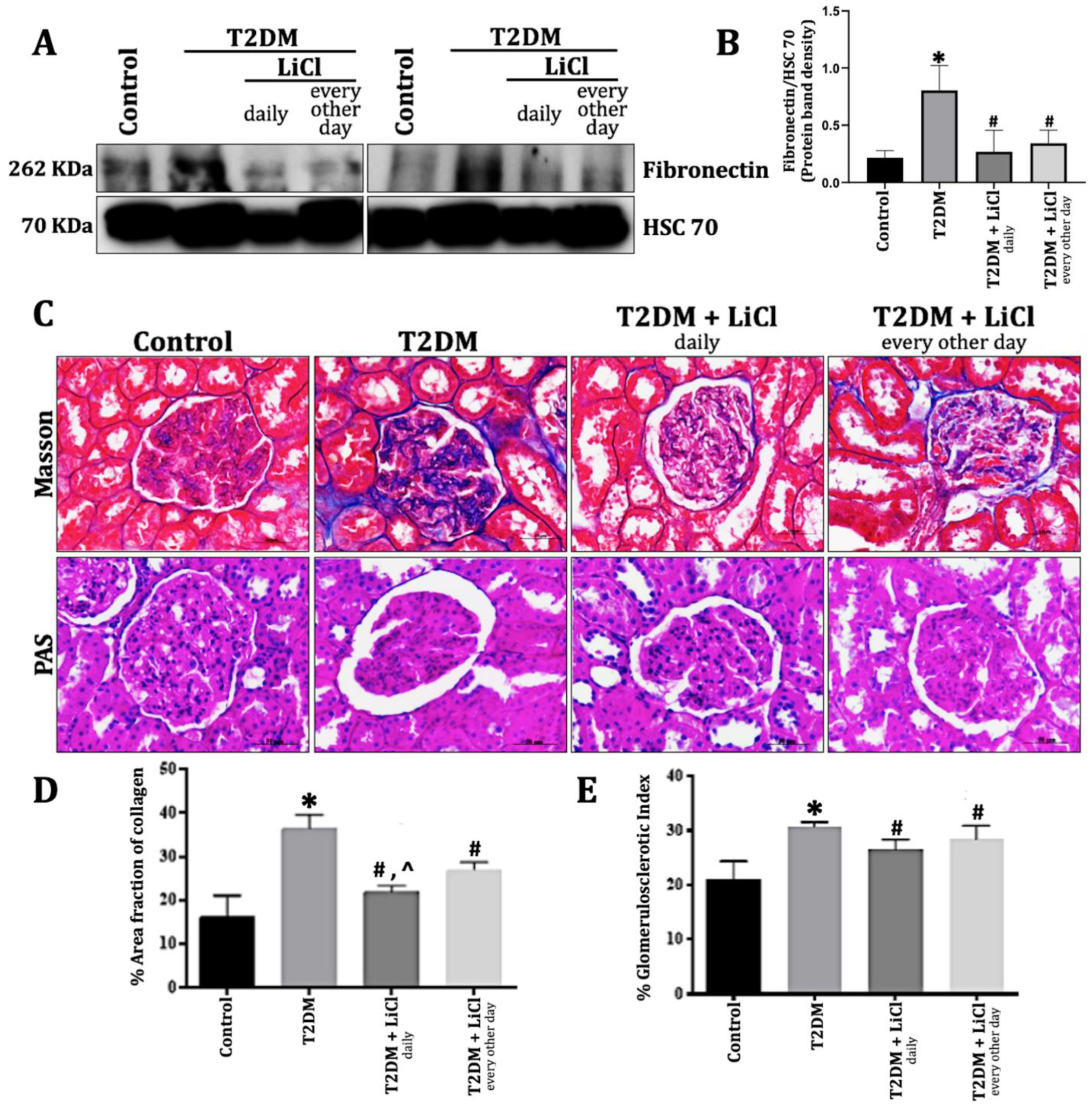
Glomerulosclerosis in diabetic kidney disease is defined by the deposition of extracellular matrix (ECM) components, such as fibronectin and collagen, along with the intracellular accumulation of glycogen, thus altering the kidney’s filtration apparatus [4,7]. Herein, we examined variations in fibronectin protein expression levels in the control, untreated T2DM group, and T2DM rats administered 1 mmol/kg LiCl either daily or every other day. Fibronectin levels (Figure 2A,E) were significantly elevated in T2DM rats compared to the control group (p < 0.05), indicating that hyperglycemia promotes ECM accumulation. However, treatment with LiCl administered either daily or every other day significantly decreased fibronectin protein expression. This reduction suggests that LiCl may attenuate fibronectin-mediated fibrosis by inhibiting upstream signaling pathways involved in ECM production.
Additionally, Masson’s trichrome staining (Figure 2B,C) and PAS staining (Figure 2B,D) showed an increase in renal fibrosis and the glomerulosclerotic index in the untreated T2D rats compared to the control rats (p < 0.05). This finding highlights how T2DM exacerbates renal structural changes through ECM deposition and inflammation. However, this was reversed when treated with LiCl, indicating its potential role in restoring renal architecture.

3.4. Tau Hyperphosphorylation Induced Kidney Inflammation via TGF-β1 Overexpression in T2DM Rats

The transforming growth factor-β (TGF-β) signaling pathways have exhibited multifunctionality, demonstrating both profibrotic and protective effects in kidney disease models. It is a cytokine that regulates several processes, mainly wound repair and fibrosis [7]. TGF-β1, the most characterized isoform present in the kidney, has been linked to the progression of glomerulonephritis and glomerulosclerosis (8). TGF-β1 biological activities are mediated by ligand-receptor binding; however, this process is disrupted in hyperglycemia, leading to an increased production of fibronectin and collagen and therefore ECM accumulation [7].
In this study, we examined the variations in the TGF-β1 protein expression levels in the control, untreated T2DM, and LiCl-treated groups. The levels of TGF-β1 (Figure 3A,B) were significantly elevated in T2D rats compared to control rats (p < 0.05), suggesting that hyperglycemia disrupts normal signaling pathways leading to increased ECM production.
The inhibition of Tau hyperphosphorylation through LiCl treatment exhibited a reduction in renal TGF-β1 expression, with better improvements obtained during daily treatments. This suggests that inhibiting Tau hyperphosphorylation through LiCl treatment mitigates TGF-β signaling dysregulation associated with DKD progression. This relationship highlights a potential therapeutic pathway, where the modulation of Tau phosphorylation can influence TGF-β signaling dynamics, ultimately impacting renal health.

4. Discussion

This study determined the connection between Tau hyperphosphorylation and the onset and progression of DKD through the activation of the TGF-β signaling pathway. Excess glucose has been shown to trigger systemic and intrarenal inflammation through altered metabolic pathways that promote the accumulation of the ECM and the development of renal fibrosis, with TGF-β being a potent profibrotic factor in this process [2,7,31,32]. Prior human and animal studies on the brain have shown that insulin resistance triggers Tau hyperphosphorylation, leading to Tau pathology with an unknown mechanism [21,33,34,35,36]. Treatment with LiCl, which effectively inhibited Tau protein hyperphosphorylation in T2D rats, attenuated renal fibrosis and inflammation and improved kidney function. This was accompanied by a reduction in TGF-β expression, previously upregulated in response to DKD.
While previous studies have established the role of TGF-β in renal fibrosis, our findings uniquely demonstrate how Tau hyperphosphorylation acts as a mediator in this process, filling a critical gap in understanding DKD pathogenesis. The results indicate that Tau hyperphosphorylation contributes to renal inflammation and fibrosis by activating TGF-β signaling pathways, highlighting significant biological implications.
The HFD/STZ model was utilized to mimic type 2 diabetes. It is performed through continuous HFD feeding until a plateau of hyperglycemia and weight gain, thus inducing insulin resistance. This was followed by a low dose of 35 mg/kg STZ to initiate β-cell dysfunction, reflected in a decrease in plasma insulin concentration [37,38,39]. Our T2DM diabetic model was demonstrated by elevated blood glucose levels, insulin levels, and an increased KW/BW ratio, confirming diabetic renal pathological changes and kidney injury [40]. Several studies have verified the protective role of LiCl, the GSK3β inhibitor, not only against Tau hyperphosphorylation in diabetic neuropathy but also in mitigating acute kidney injury and providing long-term benefits at low concentrations in chronic kidney disease [14,24,41,42,43,44,45,46,47,48,49,50]. Additionally, both free fatty acids and triglyceride levels were elevated, confirming metabolic dysregulation associated with T2DM. Both parameters were significantly decreased after LiCl treatment, highlighting the protective effect of LiCl in mitigating hyperlipidemia [51].
Diabetic nephropathy progression has been distinguished by the degree of proteinuria, which is followed by a decrease in renal function [52]. It is worth noting that the early identification of diabetes would prevent the onset of proteinuria [53]. Hyperglycemia has been well documented to lead to systemic and intraglomerular hypertension-inducing proteinuria that would eventually worsen over time, leading to chronic tubular injury [53,54]. Several trials using different treatments for diabetic kidney disease have shown conflicting results; however, the majority have reported a reduction in proteinuria, supporting the fact that its reduction remains the key in treating diabetic kidney disease and other nephropathies [54,55].
Secondary to proteinuria, DKD is correlated with glomerular morphological changes, including mesangial expansion in the early stage of DKD, which is directly related to increased ECM deposition, mesangial cell hypertrophy, and mesangial cellularity [4,56,57]. The progression of DKD is characterized by a nodular glomerulosclerosis pattern that is known as Kimmelstiel–Wilson nodules and GMB thickening [4]. The histological evaluation of renal fibrosis has been used as the sole definitive method for quantifying renal fibrosis [8]. Several studies have used renal biopsy for routine diagnosis using Sirius Red or Masson’s trichrome stain [58,59,60]. Our staining procedures have confirmed the morphological pathologies in terms of quantification of the fibrotic area using Masson’s trichrome and PAS stains for excessive accumulation of collagen and glycogen in diabetic models. Interestingly, LiCl treatments have attenuated fibrosis in the renal glomeruli. In support of our results, lithium protective therapies have been widely used against renal fibrosis in various models of diabetic nephropathy [61,62,63,64,65].
The Tau protein has been extensively studied in the brain since 1975, until Gu et al. affirmed its high expression in the kidneys in 1996 [11,20]. Recent studies have highlighted the association between insulin resistance and Tau hyperphosphorylation in the brain and pancreas, while it is not as prominently as observed in other tissues. [21,33,34,35,36,66,67,68]. In the pancreas, Tau phosphorylation was overexpressed in T2D patients [67]. This was followed by the study by Wijesekara et al., showing a significant hyperphosphorylation of Tau in pancreatic islets extracted from AD and T2D transgenic mouse models, proposing a potential role for Tau protein in metabolic regulation [68]. Our results confirmed Tau hyperphosphorylation in the kidneys of diabetic rats, suggesting a new promising therapeutic target. This is also consistent with the findings of other studies confirming that high glucose levels enhance GSK3β-mediated Tau phosphorylation in the brain [21,69,70]. LiCl has worked as an inhibitor of GSK3β, an expressed serine/threonine protein kinase, against Tau hyperphosphorylation observed in neurons [24,71], against axonal degradation during the early stage of tangle development [49], and as a mood stabilizer of several mental illnesses [50], supporting its protective role against tauopathies. Similarly, Tau hyperphosphorylation was significantly reduced upon LiCl treatment in the kidneys. Furthermore, Tau phosphorylation is recognized at multiple sites in human neuronal NT2N cells and prevented by the GSK3β inhibitor, lithium [24]. Similar studies have used lithium to suppress GSK3β activity and inhibit hyperphosphorylation, resulting in refined effects and reduced tauopathy [13,49,72].
Several experimental models have been used to distinguish between different phosphorylated sites and discover the site of more diagnostic power for a related tauopathy [73]. Herein, we used phosphorylated Tau, specifically at Ser214 and Ser404, which reflected the same hyperphosphorylated pattern in the glomeruli and tubules of untreated diabetic models. It has been well indicated that S214 and S404 are phosphorylated in paired helical filaments of the brain out of 37 serine and threonine phosphorylated residues in tauopathies [74], although S214 phosphorylation has only been seen in paired helical filaments and not in normal brains [75]. S404 was among 10 other sites that were markedly phosphorylated after STZ injections into rat brains, suggesting the specificity of site phosphorylation in insulin signaling and glucose metabolism [33]. However, the hyperphosphorylation of Tau in S404 and S396 decreased its binding affinity to microtubules [33,76], and a similar effect was obtained with an increased phosphorylation in S214 and T241 in the proline-rich domain, thus disrupting the biological activity of normal Tau [77]. GSK3β inhibition has affirmed its ability to reduce Tau pathologies in several in vivo and in vitro models [78]. Lithium has been used primarily as a GSK3β inhibitor for clinical treatments of bipolar disorder and major depression [79], resulting in reduced phosphorylated Tau levels in cerebrospinal fluid along with improvements in cognitive parameters [80]. This was consistent with our results at the level of the kidney glomeruli and tubules, where the protein expressions of phosphorylated Tau at S214 and S404 decreased after treatment with 1 mmol/kg LiCl. Daily treatment exhibited a more significant reduction than every other day, while phospho-tau-S404 showed approximately similar reductions in its expression with lithium treatments in both the glomeruli and tubules. These results have established the protective effects of LiCl against hyperphosphorylation pathologies. Confirmatively, GSK3β inhibition by LiCl has shown its ability to reverse the axonal transport defects and locomotor phenotypes in Drosophila transgenic flies carried through Tau overexpression, as well as in rat cortical neurons [81,82]. In the same manner, LiCl treatment has reduced Tau phosphorylation at Ser-396/404, resulting in decreased axonal degeneration and Tau aggregation in the overexpressed human Tau protein of JNPL3 transgenic mice [49]. Tau phosphorylation by GSK3β has been associated with lithium-specific sensitive sites, including S214 and S404 in primary cortical rat neurons and cultured hippocampal neurons, suggesting the direct inhibitory effect on Tau phosphorylation by GSK3β at these specific sites [49,81].
TGF-β is an essential mediator in the pathogenesis of glomerular diseases through intracellular signaling pathways that induce renal pathophysiological changes. TGF-β1 is recognized as the major inflammatory cytokine being overexpressed upon glomerulonephritis and glomerulosclerosis development in various diabetic animal models [83]. Under hyperglycemic conditions, TGF-β1 is released to its free form and activated upon binding to its cellular receptor, thus promoting the transcription of several genes, including fibronectin and collagen, eventually accumulating ECM molecules [7]. There is growing evidence underscoring the role of TGF-β1 in the progression of DN [84,85], as well as in the repair response following LiCl treatment. Several treatments were adopted to neutralize TGF-β in STZ-induced and db/db diabetic animal models, showing suppressed mesangial matrix expansion in the glomeruli along with a reduced expression of collagen and fibronectin [86,87,88]. Collectively, our results affirm that the hyperphosphorylation of the Tau protein is balanced by regulating TGF- β, thus reducing renal Tau protein pathogenesis. Interestingly, treatment has reduced high blood glucose levels, preserved glomerular structure and function, and demonstrated the protective effect of TGF-β on renal function.
This study strengthens the mechanistic link between Tau hyperphosphorylation and TGF-β signaling in DKD, demonstrating that LiCl treatment effectively reduces Tau hyperphosphorylation and downregulates TGF-β1 expression. This interplay suggests that Tau hyperphosphorylation amplifies TGF-β signaling, triggering inflammation and fibrosis hallmarks of DKD progression. Our findings align with previous studies demonstrating the role of TGF-β1 in ECM accumulation and renal fibrosis [7,8]; however, unlike prior research, we establish a direct link between Tau hyperphosphorylation and TGF-β1 dysregulation in diabetic kidneys.
Recent studies have highlighted the protective effects of LiCl against Tau hyperphosphorylation in diabetic neuropathy, but its impact on renal pathology has not been previously investigated [24]. By addressing this gap, our study expands the understanding of LiCl’s therapeutic potential beyond neurological disorders to include DKD [2,6]. Our findings indicate that inhibiting Tau hyperphosphorylation with LiCl reduces kidney hypertrophy, proteinuria, and fibrosis while modulating key molecular pathways involved in disease progression.
While our study provides compelling evidence for the role of Tau hyperphosphorylation in DKD, certain limitations must be acknowledged. First, although LiCl effectively reduced Tau phosphorylation and improved renal outcomes, the precise molecular mechanisms underlying this effect remain unclear. Additionally, variability in individual responses to treatment was observed, which may be attributed to differences in baseline metabolic or renal function among experimental animals [6,31]. Future studies should aim to elucidate these mechanisms and explore strategies to minimize variability.
These findings highlight the therapeutic potential of LiCl as a novel strategy for managing DKD, particularly in patients with T2DM, by mitigating renal fibrosis, inflammation, and metabolic dysregulation. However, long-term safety considerations for LiCl therapy remain critical. While previous studies have shown that low-dose lithium treatment does not impair renal function over extended periods, the comprehensive monitoring of renal parameters is essential to ensure patient safety during prolonged use. Future clinical trials should focus on optimizing dosing regimens and evaluating the efficacy and safety of LiCl in diabetic populations to establish its role as a viable therapeutic option.

5. Conclusions

In conclusion, this study sheds light on hyperglycemia-induced Tau hyperphosphorylation in rodents’ kidneys and its crosslinking with the highly protective role of the TGF-β pathway. This can provide new insights into developing a promising therapeutic target for the Tau protein against DKD to ameliorate the progression of renal injury in the case of T2DM.

Author Contributions

Methodology, investigation, formal analysis, data curation, software, writing—original draft preparation, L.A.A.; writing—review and editing, L.A.A., F.A.S. and M.I.K.; validation, visualization, supervision, A.A.E., F.A.S. and M.I.K.; conceptualization, resources, project administration, funding acquisition, A.A.E. All authors have read and agreed to the published version of the manuscript.

Funding

This study was supported by a regular research grant provided by the Medical Practice Plan (MPP) at the American University of Beirut and awarded to A.A.E.

Institutional Review Board Statement

The protocol for the animal study protocol was reviewed and approved by the Institutional Animal Care and Use Committee (IACUC) of the American University of Beirut (approval number: IACUC 17-03-604).

Informed Consent Statement

Not applicable.

Data Availability Statement

The data generated and analyzed during this study are available from the corresponding author upon reasonable request.

Conflicts of Interest

The authors declare no conflicts of interest.

References

  1. Saeedi, P.; Petersohn, I.; Salpea, P.; Malanda, B.; Karuranga, S.; Unwin, N.; Colagiuri, S.; Guariguata, L.; Motala, A.A.; Ogurtsova, K.; et al. Global and Regional Diabetes Prevalence Estimates for 2019 and Projections for 2030 and 2045: Results from the International Diabetes Federation Diabetes Atlas, 9th Edition. Diabetes Res. Clin. Pract. 2019, 157, 107843. [Google Scholar] [CrossRef] [PubMed]
  2. Samsu, N. Diabetic Nephropathy: Challenges in Pathogenesis, Diagnosis, and Treatment. BioMed Res. Int. 2021, 2021, 1497449. [Google Scholar] [CrossRef]
  3. Liman, M.N.P.; Jialal, I. Physiology, Glycosuria; StatPearls Publishing: Treasure Island, FL, USA, 2023; Available online: https://www.ncbi.nlm.nih.gov/books/NBK557441 (accessed on 13 March 2023).
  4. Alsaad, K.O.; Herzenberg, A.M. Distinguishing Diabetic Nephropathy from Other Causes of Glomerulosclerosis: An Update. J. Clin. Pathol. 2007, 60, 18–26. [Google Scholar] [CrossRef]
  5. Murray, I.; Paolini, M.A. Histology, Kidney and Glomerulus; StatPearls Publishing: Treasure Island, FL, USA, 2023; Available online: https://pubmed.ncbi.nlm.nih.gov/32119431/ (accessed on 17 April 2023).
  6. Pérez-Morales, R.E.; Del Pino, M.D.; Valdivielso, J.M.; Ortiz, A.; Mora-Fernández, C.; Navarro-González, J.F. Inflammation in Diabetic Kidney Disease. Nephron 2018, 143, 12–16. [Google Scholar] [CrossRef]
  7. Sureshbabu, A.; Muhsin, S.A.; Choi, M.E. TGF-β Signaling in the Kidney: Profibrotic and Protective Effects. Am. J. Physiol.-Ren. Physiol. 2016, 310, F596–F606. [Google Scholar] [CrossRef]
  8. Vallée, A.; Lecarpentier, Y. TGF-β in Fibrosis by Acting as a Conductor for Contractile Properties of Myofibroblasts. Cell Biosci. 2019, 9, 98. [Google Scholar] [CrossRef]
  9. Bülow, R.D.; Boor, P. Extracellular Matrix in Kidney Fibrosis: More than Just a Scaffold. J. Histochem. Cytochem. 2019, 67, 643–661. [Google Scholar] [CrossRef]
  10. Barbier, P.; Zejneli, O.; Martinho, M.; Lasorsa, A.; Belle, V.; Smet-Nocca, C.; Tsvetkov, P.O.; Devred, F.; Landrieu, I. Role of Tau as a Microtubule-Associated Protein: Structural and Functional Aspects. Front. Aging Neurosci. 2019, 11, 204. [Google Scholar] [CrossRef]
  11. Gu, Y.; Oyama, F.; Ihara, Y. τ Is Widely Expressed in Rat Tissues. J. Neurochem. 2002, 67, 1235–1244. [Google Scholar] [CrossRef]
  12. Kenner, L.; El-Shabrawi, Y.; Hutter, H.; Forstner, M.; Zatloukal, K.; Hoefler, G.; Preisegger, K.-H.; Kurzbauer, R.; Denk, H. Expression of Three- and Four-Repeat Tau Isoforms in Mouse Liver. Hepatology 1994, 20, 1086–1089. [Google Scholar] [CrossRef]
  13. Hanger, D.P.; Noble, W. Functional Implications of Glycogen Synthase Kinase-3-Mediated Tau Phosphorylation. Int. J. Alzheimer’s Dis. 2011, 2011, 352805. [Google Scholar] [CrossRef]
  14. Pei, J.-J.; Braak, E.; Braak, H.; Grundke-Iqbal, I.; Iqbal, K.; Winblad, B.; Cowburn, R.F. Distribution of Active Glycogen Synthase Kinase 3β (GSK-3β) in Brains Staged for Alzheimer Disease Neurofibrillary Changes. J. Neuropathol. Exp. Neurol. 1999, 58, 1010–1019. [Google Scholar] [CrossRef] [PubMed]
  15. Zhu, B.; Zhang, L.; Creighton, J.; Alexeyev, M.; Strada, S.J.; Stevens, T. Protein Kinase a Phosphorylation of Tau-Serine 214 Reorganizes Microtubules and Disrupts the Endothelial Cell Barrier. Am. J. Physiol.-Lung Cell. Mol. Physiol. 2010, 299, L493–L501. [Google Scholar] [CrossRef] [PubMed]
  16. Liu, S.; Zhang, J.-Y.; Li, H.; Fang, Z.; Wang, Q.; Deng, H.; Gong, C.-X.; Grundke-Iqbal, I.; Iqbal, K.; Wang, J.-Z. Tau Becomes a More Favorable Substrate for GSK-3 When It Is Prephosphorylated by PKA in Rat Brain. J. Biol. Chem. 2004, 279, 50078–50088. [Google Scholar] [CrossRef]
  17. Morfini, G.; Szebenyi, G.; Elluru, R.; Ratner, N.; Brady, S.T. Glycogen Synthase Kinase 3 Phosphorylates Kinesin Light Chains and Negatively Regulates Kinesin-Based Motility. EMBO J. 2002, 21, 281–293. [Google Scholar] [CrossRef]
  18. Biernat, J.; Mandelkow, E.-M. The Development of Cell Processes Induced by Tau Protein Requires Phosphorylation of Serine 262 and 356 in the Repeat Domain and Is Inhibited by Phosphorylation in the Proline-Rich Domains. Mol. Biol. Cell 1999, 10, 727–740. [Google Scholar] [CrossRef]
  19. Combs, B.; Mueller, R.L.; Morfini, G.; Brady, S.T.; Kanaan, N.M. Tau and Axonal Transport Misregulation in Tauopathies. Adv. Exp. Med. Biol. 2019, 1184, 81–95. [Google Scholar] [CrossRef]
  20. Mueller, R.L.; Combs, B.; Alhadidy, M.M.; Brady, S.T.; Morfini, G.; Kanaan, N.M. Tau: A Signaling Hub Protein. Front. Mol. Neurosci. 2021, 14, 647054. [Google Scholar] [CrossRef]
  21. Ho, K.-H.; Yang, X.; Osipovich, A.B.; Cabrera, O.; Hayashi, M.L.; Magnuson, M.A.; Gu, G.; Kaverina, I. Glucose Regulates Microtubule Disassembly and the Dose of Insulin Secretion via Tau Phosphorylation. Diabetes 2020, 69, 1936–1947. [Google Scholar] [CrossRef]
  22. Ozturk, Z. High-Fat Diet and Low-Dose Streptozotocin Induced Type 2 Diabetes: A Methodological Critique. Appl. Med. Res. 2016, 2, 41. [Google Scholar] [CrossRef]
  23. Rush, B.M.; Small, S.A.; Stolz, D.B.; Tan, R.J. An Efficient Sieving Method to Isolate Intact Glomeruli from Adult Rat Kidney. J. Vis. Exp. 2018, e58162. [Google Scholar] [CrossRef]
  24. Hong, M.; Chen, D.C.; Klein, P.S.; Lee, V.M. Lithium Reduces Tau Phosphorylation by Inhibition of Glycogen Synthase Kinase-3. J. Biol. Chem. 1997, 272, 25326–25332. [Google Scholar] [CrossRef]
  25. Farris, A.B.; Alpers, C.E. What Is the Best Way to Measure Renal Fibrosis?: A Pathologist’s Perspective. Kidney Int. Suppl. 2014, 4, 9–15. [Google Scholar] [CrossRef] [PubMed]
  26. Mcmanus, J.F.A. Histological Demonstration of Mucin after Periodic Acid. Nature 1946, 158, 202. [Google Scholar] [CrossRef]
  27. Ngokere, A.; Choji, T.; Ogenyi, S.; Kumbish, P.; Moses, G.; Ahmed, J.; Suleiman, I.; Zamfara, R.; Bukar, S.; Gwong, V. Periodic Acid Schiff Reactions and General Tissue Morphology of Conventionally-Processed versus Two Rapid Microwave-Processed Tissues. Br. J. Appl. Sci. Technol. 2016, 12, 1. [Google Scholar] [CrossRef]
  28. Heyderman, E. Immunoperoxidase Technique in Histopathology: Applications, Methods, and Controls. J. Clin. Pathol. 1979, 32, 971–978. [Google Scholar] [CrossRef] [PubMed]
  29. Lowry, O.H.; Rosebrough, N.J.; Farr, A.L.; Randall, R.J. Protein Measurement with the Folin Phenol Reagent. J. Biol. Chem. 1951, 193, 265–275. [Google Scholar] [CrossRef]
  30. Yang, P.-C.; Mahmood, T. Western Blot: Technique, Theory, and Trouble Shooting. N. Am. J. Med. Sci. 2012, 4, 429. [Google Scholar] [CrossRef]
  31. Amorim, R.G.; Guedes, G.D.S.; Vasconcelos, S.M.D.L.; Santos, J.C.D.F. Kidney Disease in Diabetes Mellitus: Cross-Linking between Hyperglycemia, Redox Imbalance and Inflammation. Arq. Bras. Cardiol. 2019, 112, 577–587. [Google Scholar] [CrossRef]
  32. De Cosmo, S.; Menzaghi, C.; Prudente, S.; Trischitta, V. Role of Insulin Resistance in Kidney Dysfunction: Insights into the Mechanism and Epidemiological Evidence. Nephrol. Dial. Transplant. 2012, 28, 29–36. [Google Scholar] [CrossRef]
  33. Deng, Y.; Li, B.; Liu, Y.; Iqbal, K.; Grundke-Iqbal, I.; Gong, C.-X. Dysregulation of Insulin Signaling, Glucose Transporters, O-GlcNAcylation, and Phosphorylation of Tau and Neurofilaments in the Brain. Am. J. Pathol. 2009, 175, 2089–2098. [Google Scholar] [CrossRef] [PubMed]
  34. Marciniak, E.; Leboucher, A.; Caron, E.; Ahmed, T.; Tailleux, A.; Dumont, J.; Issad, T.; Gerhardt, E.; Pagesy, P.; Vileno, M.; et al. Tau Deletion Promotes Brain Insulin Resistance. J. Exp. Med. 2017, 214, 2257–2269. [Google Scholar] [CrossRef] [PubMed]
  35. Yarchoan, M.; Toledo, J.B.; Lee, E.B.; Arvanitakis, Z.; Kazi, H.; Han, L.-Y.; Louneva, N.; Lee, V.M.-Y.; Kim, S.F.; Trojanowski, J.Q.; et al. Abnormal Serine Phosphorylation of Insulin Receptor Substrate 1 Is Associated with Tau Pathology in Alzheimer’s Disease and Tauopathies. Acta Neuropathol. 2014, 128, 679–689. [Google Scholar] [CrossRef] [PubMed]
  36. Benedict, C.; Grillo, C.A. Insulin Resistance as a Therapeutic Target in the Treatment of Alzheimer’s Disease: A State-of-The-Art Review. Front. Neurosci. 2018, 12, 215. [Google Scholar] [CrossRef]
  37. Heydemann, A. An Overview of Murine High Fat Diet as a Model for Type 2 Diabetes Mellitus. J. Diabetes Res. 2016, 2016, 2902351. [Google Scholar] [CrossRef]
  38. Mull, A.J.; Berhanu, T.K.; Roberts, N.W.; Heydemann, A. The Murphy Roths Large (MRL) Mouse Strain Is Naturally Resistant to High Fat Diet-Induced Hyperglycemia. Metab. Clin. Exp. 2014, 63, 1577–1586. [Google Scholar] [CrossRef]
  39. Zhang, M.; Feng, L.; Zhu, M.; Gu, J.-F.; Jiang, J.; Zhang, H.; Ding, S.; Wu, C.; Jia, X. The Anti-Inflammation Effect of Moutan Cortex on Advanced Glycation End Products-Induced Rat Mesangial Cells Dysfunction and High-Glucose–Fat Diet and Streptozotocin-Induced Diabetic Nephropathy Rats. J. Ethnopharmacol. 2014, 151, 591–600. [Google Scholar] [CrossRef]
  40. Wu, D.; Wen, W.; Qi, C.-L.; Zhao, R.-X.; Lü, J.-H.; Zhong, C.-Y.; Chen, Y.-Y. Ameliorative Effect of Berberine on Renal Damage in Rats with Diabetes Induced by High-Fat Diet and Streptozotocin. Phytomedicine 2012, 19, 712–718. [Google Scholar] [CrossRef]
  41. Plotnikov, E.Y.; Grebenchikov, O.A.; Babenko, V.A.; Pevzner, I.B.; Zorova, L.D.; Likhvantsev, V.V.; Zorov, D.B. Nephroprotective Effect of GSK-3β Inhibition by Lithium Ions and δ-Opioid Receptor Agonist Dalargin on Gentamicin-Induced Nephrotoxicity. Toxicol. Lett. 2013, 220, 303–308. [Google Scholar] [CrossRef]
  42. Xu, W.; Ge, Y.; Liu, F.; Gong, R. Glycogen Synthase Kinase 3β Orchestrates Microtubule Remodeling in Compensatory Glomerular Adaptation to Podocyte Depletion. J. Biol. Chem. 2015, 290, 1348–1363. [Google Scholar] [CrossRef]
  43. Xu, W.; Ge, Y.; Liu, F.; Gong, R. Glycogen Synthase Kinase 3β Dictates Podocyte Motility and Focal Adhesion Turnover by Modulating Paxillin Activity. Am. J. Pathol. 2014, 184, 2742–2756. [Google Scholar] [CrossRef] [PubMed]
  44. Bao, H.; Ge, Y.; Wang, Z.; Zhuang, S.; Dworkin, L.D.; Peng, A.; Gong, R. Delayed Administration of a Single Dose of Lithium Promotes Recovery from AKI. J. Am. Soc. Nephrol. 2014, 25, 488–500. [Google Scholar] [CrossRef] [PubMed]
  45. Kalita-De Croft, P.; Bedford, J.J.; Leader, J.P.; Walker, R.J. Amiloride Modifies the Progression of Lithium-Induced Renal Interstitial Fibrosis. Nephrology 2018, 23, 20–30. [Google Scholar] [CrossRef]
  46. Wang, Y.; Huang, W.; Wang, C.; Tsai, C.; Chen, C.; Chang, Y.; Kai, J.; Lin, C. Inhibiting Glycogen Synthase Kinase-3 Reduces Endotoxaemic Acute Renal Failure by Down-Regulating Inflammation and Renal Cell Apoptosis. Br. J. Pharmacol. 2009, 157, 1004–1013. [Google Scholar] [CrossRef] [PubMed]
  47. Sinha, D.; Wang, Z.; Ruchalski, K.L.; Levine, J.S.; Krishnan, S.; Lieberthal, W.; Schwartz, J.H.; Borkan, S.C. Lithium Activates the Wnt and Phosphatidylinositol 3-Kinase Akt Signaling Pathways to Promote Cell Survival in the Absence of Soluble Survival Factors. Am. J. Physiol.-Ren. Physiol. 2005, 288, F703–F713. [Google Scholar] [CrossRef]
  48. Alsady, M.; Baumgarten, R.; Deen, P.M.T.; de Groot, T. Lithium in the Kidney: Friend and Foe? J. Am. Soc. Nephrol. 2015, 27, 1587–1595. [Google Scholar] [CrossRef]
  49. Noble, W.; Planel, E.; Zehr, C.; Olm, V.; Meyerson, J.; Suleman, F.; Gaynor, K.; Wang, L.; LaFrancois, J.; Feinstein, B.; et al. Inhibition of Glycogen Synthase Kinase-3 by Lithium Correlates with Reduced Tauopathy and Degeneration in Vivo. Proc. Natl. Acad. Sci. USA 2005, 102, 6990–6995. [Google Scholar] [CrossRef]
  50. Freland, L.; Beaulieu, J.-M. Inhibition of GSK3 by Lithium, from Single Molecules to Signaling Networks. Front. Mol. Neurosci. 2012, 5, 14. [Google Scholar] [CrossRef]
  51. Krauss, R.M. Lipids and Lipoproteins in Patients with Type 2 Diabetes. Diabetes Care 2004, 27, 1496–1504. [Google Scholar] [CrossRef]
  52. Bao, H.-F.; Ge, Y.; Peng, A.; Gong, R. Fine-Tuning of NFκB by Glycogen Synthase Kinase 3β Directs the Fate of Glomerular Podocytes upon Injury. Kidney Int. 2015, 87, 1176–1190. [Google Scholar] [CrossRef]
  53. Ruggenenti, P.; Fassi, A.; Ilieva, A.P.; Bruno, S.; Iliev, I.P.; Brusegan, V.; Rubis, N.; Gherardi, G.; Arnoldi, F.; Ganeva, M.; et al. Preventing Microalbuminuria in Type 2 Diabetes. N. Engl. J. Med. 2004, 351, 1941–1951. [Google Scholar] [CrossRef]
  54. Sharma, S.; Smyth, B. From Proteinuria to Fibrosis: An Update on Pathophysiology and Treatment Options. Kidney Blood Press. Res. 2021, 46, 411–420. [Google Scholar] [CrossRef]
  55. Naaman, S.; Bakris, G.L. Diabetic Nephropathy: Update on Pillars of Therapy Slowing Progression. Diabetes Care 2023, 46, 1574–1586. [Google Scholar] [CrossRef] [PubMed]
  56. Varghese, R.T.; Jialal, I. Diabetic Nephropathy; StatPearls Publishing: Treasure Island, FL, USA, 2025; Available online: https://pubmed.ncbi.nlm.nih.gov/30480939/ (accessed on 9 January 2025).
  57. Wolf, G.; Reinking, R.; Zahner, G.; Stahl, K.; Shankland, S.J. Erk 1,2 Phosphorylates P27 Kip1: Functional Evidence for a Role in High Glucose-Induced Hypertrophy of Mesangial Cells. Diabetologia 2003, 46, 1090–1099. [Google Scholar] [CrossRef] [PubMed]
  58. Azancot, M.A.; Moreso, F.; Salcedo, M.; Cantarell, C.; Perello, M.; Torres, I.B.; Montero, A.; Trilla, E.; Sellarés, J.; Morote, J.; et al. The Reproducibility and Predictive Value on Outcome of Renal Biopsies from Expanded Criteria Donors. Kidney Int. 2014, 85, 1161–1168. [Google Scholar] [CrossRef]
  59. Cohen, A.H. Masson’s Trichrome Stain in the Evaluation of Renal Biopsies: An Appraisal. Am. J. Clin. Pathol. 1976, 65, 631–643. [Google Scholar] [CrossRef] [PubMed]
  60. Farris, A.B.; Adams, C.D.; Brousaides, N.; Della Pelle, P.A.; Collins, A.B.; Moradi, E.; Smith, R.N.; Grimm, P.C.; Colvin, R.B. Morphometric and Visual Evaluation of Fibrosis in Renal Biopsies. J. Am. Soc. Nephrol. 2010, 22, 176–186. [Google Scholar] [CrossRef]
  61. Lin, C.L.; Wang, J.Y.; Huang, Y.T.; Kuo, Y.H.; Surendran, K.; Wang, F.S. Wnt/β-Catenin Signaling Modulates Survival of High Glucose–Stressed Mesangial Cells. J. Am. Soc. Nephrol. 2006, 17, 2812–2820. [Google Scholar] [CrossRef]
  62. Ho, C.; Lee, P.-H.; Hsu, Y.-C.; Huang, Y.-T.; Wang, F.-S.; Lin, C.-L. Sustained Wnt/β-Catenin Signaling Rescues High Glucose Induction of Transforming Growth Factor-β1-Mediated Renal Fibrosis. Am. J. Med. Sci. 2012, 344, 374–382. [Google Scholar] [CrossRef]
  63. Wolf, G.; Ziyadeh, F.N. Cellular and Molecular Mechanisms of Proteinuria in Diabetic Nephropathy. Nephron Physiol. 2007, 106, p26–p31. [Google Scholar] [CrossRef]
  64. Zhang, J.; Anshul, F.; Malhotra, D.K.; Jaume, J.; Dworkin, L.D.; Gong, R. Microdose Lithium Protects against Pancreatic Islet Destruction and Renal Impairment in Streptozotocin-Elicited Diabetes. Antioxidants 2021, 10, 138. [Google Scholar] [CrossRef]
  65. Vega, M.E.; Kastberger, B.; Wehrle-Haller, B.; Schwarzbauer, J.E. Stimulation of Fibronectin Matrix Assembly by Lysine Acetylation. Cells 2020, 9, 655. [Google Scholar] [CrossRef] [PubMed]
  66. Madhusudhanan, J.; Suresh, G.; Devanathan, V. Neurodegeneration in Type 2 Diabetes: Alzheimer’s as a Case Study. Brain Behav. 2020, 10, e01577. [Google Scholar] [CrossRef]
  67. Wijesekara, N.; Ahrens, R.; Sabale, M.; Wu, L.; Ha, K.; Verdile, G.; Fraser, P.E. Amyloid-β and Islet Amyloid Pathologies Link Alzheimer’s Disease and Type 2 Diabetes in a Transgenic Model. FASEB J. Off. Publ. Fed. Am. Soc. Exp. Biol. 2017, 31, 5409–5418. [Google Scholar] [CrossRef] [PubMed]
  68. Miklossy, J.; Qing, H.; Radenovic, A.; Kis, A.; Vileno, B.; Làszló, F.; Miller, L.; Martins, R.N.; Waeber, G.; Mooser, V.; et al. Beta Amyloid and Hyperphosphorylated Tau Deposits in the Pancreas in Type 2 Diabetes. Neurobiol. Aging 2010, 31, 1503–1515. [Google Scholar] [CrossRef] [PubMed]
  69. Leng, S.; Zhang, W.; Zheng, Y.; Liberman, Z.; Rhodes, C.J.; Eldar-Finkelman, H.; Sun, X.J. Glycogen Synthase Kinase 3β Mediates High Glucose-Induced Ubiquitination and Proteasome Degradation of Insulin Receptor Substrate 1. J. Endocrinol. 2010, 206, 171–181. [Google Scholar] [CrossRef]
  70. Sacco, F.; Seelig, A.; Humphrey, S.J.; Krahmer, N.; Volta, F.; Reggio, A.; Marchetti, P.; Gerdes, J.; Mann, M. Phosphoproteomics Reveals the GSK3-PDX1 Axis as a Key Pathogenic Signaling Node in Diabetic Islets. Cell Metab. 2019, 29, 1422–1432.e3. [Google Scholar] [CrossRef]
  71. Muñoz-Montaño, J.R.; Moreno, F.J.; Avila, J.; Díaz-Nido, J. Lithium Inhibits Alzheimer’s Disease-like Tau Protein Phosphorylation in Neurons. FEBS Lett. 1997, 411, 183–188. [Google Scholar] [CrossRef]
  72. Khanna, M.R.; Kovalevich, J.; Lee, V.M.-Y.; Trojanowski, J.Q.; Brunden, K.R. Therapeutic Strategies for the Treatment of Tauopathies: Hopes and Challenges. Alzheimer’s Dement. 2016, 12, 1051–1065. [Google Scholar] [CrossRef]
  73. Stepanov, A.; Karelina, T.; Markevich, N.; Demin, O.; Nicholas, T. A Mathematical Model of Multisite Phosphorylation of Tau Protein. PLoS ONE 2018, 13, e0192519. [Google Scholar] [CrossRef]
  74. Gong, C.-X.; Liu, F.; Grundke-Iqbal, I.; Iqbal, K. Post-Translational Modifications of Tau Protein in Alzheimer’s Disease. J. Neural Transm. 2004, 112, 813–838. [Google Scholar] [CrossRef]
  75. Hoffmann, R.; Lee, V.M.-Y.; Leight, S.; Varga, I.; Otvos, L. Unique Alzheimer’s Disease Paired Helical Filament Specific Epitopes Involve Double Phosphorylation at Specific Sites†. Biochemistry 1997, 36, 8114–8124. [Google Scholar] [CrossRef] [PubMed]
  76. Evans, D.B.; Rank, K.B.; Bhattacharya, K.; Thomsen, D.R.; Gurney, M.E.; Sharma, S.K. Tau Phosphorylation at Serine 396 and Serine 404 by Human Recombinant Tau Protein Kinase II Inhibits Tau’s Ability to Promote Microtubule Assembly. J. Biol. Chem. 2000, 275, 24977–24983. [Google Scholar] [CrossRef] [PubMed]
  77. Cho, J.-H.; Johnson, G.V.W. Glycogen Synthase Kinase 3beta Phosphorylates Tau at Both Primed and Unprimed Sites. Differential Impact on Microtubule Binding. J. Biol. Chem. 2003, 278, 187–193. [Google Scholar] [CrossRef] [PubMed]
  78. Medina, M.; Garrido, J.J.; Wandosell, F.G. Modulation of GSK-3 as a Therapeutic Strategy on Tau Pathologies. Front. Mol. Neurosci. 2011, 4, 24. [Google Scholar] [CrossRef]
  79. Tondo, L.; Alda, M.; Bauer, M.; Bergink, V.; Grof, P.; Hajek, T.; Lewitka, U.; Licht, R.W.; Manchia, M.; Müller-Oerlinghausen, B.; et al. Clinical Use of Lithium Salts: Guide for Users and Prescribers. Int. J. Bipolar Disord. 2019, 7, 16. [Google Scholar] [CrossRef]
  80. Forlenza, O.V.; De-Paula, V.J.R.; Diniz, B.S.O. Neuroprotective Effects of Lithium: Implications for the Treatment of Alzheimer’s Disease and Related Neurodegenerative Disorders. ACS Chem. Neurosci. 2014, 5, 443–450. [Google Scholar] [CrossRef]
  81. Mudher, A.; Shepherd, D.; Newman, T.A.; Mildren, P.; Jukes, J.P.; Squire, A.; Mears, A.; Berg, S.; MacKay, D.; Asuni, A.A.; et al. GSK-3β Inhibition Reverses Axonal Transport Defects and Behavioural Phenotypes in Drosophila. Mol. Psychiatry 2004, 9, 522–530. [Google Scholar] [CrossRef]
  82. Takahashi, M.; Yasutake, K.; Tomizawa, K. Lithium Inhibits Neurite Growth and Tau Protein Kinase I/Glycogen Synthase Kinase-3beta-Dependent Phosphorylation of Juvenile Tau in Cultured Hippocampal Neurons. J. Neurochem. 1999, 73, 2073–2083. [Google Scholar]
  83. Ziyadeh, F.N. Mediators of Diabetic Renal Disease: The Case for TGF- as the Major Mediator. J. Am. Soc. Nephrol. 2004, 15, S55–S57. [Google Scholar] [CrossRef]
  84. Langham, R.G.; Kelly, D.J.; Gow, R.M.; Zhang, Y.; Cordonnier, D.J.; Pinel, N.; Zaoui, P.; Gilbert, R.E. Transforming Growth Factor-β in Human Diabetic Nephropathy. Diabetes Care 2006, 29, 2670–2675. [Google Scholar] [CrossRef] [PubMed]
  85. García-Sánchez, O.; López-Hernández, F.J.; López-Novoa, J.M. An Integrative View on the Role of TGF-β in the Progressive Tubular Deletion Associated with Chronic Kidney Disease. Kidney Int. 2010, 77, 950–955. [Google Scholar] [CrossRef] [PubMed]
  86. Chen, S.; Carmen Iglesias-de la Cruz, M.; Jim, B.; Hong, S.W.; Isono, M.; Ziyadeh, F.N. Reversibility of Established Diabetic Glomerulopathy by Anti-TGF-β Antibodies in Db/Db Mice. Biochem. Biophys. Res. Commun. 2003, 300, 16–22. [Google Scholar] [CrossRef] [PubMed]
  87. Sharma, K.; Jin, Y.; Guo, J.; Ziyadeh, F.N. Neutralization of TGF-β by Anti-TGF-β Antibody Attenuates Kidney Hypertrophy and the Enhanced Extracellular Matrix Gene Expression in STZ-Induced Diabetic Mice. Diabetes 1996, 45, 522–530. [Google Scholar] [CrossRef]
  88. Ziyadeh, F.N.; Hoffman, B.B.; Han, D.C.; Iglesias-De la Cruz, M.C.; Hong, S.W.; Isono, M.; Chen, S.; McGowan, T.A.; Sharma, K. Long-Term Prevention of Renal Insufficiency, Excess Matrix Gene Expression, and Glomerular Mesangial Matrix Expansion by Treatment with Monoclonal Antitransforming Growth Factor-β Antibody in Db/Db Diabetic Mice. Proc. Natl. Acad. Sci. USA 2000, 97, 8015–8020. [Google Scholar] [CrossRef]
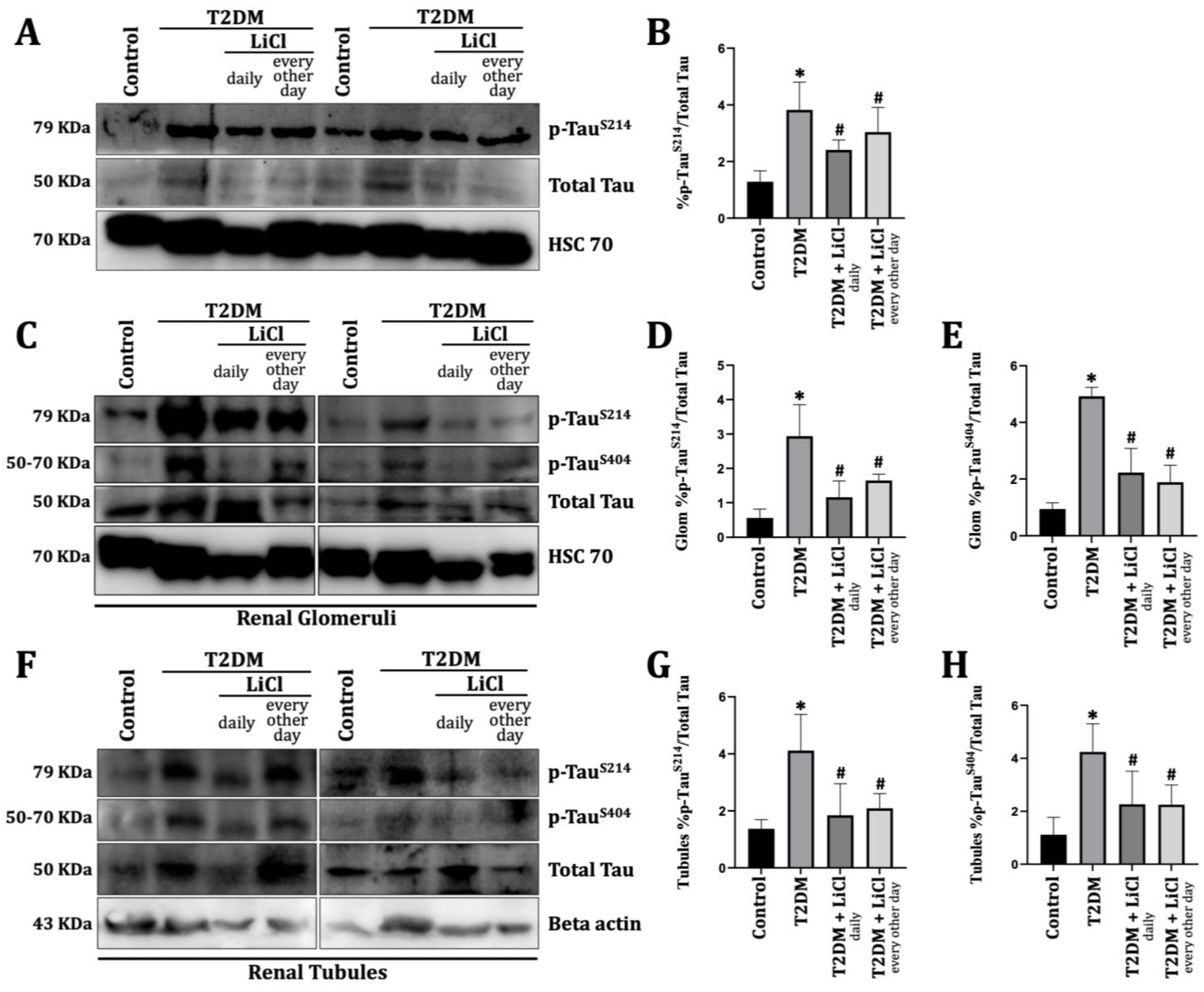
Figure 1. LiCl restored the hyperphosphorylated expression of the Tau protein in the glomeruli and tubules of the T2DM group. (A) Western blots representing Tau and Hsc70 protein levels. (B) Bar graph showing Western blot quantification; results from 20 rats. (C) Representative Western blots collected glomerul. (D,E) Bar graph showing quantification of the (D) Glom p-TauS214/total Tau and (E) Glom p-TauS404/total Tau results from 20 rats. (F) Representative Western blot-collected tubules. (G,H) Bar graph showing quantification of the (G) tubules p-TauS214/total Tau; (H) tubules p-TauS404/total Tau results from 20 rats. Data are shown as mean ± SEM with n = 5 per group. The following indicates statistical significance: * p < 0.05 compared to control rats; and # p < 0.05 compared to diabetic rats.
Diabetology 06 00026 g001
Figure 2. LiCL inhibits fibronectin expression and reduces renal fibrosis in T2DM rats. (A) Western blot demonstrating the expression levels of fibronectin and Hsc70. Densitometric analysis was used to quantify the protein levels relative to Hsc70. (B) Bar graph illustrating the quantification of fibronectin by Western blotting; n = 20 rats. ((C)-Masson): A representative micrograph (20× objective) of kidney sections stained with Masson’s trichrome to visualize collagen deposition in renal fibrotic areas after a 22-week study duration. In these sections, the blue-stained extracellular matrix contrasts with red muscle fibers and the cytoplasm, highlighting the extent of fibrosis. Scale bar = 50 µm. ((C)-PAS): A representative micrograph (20× objective) of kidney sections stained with PAS to assess the distribution and intensity of glycogen, indicative of glomerulosclerotic changes and mesangial accumulation after a 22-week study duration. Scale bar = 50 µm. (D) Bar graph illustrating the quantification of collagen deposition intensity in severely fibrotic areas. Quantification was based on the percentage of the area covered by blue staining in high-power fields, with data averaged over fifteen fields per section; n = 20 rats. (E) Bar graph depicting the quantification of glycogen deposition intensity, assessed by the area fraction of PAS-positive staining relative to the total tissue area in each high-power field. Results are based on 20 rats, with data averaged over fifteen fields per section. Data are shown as mean ± SEM with n = 5 per group. The following indicates statistical significance: * p < 0.05 compared to control rats; # p < 0.05 compared to diabetic rats; and ^ p < 0.05 compared to T2DM treated with LiCl daily.
Diabetology 06 00026 g002
Figure 3. LiCl reduces TGF- β1 expression in the kidney of T2DM. (A) Representative kidney micrograph sections of TGF-β1 expression were detected by immunoperoxidase staining (20× objective; fibrosis indicated by brown coloration) in the experimental groups. Scale bar = 50 µm. (B) Bar graph illustrating the quantification of TGF-β1 immunoperoxidase staining expressed as a percentage of the total area analyzed. Quantification was performed using ImageJ software (1.53e), with positive staining determined based on a defined intensity threshold. A total of 15 sections per rat were analyzed for each group. Data are shown as mean ± SEM based on results from 20 rats, with n = 5 per group. The following indicates statistical significance: * p < 0.05 compared to control rats; and # p < 0.05 compared to diabetic rats.
Diabetology 06 00026 g003
Table 1. Functional parameters of the four groups.
ParameterControlT2DMT2DM Treated with LiCl DailyT2DM Treated with LiCl Every Other Day
Metabolic Parameters
Body weight (g)350.10 ± 62.07456.40 ± 20.59 *347.20 ± 24.82 #491.3 ± 4.93 *^
Blood glucose (mg/dL)114.58 ± 5.81355.45 ± 22.69 *302.04 ± 40.03 *347.93 ± 34.20 *
Insulin (pmol/L)120 ± 16207 ± 21 *167 ± 18 *#158 ± 16 *#
Free Fatty Acids (mmol/L)1.7 ± 0.055.2 ± 0.12 *3.4 ± 0.09 *#3.7 ± 0.07 *#
Triglycerides (mmol/L)1.2 ± 0.029.9 ± 0.36 *5.3 ± 0.44 *#4.9 ± 0.33 *#
Renal Parameters
Kidney weight (g)1.94 ± 0.102.50 ± 0.14 *1.73 ± 0.11 #2.09 ± 0.13
KW/BW ratio3.26 × 10−3 ± 2.1 × 10−44.33 × 10−3 ± 2.3 × 10−4 *3.27 × 10−3 ± 1.4 × 10⁻⁴ #3.7 × 10⁻³ ± 2 × 10⁻⁴
Proteinuria (mg/g)228.92 ± 25.101099.10 ± 359.79 *377.92 ± 103.71 #611.85 ± 72.06 #
Body weight (g) and blood glucose (mg/dL) were recorded weekly throughout the 22-week study duration. Post-sacrifice measurements of insulin (pmol/L), free fatty acids (FFAs, mmol/L), triglycerides (TGs, mmol/L), and kidney weight (g). Proteinuria levels were assessed following a 24 h urine collection. Data presented are means ± SE, with n = 5 per group. Statistical significance is denoted as follows: * p < 0.05 compared to control rats; # p < 0.05 compared to T2DM rats; ^ p < 0.05 compared to T2DM treated with LiCl daily.
Disclaimer/Publisher’s Note: The statements, opinions and data contained in all publications are solely those of the individual author(s) and contributor(s) and not of MDPI and/or the editor(s). MDPI and/or the editor(s) disclaim responsibility for any injury to people or property resulting from any ideas, methods, instructions or products referred to in the content.