Brief Report

The Von Hippel–Lindau Protein (pVHL) Is Downregulated in the Pancreatic Islets of Mice with Type 2 Diabetes Induced by a High-Calorie Diet

by
Alma Nelly Diaz-Herreros
1,2,
Alberto Granados-Galeana
1,
Isaí Martínez-Torres
1,
Elba Reyes-Maldonado
3,
Erika Rosales-Cruz
3,
Fernando Gómez-Chávez
4,
Gabriel Betanzos-Cabrera
5,
Amaranta Sarai Valdez-Guerrero
6,
Juan Carlos Cancino-Diaz
7,* and
Mario Eugenio Cancino-Diaz
1,*
1
Laboratorio de Inmunología Aplicada, Departamento de Inmunología, Escuela Nacional de Ciencias Biológicas del Instituto Politécnico Nacional, Mexico City 11340, Mexico
2
Centro de Investigación Científica Serendipia A.C., Tláhuac, México City 13210, Mexico
3
Laboratorio de Hematología, Departamento de Morfología, Escuela Nacional de Ciencias Biológicas del Instituto Politécnico Nacional, Mexico City 11340, Mexico
4
Laboratorio de Enfermedades Osteoarticulares e Inmunológicas, Sección de Estudios de Posgrado e Investigación, Escuela Nacional de Medicina y Homeopatia del Instituto Politécnico Nacional, Mexico City 07320, Mexico
5
Área Académica de Nutrición, Instituto de Ciencias de la Salud, Universidad Autónoma del Estado de Hidalgo, Pachuca City 42160, Mexico
6
Laboratorio de Bioquímica Aplicada, Escuela Superior de Medicina del Instituto Politécnico Nacional, Mexico City 11340, Mexico
7
Laboratorio de Inmunomicrobiología, Departamento de Microbiología, Escuela Nacional de Ciencias Biológicas del Instituto Politécnico Nacional, Mexico City 11340, Mexico
*
Authors to whom correspondence should be addressed.
Diabetology 2025, 6(12), 143; https://doi.org/10.3390/diabetology6120143
Submission received: 16 September 2025 / Revised: 7 November 2025 / Accepted: 17 November 2025 / Published: 27 November 2025

Abstract

Background/Objectives: Knock-out mice lacking von Hippel–Lindau protein (pVHL) in pancreatic beta cells exhibit glucose intolerance and low insulin production, indicating a possible association between pVHL and diabetes mellitus (DM). It is currently unknown whether DM causes a decrease in pVHL expression. In this study, we determined the level of pVHL expression in the pancreas of mice with type 2 DM (T2DM) induced by a high-calorie diet (HCD). Methods: Mice of the C57BL/6 and BALB/c strains were fed with a HCD for 10 weeks, and another group of mice of both strains were fed a standard diet (SD). The mice were monitored for body weight and glucose levels until the end of the treatment. Immunodetection for pVHL, HIF-1α, Insulin and GLUT-1 was performed. Results: A significant increase in body weight in C57BL/6 mice fed HCD at week 10 compared to mice fed a SD (p < 0.05), with similar results for the BALB/c strain. The glucose level was significantly higher in the C57BL/6 strain and the BALB/c strain fed with HCD compared to mice on a SD (p < 0.05). There was lower pVHL and insulin expression in the pancreatic islets of both strains fed HCD. In contrast, there was higher pVHL expression in the pancreatic islets of both strains of mice fed a SD. HIF-1α and GLUT-1 expression was higher in mice fed HCD than in mice fed a SD. Conclusions: HCD-induced T2DM causes low pVHL expression in the pancreatic islets of C57BL/6 and BALB/c mice, suggesting that low pVHL expression is related to the development of T2DM in mice.

Graphical Abstract

1. Introduction

In 2006, Li et al. reported that an increase in the expression of hypoxia-related genes was observed in the pancreatic islets of 7- and 12-week-old male Zucker diabetic fatty (ZDF) rats. For instance, these rats showed approximately threefold and twofold increases in vascular endothelial growth factor (VEGF)-A mRNA and VEGF protein secretion, respectively, compared to control rats [1]. This result correlates with the increase in islet mass observed in the diabetic animal [1]. It is known that VEGF expression is one of the proteins regulated by hypoxia-inducible factor 1-α (HIF-1α), whose activity is negatively regulated by von Hippel–Lindau protein (pVHL) [2]. Furthermore, it is also known that the oxygen-sensing pathway, such as mitochondrial oxidative phosphorylation, controls insulin release in beta cells when there is a high level of glucose around beta cells, Glut2 and Glut1 proteins transport glucose into beta cells, where the internalized glucose is phosphorylated by glucokinase, activating the glycolytic pathway that activates mitochondrial metabolism to produce ATP, which induces insulin secretion through Ca2+ channels [3]. To investigate the role of the VHL-HIF axis in increasing pancreatic islet mass [4], in insulin production and secretion in beta cells [5], as well as to investigate the role of pVHL in the pancreas [6], three independent studies were conducted in which knock-out mice were produced in the vhlh gene in pancreatic beta cells and the pancreas (betaVhlhKO and PVhlhKO), respectively. All three reports state that all VHL knock-out mice have normal feeding behavior and normal fasting blood glucose levels. However, the mouse’s response to a glucose challenge shows impaired glucose tolerance [4,5,6]. These mutant mice reach normal blood glucose levels up to 3 h after the glucose challenge. This glucose intolerance phenotype develops at 6–8 weeks. In addition, the three studies agree that the loss of pVHL function in pancreatic beta cells affects glucose homeostasis, possibly due to impaired insulin production and secretion [4,5,6], indicating a strong association between pVHL function and glucose level regulation in animals. On the other hand, it has also been reported that mice with VHL deletion (upon Cre recombinase expression under the control of the SGLT2 promoter) in the cells of the proximal tubule, which are adjacent to the glomerulus, show morphological and gene expression changes resembling those of streptozotocin-induced type 1 DM; these mice exhibit early stages of diabetic kidney disease [7].
In humans, the allelic mutation of the vhl gene naturally causes VHL syndrome, which is characterized by the formation of multiple benign and malignant tumors, as well as the production of cysts in the central nervous system and other visceral organs such as the pancreas. There appears to be a relationship between VHL disease and diabetes mellitus (DM). In a study conducted by Hammel et al. in 2000, it was reported that DM is present in 3 out of 158 patients with VHL disease [8]. Another study reports that a patient initially diagnosed with gestational DM presented clinical symptoms of VHL disease years later, and a new mutation (del291C) was found in the vhl gene in this person [9]. Currently, there is no evidence of mutation in pVHL in patients with DM without VHL disease.
Considering that the elimination of the vhlh gene in the beta cell of a normal mouse causes an alteration in blood glucose regulation and that the mutation of the vhl gene in the human genome in VHL syndrome can cause DM, it suggests that the loss of pVHL function due to genetic mutation is an initiating factor in hyperglycemia. However, to date, in healthy mice (without any mutation in pVHL) in which DM is induced, it is unknown whether there is a loss of pVHL function or an alteration in the expression level of this protein. Currently, there are no reports comparing pVHL expression levels in the pancreatic islets of patients or animals with DM with those in the pancreatic islets of healthy individuals or animals. Therefore, this study determined the expression pattern of pVHL in the pancreatic beta islets of mice with type 2 DM (T2DM) induced by a high-calorie diet (HCD). The result was that pVHL expression is decreased in the islets of mice with T2DM compared to the islets of non-diabetic mice.

2. Materials and Methods

2.1. Murine Model of T2DM Induced by a High-Calorie Diet (HCD)

Several studies report that the prevalence of T2 DM is similar in men and women worldwide. Similar results have been observed when using animals of both sexes to study DM models; both males and females can develop DM without any difference. Based on the above, to select the sex of the mice in our study, groups of healthy male and female mice from the C57BL/6 and BALB/c strains were initially placed on a high-calorie, high-fat diet ad libitum for 10 weeks. In the case of our male mice, the diet caused more obesity than in females; however, they did not develop hyperglycemia to the same extent as the females. Therefore, it was decided to work only with female mice [10,11].
First, we worked with female C57BL/6 mice, as this strain is known to be more susceptible to T2DM induction. The HCD administered to C57BL/6 mice was prepared as follows: 1 kg of ground pellets (Lab Rodent diet 5001), 8 eggs, 400 g of lard, and 260 g of wheat flour were mixed. All ingredients were homogenized, poured into molds, and frozen for storage. The composition of this diet is approximately: protein 12.63%, fat 29.16%, carbohydrates 29.67%, and fiber 5.72%. In addition, water with 20% sucrose was provided.
For female BALB/c mice, considered less susceptible to type T2DM induction, the HCD was different, and the preparation consisted of the following ingredients: 1 kg of ground pellets (Lab Rodent diet 5001), 6 eggs, 300 g of wheat flour, and 600 g of lard. All ingredients were homogenized, poured into molds, and frozen for preservation. The nutritional composition of the diet administered to BALB/c mice was protein 11.31%, fat 37.3%, carbohydrates 27.02%, and fiber 6.11%. All of this was accompanied by water with 30% sucrose. The standard diet (SD) was protein 24.1%, fat 6.4%, carbohydrates 57.94%, and fiber 5.4%.
The approximate calorie intake of each diet is as follows: SD 3.35 Cal/g; HCD for BALB/c 4.89 Cal/g, and HCD for C57BL/6, 4.31 Cal/g. Each milliliter of water with 30% sucrose provides 1.2 calories, and 20% provides 0.8 calories. BALB/c mice intake an average of 75.14 g of SD and sucrose 30%, 77.64 mL (in total 251.72 KCal), and 41.89 g of HCD and sucrose 30%, 71.64 mL (in total 297.72 KCal). C57BL/6 mice intake an average of 108.3 g of SD and sucrose 20%, 131.2 mL (in total 362.43 KCal), and 71.45 g of HCD and sucrose 20%, 217.2 mL (in total 482.5 KCal).
The experimental design consisted of a sequence of three stages or independent experiments using mice from batches with different breeding dates (at least three months apart). Pilot experiment 1 consisted of using a single female mouse (aged four months and weighing between 23 and 24 g) of the C57BL/6 strain fed with HCD and another mouse of the same strain fed with an SD; these mice were taken from a mouse breeding batch. Other breeding batches with different breeding dates for the two strains, C57BL/6 and BAlB/c (both female mice, aged four months and weighing between 23 and 24 g) were taken for experiment 2, which used a single mouse for both strains fed HCD and another mouse from both strains on an SD. Finally, another batch of mice from the two strains with different breeding dates was used for experiment 3 with a larger number of animals per group. One group of eight mice was fed HCD, and another group of eight mice was on an SD from both strains. In experiment 3, both C57BL/6 and BALB/c mice were female, aged four months, and weighing between 23 and 24 g.
Weight, blood glucose, and calorie intake measurements were taken over 10 weeks, and those reported are from experiment 3.
At the end of the study (10 weeks), animals were euthanized with a lethal dose of pentobarbital following ENCB-IPN ethical guidelines to develop the immunohistochemistry assay.

2.2. Body Weight Monitoring

To monitor the body weight of the mice, they were weighed on a conventional scale at the start of the fasting period at approximately 9:00 a.m. each day, every week.

2.3. Blood Glucose Measurement

After 5 h of fasting, all mice in the study were punctured at the distal end of the tail with a 25 G × 16 mm needle to collect blood. Blood glucose was determined with the help of an Accu-Check Instant glucometer and test strips each week.

2.4. Calorie Intake Measurement

Food consumption was recorded twice a week, ensuring adequate replacement of water and perishable food to prevent bacterial growth in both food and water. Subsequently, the sum of the volume of water and food ingested was multiplied by the calorie values per gram and milliliter calculated for each diet and sucrose concentration. In this way, we obtained the total calorie consumption for each week.

2.5. Immunohistochemistry of pVHL and HIF-1α

The pancreases from each group were analyzed by immunohistochemistry using the streptavidin–biotin–peroxidase method to determine the expression levels of pVHL and HIF-1α. The protocol was as follows.
The tissue sections from the control group were considered positive controls, and a section without primary antibody addition was used as a negative control. From each tissue to be evaluated, two or three serial sections 5 μm thick were made using a standard microtome. The sections were placed on electrocharged slides and then heat-deparaffinized, followed by two washes with xylene. Peroxidase was inhibited in the tissue sections with 30% hydrogen peroxide in absolute methanol (1:8 ratio). The tissues were gradually rehydrated with ethanol (96%, 80%, and 70%) and distilled water for three minutes each time.
For antigen retrieval, the slides were placed in a pressure cooker for 18 min in citrate buffer. Endogenous avidin and biotin were blocked with commercial solutions (Zymed) by applying 20 μL of solution to each tissue and incubating in a humidified chamber at 37 °C for 15 min. Similarly, non-specific binding sites of the primary and secondary antibodies were blocked with 5% non-immune rabbit serum (Sigma-Aldrich) by applying 50 μL of solution to each tissue section and incubating in a humidified chamber at 37 °C for 20 min. Antibody dilutions (VHL 1:100, Novus biologicals NB100-41384 and HIF1-α 1:300, Novus biologicals NB100-479) were prepared by calculating a volume of 70–100 μL for each slide. For both primary antibodies, a commercial detection system (Histostain Invitrogen) was used, initially adding biotinylated antibody to each slide (70–100 μL for 20 min in a humid chamber at 37 °C). After this time, the slides were washed in PBS for 5 min. Next, the streptavidin–peroxidase complex was added to each slide under similar conditions. To determine the presence or absence of antigen in the tissues, a commercial solution of chromogen developing compounds (DAB, diaminobenzidine) and a reaction buffer (Biocare) was added to each section, mixing one drop of the chromogen in 1 mL of the buffer. Once the developing solution was prepared, 100 μL was applied to each tissue section, observing the dye reaction under optical microscopy (for 50 to 60 s) until it changed. The reaction was stopped by placing the slide in distilled water. The sections were counterstained with Meyer’s hematoxylin by applying 50 μL to each section for 1 min, washed with distilled water and then with PBS, and covered with conventional coverslips. All stains were analyzed using conventional optical microscopy.
For the quantitative analysis of the immunohistochemistry assays, three fields from the pancreatic section of each mouse were taken and analyzed with Fiji ImageJ software version 1.54p. The color deconvolution function with the H&E DAB setting was used to separate the positive stain (Channel 3), and the percentage of the stained area was quantified with a threshold of 11 to 250. To avoid bias, the area with blood vessels, ducts, and spaces without tissue was excluded using ROI masks. The results were graphically represented, and statistical comparisons were made using the unpaired t-test.

2.6. Detection of Insulin and GLUT-1 by Immunofluorescence

Pancreas sections of 5 μm were obtained and placed on electrocharged glass slides and fixed in 4% paraformaldehyde, and rehydrated with a series of solvents. Heat antigen retrieval was performed using a citrate buffer at pH 6.0 for 18 min at 90 °C, and the tissue slides were blocked with a solution of BSA/FBS 5% and sodium azide 0.02% in PBS for 30 min. Primary antibody anti-insulin (1:100; EPR17359, Abcam) or anti-GLUT-1 (1:200; NB110-39113AF594, Novus biologicals) was incubated for 1 h at 37 °C, and the donkey anti-rabbit IgG secondary antibody (1:500, AB150075, Abcam) was then incubated for 30 min at room temperature. Slides were counterstained and mounted with DAPI VectaShield (Vector Laboratories, Burlingame, CA, USA). Images were obtained on a Nikon Ti Eclipse inverted confocal microscope (Nikon Corporation, Minato, Tokyo, Japan). All images were analyzed using Fiji ImageJ software, focusing on endocrine tissues by ROI masks. Mean intensity fluorescence was determined using a threshold from 1650 to 4000.

2.7. Statistical Analysis

Sample size was calculated using GPower software version 3.1.9.7, with the following settings: t tests, means—difference between two independent groups, one-tailed, effect size 0.9, alpha value 0.20, 1-beta value 0.80, allocation ratio 1. Statistical analysis was performed using Graphpad software 8.0.2. Normality was evaluated by the Shapiro–Wilk test with an alpha value of 0.05. The difference between groups was calculated by an unpaired t test or U Mann–Whitney test as appropriate.

3. Results

3.1. Validation of the Diet-Induced T2DM Model in Mice

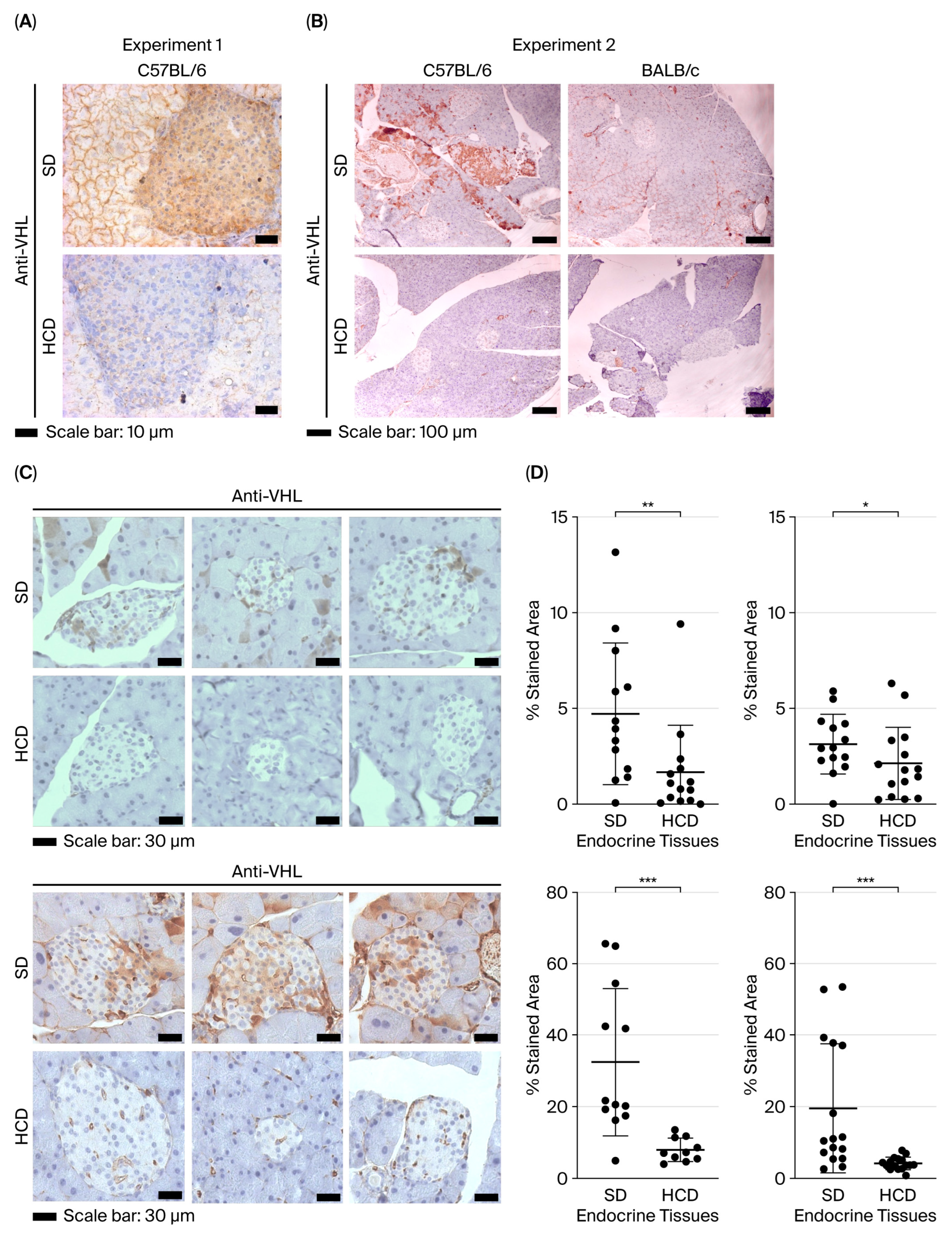
The hypercaloric diet (HCD)-induced T2DM model was obtained in previous work by our group. Based on the previous work, we reproduced T2DM in both C57BL/6 and BALB/c mouse strains (experiments 1 and 2). In all mice fed with HCD, significant hyperglycemia was observed in both mouse strains at week 10. Subsequently, in experiment 3, the number of mice used was increased for both strains, with one group of eight mice fed a standard diet (SD) and another group of eight mice fed HCD for 10 weeks. During the 10 weeks, the weight of the mice and fasting blood glucose levels were measured. Figure 1A shows the average weight of the mice of both strains. In the C57BL/6 strain, a marked significant increase in body weight was observed starting in week 3 in the group of mice fed HCD compared to the group of mice fed an SD (p < 0.05; Figure 1A). For the BALB/c mouse strain, the group of mice fed HCD did not show a significant increase in body weight compared to the group of mice on an SD up to 10 weeks (p < 0.05; Figure 1A).
Regarding blood glucose, the C57BL/6 mice fed HCD showed a significant increase in blood glucose levels starting at week 4 and lasting until week 10, compared to C57BL/6 mice fed an SD (p < 0.05; Figure 1B). Like the C57BL/6 mice, the BALB/c mice fed HCD exhibited a significant rise in blood glucose levels, beginning at week 2 and continuing until week 10, compared to BALB/c mice fed an SD (p < 0.05; Figure 1B). Other parameters were also measured to determine the development of type 2 DM in the animals. All mice fed HCD had elevated levels of triglycerides, cholesterol, and insulin in serum compared to the mice fed an SD. Additionally, histological analysis revealed signs of hepatic steatosis and hypertrophy of adipose tissues. These results indicate that both strains of mice fed HCD were capable of developing type 2 DM.

3.2. pVHL Expression Pattern in the Pancreas of Non-Diabetic Mice and Diabetic Mice Induced by a High-Calorie Diet

The pancreases of the mice in the study were tested to determine the level of pVHL expression using immunohistochemistry. Experiment 1 (pilot) first consisted of using the C57BL/6 mouse strain (described as a strain highly susceptible to inducing DM). In this experiment, it was observed that the pancreatic islets of C57BL/6 mice fed an HCD showed low immunodetection with the anti-pVHL antibody compared to the immunodetection of the islets of C57BL/6 mice fed an SD, where strong pVHL detection was observed (Figure 2A). In experiment 2, where two mouse strains were analyzed, both the C57BL/6 strain and the BALB/c strain (which has low susceptibility to DM), a similar result to experiment 1 is observed; in both mouse strains, lower immunodetection with the anti-pVHL antibody was observed in the islets of mice fed HCD compared to mice fed an SD (Figure 2B). Interestingly, the C57BL/6 strain of mice fed an SD had higher pVHL positivity than the BALB/c strain on the same diet. These pVHL immunodetection results suggest that there is low pVHL expression in the pancreatic islets of HCD-fed mice of both strains compared to the islets of mice fed an SD (non-hyperglycemic).
In order to confirm the level of pVHL expression in the pancreatic islets of animals with and without T2DM, an increase in the number of mice (eight mice) for both strains was made (experiment 3). Figure 2C shows representative results from three immunohistochemistry assays for pVHL in the pancreas of the three mice under study. In the pancreas of BALB/c mice on an SD, similar to experiment 2, the anti-VHL antibody detects pVHL in the islet and even in the exocrine tissue of the pancreas. In contrast, in the pancreas of BALB/c mice with HCD, this immunodetection of pVHL is lost, and this loss is more evident in the beta islet (Figure 2C). About the C57BL/6 mouse strain, in the pancreas of mice on an SD, immunodetection with the anti-VHL antibody is more intense than in BALB/c mice in the pancreatic islets as well as in the exocrine tissues. In some areas of the parenchyma, this immunodetection is stronger. In contrast, in the pancreas of mice with HCD, very weak immunodetection towards pVHL is observed (Figure 2C), similar to that observed in the islets of BALB/c mice fed with HCD.
Immunostaining semiquantitative analysis of pVHL in experiment 3 (Figure 2D) shows that the group of BALB/c mice fed a standard diet (SD) has greater immunopositivity in both pancreatic tissues, endocrine and exocrine, for pVHL compared to the group of mice fed a high-carb diet (HCD) (p < 0.05). The same result is observed for the C57BL/6 strain (p < 0.05; Figure 2D). As mentioned above, Figure 2D shows that this difference in pVHL expression in endocrine and exocrine tissues is ten times higher in C57BL/6 than in BALB/c. These results suggest that pVHL expression decreases significantly in the beta islets (endocrine tissues) of the pancreas when mice are fed an HCD, which induces Type 2 DM.

3.3. HIF-1alpha, Insulin, and GLUT-1 Expression Pattern in the Pancreas of Non-Diabetic and Diabetic Mice Induced by a High-Calorie Diet

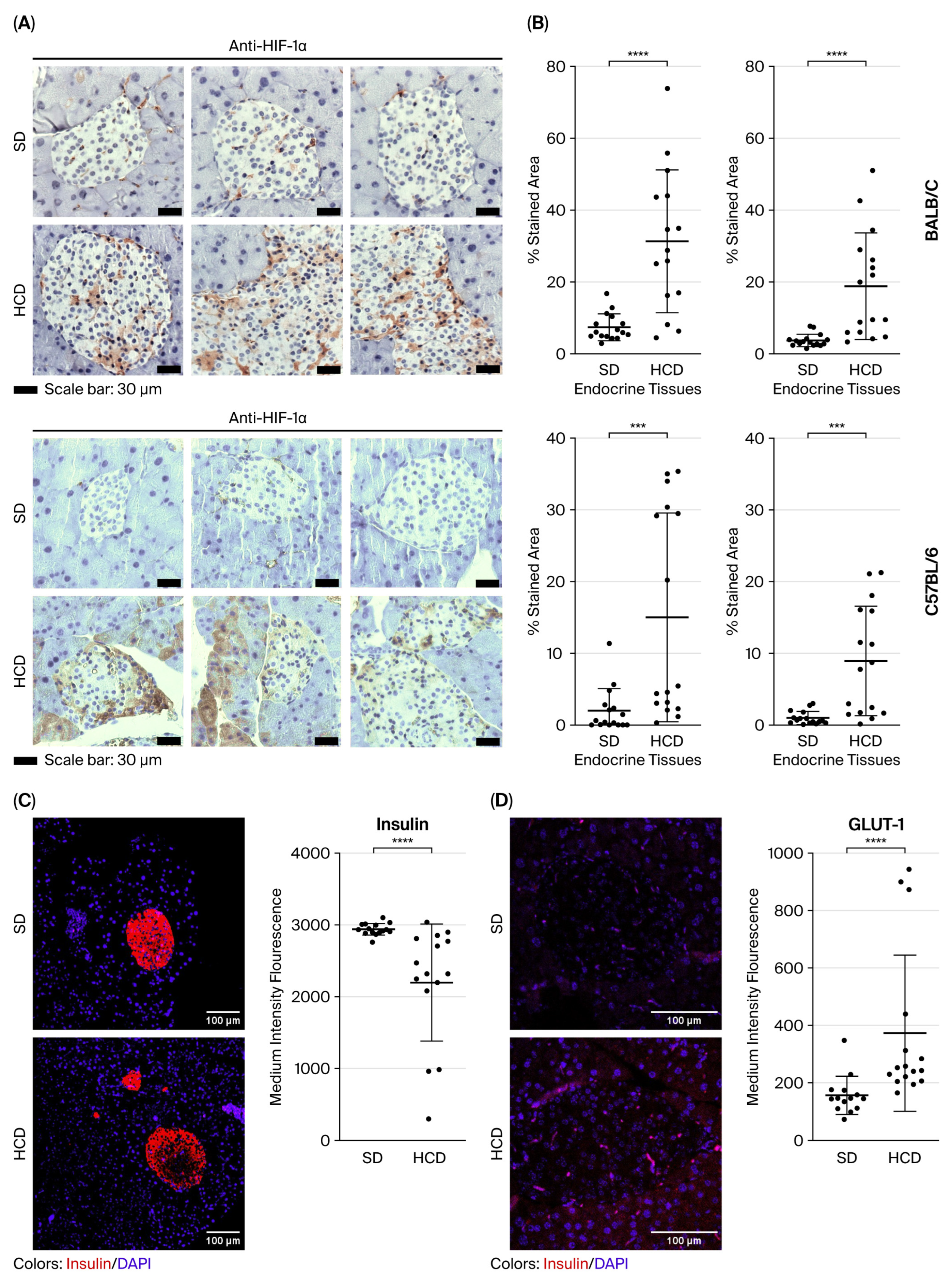
To verify the VHL-HIF-1alpha axis in mice fed HCD, the level of HIF-1α expression was measured and compared with pVHL expression levels. Additionally, GLUT-1 was assessed because it is known to be positively regulated by HIF-1alpha [12]. Insulin was also measured because the KO-VHL beta-cells showed decreased insulin expression [4,5,6]. Figure 3A shows strong immunostaining with the anti-HIF-1α antibody in the pancreatic islets and exocrine tissues of BALB/c mice fed HCD, especially when pVHL immunostaining is low. In contrast, low HIF-1α immunostaining is observed in BALB/c mice fed with a standard diet (SD). For the C57BL/6 strain, a strong HIF-1α immunodetection is also observed in the pancreas of mice fed HCD compared to those fed SD (Figure 3A). Analysis of HIF-1α immunostaining intensity indicates that both endocrine and exocrine tissues show a significant increase in the HCD-fed group for both strains compared to the SD group (Figure 3B). These results suggest that HCD feeding increases HIF-1α activity and correlates with low pVHL expression in a hyperglycemic state. Similarly, in the endocrine and exocrine tissues of C57BL/6 mice fed HCD, HIF-1α expression is more abundant than in BALB/c mice fed HCD.
Regarding insulin, which is produced only by endocrine tissues, mice fed an HCD show lower insulin signals than mice fed an SD in both strains (Figure 3C). This figure, shown in an image and through an analysis of fluorescence intensity, indicates that insulin production in endocrine tissues is reduced in animals fed HCD. This result suggests that decreased pVHL activity in hyperglycemic mice also affects insulin production in pancreatic beta cells.
To corroborate the function of the VHL-HIF-1alpha axis in pancreatic tissues, GLUT-1 was measured because HIF-1alpha regulates its protein expression. Figure 3D shows that in the pancreatic tissues of animals fed an HCD, where HIF-1alpha expression is high (Figure 3A,B), GLUT-1 expression is also high compared to animals fed an SD (Figure 3D).

4. Discussion

The mutation of the vhl gene in humans causes VHL syndrome, which leads to tumors or cysts in multiple organs, indicating that pVHL is an important factor in the regulation of various diseases. In the case of the pancreas, patients with VHL disease develop abnormalities such as pancreatic cysts or tumors [13] and, in a small number of cases, type 2 diabetes [8,14]. Cases of VHL syndrome patients with DM, pulmonary, and thyroid nodules are also reported [15]. One of the main functions of pVHL is the regulation of HIF-1α under normal oxygen conditions. In this regard, much attention has been paid to the relationship between HIF-α and various diseases such as cancer, inflammation, and angiogenesis, among others [16,17,18]. Few studies have linked pVHL as a regulatory factor in disease, and therefore, its role has received little attention. In diseases unrelated to VHL, such as psoriasis [19] or glioblastoma [20], patients have been reported to have low or no expression of pVHL and an increase in HIF-1α. In this same vein, we are reporting for the first time that pVHL is under-expressed in the endocrine and exocrine pancreatic tissues of mice with HCD-induced T2DM. This result suggests that pVHL could be an important modulator in glucose regulation, as has already been proven in betaVhlhKO [4,5,6].
In the case of psoriasis, it has been shown that pVHL is not expressed in the damaged skin of patients, whereas there is expression of pVHL in the healthy skin of healthy subjects [19]. This lack of pVHL expression is also present in the damaged skin of murine psoriasis induced by imiquimod [21]. This strongly suggests that pVHL has a role in preventing psoriasis in susceptible individuals. The lack of pVHL expression in psoriasis is not due to a mutation in the vhl gene. It is currently thought that pVHL is regulated by other molecules to inhibit its expression, but the molecules that inhibit pVHL expression in psoriasis are still unknown. The negative regulatory function of pVHL in psoriasis has been proven by restoring pVHL expression in skin damaged by imiquimod-induced psoriasis. This restoration of pVHL expression prevents the onset of murine psoriasis and reduces the molecules involved in psoriasis [21]. This indicates that in the pathology of psoriasis, keratinocytes induce the expression of proteins that inhibit pVHL expression, leading to disease development.
In the case of glioblastoma, it has been documented that cancer cells lose pVHL function because its production is inhibited [22]. Glioblastomas are highly vascularized and hypoxic cancers in which HIF-1α and VEGF are important elements for tumor maintenance. Patients with glioblastoma do not have mutations in the vhl gene, suggesting that the lack of the protein in cancer is due to molecules that negatively regulate pVHL expression. Recently, some proteins that inhibit the activity of pVHL have been reported; for example, the DAAM2 protein is overexpressed in glioblastoma and participates in tumorigenesis [23]; DAAM2 binds to pVHL to ubiquitinate and degrade it via the proteasome [22]. Another molecule involved in glioblastoma is FBXO22, which, like DAAM2, is overexpressed in this cancer and participates in tumor proliferation and invasion [18]. FBXO22 also binds to pVHL to degrade it [24]. Another negative regulation of pVHL in glioblastoma is through microRNAs (miRNAs). It has been observed that miR-23b, miR-150, and miR-566 are overexpressed in glioblastoma and cause low expression of pVHL [23,25,26]. The functionality of pVHL in glioblastoma has been studied by restoring pVHL function through gene introduction by plasmids or viral vectors carrying the vhl gene. In glioblastoma cells transfected with the vhl gene, there is a decrease in the expression of HIF-1α, VEGF, and Bcl-2, as well as a decrease in angiogenesis and an increase in apoptosis of these cells [27]. A decrease in the proliferation rate and invasive capacity is also observed [28]. Furthermore, when glioblastoma cells transfected with the vhl gene are implanted into nude mice, the tumor growth rate is lower than that of non-transfected cells [27,28]. These results demonstrate that pVHL is an important modulator in cell biology and that low expression of this protein is related to different pathologies.
The low expression of pVHL in the endocrine and exocrine tissues of the pancreas of mice with DM induced by a high-calorie diet and the increase in HIF-1α and GLUT-1 expression reported in this study confirm what has been demonstrated with pVHL knock-out mice in the pancreas [4,5,6], i.e., that the lack of pVHL functionality in beta cells is possibly an important factor in the development of DM. This is a relevant result because a high-calorie diet induces an increase in blood glucose levels in mice, and this increase in glucose affects the pancreatic islets, causing the manifestations of DM and also inducing low pVHL expression. Furthermore, this result is supported by two mouse strains: the C57BL/6 strain, which is highly susceptible to DM due to a high-calorie diet, and the less susceptible BALB/c strain. We believe that high blood glucose may affect pancreatic beta cells, causing an alteration in pVHL expression, possibly due to epigenetic events or pVHL-degrading molecules. Our results are also consistent with those of research groups that report an increase in pancreatic islet mass due to high expression of HIF-1α and VEGF in individuals with T2DM [4,5,6].
Recently, Halperin et al. reported another interesting association between a possible decrease in pVHL activity and DM, which aligns with our findings in this work. Analyzing heterozygous VHL alterations in nearly 500,000 participants from the UK Biobank database, they found alterations in the VHL gene locus in nearly 0.5% of the population, which were linked to an increased risk of diabetes and cerebrovascular accidents. This risk appears to grow with the severity of the variants. They suggest that these heterozygous VHL alterations can induce pseudohypoxia in pancreatic and cerebral tissues [29].
One pathway for the development of human DM is a high-calorie diet in a normal subject [30]. In this regard, our results indicate that healthy mice on this diet produce alterations in blood glucose levels, similar to what occurs in humans. Therefore, the results obtained in this study could be projected to patients with T2DM (not associated with VHL disease), where DM patients would have low expression of pVHL in the pancreatic tissues. However, we understand that T2DM is complex and diverse in its onset and progression. It is important to mention that it is necessary to conduct studies of pVHL expression in patients with human T2DM to confirm the importance of pVHL in DM.
Our results suggest that low expression of pVHL in the pancreatic islets of mice with T2DM induced by a high-calorie diet promotes the pathology of DM. Some molecules, such as DAAM2, FBXO22, and miRNAs, may be involved in the downregulation of pVHL, as occurs in glioblastoma cells [22,23,24,25,26,31,32]. In addition, high glucose may also be involved in the downregulation of pVHL, possibly through epigenetic events. In the future, it will be important to verify whether these molecules or glucose are the key elements in the regulation of pVHL functionality in the pancreatic islet. Furthermore, these results open the door to the use of pVHL as a therapeutic tool for type 2 DM.

5. Conclusions

In conclusion, T2DM induced by a high-calorie diet in mice causes low expression of pVHL in the pancreatic islets of C57BL/6 and BALB/c mice, supporting the finding of pVHL knock-out mice in the pancreas. Furthermore, low expression of pVHL is a possible mechanism for the development of DM in mice and has therapeutic prospects.

Author Contributions

Conceptualization, J.C.C.-D. and M.E.C.-D.; methodology, A.N.D.-H., A.G.-G., I.M.-T., E.R.-M., A.S.V.-G. and E.R.-C.; software, F.G.-C.; validation, J.C.C.-D. and M.E.C.-D.; formal analysis, A.N.D.-H.; investigation, F.G.-C.; data curation, A.N.D.-H., G.B.-C., and A.G.-G.; writing—original draft preparation, G.B.-C.; writing—review and editing, J.C.C.-D.; visualization, M.E.C.-D.; supervision, M.E.C.-D.; project administration, M.E.C.-D.; funding acquisition, M.E.C.-D. All authors have read and agreed to the published version of the manuscript. All authors have read and agreed to the published version of the manuscrip.

Funding

This research was funded by Instituto Politécnico Nacional project number SIP-20253840 and by SECIHTI project number CF-2023-I-1131.

Institutional Review Board Statement

The animal study protocol was approved by the Ethics Committee for the Welfare of Experimental Animals at the Escuela Nacional de Ciencias Biológicas of the Instituto Politécnico Nacional, Mexico, No. Z00-001-2021, approval date: 29 June 2021.

Informed Consent Statement

Not applicable.

Data Availability Statement

Additional data can be obtained upon request to the corresponding author.

Acknowledgments

M.E. Cancino-Diaz, F. Gómez-Chávez, S., E. Reyes-Maldonado, E. Rosales-Cruz, and J.C. Cancino-Diaz appreciate the COFAA and EDI-IPN fellowships. All authors are SNII-SECIHTI fellows.

Conflicts of Interest

The authors declare no conflicts of interest. The funders had no role in the design of the study; in the collection, analyses, or interpretation of data; in the writing of the manuscript; or in the decision to publish the results.

Abbreviations

The following abbreviations are used in this manuscript:
MDdiabetes mellitus
pVHLvon Hippel–Lindau protein
HIF-1αhypoxia-inducible factor 1-alpha
VEGFvascular endothelial growth factor
HCDhigh-calorie diet
SDstandard diet

References

  1. Li, X.; Zhang, L.; Meshinchi, S.; Dias-Leme, C.; Raffin, D.; Johnson, J.D.; Treutelaar, M.K.; Burant, C.F. Islet Microvasculature in Islet Hyperplasia and Failure in a Model of Type 2 Diabetes. Diabetes 2006, 55, 2965–2973. [Google Scholar] [CrossRef]
  2. Mahon, P.C.; Hirota, K.; Semenza, G.L. FIH-1: A novel protein that interacts with HIF-1α and VHL to mediate repression of HIF-1 transcriptional activity. Genes Dev. 2001, 15, 2675–2686. [Google Scholar] [CrossRef]
  3. Fu, Z.; Gilbert, E.R.; Liu, D. Regulation of insulin synthesis and secretion and pancreatic Beta-cell dysfunction in diabetes. Curr. Diabetes Rev. 2013, 9, 25–53. [Google Scholar] [CrossRef] [PubMed]
  4. Zehetner, J.; Danzer, C.; Collins, S.; Eckhardt, K.; Gerber, P.A.; Ballschmieter, P.; Galvanovskis, J.; Shimomura, K.; Ashcroft, F.M.; Thorens, B.; et al. pVHL is a regulator of glucose metabolism and insulin secretion in pancreatic β cells. Genes Dev. 2008, 22, 3135–3146. [Google Scholar] [CrossRef] [PubMed]
  5. Cantley, J.; Selman, C.; Shukla, D.; Abramov, A.Y.; Forstreuter, F.; Esteban, M.A.; Claret, M.; Lingard, S.J.; Clements, M.; Harten, S.K.; et al. Deletion of the von Hippel–Lindau gene in pancreatic β cells impairs glucose homeostasis in mice. J. Clin. Investig. 2008, 119, 125–135. [Google Scholar] [CrossRef] [PubMed]
  6. Puri, S.; Cano, D.A.; Hebrok, M. A Role for von Hippel-Lindau Protein in Pancreatic β-Cell Function. Diabetes 2009, 58, 433–441. [Google Scholar] [CrossRef]
  7. Kunke, M.; Knöfler, H.; Dahlke, E.; Zanon Rodriguez, L.; Böttner, M.; Larionov, A.; Saudenova, M.; Ohrenschall, G.M.; Westermann, M.; Porubsky, S.; et al. Targeted deletion of von-Hippel-Lindau in the proximal tubule conditions the kidney against early diabetic kidney disease. Cell Death Dis. 2023, 14. [Google Scholar] [CrossRef]
  8. Hammel, P.R.; Vilgrain, V.; Terris, B.; Penfornis, A.; Sauvanet, A.; Correas, J.M.; Chauveau, D.; Balian, A.; Beigelman, C.; O’Toole, D.; et al. Pancreatic involvement in von Hippel–Lindau disease. Gastroenterology 2000, 119, 1087–1095. [Google Scholar] [CrossRef]
  9. Ku, Y.H.; Ahn, C.H.; Jung, C.-H.; Lee, J.E.; Kim, L.-K.; Kwak, S.H.; Jung, H.S.; Park, K.S.; Cho, Y.M. A Novel Mutation in the Von Hippel-Lindau Tumor Suppressor Gene Identified in a Patient Presenting with Gestational Diabetes Mellitus. Endocrinol. Metab. 2013, 28, 320–325. [Google Scholar] [CrossRef]
  10. Wild, S.; Roglic, G.; Green, A.; Sicree, R.; King, H. Global Prevalence of Diabetes. Diabetes Care 2004, 27, 1047–1053. [Google Scholar] [CrossRef]
  11. Ma, Y.; Li, W.; Yazdizadeh Shotorbani, P.; Dubansky, B.H.; Huang, L.; Chaudhari, S.; Wu, P.; Wang, L.A.; Ryou, M.-G.; Zhou, Z.; et al. Comparison of diabetic nephropathy between male and female eNOS−/− db/db mice. Am. J. Physiol.-Ren. Physiol. 2019, 316, F889–F897. [Google Scholar] [CrossRef] [PubMed]
  12. Nishioku, T.; Nakao, S.; Anzai, R.; Hiramatsu, S.; Momono, A.; Moriyama, M.; Nakao, M.; Terazono, A. HIF-1α stabilization in osteoclasts induces the expression of aerobic glycolysis-related proteins GLUT1, LDHA, and MCT4. J. Pharmacol. Sci. 2025, 158, 336–342. [Google Scholar] [CrossRef]
  13. Onishi, Y.; Shimizu, H.; Koyasu, S.; Taura, D.; Takahashi, A.; Uza, N.; Isoda, H.; Nakamoto, Y. Association Between Pancreatic Cysts and Diabetes Mellitus in Von Hippel-Lindau Disease. Cureus 2024, 16, e54781. [Google Scholar] [CrossRef]
  14. Wang, Y.; Liu, Z.; Zhao, W.; Cao, C.; Xiao, L.; Xiao, J. Diversities of Mechanism in Patients with VHL Syndrome and diabetes: A Report of Two Cases and Literature Review. Diabetes Metab. Syndr. Obes. 2024, 17, 1611–1619. [Google Scholar] [CrossRef]
  15. Peng, Z.; Hua, C.; Liu, W.; Zhou, M.; Yu, X.; Zhao, Y.; Zuo, X. VHL Syndrome with Diabetes Mellitus, and Pulmonary and Thyroid Nodules: A Case Report. J. Kidney Cancer VHL 2025, 12, 37–46. [Google Scholar] [CrossRef]
  16. Alvarado-Ortiz, E.; Sarabia-SáNchez, M.A. Hypoxic link between cancer cells and the immune system: The role of adenosine and lactate. Oncol. Res. 2025, 33, 1803–1818. [Google Scholar] [CrossRef] [PubMed]
  17. Sandau, K.B.; Zhou, J.; Kietzmann, T.; Brüne, B. Regulation of the Hypoxia-inducible Factor 1α by the Inflammatory Mediators Nitric Oxide and Tumor Necrosis Factor-α in Contrast to Desferroxamine and Phenylarsine Oxide. J. Biol. Chem. 2001, 276, 39805–39811. [Google Scholar] [CrossRef] [PubMed]
  18. Maxwell, P.H.; Dachs, G.U.; Gleadle, J.M.; Nicholls, L.G.; Harris, A.L.; Stratford, I.J.; Hankinson, O.; Pugh, C.W.; Ratcliffe, P.J. Hypoxia-inducible factor-1 modulates gene expression in solid tumors and influences both angiogenesis and tumor growth. Proc. Natl. Acad. Sci. USA 1997, 94, 8104–8109. [Google Scholar] [CrossRef]
  19. Tovar-Castillo, L.E.; Cancino-Díaz, J.C.; García-Vázquez, F.; Cancino-Gómez, F.G.; León-Dorantes, G.; Blancas-González, F.; Jiménez-Zamudio, L.; García-Latorre, E.; Cancino-Díaz, M.E. Under-expression of VHL and over-expression of HDAC-1, HIF-1α, LL-37, and IAP-2 in affected skin biopsies of patients with psoriasis. Int. J. Dermatol. 2007, 46, 239–246. [Google Scholar] [CrossRef]
  20. Assimakopoulou, M.; Androutsopoulou, C.; Zolota, V.; Matsoukas, J. Immunoexpression patterns for Hypoxia-inducible Factor-1alpha and von Hippel-Lindau protein, in relation to Hsp90, of human brain tumors. Histol. Histopathol. 2016, 31, 535–546. [Google Scholar] [CrossRef]
  21. Martínez-Torres, I.; Tepale-Segura, A.; Castro-Escamilla, O.; Cancino-Diaz, J.C.; Rodríguez-Martínez, S.; Perez-Tapia, S.M.; Bonifaz, L.C.; Cancino-Diaz, M.E. The Protective Role of pVHL in Imiquimod-Induced Psoriasis-like Skin Inflammation. Int. J. Mol. Sci. 2022, 23, 5226. [Google Scholar] [CrossRef]
  22. Zhu, W.; Krishna, S.; Garcia, C.; Lin, C.-C.J.; Mitchell, B.D.; Scott, K.L.; Mohila, C.A.; Creighton, C.J.; Yoo, S.-H.; Lee, H.K.; et al. Daam2 driven degradation of VHL promotes gliomagenesis. eLife 2017, 6, 31926. [Google Scholar] [CrossRef]
  23. Li, Z.; Wei, X.; Zhu, Y. The prognostic value of DAAM2 in lower grade glioma, liver cancer, and breast cancer. Clin. Transl. Oncol. 2023, 25, 2224–2238. [Google Scholar] [CrossRef] [PubMed]
  24. Shen, Z.; Dong, T.; Yong, H.; Deng, C.; Chen, C.; Chen, X.; Chen, M.; Chu, S.; Zheng, J.; Li, Z.; et al. FBXO22 promotes glioblastoma malignant progression by mediating VHL ubiquitination and degradation. Cell Death Discov. 2024, 10, 151. [Google Scholar] [CrossRef]
  25. Chen, L.; Han, L.; Zhang, K.; Shi, Z.; Zhang, J.; Zhang, A.; Wang, Y.; Song, Y.; Li, Y.; Jiang, T.; et al. VHL regulates the effects of miR-23b on glioma survival and invasion via suppression of HIF-1α/VEGF and β-catenin/Tcf-4 signaling. Neuro-Oncol. 2012, 14, 1026–1036. [Google Scholar] [CrossRef] [PubMed]
  26. Xiao, B.; Zhou, X.; Ye, M.; Lv, S.; Wu, M.; Liao, C.; Han, L.E.I.; Kang, C.; Zhu, X. MicroRNA-566 modulates vascular endothelial growth factor by targeting Von Hippel-Landau in human glioblastoma in vitro and in vivo. Mol. Med. Rep. 2016, 13, 379–385. [Google Scholar] [CrossRef] [PubMed]
  27. Sun, X.; Liu, M.; Wei, Y.; Liu, F.; Zhi, X.; Xu, R.; Krissansen, G.W. Overexpression of von Hippel-Lindau tumor suppressor protein and antisense HIF-1α eradicates gliomas. Cancer Gene Ther. 2005, 13, 428–435. [Google Scholar] [CrossRef]
  28. Kanno, H.; Sato, H.; Yokoyama, T.-A.; Yoshizumi, T.; Yamada, S. The VHL tumor suppressor protein regulates tumorigenicity of U87-derived glioma stem-like cells by inhibiting the JAK/STAT signaling pathway. Int. J. Oncol. 2013, 42, 881–886. [Google Scholar] [CrossRef]
  29. Halperin, R.; Horwitz, R.; Schwarz, Y.; Tirosh, A. Pseudohypoxia caused by germline genetic alterations in the VHL gene is associated with increased diabetes and cardiovascular risk: A UK biobank study. Cardiovasc. Diabetol. 2025, 24, 239. [Google Scholar] [CrossRef]
  30. Elsakr, J.M.; Dunn, J.C.; Tennant, K.; Zhao, S.K.; Kroeten, K.; Pasek, R.C.; Takahashi, D.L.; Dean, T.A.; Velez Edwards, D.R.; McCurdy, C.E.; et al. Maternal Western-style diet affects offspring islet composition and function in a non-human primate model of maternal over-nutrition. Mol. Metab. 2019, 25, 73–82. [Google Scholar] [CrossRef]
  31. Cheng, J.; Lin, M.; Chu, M.; Gong, L.; Bi, Y.; Zhao, Y. Emerging role of FBXO22 in carcinogenesis. Cell Death Discov. 2020, 6, 66. [Google Scholar] [CrossRef] [PubMed]
  32. Li, S.J.; Liu, H.L.; Tang, S.L.; Li, X.J.; Wang, X.Y. MicroRNA-150 regulates glycolysis by targeting von Hippel-Lindau in glioma cells. Am. J. Transl. Res. 2017, 9, 1058–1066. [Google Scholar] [PubMed]
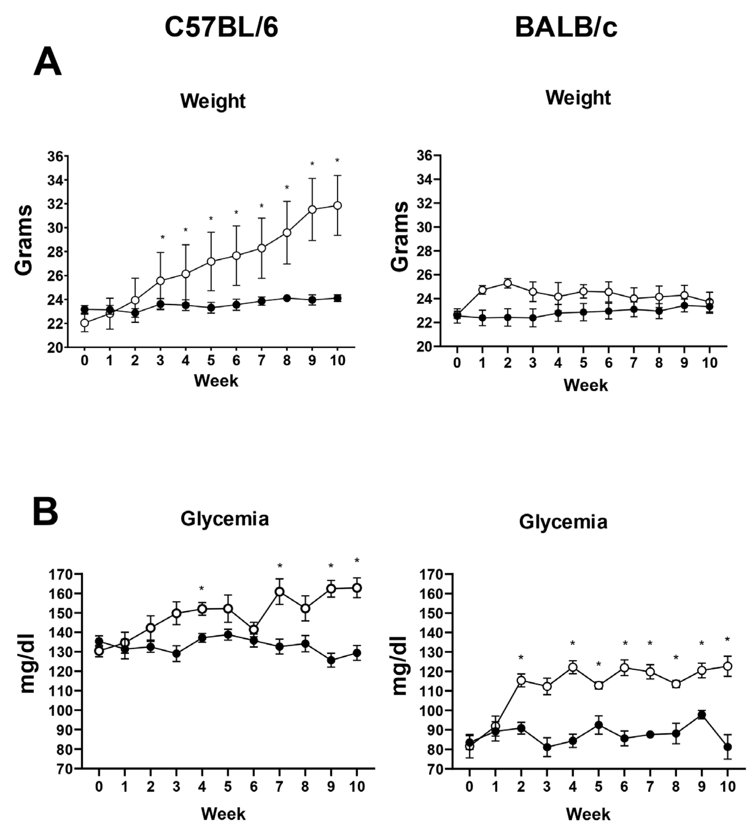
Figure 1. Body weight and blood glucose levels of mice fed a high-calorie diet (HCD). Mice from the C57BL/6 and BALB/c strains were given either a high-calorie diet (HCD) or a standard diet (SD) for 10 weeks. Weight (A) and blood glucose levels (B) were measured weekly. In all panels, data from mice fed the HCD are shown as white circles, while data from mice fed the SD are shown as black circles. Eight mice per group and strain (experiment 3) were tested. The asterisk indicates a significant difference based on an ANOVA two-way or mixed-model test as appropriate * = p < 0.05. For weight: Week factor p < 0.0001, Week × Diet p < 0.000, interaction p < 0.0001. For glycemia: Week factor p < 0.01, Week × Diet p < 0.001, interaction p < 0.0001.
Diabetology 06 00143 g001
Figure 2. pVHL expression levels in islets of mice fed a high-calorie diet (HCD) and standard diet (SD). Immunohistochemistry with anti-pVHL antibody was performed on the pancreas of mice fed a high-calorie diet (HCD) and a standard diet (SD). Experiment 1 (A) was performed with a single mouse of the C57BL/6 strain fed an HCD and another mouse of the same strain fed an SD. Images were acquired at 40× Magnification. Experiment 2 (B) consisted of C57BL/6 and BALB/c mouse strains obtained from a different breeding batch than experiment 1, with one mouse fed an HCD and another mouse fed an SD. All Images were acquired at 10× Magnification. Experiment 3: A group of 8 mice from the two strains (BALB/c and C57BL/6) were fed HCD, and a group of 8 mice from the two strains were fed SD. The mice in experiment 3 (C,D) were taken from a different breeding batch than the previous experiments. Representative immunohistochemistry results from three mice fed with both diets are shown in (C) (the six images above correspond to BALB/c mice and the six images below correspond to C56BL/6 mice). Images were acquired at 40× Magnification. (D): A semiquantitative analysis of the immunostaining level for pVHL was performed using the FijiImageJ software with a group of 8 mice from experiment. The two graphs above correspond to BALB/c mice, and the two graphs below correspond to C56BL/6 mice. For each strain, the percentage of the stained area in endocrine and exocrine tissues was measured. The asterisk indicates a significant difference according to an unpaired t-test (* = p < 0.05; ** = p < 0.01; *** = p < 0.001).
Diabetology 06 00143 g002
Figure 3. Expression levels of HIF-1α, insulin, and GLUT-1 in the pancreas of mice fed a high-calorie diet (HCD) and a standard diet (SD). Immunodetection was performed on the pancreas using anti-HIF-1α, anti-insulin, and anti-GLUT-1 antibodies in mice from both diet groups. A total of 8 mice from each strain (BALB/c and C57BL/6) were fed HCD, and 8 mice from each strain were fed SD. (A) shows a representative image from one of three mice analyzing HIF-1α, using the same pancreases as in Figure 2C. All images were acquired at 40×. The level of immunostaining for HIF-1α was quantified using Fiji ImageJ software, and the percentage of stained area in endocrine and exocrine regions is shown for both strains (B). An asterisk indicates a significant difference, determined by U Mann–Whitney test (*** = p < 0.001 **** = p < 0.0001). Immunodetection of insulin (C) and Glut1 (D) in the pancreas of C57BL/6 mice fed HCD and SD was performed on eight mice, with one representative assay shown for each protein. The images of insulin were acquired at 20× magnification and of GLUT1 40×. The medium intensity fluorescence was also calculated using Fiji ImageJ. The asterisk indicates a significant difference according to a Mann–Whitney test (*** = p < 0.001 **** = p < 0.0001).
Diabetology 06 00143 g003
Disclaimer/Publisher’s Note: The statements, opinions and data contained in all publications are solely those of the individual author(s) and contributor(s) and not of MDPI and/or the editor(s). MDPI and/or the editor(s) disclaim responsibility for any injury to people or property resulting from any ideas, methods, instructions or products referred to in the content.