Article

Evaluation of Tribulus terrestris Extracts Relative to Metformin on Oxidative Stress and Histopathology of the Liver for Diabetic Male Rats

by
Rasha A. Al-Eisa
1,
Hend M. Tag
2,3,
Mohamed Salah ElNaggar
3,
Heba M. A. Abdelrazek
4 and
Nahla S. El-Shenawy
3,*
1
Biology Department, College of Science, Taif University, P.O. Box 11099, Taif 21944, Saudi Arabia
2
Department of Biology, College of Science and Arts at Khulais, University of Jeddah, Jeddah 21577, Saudi Arabia
3
Department of Zoology, Faculty of Sciences, Suez Canal University, Ismailia 41522, Egypt
4
Faculty of Veterinary Medicine, Suez Canal University, Ismailia 41522, Egypt
*
Author to whom correspondence should be addressed.
Diabetology 2022, 3(1), 46-55; https://doi.org/10.3390/diabetology3010004
Submission received: 9 November 2021 / Revised: 17 December 2021 / Accepted: 10 January 2022 / Published: 12 January 2022

Abstract

:
Insulin-dependent diabetes mellitus (IDDM) is a metabolic condition that induces blood glucose levels to rise due to insulin deficiency and the formation of reactive oxygen species (ROS). The purpose of this study is to assess how efficient the antioxidant extracts Tribulus terrestris (TT) and metformin (MET) are in reducing oxidative stress and histopathology produced by streptozotocin in rat hepatocytes. The 36 male rats weighing 170–190 g of this study were randomly sorted into 6 groups. The first group was considered a normal control group, and the second and third groups were normal and remedy with MET and TT extract, respectively. The fourth group was positive diabetic, and the fifth and sixth groups were diabetic rats that were treated with MET and TT extract, respectively. Lipid peroxidation (LPO), catalase (CAT), glutathione-S-transferase (GST), and glutathione (GSH) were detected, and the histopathology of the liver was evaluated after 8 weeks of treatment. Compared to regulation, morphological changes in the liver were found in diabetic animals, with a rise in LPO and a change in GSH levels as well as CAT and GST activities. The oxidative stress and histological architecture of the hepatocytes caused by hyperglycemia were improved as a result of therapy in the rats with MET and TT extract. Because of its antioxidant activities, diabetic rats with TT extract are more effective than MET in normoglycemia and hepatocyte reconditioning. Beneficial intervention tends to benefit primarily from direct ROS scavenging and CAT, GST, and GSH regeneration.

1. Introduction

The number of patients with diabetes mellitus (DM) will increase to 592 million in 2035 [1]. As a result of glucose autoxidation and protein glycosylation, persistent diabetic hyperglycemia produces O2 radicals [2], causing oxidative stress in tandem with numerous health problems, including antipathy, cardiovascular diseases, kidney dysfunction, atherosclerosis, and neuropathy [3]. Trigger factors for the occurrence of diabetic complications include decreased endogenous antioxidants and enhanced oxidative stress [4,5].
A big source of medicines for the treatment of different health conditions is medicinal plants. Approximately 400,000 higher plant species worldwide contain active ingredients that, if used properly, act against human and animal diseases and infections [6].
Drug formulation from herbal plants to cure DM and other diseases has drawn the interest of many researchers [7,8]. Tribulus terrestris L (TT) is a medicinal agent used against sexual impotence, edema, intestinal distention, and cardiovascular disorders belonging to the Zygophyllaceae family of plants [9]. It is spread in Western Asia, China, Africa, Japan, Korea, Europe, and the Mediterranean region [10]. It has no notable negative effects when taken in the healthy range of 250 to 750 mg per day [11]. Many chemicals, such as flavonoids, oil, saponins, alkaloids, and resins, as well as nitrates are found in TT extract [12,13]. Additionally, noted for its hypoglycemic influence is TT saponin [14]. In our previous research, TT extracts showed to have a possible effect in diabetic male rats against spermatotoxicity and testicular toxicity [13]. TT ethanolic extract exhibited substantial antioxidant function in testis tissues against STZ-induced diabetes [15]. Additionally, Kamboj et al. [16] found that TT has antioxidant properties and reduces oxidative stress; therefore, it can mitigate renal injury. The aqueous fruit extract of TT can protect the heart and circulatory system and maintain the architecture of liver tissue [17]. However, the aqueous extract of TT did not test before on the hepatic of diabetic rats.
Metformin (MET), a glucose-lowering medication, is used on a daily basis to treat type 2 diabetes. Decreasing the development of hepatic glucose by suppressing gluconeogenesis and enabling the use of peripheral glucose in the liver, intestine, and muscle has been identified as MET’s mechanism to decrease glucose [18]. Linden et al. [19] found that MET reduces liver enzymes in type 2 diabetes.
Brownlee [20] stated that supraphysiological glucose levels are typical for inspiring oxidative damage in the hepatocytes of diabetic rats. In oxidative and detoxifying processes and free radical reactions, the liver is the primary organ. In several disorders, including DM [21], oxidative stress biomarkers are increased in the hepatocytes at an early stage. Additionally, using gluconeogenesis and glycogenolysis, the liver is important in glucose metabolism regulation [22].
The current study focuses on the effect of aqueous TT extract on oxidative/antioxidant parameters of the liver tissues of streptozotocin-induced diabetic male rats to identify their therapeutic capacity for the treatment of DM. Through the study, the histological lesion of the liver of diabetic rats caused by streptozotocin relative to controls could help to better explain the histopathological changes in DM and stress the protective effects of the extract. To explain the disparity in pathways between them when it comes to therapeutic application to treat diabetes, the TT extract is juxtaposed with MET.

2. Materials and Methods

2.1. Chemicals

Streptozotocin (STZ) was bought from the company Sigma (St. Louis, MO, USA). The aqueous solution of thiobarbituric acid (TBA), n-butanol, pyridine, standard 1,1,3,3-tetramethoxypropane, phosphate buffer, trichloroacetic acid (TCA), the usual Fluka products 5,5-dithiobis (2-nitrobenzoic acid) (DTNB), and reduced glutathione (GSH) were acquired (Taufkirchen in Bavaria, D-B City, South Germany). All other compounds utilized in the experiment were of analytical grade.

2.2. Preparation of Tribulus terrestris Aqueous Extract

The fruits of Tribulus terrestris were delivered from the nearby herbal market in Ismailia, Egypt. The plant was described and authenticated by the Department of Botany, Suez Canal University Faculty of Science, Ismailia, Egypt. A hundred grams of powdered fruit was extracted for aqueous extraction using distilled water (200 mL) in the Soxhlet extraction method for 12 h. Using a rotating evaporator at 40 °C and reduced heat, the extract was almost completely evaporated (gummy residue). It was found that the yield was 12 percent. The gum residue was dissolved in a sufficient quantity of sterile water and preserved at a temperature of −20 °C before use [23].

2.3. Phytochemical Screening of the Extract

As defined by Costa [24], a phytochemical profile was carried out. Centered on the chemical community, it was defined by recognition reactions. The qualitative phytochemical screening of TT extracts revealed tannins, alkaloids, saponins, and glycosides [13].

2.4. Preparation of Metformin Drug (MET)

Metformin HCl (500 mg/tablet) was acquired from a local pharmacy. It was dissolve in 100 mL of distilled water after grinding it into a fine powder. The rat dosage of MET based on the surface area was determined from the normal clinical human dose [rat dose = ((human dose/average bodyweight of rats) × 7) [25]].

2.5. Animals Care

The Animal Center, Faculty of Veterinary Medicine, Suez Canal University, Ismailia, Egypt, supplied stable adult male rats, aged 4–5 months and weighing 170–190 g. They were sustained with natural light at room temperature: dark period (12 h:12 h) and supplied with a normal diet and water ad libitum. The studies were carried out in compliance with the generally agreed normative ethical standards for the use and treatment of laboratory animals as set out in the Guidelines of the European Community; EEC Directive 86/609/EEC of 24 November 1986 [26]. Rats were acclimatized to the laboratory setting for a week before the commencement of the testing. Suez Canal University’s Faculty of Science gave its approval to this project.

2.6. Induction of Diabetes Mellitus by Streptozotocin

The animals were starved overnight before being given an intraperitoneal injection of streptozotocin at a dose of 55 mg/kg in a buffered solution (0.1 M citrate, pH 4.5), which produced diabetes [27]. The blood glucose levels were assessed 3 days after streptozotocin administration using reagent strips (Accu-Chek®, Roche Diabetes Care, Inc., Rotkreuz, Switzerland). Blood from the tail vein was obtained and rats with fasting blood glucose levels more than 200 mg/dL were recognized to be diabetic.

2.7. Experimental Design

A total of 36 animals (18 diabetic and 18 regular rats) were equally subdivided into 6 groups of 6 animals each and administered as follows: group I, vehicle-treated normal rats (10 mL/kg purified water); group II, MET-treated normal animals (350 mg/kg/day) [28]; group III, aqueous extracts of TT-treated normal animals (10 mg/kg) [13,29]. Diabetic rats were treated in groups 4, 5, and 6 with the vehicle, MET, and TT aqueous extract, respectively, as shown in the above doses. All the rats were treated daily for eight weeks orally via a gastric drain.

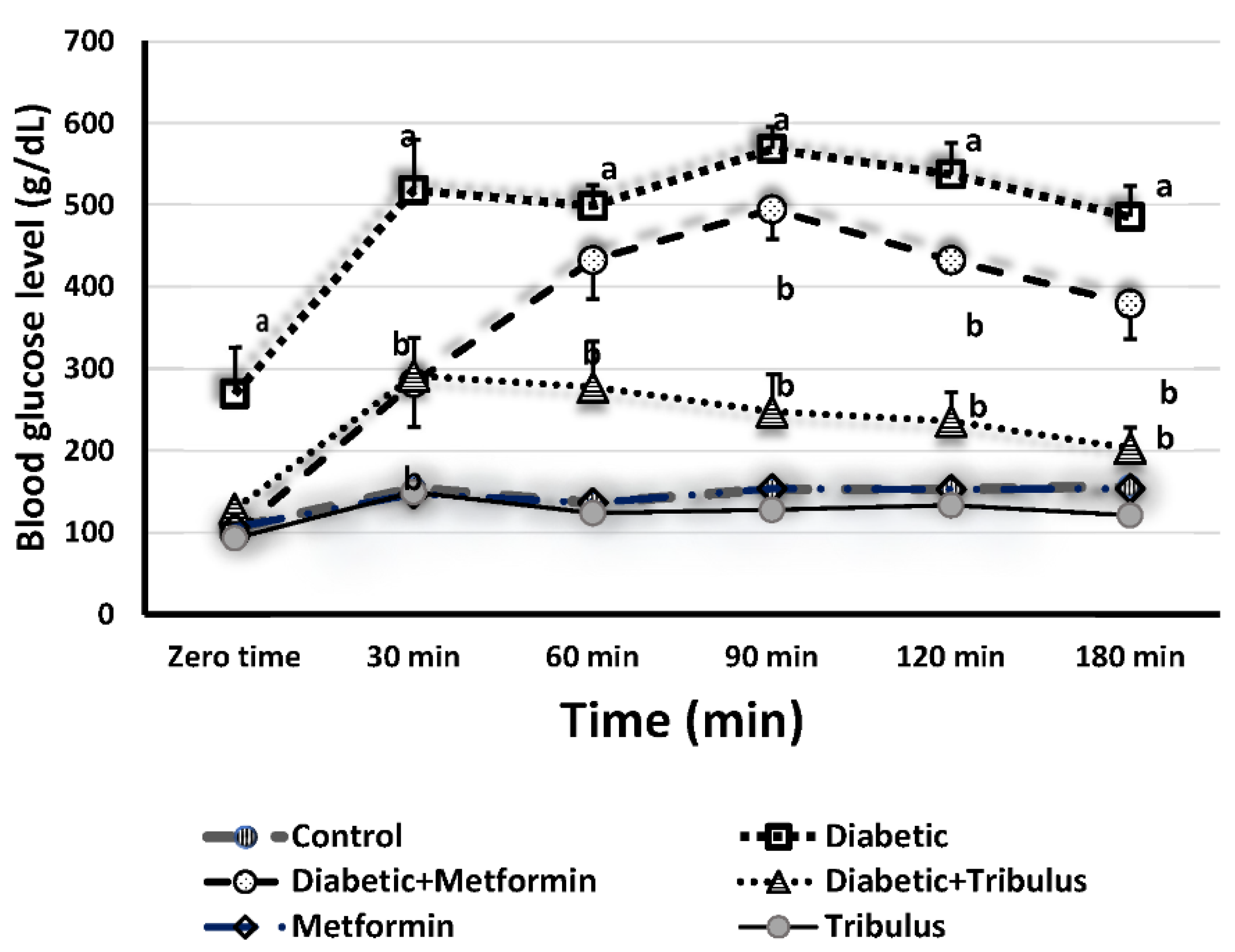
2.8. Glucose Tolerance Test

A glucose tolerance test (GTT) was used to assess the effects of TT and MET. Briefly, rats were given glucose (2 g/kg body weight) intraperitoneal after overnight fasting, according to Islam et al. [30]. Blood samples were removed from the tail vein at 0, 30, 60, 90, 120 and 180 min, and reagent strips (Accu-Chek®, Roche, Rotkreuz, Switzerland) were used to test blood glucose levels.

2.9. Preparation of Liver Homogenate for Measuring the Oxidative/Antioxidant Parameters

Before dissection, the tissue was injected with 50 mM/L (sodium phosphate-buffered saline; 100 mM/L Na2HPO4/NaH2PO4) at pH 7.4 and 0.1 M ethylenediaminetetraacetic acid (EDTA) to remove all red blood cells and clots. After homogenizing the liver tissue (0.25 g) in a 5 mL cold buffer/g tissue, it was centrifuged for 1/2 h at 2700× g. The resultant supernatant was transferred to Eppendorf tubes and kept in the deep freezer at −18 °C until being employed in various oxidative/antioxidant tests [31].

2.10. Determination of Oxidative/Antioxidant Parameters

The degree of lipid peroxidation (LPO) was measured as malondialdehyde (MDA) using the Ohkawa et al. method [32]. MDA concentrations were determined using 1,1,3,3-tetraethoxypropane as a standard and represented as μM/g tissue.
The activity of catalase (CAT) was calculated according to Aebi [33]. A total of 1:9 samples of 1 percent (v/v) Triton X-100 and CAT movements expressed as μM/min/g tissue were diluted. Glutathione-S-transferase (GST) was calculated [34] using a spectrophotometric assay. By calculating the conjugation of 1-chloro-2,4-dinitrobenzene (CDNB) with reduced glutathione (GSH), we obtained the GST activity at 340 nm. The activity of GST was expressed as U/g tissue (the quantity of enzyme conjugating 1 nM of CDNB with GSH/min is 1 unit). For the measurements (μM/g tissue), an extinction coefficient of 9.6 mM/Cm/CDNB was used.
Beutler [35] conducted a decreased GSH content of the supernatant using GSH reduced kits (Bio-diagnostic for diagnostic reagents, Dokki, Giza, Egypt). In the liver tissue, the volume of GSH present was measured as mM/g tissue.

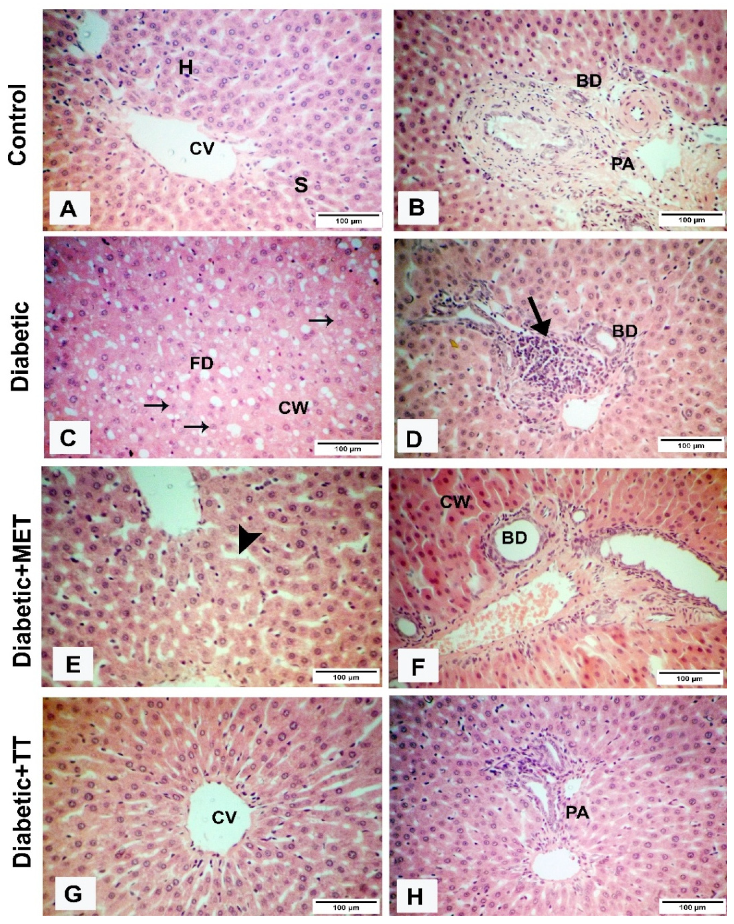
2.11. Histological Evaluation

Using a 10 percent neutral-buffered formalin solution for 24 h, a histological inspection of the tissues was conducted and the fixative was extracted by washing overnight with flowing tap water. Methyl benzoate was used to clean the tissues and they were embedded in paraffin after dehydration by a graduated sequence of alcohols. Sections were cut at 6 μm thickness by a microtome and stained with hematoxylin and eosin (H&E) [36].
According to Thoolen et al. [37], histopathological scoring research was conducted. The evaluation was represented as the sum of the individual grades of 1 (minimum), 2 (mild), 3 (moderate), and 4 (marked) for each of the following liver parameters: necrosis of hepatocytes, cholestasis, hyperplasia, and shift in hepatocyte fat.

2.12. Statistical Analysis

The data are expressed as ±SE (n = 6) mean values. To determine substantial variations between patient groups, regression research was conducted using a one-way analysis of variance (ANOVA). The post hoc Tukey’s test was used for comparative purposes for each relevant impact of medication. The threshold was set at p < 0.05 for statistical significance. The statistical studies were carried out using the SPSS statistics version 20 software package (SPSS Inc., Chicago, IL, USA). The correlations between various parameters were tested by the correlation test by Pearson [38].

3. Results

The TT- and MET-treated normal group’s glucose tolerance curve was decreased as compared to the control group, suggesting that TT and MET decreased the influence of the GTT in normal mice (Figure 1). The diabetic group’s blood glucose levels responded to a glucose loading in a normal diabetic manner. The rats treated with standard drug show a decrease in glucose levels at 30 min, which increased again to reach the maximum level (494.5 g/dL) at 90 min, and then started to decrease again at 120 and 180 min. The TT extract showed hypoglycemic activity at 30 min (291.3 g/dL) and produced a maximum fall after 180 min (approximately 200 g/dL) after glucose administration (Figure 1).
The activities of CAT and GST were significantly reduced in diabetic rats by 70.5% and 37.0% compared to the control group, respectively (Table 1). In diabetic animals, the LPO level was significantly increased by 2.1-fold while the level of GSH decreased by 40.5% in comparison to the normal rats. The CAT activities of diabetic rats were increased by 1.9- and 2.5-fold when treated with MET and TT extract, respectively, as compared to the diabetic group. The activities of GST in diabetic animals were increased by 1.3-fold as the effect of both MET or TT extract against diabetic rats (Table 1).
In MET- and TT-treated diabetic rats, the LPO levels decreased by 53.2% and 66.8%, respectively, compared to diabetic rats. The MET and TT extract increased the GSH concentrations by 1.1- and 1.3-folds as compared to control animals, respectively (Table 1). However, the administration of MET or TT to the diabetic group caused a significant elevation in the levels of GSH by 1.5- and 1.7-fold compared to diabetic rats, respectively. The administration of TT extract significantly improved the above parameters more than MET.
Correlations were computed between CAT, GSH and GST activities with MDA content of liver tissue in normal and diabetic rats treated with the TT extract and MET. There were significant negative correlations among the activities of hepatic CAT, GSH and GST with MDA content (r = −0.533, p < 0.02; r = −0.0.786, p < 0.01 and r = −0.625, p < 0.01, respectively). In addition, significant positive correlations were found between activity of CAT with activities of GST and GSH (r = 0.629, p < 0.05 and r = 0.774, p < 0.05, respectively).
Histological examinations of liver tissue are seen in Figure 2. Light liver tissue micrographs revealed normal hepatic cell and central vein architecture and normal blood sinusoids in the MET- and TT-treated animals (Figure 2A,B). STZ-diabetic rats, however, showed significant pathological changes, including degeneration of hepatocytes and fatty degeneration (Figure 2C). Moreover, in diabetic rats, focal aggregations of lymphocytes were also found. Degenerative and necrotic modifications were seen in the hepatic cells of diabetic animals (Figure 2D). However, the livers of diabetic rats treated with TT (Figure 2G,H) and MET (Figure 2E,F) extracts greatly decreased and attenuated the histological modifications attributable to mild changes.
In the injected diabetic rats, histological scores dependent on hepatic necrosis, lipid degeneration, and mononuclear cell inflammation were substantially improved relative to the injected control animals (Figure 3). In contrast to diabetic animals, therapy with TT decreased liver damage and necrosis substantially more than MET.

4. Discussion

Oxidative stress is increased in diabetic animals due to glucose oxidation, protein glycation, lipid peroxidation, and decreased antioxidant enzyme activity [39]. In line with this observation, the current research found that increased levels of LPO, decreased levels of GSH, and decreased antioxidant enzyme activity, such as CAT and GST, were shown in the liver of diabetic animals induced by STZ. These findings corroborate those of a recent study that found a decrease in GSH levels as diabetes progressed [40]. Protein glycation and suppression of the actions of antioxidant enzymes (superoxide dismutase and glutathione peroxidase) [41] may be the cause for the process of increased oxidative stress. TT extract alleviated oxidative stress in the current research by inhibiting the activities of CAT and GST in diabetic rats. The presence of flavonoid components [13], such as quercetin, which protects rats from oxidative stress assaults, may explain the antioxidant action of the TT extract.
In diabetic rats caused by STZ, LPO increased as a result of elevation the activity of fatty acyl-coenzyme A oxidase, which initiates ß-fatty acid oxidation [42]. In the current research, STZ-induced diabetics treated with TT extract reported a reduced level of liver LPO relative to non-treated diabetic rats. In normal rats, the extract also greatly decreased liver MDA levels. Similar reductions in liver LPO levels were also observed following TT therapy in diabetic and normal rats [15].
Deficiency in CAT activity in diabetic rats was found in the current research, as previously reported in diabetic patients [43]. Pancreatic β-cells are protected by CAT from destruction by hydrogen peroxide. Low catalase activity increases β-cell mitochondrial ROS, causing methemoglobinemia and hemolytic anemia, which can be due to glucose-6-phosphate dehydrogenase deficiency.
In the present investigation, which was previously reported by Lapshina et al. [44], a decline in GST activity was observed in diabetic rats. The re-balancing of the GST activity with TT extract or MET is similar, but the CAT activity with TT extract increased more than with MET therapy. It can be verified that TT extract defends against free radicals.
GSH is formed mainly in the liver, as one of the vital non-enzymatic antioxidants in the redox protection system [45]. It is involved in the synthesis and defense against reactive O2 compounds of essential macromolecules. The reduced concentration of GSH leads to the pathogenesis of chronic diabetic state-related complications. A sudden drop in the GSH content of the liver caused by STZ-injection may be one of the factors in the oxidative DNA damage in type 2 diabetics [46]. The depleted GSH in diabetic rats was restored by MET and TT extract in the current work. A similar result has been reported [15], suggesting that TT extracts increased the quality of GSH. The research showed that liver GSH levels were significantly enhanced in STZ-induced diabetic rats treated with the extract and that the rise was equivalent in normal rats treated with the TT extract. These findings are in line with another study stating that the herbal A. Maurorum ethanolic extracts decrease oxidative stress by elevating the GSH level and reducing the level of LPO [47]. The TT-extract decreases the complications of diabetes in different ways as mentioned before [48].
There was a relationship between histological alterations in the liver and diabetes-like oxidative stress. Dilatation of blood vessels, congestion of lobules, specific hemorrhagic coagulative foci in the hepatic parenchyma, and mixed inflammatory cell penetration of necrotic hepatocytes are all seen in STZ-induced diabetic rats. In this study, the use of TT ethanolic extract in STZ-induced diabetic rats resulted in significant restoration of the normal hepatic architecture.

5. Conclusions

Oxidative stress is a key factor in the development of diabetes and its consequences. According to this study, oxidative stress inhibition can mediate the protective effect of TT in STZ-induced diabetic rats more than MET. A histological examination of TT-treated rats revealed significant liver regrowth. As a result, TT extract might be utilized to treat diabetes and alleviate its symptoms in the liver.

Author Contributions

All the authors have responsibility for the entire content of this manuscript and approve its submission. Conceptualization, H.M.T.; methodology, H.M.A.A.; software, M.S.E.; validation, N.S.E.-S., R.A.A.-E. and M.S.E.; formal analysis, H.M.A.A.; investigation, H.M.T.; resources, R.A.A.-E.; data curation, M.S.E.; writing—original draft preparation, N.S.E.-S.; writing—review and editing, H.M.T.; visualization, H.M.A.A.; supervision, H.M.T.; project administration, R.A.A.-E.; funding acquisition, R.A.A.-E. All authors have read and agreed to the published version of the manuscript.

Data Availability Statement

The used to support the findings of this study are available from the corresponding author upon request.

Acknowledgments

Taif University Researcher Supporting Project Number (TURSP-2020/112), Taif University, Taif, Saudi Arabia.

Conflicts of Interest

The authors declare no conflict of interest.

References

  1. International Diabetes Federation (IDF). IDF Diabetes; International Diabetes Federation: Brussels, Belgium, 2013. [Google Scholar]
  2. Hunt, J.V.; Smith, C.C.; Wolff, S.P. Autoxidative glycosylation and possible involvement of peroxides and free radicals in LDL modification by glucose. Diabetes 1990, 39, 1420–1424. [Google Scholar] [CrossRef] [PubMed]
  3. Mansour, H.A.; Newairy, A.S.; Yousef, M.I.; Sheweita, S.A. Biochemical study on the effects of some Egyptian herbs in alloxan-induced diabetic rats. Toxicology 2002, 170, 221–228. [Google Scholar] [CrossRef]
  4. Maritim, A.C.; Sanders, A.; Watkins, J.B., III. Diabetes, oxidative stress, and antioxidants: A review. J. Biochem. Mol. Toxicol. 2003, 17, 24–38. [Google Scholar] [CrossRef]
  5. El-Shenawy, N.S.; Refat, M.S.; Fakihi, F.H. Decreasing the diabetic complication by vanadyl(VO)2+/vitamin B 6 complex in alloxan-induced diabetic mice. J. Mater. Sci. Mater. Med. 2013, 24, 911–930. [Google Scholar] [CrossRef]
  6. El-Said, S.M.; Al-Barak, A.S. Extraction of insulin like compounds from bitter melon plants. Am. J. Drug Discov. Dev. 2011, 1, 1–7. [Google Scholar] [CrossRef]
  7. Loizzo, M.R.; Rashed, K.; Said, A.; Bonesi, M.; Menichini, F.; Tundis, R. Antiproliferative and antioxidant properties of Alhagi maurorum Boiss (Leguminosae) aerial parts. Ind. Crops Prod. 2014, 53, 289–295. [Google Scholar] [CrossRef]
  8. Sheweita, S.A.; Mashaly, S.; Newairy, A.A.; Abdou, H.M.; Eweda, S.M. Changes in oxidative stress and antioxidant enzyme activities in streptozotocin-induced diabetes mellitus in rats: Role of Alhagi maurorum extracts. Oxidative Med. Cell. Longev. 2016, 2016, 5264064. [Google Scholar] [CrossRef] [PubMed] [Green Version]
  9. Chhatre, S.; Nesari, T.; Somani, G.; Kanchan, D.; Sathaye, S. Phytopharmacological overview of Tribulus terrestris. Pharmacogn. Rev. 2014, 8, 45–51. [Google Scholar] [CrossRef] [Green Version]
  10. Mohammed, M.M.; Shaddad, S.A.I.; Mudathir, A.E.; Elsharif, B.A.; Abu Algasem, A.A.E. Effects of Tribulus terrestris ethanolic extract in male rats and cocks fertility. J. Pharm. Biomed. Sci. 2013, 30 (Suppl. 1), S13–S18. [Google Scholar]
  11. Heidari, M.R.; Mehrabani, M.; Pardakhty, A.; Khazaeli, P.; Zahedi, M.J.; Yakhchali, M.; Vahedian, M. The analgesic effect of Tribulus terrestris extract and comparison of gastric ulcerogenicity of the extract with indomethacin in animal experiments. Ann. N. Y. Acad. Sci. 2007, 1095, 418–427. [Google Scholar] [CrossRef]
  12. Adaay, M.H.; Mosa, A.R. Evaluation of the effect of aqueous extract of Tribulus terrestris on some reproductive parameters in female mice. J. Mater. Environ. Sci. 2012, 3, 1153–1162. [Google Scholar]
  13. Tag, H.M.; Abdelazek, H.M.; Mahoud, Y.S.; EL-Shenawy, N.S. Efficacy of Tribulus terrestris extract and metformin on fertility indices and oxidative stress of testicular tissue in streptozotocin-induced diabetic male rats. Afr. J. Pharm. Pharmacol. 2015, 9, 1088–1098. [Google Scholar]
  14. Li, M.; Qu, W.; Wang, Y.; Wan, H.; Tian, C. Hypoglycemic effect of saponin from Tribulus terrestris. Zhong Yao cai = Zhongyaocai = J. Chin. Med. Mater. 2002, 25, 420–422. [Google Scholar]
  15. Amin, A.M.; Lotfy, M.; Shafiullah, M.; Adeghate, E. The protective effect of Tribulus terrestris in diabetes. Ann. N. Y. Acad. Sci. 2006, 1084, 391–401. [Google Scholar] [CrossRef]
  16. Kamboj, P.; Aggarwal, M.; Puri, S.; Singla, S.K. Effect of aqueous extract of Tribulus terrestris on oxalate-induced oxidative stress in rats. Indian J. Nephrol. 2011, 21, 154–159. [Google Scholar]
  17. Rahmathulla, S.M.; Maruthi, E.; Bheemewsaraiah, K.; Manjunatha, S.; Devi, K.L. Effect of Tribulus terrestris (L.) on liver in ISO-induced myocardial infarction. Int. J. Res. Biochem. Biophys. 2012, 2, 10–12. [Google Scholar]
  18. Yakubu, M.T.; Afolayan, A.J. Effect of aqueous extract of Bulbine natalensis (Baker) stem on the sexual behavior of male rats. Int. J. Androl. 2009, 32, 629–636. [Google Scholar] [CrossRef] [PubMed]
  19. Linden, M.A.; Lopez, K.T.; Fletcher, J.A.; Morris, E.M.; Meers, G.M.; Siddique, S.; Laughlin, M.H.; Sowers, J.R.; Thyfault, J.P.; Ibdah, J.A.; et al. Combining metformin therapy with caloric restriction for the management of type 2 diabetes and nonalcoholic fatty liver disease in obese rats. Appl. Physiol. Nutr. Metab. 2015, 40, 1038–1047. [Google Scholar] [CrossRef] [PubMed]
  20. Brownlee, M. A radical explanation for glucose-induced β cell dysfunction. J. Clin. Investig. 2003, 112, 1788–1790. [Google Scholar] [CrossRef]
  21. Shao, J.; Qiao, L.; Janssen, R.C.; Pagliassotti, M.; Friedman, J.E. Chronic hyperglycemia enhances PEPCK gene expression and hepatocellular glucose production via elevated liver activating protein/liver inhibitory protein ratio. Diabetes 2005, 54, 976–984. [Google Scholar] [CrossRef] [Green Version]
  22. Raddatz, D.; Ramadori, G. Carbohydrate metabolism and the liver: Actual aspects from physiology and disease. Z. Gastroenterol. 2007, 45, 51–62. [Google Scholar] [CrossRef]
  23. Eagappan, K.; Sasikumar, S.; Brindha, D. Antioxidant capacity insolvent and cooked extracts of Tribulus Terrestris L Fruit. Int. J. Biol. Pharm. Res. 2015, 6, 63–67. [Google Scholar]
  24. Costa, A.F. Farmacognosia, 2nd ed.; Fundacao Calouste Gulbenkian: Lisbon, Portugal, 1977; Volume 1–2. [Google Scholar]
  25. Freireich, E.J.; Gehan, E.A.; Rall, D.; Schmidt, L.H.; Skipper, H.E. Quantitative comparison of toxicity of anticancer agents in mouse, rat, hamster, dog, monkey, and man. Cancer Chemother. Rep. 1966, 50, 219–224. [Google Scholar]
  26. European Communities (EC). Council Directive 86/609/EEC of 24 November 1986 on the approximation of laws, regulations, and administrative provisions of the Member States regarding the protection of animals used for experimental and other scientific purposes. Off. J. Eur. Communities 1986, L 358, 1–28. [Google Scholar]
  27. Akbarzadeh, A.; Norouzian, D.; Mehrabi, M.R.; Jamshidi, S.H.; Farhangi, A.; Verdi, A.A.; Mofidian, S.M.; Rad, B.L. Induction of diabetes by streptozotocin in rats. Indian J. Clin. Biochem. 2007, 22, 60–64. [Google Scholar] [CrossRef] [PubMed] [Green Version]
  28. Owolabi, O.J.; Omogbai, E.K. Effect of metformin on potassium-adapted and Nonadapted Diabetic Rats. Trop. J. Pharm. Res. 2012, 11, 747–752. [Google Scholar] [CrossRef] [Green Version]
  29. Gauthaman, K.; Ganesan, A.P. The hormonal effects of Tribulus terrestris and its role in the management of male erectile dysfunction–an evaluation using primates, rabbit, and rat. Phytomedicine 2008, 15, 44–54. [Google Scholar] [CrossRef]
  30. Islam, M.A.; Akhtar, M.A.; Khan, M.R.; Hossain, M.S.; Alam, A.H.; Ibne-Wahed, M.I.; Amran, M.S.; Rahman, B.M.; Ahmed, M. Oral glucose tolerance test (OGTT) in normal control and glucose induced hyperglycemic rats with Coccinia cordifolia L. and Catharanthus roseus L. Pak. J. Pharm. Sci. 2009, 22, 402–404. [Google Scholar] [PubMed]
  31. Hamza, R.Z.; Ismail, H.A.; Khaled, H.E.; El-Shenawy, N.S. Antioxidant effect of L-cysteine on sodium-valproate-induced oxidative stress in rat liver: Biochemical and ultrastructural approaches. Toxicol. Environ. Chem. 2015, 97, 1041–1056. [Google Scholar] [CrossRef]
  32. Ohkawa, H.; Ohishi, N.; Yagi, K. Assay for lipid peroxides in animal tissues by thiobarbituric acid reaction. Anal. Biochem. 1979, 95, 351–358. [Google Scholar] [CrossRef]
  33. Aebi, M.; Furter, R.; Prand, F.; Niederberger, P.; Hütter, R. Structure and function of the TRP3 gene of Saccharomyces cerevisiae: Analysis of transcription, promoter sequence, and sequence coding for a glutamine amidotransferase. Curr. Genet. 1984, 8, 165–172. [Google Scholar] [CrossRef] [PubMed]
  34. Ålin, P.; Danielson, U.H.; Mannervik, B. 4-Hydroxyalk-2-enals are substrates for glutathione transferase. FEBS Lett. 1985, 179, 267–270. [Google Scholar] [CrossRef] [Green Version]
  35. Beutler, E.; Duron, O.; Kelly, B.M. Improved method for the determination of blood glutathione. J. Lab. Clin. Med. 1963, 61, 882–888. [Google Scholar] [PubMed]
  36. Bancroft, J.D.; Gamble, M. (Eds.) Theory and Practice of Histological Techniques, 6th ed.; Elsevier Health Sciences: Amsterdam, The Netherlands, 2008. [Google Scholar]
  37. Thoolen, B.; Maronpot, R.R.; Harada, T.; Nyska, A.; Rousseaux, C.; Nolte, T.; Malarkey, D.E.; Kaufmann, W.; Küttler, K.; Deschl, U.; et al. Proliferative and nonproliferative lesions of the rat and mouse hepatobiliary system. Toxicol. Pathol. 2010, 38 (Suppl. 7), S5–S81. [Google Scholar] [CrossRef] [PubMed]
  38. Filed, A.P. Discovering Statistics Using SPSS for Windows: Advanced Techniques for the Beginner; Sage publications: London, UK, 2000. [Google Scholar]
  39. Matough, F.A.; Budin, S.B.; Hamid, Z.A.; Alwahaibi, N.; Mohamed, J. The role of oxidative stress and antioxidants in diabetic complications. Sultan Qaboos Univ. Med. J. 2012, 12, 5–18. [Google Scholar] [CrossRef] [PubMed]
  40. Rahman, K. Studies on free radicals, antioxidants, and co-factors. Clin. Interv. Aging 2007, 2, 219–236. [Google Scholar]
  41. Wolf, S.P. Diabetes mellitus and free radicals. Br. Med. Bull. 1993, 49, 642–652. [Google Scholar] [CrossRef]
  42. Afshari, A.T.; Shirpoor, A.; Farshid, A.; Saadatian, R.; Rasmi, Y.; Saboory, E.; Ilkhanizadeh, B.; Allameh, A. The effect of ginger on diabetic nephropathy, plasma antioxidant capacity, and lipid peroxidation in rats. Food Chem. 2007, 101, 148–153. [Google Scholar] [CrossRef]
  43. Takemoto, K.; Tanaka, M.; Iwata, H.; Nishihara, R.; Ishihara, K.; Wang, D.H.; Ogino, K.; Taniuchi, K.; Masuoka, N. Low catalase activity in blood is associated with the diabetes caused by alloxan. Clin. Chim. Acta 2009, 407, 43–46. [Google Scholar] [CrossRef]
  44. Lapshina, E.A.; Sudnikovich, E.J.; Maksimchik, J.Z.; Zabrodskaya, S.V.; Zavodnik, L.B.; Kubyshin, V.L.; Nocun, M.; Kazmierczak, P.; Dobaczewski, M.; Watala, C.; et al. Antioxidative enzyme and glutathione S-transferase activities in diabetic rats exposed to long-term ASA treatment. Life Sci. 2006, 79, 1804–1811. [Google Scholar] [CrossRef]
  45. Li, S.; Tan, H.Y.; Wang, N.; Zhang, Z.J.; Lao, L.; Wong, C.W.; Feng, Y. The role of oxidative stress and antioxidants in liver diseases. Int. J. Mol. Sci. 2015, 16, 26087–260124. [Google Scholar] [CrossRef] [PubMed] [Green Version]
  46. Tiwari, B.K.; Pandey, K.B.; Abidi, A.B.; Rizvi, S.I. Markers of oxidative stress during diabetes mellitus. J. Biomark. 2013, 2013, 378790. [Google Scholar] [CrossRef] [PubMed] [Green Version]
  47. Shaker, E.; Mahmoud, H.; Mnaa, S. Anti-inflammatory and anti-ulcer activity of the extract from Alhagi maurorum (camelthorn). Food Chem. Toxicol. 2010, 48, 2785–2790. [Google Scholar] [CrossRef] [PubMed]
  48. Ștefănescu, R.; Farczadi, L.; Huțanu, A.; Ősz, B.E.; Mărușteri, M.; Negroiu, A.; Vari, C.E. Tribulus terrestris efficacy and safety concerns in diabetes and erectile dysfunction, assessed in an experimental model. Plants 2021, 10, 744. [Google Scholar] [CrossRef] [PubMed]
Figure 1. Glucose tolerance test (GTT) in normal and streptozotocin-induced diabetic rats after 8 weeks of treatment of Tribulus terrestris extract and metformin. A glucose solution (2 g/kg body weight) was given after a 16 h fast. The blood samples were taken at 30, 60, 90, 120 and 180 min after giving glucose. Data are shown as the mean ± SE (n = 6/group). a p < 0.05 when compared with control, b p < 0.05 when compared with the diabetic group.
Diabetology 03 00004 g001
Figure 2. Influence of metformin (MET) and Tribulus terrestris (TT) extract on liver histopathology in normal and diabetic rats. (A,B): Section from the liver showing normal hepatic tissue displayed in MET and TT extract treated rats. (C): Section from the liver of the diabetic group showed cloudy swelling of hepatocytes (CW) and fatty degeneration (FD); additionally, some degenerated hepatocytes displayed karyolized nuclei (arrows). (D): Section from the liver of the diabetic group showing focal necrosis associated with lymphatic infiltration in the portal area (arrow). (E,F): Section from the liver of diabetic rats treated with 350 mg/kg of MET showing dilatation of sinusoids (arrowhead). (G,H): Section from the liver of diabetic rats treated with 20 mg/kg of TT extract showing the normal histological structure of the hepatic tissue. Scale bars: 100 µm. H: hepatocytes, S: sinusoids, CV: central vein, BD: bile duct, PA: portal area.
Diabetology 03 00004 g002
Figure 3. Histological score of the liver in normal and streptozotocin-induced diabetic rats after 8 weeks of treatment with Tribulus terrestris (TT) extract or metformin (MET). a p < 0.05 when compared with the normal group, b p < 0.05 when compared with the diabetic group.
Diabetology 03 00004 g003
Table 1. Influence of MET and TT fruit extract on oxidative/antioxidant of normal and diabetic rats.
GroupsMDA (µM/g)CAT Activity (µM/min/g)GST (µM/g)GSH (mM/g)
Control87.40 ± 3.1243.39 ± 5.8134.98 ± 1.9749.21 ± 4.89
MET42.80 ± 10.23 a42.89 ± 5.5533.88 ± 3.2251.72 ± 3.28
TT extract31.00 ± 7.18 a51.33 ± 2.5332.61 ± 3.0061.79 ± 3.39
Diabetic184.20 ± 12.22 a12.80 ± 0.54 a22.03 ± 0.82 a29.26 ± 1.90 a
Diabetic and MET86.20 ± 2.65 b24.37 ± 1.23 b29.05 ± 0.57 b44.01 ± 1.23 b
Diabetic and TT61.20 ± 4.74 b,c32.53 ± 2.62 b,c29.85 ± 0.88 b50.50 ± 1.58 b,c
Values presented as mean ± SE (n = 6/group). a p < 0.05 compared to control animals, b p < 0.05 compared to the diabetic rats and c p < 0.05 compared to diabetic and MET group. MET; Metformin, TT; Tribulus terrestris, MDA; malondialdehyde, CAT; catalase, GST; glutathione-S-transferase, and GSH; glutathione.
Publisher’s Note: MDPI stays neutral with regard to jurisdictional claims in published maps and institutional affiliations.

