Perspective

Deciphering the Neurosensory Olfactory Pathway and Associated Neo-Immunometabolic Vulnerabilities Implicated in COVID-Associated Mucormycosis (CAM) and COVID-19 in a Diabetes Backdrop—A Novel Perspective

by
Maryada Sharma
1,*,
Hari Pankaj Vanam
2,
Naresh K. Panda
1,
Sourabha K. Patro
1,
Rhythm Arora
1,
Sanjay K. Bhadada
3,
Shivaprakash M. Rudramurthy
4,
Mini P. Singh
5 and
Purushotham Reddy Koppula
6,*
1
Department of Otolaryngology and Head & Neck Surgery, Postgraduate Institute of Medical Education and Research, Chandigarh 160012, India
2
(Former) Department of Microbiology, Bhaskar Medical College and General Hospital, Hyderabad 500075, India
3
Department of Endocrinology, Postgraduate Institute of Medical Education and Research, Chandigarh 160012, India
4
Department of Microbiology, Postgraduate Institute of Medical Education and Research, Chandigarh 160012, India
5
Department of Virology, Postgraduate Institute of Medical Education and Research, Chandigarh 160012, India
6
Regeneron Pharmaceuticals Inc., Tarrytown, NY 10591, USA
*
Authors to whom correspondence should be addressed.
Diabetology 2022, 3(1), 193-235; https://doi.org/10.3390/diabetology3010013
Submission received: 25 January 2022 / Revised: 23 February 2022 / Accepted: 24 February 2022 / Published: 4 March 2022
(This article belongs to the Special Issue Diabetology: Feature Papers 2021)

Abstract

:
Recent Mucorales-mediated outbreaks of infections and an association of fungal infection with COVID-19 cases, as observed for COVID-19-associated mucormycosis (CAM), have posed new challenges for the management of patients in critical care units. Diabetes and hyperglycemia are integrally linked to the severity of COVID-19, and uncontrolled diabetes mellitus and COVID-19 have recently been (independently or in combination) associated with the emergence of aggressive mucormycosis due to attendant defects in innate immune recognition pathways. Therefore, the identification of novel global cellular stressors upregulated during diabetes to understand the contribution of diabetes-associated metabolic vulnerabilities can help build a Metabolic-Stress-Associated Interactome (MSAI). This interactome can help reshape the metabolic inflammation (meta-inflammation) underlying the clinical manifestations of COVID-19 to facilitate the rational design of effective therapies for COVID-19 and CAM. Accordingly, an important area of research in COVID-19 therapeutics is engaged with identifying diabetes-associated pan-cellular stressors to understand their role in immune deregulation during COVID-19 and CAM, including investigating the distant trans-neuro-vascular–endocrine axis’s role in coordinating cellular-stress recognition, transmission, compensation, and decompensation during inter-organ regulation of metabolic homeostasis in diabetes. We reviewed clinico-pathological and laboratory data to propose potential diabetes-linked novel neo-vulnerabilities that can reshape the olfactory mucosal immune landscape during airway infections such as COVID-19 and CAM.

1. Introduction

1.1. Current Taxonomy of Mucorales and Congeners

The Mucoromycota phylum comprises the Mucormycotina, Glomeromycotina, and Mortierellomycotina sub-phyla that fall into the Mucorales, Umbelopsidales, and Endogonales orders, respectively. Mucorales comprise a group of versatile and ecologically highly diverse environmental molds. They are considered to be the cosmopolitan saprobes with a variety of unique features—they are omnipresent in human habitation because they thrive in the same environmental conditions as mankind. Generally, they are closely associated with the organic substrates, and the spores may be released into the soil or remained suspended in air. The medically important order Mucorales comprises 55 genera and 261 species. With the advent of molecular sequencing tools such as the barcoding of Mucorales using internal transcribed spacer (ITS) region and phylogenetic analyses, new species are continuously being identified and added to the list [1,2,3,4].
Recently, the identification of Cunninghamella (C.) arunalokei sp. nov. (as seen in an infection in an immunocompetent host) potentially expanded the list of medically important Mucorales to >38 etiological agents causing a life-threatening condition coiled as mucormycosis [2]. Hoffman et al. assigned various genera of Mucorales into family and species by clinical relevance. The opportunistic Mucorales that cause mucormycosis are usually thermotolerant and may include opportunistic genera and species such as Actinomucor, Apophysomyces, Cokeromyces, Cunninghamella, Lichtheimia, Mucor, Rhizomucor, Rhizopus (R.), Saksenaea, Syncephalastrum, and Thamnostylum. Two species of the genera Rhizopus-R. arrhizus (syn. R. oryaze) and R. microsporus cause 70% of worldwide mucormycosis and predominant COVID-19-associated mucormycosis (CAM). The three most virulent Mucorales primarily associated with diabetes mellitus (DM) are C. bertholletiae (77%), Rhizopus spp. (57%), and Mucor spp. (41%) [1,3,4]. Furthermore, the prevalence of rare species such as R. homothallicus, Saksenaea vasiformis, Apophysomyces variabilis, Thamostylum lucknowense, Mucor irregularis, and (most recently) C. arunlalokei nov. sp. in the Indian subcontinent suggests the unique ecological niche and epidemiological significance of mucormycosis and its implications and burden on public health [5,6,7].
Uncontrolled DM is the major comorbid risk factor along with a high spore burden for acquiring mucormycosis. There has been an increase in the trend of DM prevalence worldwide, and the situation in India is alarmingly high, as nearly 8% of adults aged ≤20 years have DM [8]. Inarguably, infectious diseases alone attribute to over 12% of mortality in DM cases worldwide [3]. Secondly, 7–14% of COVID-19 patients have DM as their predominant comorbidity, and COVID-19-associated acute or stress-induced hyperglycemia cases comprise half of the hospitalized patients in the ongoing pandemic [3,9,10,11,12,13]. Ergo, COVID-19 itself leads to new-onset DM and precipitates into diabetic keto acidosis (DKA) due to the severe acute respiratory syndrome coronavirus 2 (SARS-CoV-2) virus and the inappropriate use of systemic corticosteroids (CS) [6,12,14]. Furthermore, CAM, a complication in individuals with severe or critical COVID-19 as a virus-induced endothelial dysfunction, has proven detrimental with worsening outcome [7,15,16]. The emergence of CAM in India is multifactorial and involves the immediate environment, as an abundance of mucoralean spores—indoors, outdoor, and in the soil—can be acquired by air inhalation, ingestion, or traumatic inoculation. Host factors include uncontrolled DM, inappropriate SC therapy, iron overload, iatrogenic factors, and COVID-19 virus involving endothelial dysfunction and immune dysregulation [7]. Similar to Coxsackie, Influenzae, and SARS-CoV-1 viruses, SARS-CoV-2 can also induce an acute diabetes state mediated by the high-level expression of angiotensin-converting enzyme 2 (ACE2) receptors in the pancreatic islet cells, which results in the widespread destruction of beta cells in the pancreas, the diminution of insulin production, an enhanced secretion of cortisol, and resultant stress exacerbating the progression of insulin resistance. Furthermore, in type 2 DM and DKA, along with a chronic inflammatory state, there is a reduction in the master regulatory cytokine interleukin-10 (IL-10) synthesis by lymphocytes and macrophages in place of increased glycosylation, which also hampers the extravasation and chemotactic abilities of phagocytic cells on the path to overall dysregulated and dysfunctional immune responses. All these events fail to arrest spore germination and resultant morbid culmination of the pathogenesis of CAM [17,18].

1.2. COVID-Associated Mucormycosis (CAM)

The estimated global cases of mucormycosis per year pre-COVID era were 10,000. Startlingly, during the second wave of the COVID-19 pandemic, mediated by the delta variant, the averages rose 2–3 times, with more than 47,000 CAM cases being reported alone from the Indian subcontinent in just three months, i.e., May to July 2021, and continuing to wreak havoc and straining healthcare systems to the breaking point [2,6,7,10,11]. Furthermore, the prevalence of cases per million inhabitants of mucormycosis was 14.0 per year, which was highest global average, and Asia stands in the highest prevalence zone, with 29.9 cases per million inhabitants per year [19]. Mucormycosis usually develops in 10–14 days after hospitalization according to Muthu et al., who systematically reviewed the cases first reported during COVID-19 from India and compared them with the rest of the world [6]. They categorized cases into early CAM (wherein mucormycosis is diagnosed simultaneously with COVID-19) and late CAM at proportions of 25% and 36%, respectively; in other parts of the world, late CAM develops from seven days to 3 months following COVID-19 infection, with a mean average of 19.5 days. Similarly, the predominant presentation of CAM was rhino-orbital-cerebral-mucormycosis (ROCM), reaching 89% in India and 64% globally [20]. A study post-June 2021 from India revealed that the case fatality rate reported from India versus the rest of the global averages was comparatively lower, i.e., 36.5% vs. 61.9%, respectively. This may have been due to a diagnostic dilemma in picking pulmonary mucormycosis (PM) and probably due to the predominance of ROCM in the Indian context, wherein healthcare is overburdened with radical surgeries, orbital exenteration, the extensive dissection of the sinuses, and critical shortages of antifungal drugs [6,11]. First, the phenomenon was unique to India, but soon several countries of various continents started reporting a similar surge in CAM cases including Pakistan, Iran, Egypt, Brazil, USA, Mexico, Chile, Honduras, Paraguay, Uruguay, and European countries. The list is increasing in the wake of yet another wave of COVID-19 due to the delta and newly emerged omicron variants [21,22,23]. The analysis of CAM in 18 countries by Hoenigl et al. revealed that 53% of the cases were from India. A multi-center study from the first wave of COVID-19 concluded that CAM predominately presented as ROCM followed by the PM type and was caused by three important comorbid factors, diabetes, inappropriate SC (more than 6 mg of dexamethasone use for more than recommended duration), and SARS-CoV-2 itself [24,25]. The estimated prevalence of CAM in India at epidemic proportions during the pandemic is unprecedented and nearly 70 times that of the global average [26]. This uncanny upsurge was termed by many mycologists and infectious disease experts as a “tsunami of black fungus in COVID stricken India.” This maiden mayhem of CAM in India raised alarm bells, warranted swift federal government interventions with rapid response groups, and led to the naming of mucormycosis as the national “notifiable disease” [27]. This initiative further developed a comprehensive framework by strengthening the national registry of mucormycosis cases, monitoring systems, early recommendations for diagnosis, treatment (in places with shortages of antifungals), and management strategies. Under the aegis of the Fungal Infection Study Forum (FISF), a systematic registry of cases was initiated under the name of “Fung-I-Reg” in India and stood as a pivotal forum in recommending the guidelines on limiting and managing CAM. Furthermore, in harmony with the International Society of Human and Animal Mycology (ISHAM), European Confederation of Medical Mycology (ECMM), and Mycoses Study Group (MSG), FISF proposed global guidelines for the diagnosis and management of mucormycosis in low and middle-income countries (LMIC) such as India [12,28].

1.3. Mucormycosis-Associated Diabetes (MAD)

Hyperglycemia may be induced by SC. During the COVID-19 pandemic, SC were indiscriminately used to circumvent oxygen desaturation, especially in the second wave [6,7]. Firstly, SCs act as a substrate of oxidative stress metabolism through lipolysis and proteolysis from hepatocytes, resulting in a hyperglycemic state, insulin resistance, and continuous ketogenesis [17]. Secondly, this results in inefficient cellular functions such as neutrophil migration, ingestion, and phagolysosome fusion, thereby suppressing the microbicidal activity of activated macrophages, the antagonism of macrophage maturation and differentiation, and the depression of inflammatory cytokines such as IL-1, IL-6, tumor necrosis factor-alpha (TNF-α), and other mediators of macrophages; virtually all phagocytic and respiratory burst mechanisms are dampened, which has broadened the scope for the establishment of mucormycosis [7,17]. Recent studies in the immunopathogenesis of mucormycosis-associated with diabetes mellitus (MAD) have documented a well-accepted hypothesis that emphasizes the pivotal role of macrophages and their subsets, i.e., M1 and M2 and their pattern recognizing receptors (PRRs) or Toll-like-receptors (TLRs), as the initiators of the efficient killing of fungal cells by phagocytosis. Among the two subsets of macrophages, those of M2 are anti-inflammatory and release IL-10, which is involved in the immune response to fungal cells. Conversely, in patients with MAD, hyperglycemia triggers stress response in the endoplasmic reticulum, the overexpression of the glucose-regulated protein-78 (GRP78 protein), increase in reactive oxygen species (ROS), and free fatty acids (FFA), and a plethora of pro-inflammatory cytokines (TNF-α and IL-1β) and chemokines from the liver, muscle, and hypertrophic cells of the adipose tissues, resulting in a persistent inflammatory state. This ensures the massive tissue recruitment of the activated M1 subset of macrophages and resultant infiltration, along with the dampening of the M2 macrophage regulatory response [17].
Furthermore, Rhizopus spp. thrives by relying on iron siderophores, high serum glucose levels, and an insulin resistance state as its energy source [3,7,17]. Incontrovertibly, DKA is a double-edged sword that Mucorales easily exploit by affecting glutathione restoration, and an increased glucose metabolism results in increased glycosylation end-products and ROS, which accumulate in organs and tissues, thus further increasing susceptibility to Rhizopus infections [3]. Alternately, these patients require hemodialysis with deferoxamine chelation therapy to correct the DKA, which in turn makes them vulnerable to mucormycosis due to augmented levels of serum-free iron readily available for Mucorales to flourish. Furthermore, COVID-19-associated hyperferritinemia may act as a lucrative source iron for thriving and invading by Mucorales. Lastly, the intensive use of antimicrobial therapy creates a favorable nidus, amplifying a conductive microenvironment for the progression of an invasive fungal infections such as mucormycosis [3,7,17].

1.4. Immuno-Pathobiology of CAM and the Interface between COVID-19 and Mucor

In the above-described subset of diabetic patients, a potentiated inflammatory state is caused by the continuous and persistent recruitment of local immune cells such as macrophages and neutrophils, which in turn recruit a variety of inflammatory cytokines [17]. This phenomenon, which paradoxically switches yet another robust inflammatory phenotype when SARS-CoV-2 is positively flagged in these subsets of patients, predisposes patients to a variety of secondary infections including mucormycosis, such as COVID-19-associated pulmonary aspergillosis (CAPA), disseminated fusariosis, and invasive candidiasis. The first was extremely devastating during the second wave of COVID-19 in the Indian subcontinent and resulted in a dual-disease term called CAM. Much of the immunopathogenesis of Mucorales in DM patients comes from the prototype species Rhizopus spp. [29].
Secondary infections in these subsets of patients caused by Mucorales are mediated via the hyper-secretion of pro-inflammatory cytokines by immune reactive cells that open up divergent pathways leading to a vicious cycle of CAM and the widespread insult of the host tissues. Moreover, a plethora of interventions in the management of COVID-19 have further potentiated serum ferritin and free iron levels, drastically restricting the efficiency of macrophage immunity. Evidently, through siderophores, Mucorales efficiently acquire free iron from patients with DKA, as higher serum levels of free iron result in acidosis and poor binding to proteins. Secondly, in a setting of uremia wherein desferrioxamine chelation may act as a source (similarly to a COVID-19-induced hyperglycemia (indiscriminate use of CS) and chronic inflammatory state), Mucorales facilitate the pivotal mechanism in the immunopathogenesis of CAM [6,7,29].
Two host cell receptors—the ACE2 receptors (which are expressed in abundance in host endothelium) and glucose-regulated protein 78 (GRP78) (in addition to its role in MAD) as a co-receptor by recognizing the spike protein (S) of SARS-CoV-2 and efficiently internalizing the virus into the host cells [30]—are also involved in endothelial cell barrier disruption, inflammation, coagulation, endotheliatis, and apoptosis. In severe cases of COVID-19, abnormally elevated levels of D-dimer, fibrinogen, and Willebrand factor (VWF) and profound increases in inflammatory cytokines such as IL-1, Il-6, IL-8, and TNF-alpha are indications of multi-organ dysfunction due to widespread venous thrombosis, systemic vasculitis, and vascular coagulopathy [29]. Rayner et al. found that the suppression of GRP78 expression resulted in the inhibition of SARS-CoV-2 replication [31]. The pre-existing hyper immune-reactiveness, endothelial damage, and multi-organ dysfunction in ongoing COVID-19 are important risks for the emergence of complications owing to angioinvasive Mucorales [7,29,30]
Recently, Franco et al. reviewed host–pathogen and molecular interactions in the pathogenesis of Rhizopus spp. in DM patients. Mucormycosis has six main clinical presentations, of which ROCM is the most predominant [3]. Though the occurrence of ROCM was preponderantly seen in immunocompromised patients in the pre-COVID-19 era, CAM has since created a similar niche by mimicking immunocompromised (IC) status in the present pandemic situation [32]. Augmenting this scenario, there have been multiple reports of mucormycosis in immunocompetent hosts, presenting as chronic ROCM, skull base invasion, and renal mucormycosis caused by these seemingly thought opportunistic invaders [33,34]. Regardless of presentation, the angioinvasiveness, dissemination to the contiguous tissues resulting in necrotic eschars and thrombosis along with mucoralean spores in IC hosts with notably high mortality rates [3]. Coincidentally, the angioinvasive process and thrombotic microangiopathy in the disease spectrum are the mimickers of both COVID-19 and mucormycosis pathogenesis. Furthermore, the striking mortality of mucormycosis in DM patients is as high as 77% due to infections by Cunninghamella bertholletiae, followed by 57% due to Rhizopus spp. and 41% due to Mucor spp., which indicates the need for the efficient management of these rapidly progressive conditions [35].
The spread pathways of Mucorales in ROCM were described by Hosseini and Bhorgei in 2005 [36]. In the inhalation mode, the pterygopalatine fossa serves as the main reservoir for mucormycosis, and the spores and hyphae lodge in the nasal passages and contiguous sinuses. GRPs are a group of immunoglobulin-binding proteins (BiPs) that include GRP78 (a molecular chaperone belonging to the heat shock protein 70 (HSP70) family located in the lumen of the endoplasmic reticulum) and its isoform GRP78va (located in the cytosol), which have pivotal roles in folding, assembly, and quality control of proteins and misfolded protein degradation. Furthermore, they are expressed in diverse types of inflammatory, cancerous, viral, and invasive fungal spores including the Mucorales [37,38]. Hyperglycemia-induced stress in Rhizopus spp. infection in DM patients results in the translocation of GRP78 onto the cell surface (cs) as csGRP78 and the overexpression of receptors for signaling, also leading to anti-csGRP78 autoantibodies. With the expression of fibronectin and type I collagen, fibrosis is a consequential event of the sequential interactions of csGRP78 with β1 integrin and the activation of the focal adhesion kinase (FAK) and downstream protein kinase Akt (van). Interestingly, a similar mechanism of receptor signaling and entry of the pathogen into the host cell was proposed for Dengue, Ebola, Coxsackie, and SARS-CoV2 viruses, along with Rhizopus spp. [39,40].
Receptor interaction studies of nasal and alveolar epithelia with homologous spore coating proteins (coat protein homolog (CotH) present on Mucorales such as Rhizopus delemar and Rhizopus spp. have revealed that csGRP78 and integrin-β1 are only overexpressed in the nasal epithelia of DM patients. This complex microenvironment, holding high glucose, iron, and DKA levels, promotes superficial tissue level invasion by favoring the interactions between CotH (specifically, CotH3) on Mucorales with that of the abundant csGRP78 and integrin-β1 on the nasal epithelium. Furthermore, this expands the virulence of Rhizopus spp. via coating with laminin and collagen IV potentiating the invasion, endothelial damage, and continuous expression of GRP78 in the contiguous tissues planes and endothelium, eventually establishing the clinical entity ROCM in DM [36,37,38,39].
Beguilingly, Mucorales have adapted to the novel acquisition of virulence factors by harboring endosymbiotic nosocomial Gram-negative bacteria including various species of Burkholderia and Ralstonia (R.) pickettii. The former releases potent toxic metabolites called rhizoxins by endosymbiosis, and the latter plays a role in the intracellular survival of macrophages and triggers a profuse pro-inflammatory cytokine release [41,42]. Intriguingly, R. pickettii endosymbiosis with Rhizopus microspores has complicated several nosocomial outbreaks of Mucorales due to their propensity to contaminate sterile water, saline medical solutions, and hospital disinfectants [40,42].

1.5. The Complex Interplay of Various Factors: Mucosal Proteases and Iron Redox Stress

Human environments, whether community settings, indoor or outdoor areas, or air-conditioned or non-air-conditioned hospital areas, allow mucoralean spores to become airborne and inhaled by immunocompetent hosts without any apparent clinical disease. However, in an event of a source with a high hyphal burden, the polymorphoneutrophils (PMNs) response is overwhelmed, thus surpassing the primary barriers of immunity. The usual modes of transmission are inhalation, ingestion, or direct inoculation. The size of sporangiospores may also contribute to their role in establishing human disease, as larger R. arrhizus spores tend to settle in the upper respiratory tract and the smaller Cunninghamella or R. microsporus spores reach the lower respiratory tract; the latter is highly virulent and leads to rapid disease [6,43,44]. Furthermore, molecular and epidemiological studies to delineating the mechanisms of acquisition of hypervirulence, Mucorales genus-specific differences in interactions with the host cells (GRP78 expression and CotH3 interactions), the role of SARS-CoV-2 in potentiating diseases leading to CAM, other co-infections, and nosocomial outbreaks in increased CAM cases in aggravating seemingly opportunistic Mucorales infection are currently underway [6,43,45].

2. Potential Immuno-Metabolic Vulnerabilities That Can Prefigure COVID-19 and CAM-Synergistic Action of Diabetes-Associated Proteolytic and Metabolic Stress

2.1. Expanding the MSAI-Proteolytic Stress as a New Player in COVID-19 Arena

The potential diabetes-associated cellular-stress pathways emerging from multi-faceted high throughput omics approach are facilitating expansions on MSAI and the development of the understanding of the pathogenesis of beta-cell dysfunction in COVID-19, which is incompletely understood even though integrated stress response (ISR) is integral to the development of diabetes. The impairment of insulin-producing pancreatic beta-cells is a key contributing factor to the diabetic pathogenesis by SARS-CoV-2. However, whether beta-cells are damaged by the coronavirus through a direct action is less clearly understood. The pancreatic cells are governed by intricate inter-organ network regulation involving soluble humoral factors from diverse organs that crosstalk with their neuro-vascular conduits. Therefore, linking the cause of pancreatic beta-cell damage in subjects with COVID-19 to the direct infection of SARS-CoV-2 or alternate indirect effects including distant organ damage, heralding inflammatory loops, and yet-unexplored cellular stressor(s) remains challenging. However, the potential of direct infection with SARS-CoV-2 and the subsequent cell fate reprograming of pancreatic cells has been recently demonstrated [46,47,48]. Notably, heterogenous host pancreatic (endocrine, exocrine, and acinar) cellular-stress responses caused by SARS-CoV-2 infections have displayed the transdifferentiation of insulin-producing beta cells into glucagon-producing alpha and trypsin-producing acinar cells in the pancreas, which has been linked to an ISR pathway. Remarkably, a trans-ISR inhibitor treatment was found to increase insulin expression and reduce glucagon, trypsin, and cell stress-associated genes in SARS-CoV-2-infected human islets [47], indicating a possible role of co-secreted pancreatic trypsin/proteolytic stress in reshaping diabetes-associated ISR.

2.2. Enhanced SARS-CoV-2 Transmissibility and Role of Olfactory Mucosal Proteases

Viral proteases are reported to have potential ancillary functions extending beyond the polyprotein/spike clipping that are linked to host immune evasion [49]. However, the biological functions (outside fusogenic potential) of cellular host proteases activated during COVID-19 remain largely unexplored. Importantly, the TMPRSS2 and trypsin serine proteases have been identified as important molecular players in the underlying pathology and emergence of coronaviruses, respectively. In particular, the trypsin treatment (proteolytic processing) of viral spike proteins has been identified as a potential species barrier for the emergence of zoonotic coronaviruses, hence posing a potential threat for future outbreaks driven by the cross-species transmission of coronaviruses [50,51,52,53,54]. It was proposed that additional small intestine proteases such as trypsin could enhance SARS-CoV-2′s viral infection and pathogenesis capability by triggering more robust cell fusion [55]. The increased transmissibility of SARS-CoV-2 compared to SARS-CoV is linked to the occurrence of a unique furin-cleavable polybasic motif (RRAR) at the S1/S2 boundary, as the cleavage results in a C-terminally exposed RRAR peptide that is capable of binding to neuropilin-1 (NRP1), which has been demonstrated to be an important host factor receptor for SARS-CoV-2 by facilitating entry and infectivity [50,56,57]. The novel core region with the SPRRAR (S680-V687) polybasic insert of SARS-CoV-2 harbors positively charged arginine at 683 and hydrophobic alanine at 684, which makes the site susceptible to promiscuous binding/cleavage by not only serine proteases such as furin or furin-like PCs but also mono- and dibasic amino acid targeting serine proteases such as matriptase, human airway trypsin, TMPRSS2, and kallikrein. Importantly, a plethora of serine proteases (SPs) such as furin, TMPRSS2, furin-like PCs, and trypsin-like proteases in the nasal microenvironment have facilitated the heightened transmissibility of emerging SARS-CoV-2 variants that have enhanced susceptibility to SP-cleavable polybasic amino acids; for instance, a mutation of non-polar proline at 681 to more positively charged arginine in delta variant (P681R) in spike protein resulted in enhanced transmissibility due to more cleavability at S1/S2 [58,59], and a mutation of proline to histidine (P681H) in omicron variant’s spike protein [60] was recently reported and affirmed the trend of enhanced proteolytic susceptibility and concomitant high transmissibility. The nasal serine proteases are therefore integrally linked to the increased infectivity of SARS-CoV-2 due to indispensable prerequisite of proteolytic-priming of spike protein by diverse nasal SPs. It is a reasonable speculation that emerging variants of concern will display increased infectivity and transmissibility due to the subsequent shedding of biologically functional cleaved fragments that tend to retain the potential to not only bind olfactory cells (e.g., RRAR binding to NPR-1) but are also secreted into the mucous and exported as respiratory droplets to transmit infection, as has been previously reported [57].

2.3. Neuro-Vascular Olfactory Mucosal Niche in Diabetes and SARS-CoV-2 Pathogenesis

The neurological symptoms of COVID-19 and long COVID including anosmia, headache, encephalitis, and neurovasculopathy are becoming increasingly recognized (reported in up to 85% of ICU patients); however, neuroinvasion/neurotropism or the presence of SARS-CoV-2 in olfactory neurons and the brain remains unproven [61,62,63,64]. The transport of viruses through the neuro-vascular niche of olfactory mucosa all the way to CNS is debatable, but damage to neuro-vascular immune units of the brain comprising neural-crest-derived vascular pericytes and neuroglial astrocytes that participate in neuro-inflammation is more accepted [61,65]. The most well-documented step in the pathophysiology of SARS-CoV-2 is the primary route of infection, the olfactory mucosa, wherein the nasal-ciliated cells and non-neuronal sustentacular cells are the primary targets of the virus replication during the early stages of COVID-19 [62,66].
Interestingly, though anosmia and olfactory dysfunction are hallmark and consistent neurological symptoms of COVID-19, mechanistic insights of how neurosensory perception is altered and the involvement of the olfactory-nerve-mediated transport of viruses to the CNS are unresolved [67,68,69,70]. Despite the abundant expression of viral entry proteases (ACE2 and TMPRSS2) in human and mouse olfactory systems and indications of sustentacular cells as prime targets of SARS-CoV-2 [71,72,73], viral replication in the non-neuronal olfactory epithelia of patient samples could be demonstrated only recently [62] due to the technical challenges of accessing the olfactory epithelium (OE) during active infection.
It is noteworthy that the major target sustentacular cells (SNCs) are glia-like in functionality. Given the paucity of literature on human SNCs, comparisons to animal models are challenging; nevertheless, in a rat model, SNCs were proposed to be glucose-transporting cells from the blood across the apical mucus to fuel olfactory sensory neuron (OSN) cilia [74,75]. Additionally, SNCs have been implicated in sheathing OSNs [76,77], which suggests a highly supportive role of SNCs in OSNs survival and maintenance, and any dysregulation in these supporting cells could lead to olfactory transduction pathway defects such as anosmia. Furthermore, the existence of a closely apposed olfactory neuro-vascular unit (not much explored) could also underlie neuro-inflammatory events resulting in massive immune dysregulation in the upper respiratory pathway during COVID-19 and paving the path for the aggressive presentation of mucormycosis. Investigating the odorant receptor repertoire is yet another potential area to inform on any association with inflammatory networks, but it is challenging to map receptor expression in COVID-19-active subjects during the acute phase of the disease marked by anosmia; nevertheless, the olfactory epithelia of deceased patients did not show any differences [62]. The strong correlation of diabetes to COVID-19 and CAM/ROCM infection suggests potential “pro-viral/pro-fungal” cellular host factors in OE, which could be linked to glucose homeostasis deregulation and immune derangement in the olfactory mucosae of diabetic patients, hence explaining the predisposition of upper airway infections in diabetic subjects. This premise may be supported by a recent study that reported the identification of a UGT2A1/UGT2A2 single locus gene encoding for UDP glucuronosyltransferase enzyme in COVID-19 patients with anosmia [78], thereby reemphasizing the involvement of a glucose stress-associated anomalous glycosylation process that is integrally linked to disturbances in mucosal homeostasis and inflammation [79]. The possible role of glycosylation in altering the olfactory mucosal immune landscape is discussed in a later section. We first discuss the association and role of proteases in inflammatory pathways triggered by protease-activated signaling cascades in infectious inflammatory diseases.

2.4. Proteases as Signal Transducers—A Role beyond Spike Clipping

Olfactory infection and dysfunction occur early on during COVID-19, overlapping with severe invasive fungal co-infections including ROCM in primarily hyperglycemic patients with compromised immune status, and CAM was recently characterized by an unprecedented upsurge in fungal cases during a delta variant outbreak in India. Given the high proteolytic priming requirement and increased infectivity of the delta variant (as discussed above), it is reasonable to propose that a protease-rich neuro-immune unit of olfactory mucosa could play a crucial role in the rapid spread and systemic dissemination of fungal infections via neuro-inflammatory responses mediated by dysregulated olfactory transduction pathways, thereby compromising the anti-fungal immunity in diabetic patients with COVID-19 during ROCM infection. Nevertheless, any strong correlation between host protease-mediated immune dysregulation has not been reported for COVID-19 so far. However, the majority of the serine proteases in OE have been reported to have cognate signaling receptors, and many such protease-receptor signaling pathways have been established for other microbial diseases.
Emerging paradigms suggest an increased trend towards “trypsin-reliance” for zoonotic coronavirus emergence that might predict the cross-species transmission of coronaviruses possibly employing alternative route(s) such as the digestive tract/gut, which has been proposed to be a potential site for future coronavirus emergence events in humans, and alternative but unidentified ACE2-independent host–cell receptors [53,54,80,81] that continue to be reported with broader host–receptor repertoire linked to an enhanced infectivity and transmissibility of SARS-CoV-2 [58]. The ACE2-independent or partially dependent entry of SARS-CoV-2 is becoming increasingly recognized by studies supporting the hypothesis that host cells infected by SARS-CoV-2 morph into virus-permissive cells that preset low ACE2 expression [82], indicating lower ACE2 thresholds for successful infection in some tissues or the presence of alternative receptors for viral entry. SARS-CoV-2-infected trachea transcriptomic signatures were found to parallel hematopoietic lineage progenitor cells, indicating a possibility of hematopoiesis induction by SARS-CoV-2 [83]. These paradigms are suggestive of potential mechanisms that might be active at the trypsin (protease)–host interface, which may extend beyond the layer of “viral-spike processing” and could be linked to “host–receptor alterations”, as previously proposed [54].
Recent findings from diverse groups support the “co-emergence/-existence of SARS-CoV-2 infection and cell fate reprogramming events”, indicating extended SARS-CoV-2-associated reprogramming trajectories including possible hematopoiesis, [83] preferred enterocyte progenitor (low ACE2) permissive replication [82], and transit-amplifying cells or intestinal stem cells supporting SARS-CoV-2 infections [84]. Furthermore, recent studies on SARS-CoV-2 infected olfactory mucosa reported that the infected nasal epithelial cells were subsequently regenerated by basal stem or other niche cells [62,66], which has been also reported for the regeneration of trachea, intrapulmonary airways, and alveoli in COVID-19 subjects [85]. The incorrectly timed growth factor responsiveness has been linked to disease severity [86], and tissue reparative growth factor signatures have been linked to recovery from moderate COVID-19 [87], thus indicating orchestrated spatio-temporal wound healing to be an important component in determining the extent and severity of COVID-19.
The regulation of the protease/antiprotease balance during pathologic insults (marked by the up- or downregulation of serine protease/SERPIN) is very complex and underlies tissue integrity and inflammation via various growth-factor-linked signaling pathways. Type II transmembrane serine proteases such as matriptase and kallikreins are known to activate protease-activated receptor-2 (PAR-2) and be regulated by SERPIN SPINT1/HAI-1 [88]. Recent studies have implicated coagulation proteases such as TF, FVIIa, and FXa in the activation of signaling receptor PAR-2, which is linked to pro-viral responses via TLR3 [89,90,91,92,93,94,95,96,97]. The authors of recent study proposed that targeting FXa and thrombin by using specific anticoagulant drugs can inhibit PARs, which may have beneficial role in human inflammatory diseases [97]. PAR-2 signaling can also be triggered by trypsin, tryptase, neutrophil elastase, GPI-anchored serine protease (PRSS8) prostatin, membrane-anchored hepsin, and TMPRSS2. PAR-2 targeting can have beneficial effects in the context of infectious pathology; notably, PAR-2-mediated NETosis was recently linked to pathogen benefits [98].

2.5. In Vitro Disease Modelling to Recapitulate COVID-19 and Diabetic Pathways

Given the potential of invoking inflammatory signaling cascades by serine proteases, as encouraged by our previous findings [99,100,101,102], we speculated that protease–antiprotease disbalance could induce concomitant metabolic reprogramming events driving viral-competent neuro-immunomodulatory pathways. We recently tested a potential hypothesis regarding whether proteolytic stress (proteolytic activity associated with COVID-19-activated TMPRSS2, furin, complement coagulation serine proteases, DPP4, HAT, neutrophil elastase, etc.) can upregulate metabolic stress and intensify metabolic reprogramming, thus resulting in altered neuro-immune responses in monocytic cells. Neural-crest-originating astrocytes and pericytes are becoming increasingly appreciated responder cells underlying neurovascular abnormalities in COVID-19 pathobiology. We explored whether developmental epithelial–mesenchymal transition (EMT) events initiated by active proteases during neural-crest generation could be recapitulated by providing exogenous proteolytic exposure to pathology competent cells in vitro, resulting in the upregulation of developmental multipotent neural-crest-like cells with ectomesodermal potential capable of upregulating neurogenesis/gliogenesis (neurons and astrocyte) and vasculogenesis (microvascular pericyte), crucial cellular processes underlying neuroinflammatory diseases [Sharma et al., unpublished data].

3. Methods

3.1. In Vitro Virus-Free Model-Establishment and Characterization of a Novel Proteolytically Tunable Plasma Based Cellular Stress Model for COVID-19 Modeling

A novel in vitro virus-free model of COVID-19 was designed to investigate the effect of serine protease trypsin as a potential cellular stressor on a disease-competent monocytic THP-1 cell line that could result in COVID-19-associated pathways upon transcriptomic profiling. The cellular model was a slight modification of our previous findings [102], as we incorporated healthy plasma in the current model in addition to the use of serum-free media and the testing of escalating doses of trypsin. Briefly, monocytic THP-1 cells were cultured on a combination of serum-free media and plasma (control, C); THP-1 cells cultured on control/C supplemented with low trypsin (30 μM) were named low proteolytic stress (LPS), and THP-1 cells cultured under control/C supplemented with high trypsin (100 μM) were named high proteolytic stress (HPS). Finally, we evaluated the growth factor responsiveness of THP-1 cells in the presence of 100 μM of trypsin. Briefly, adult retinal pigment epithelial (ARPE-19) cells were cultured in 100 μM of trypsin in serum-free media at a density of 1 million cells/mL for 72 h. The condition media were collected and used to treat THP-1 cells for a further 72 h. The ARPE-19-conditioned media well-characterized by us and is marked by the secretion of biologically active bFGF (basic fibroblast growth factor) and interleukin IL-1ß, as determined with multiplex assays [99,100,101]. The ARPE-19-conditioned media therefore contained bFGF, IL-1ß and active trypsin (as qualitatively determined by gelatin zymography—data not shown), but absolute concentrations of trypsin were not determined with a commercial trypsin substrate. We named this group HPS-GFC (high proteolytic stress growth factor cytokine). The THP-1 cells cultured under trypsin conditions were incubated for 72 h at 37 °C and 5% CO2. Each experiment was conducted in triplicate.

3.2. Transcriptomic Profiling
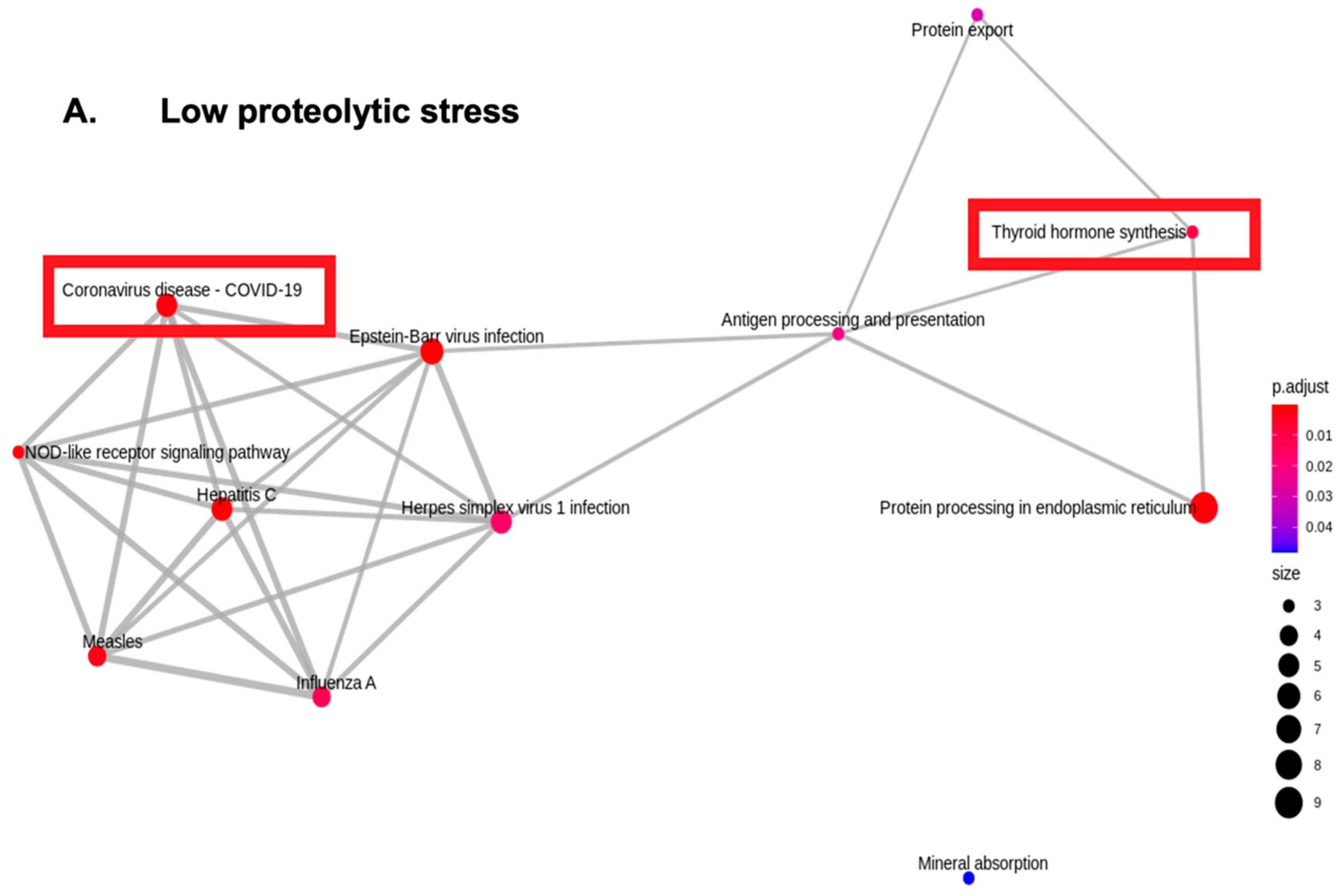
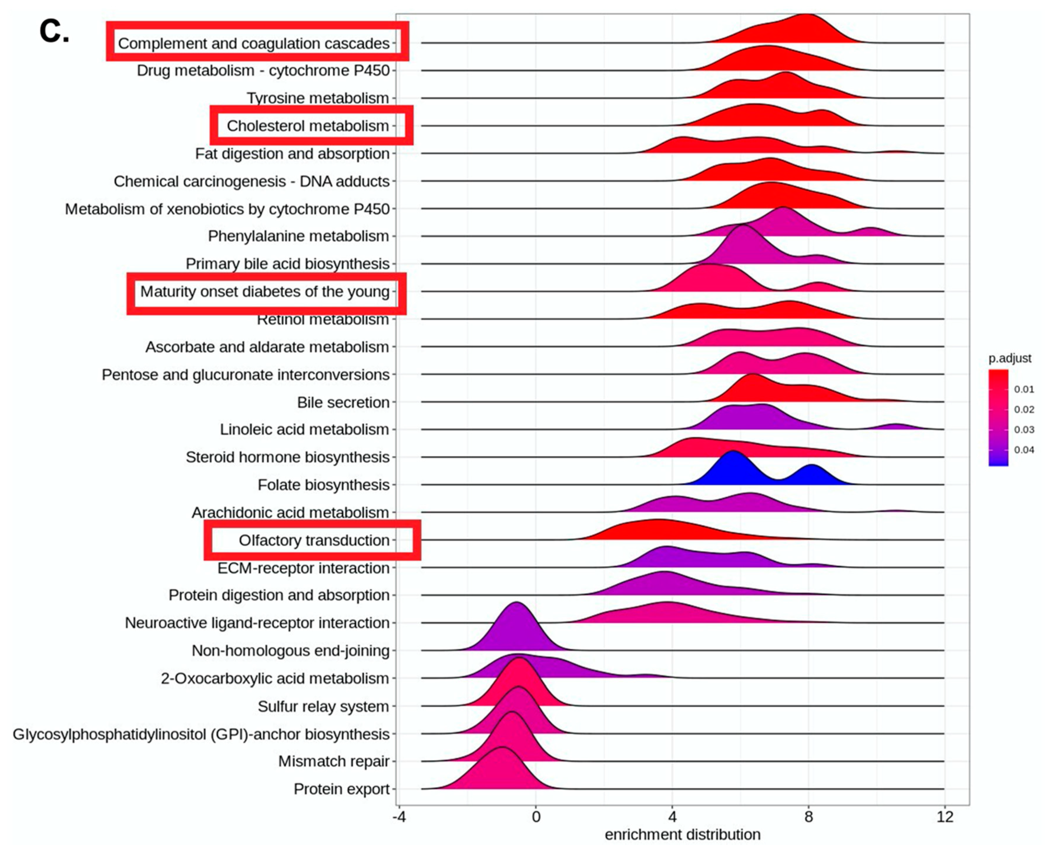
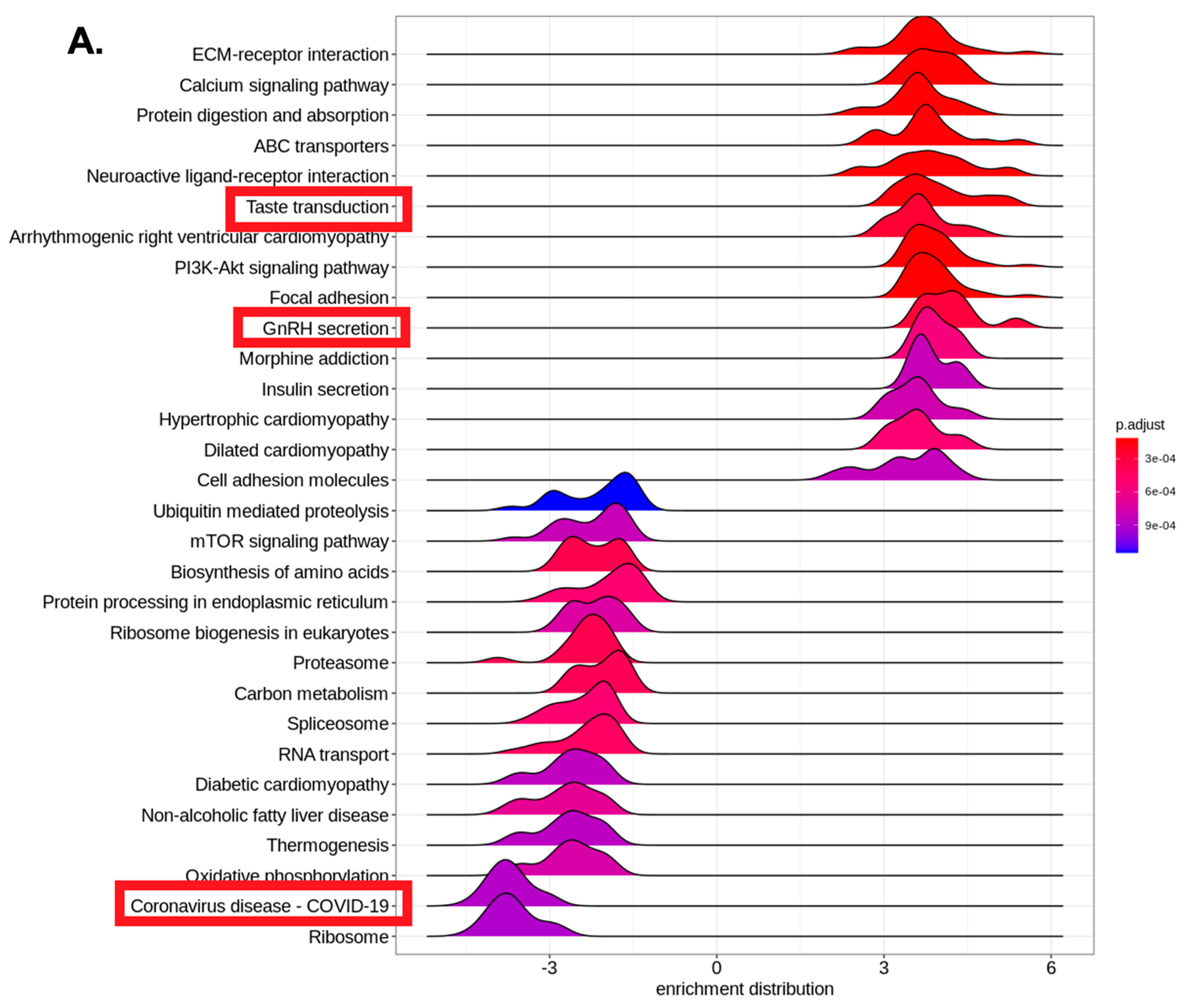
RNA sequencing was outsourced to Redcliffe Life Tech, Noida, India, which performed transcriptomics using an NGS Illumina platform with a 150 read length and paired end sequence layout. Quality control was carried out using the fastp tool (0.20.1) to provide clean data for downstream analysis. NGS sequencing reads were mapped with the Hisat2 (2.1.0) alignment tool. For abundance estimation, the data input was subjected to the Counts (2.0.1) tool in either the Sequence Alignment Map or Binary Alignment Map format. Differential gene expression and visualization were carried out using the DeSeq2 (1.8.3) tool. Proteolytic stress was tested to assess the upregulation of COVID-19-relevant inflammatory pathways using unbiased transcriptomic profiling.
Gene Set Enrichment Analysis ridge plots were used for significant KEGG terms. Ridge plots are density plots of the frequency of log2 fold-change values per gene within each enriched KEGG group, which helps to interpret the up- or downregulation of that KEGG category. Here, plots were created in clusterProfiler using KEGG orthologue annotations and log2 fold changes per gene calculated by DESeq2 during differential expression analysis. X-axis is the log2 fold change in expression for genes present in each plotted KEGG category, with positive values indicating upregulated expression in replicates and negative values indicating downregulated expression in replicates. Peaks are colored by corrected p-value, as shown by the legend, and corrected p- and q-values are shown per KEGG category.

4. Results

Interestingly, the monocytic cells upregulated COVID-19-associated pathological pathways upon escalating proteolytic stress while concomitantly upregulating metabolic stress, suggesting the importance of ensuing proteolytic stress underlying COVID-19 pathology. The combined role of metabolic and proteolytic stress resulted in the upregulation of COVID-19 pathways that co-segregated with complement–coagulation, olfactory transduction, steroid biosynthesis, iron-ion binding, ferroptosis, and maturity-onset diabetes (Figure 1, Figure 2, Figure 3, Figure 4 and Figure 5).
Furthermore, comparisons of diverse treatments allowed us to classify groups that displayed the following: (a) the COVID-19 pathway along with the co-expression of upregulated interferon response, oligoadenylate synthetase, double-stranded RNA binding, and innate immune recognition pathways and a downregulated olfactory transduction pathway, thus suggesting a mild-COVID-19-like pathological response; (b) COVID-19 pathway co-segregating with upregulated complement coagulation, acute inflammatory response, and steroid biosynthesis pathways, thus suggesting a severe COVID-like pathological response; and a unique group (c) a downregulated COVID-19 pathway co-segregated with upregulated olfactory transduction, an RNA interference pathway, and a TCR receptor pathway. We hypothesize a potential (novel) adaptive immune reshaping linked to olfactory transduction through a neuropeptide-signaling pathway that could reshape inflammatory responses by inducing antigen-specific T-cell responses by involving the RNA interference/RISC pathways of gene silencing and regulation. We are in the process of establishing the significance of these pathways in diabetic patients with COVID-19 and CAM [Sharma et al., unpublished work]. We earlier proposed pathogen-invoked proteolytic activation as a novel cellular stress-inducing process that could facilitate viral-competent immunomodulation through reprograming RNA metabolism and homeostasis in a process involving the protease-induced transcriptomic/epi-transcriptomic reshaping (PITTR) of host cells to counter cellular stress [102].
The pre-symptomatic or acute phase of COVID-19 marked by high viral loads/replication in the nasal epithelium makes it a potential target to inhibit the intensification of infection by limiting spread of the infection. Therefore, the prophylactic/therapeutic targeting of the olfactory epithelium by employing intranasal sprays is envisioned as a potential approach to prevent COVID-19 severity. The use of intranasal drug formulations are highly encouraged in COVID-19 and CAM treatment due to initially higher viral loads in the nasal microenvironment and their direct detrimental impact on infection inception and clinical course. Recently developed imidazole compounds have been reported to enhance anti-viral interferon responses upon intranasal administration in animal settings [103]. Similarly, recent phase-II clinical trials from Canada and the UK, showed the efficacy of nitric oxide nasal spray (NONS) in reducing SARS-CoV-2 viral loads for treatment of COVID-19 infections [104,105]. We envision strong anti-diabetic and anti-microbial effects of the combination of anti-metabolic and anti-proteolytic (MPI) inhibitors. Metabolic stress/hyperglycemia intensifies systemic SARS-CoV-2 and nasal/olfactory ROCM spread. We expect a prophylactic use of the MPI intranasal formulations in diabetes patients that could exhibit beneficial effects in regulating ferroptosis, promoting iron homeostasis, and facilitating the management of COVID-19 and CAM due to its anti-diabetic, anti-inflammatory, anti-oxidant, and anti-chelating properties.

5. Discussion

5.1. Hexosamine Biosynthetic Pathway of Glycosylation and Metabolic Stress Calibration in Olfactory Mucosa

Metabolic rewiring is a common mode of adapting to cellular stress to restore homeostasis due to its plasticity in cellular metabolism. As evidenced in uncontrolled hyperglycemia, persistent metabolic alterations can result in the irreversible resetting of the metabolic machinery, thus resulting in the desynchronization of the functional outputs. The primary bioenergetically tuned metabolic pathways include AMP-activated protein kinase (AMPK), mammalian target of rapamycin (mTOR), the hexosamine biosynthetic pathway (HBP), and iron handling. Major cellular calibrants of energy stress include the innate immune myeloid lineage monocyte/macrophages and neutrophils that respond to metabolic stress with remarkable resilience by adjusting to distinct metabolic states by displaying phenotypic, transcriptomic, functional, and spatial heterogeneity, thereby diversifying the inflammatory immune responses. Accordingly, burgeoning metabolic diabetes disorders are linked to myeloid cell dysfunction, which has been recognized as important feature in SARS-CoV-2 infection. Diabetes and hyperglycemia have been integrally linked to the severity of COVID-19, and uncontrolled diabetes mellitus and COVID-19 have recently been independently or in combination associated with the emergence of aggressive mucormycosis [16,19,106,107,108,109,110,111,112,113,114,115,116]. The induction of diabetes in SARS-CoV-2 infectious settings has also been demonstrated [47,48,117], suggesting an important role of metabolic resetting during COVID-19 disease course.
Myeloid lineage macrophages are central to anti-fungal/anti-Mucorales immunity. Therefore, “long COVID” and “post-infectious hyperinflammatory diseases” marked by metabolic and myeloid-immune deregulation are outcomes of COVID-19 that offer secondary fungal infections at the forefront due to ensuing immune deregulation after COVID. A consistent observation during the current unprecedented mucormycosis surge is underlying diabetes, which is also a strong comorbidity for COVID-19 severity, thus suggesting a potential role of metabolic sensors in the regulation of immune responses during infectious diseases. However, the mechanistic insights behind the correlative T2DM-associated deregulation of epithelial–endothelial barrier integrity and mucosal immunity remain relatively underexplored [20,118,119,120,121,122,123,124,125,126,127,128,129,130].
Importantly, infection-triggered (SARS-CoV-2/Mucorales) inflammasome-mediated pyroptotic cell death has been intricately linked to release of DAMPs and extracellular ATP, monocyte/macrophage activation, epithelial and endothelial cell damage, and vascular leakage, all resulting in inflammatory immune cell infiltration into the nasal/respiratory mucosa. The proinflammatory cytokines consequently lead to mucosal mucin reshaping and the activation of thromboinflammation. What remains underappreciated is the fact that the pathogenic microbes (SARS-CoV-2/Mucorales) have to traverse the bulky polyanionic–mucus barrier (20 µm) and heparan sulfate proteoglycans (HSPGs) (50 nm), embedded in the cell membranes to reach and destroy the nasal/airway epithelial cells. The mucins (terminally charged sialic acid and hydrophobic fucose) and polyanionic HSPGs offer potential structural frameworks for mediating interactions with primary binding motifs in the infecting microbes (e.g., cationic RBD groove of SARS-CoV-2 and fungal lectins attach to mucins and HSPGs to facilitate binding and entry to the host cells). Following binding, the microbes can be inactivated/cleared-off by mucociliary clearance or be subjected to infection spread, depending on the cellular/extra cellular metabolic milieu. The anti-pathogenic and rheologically flexible properties of mucus come at the cost of the high energy consumed in the process of mucous hydration in the airway system, which is highly dependent on the pathologically overexpressed metabolites (alarmin/DAMPs) including extracellular ATP and UDP-sugars derived from the HBP pathway (glucose/galactose) [131,132,133,134,135]. Therefore, it is reasonable to hypothesize that diabetes-associated metabolic pathways participate in reshaping mucin biology and innate immunity in the upper airways, thus making them susceptible to infection and the spread of microbes.
It could be speculated, that T2DM-responsive bioenergetically tuned HBP deregulation alters the “metabolic glycan-code input”; as opposed to the primary metabolic sensing by myeloid “cellular sensors” such as macrophages/monocytes. This metabolic perturbation (altered glycosylation) underlying diabetes is first read by the innate immune “structural sensors”, such as airway epithelial mucins and endothelial glycocalyx that are heavily glycosylated macromolecules. Epithelial mucins and endothelial glycocalyx are arranged as multiscale hierarchal structured fluids in nasal and respiratory epithelia, therefore acting as the frontline structural barricades of the mucosal innate immune system. The altered glycosylation patterns in nasal/respiratory mucosa and endothelial glycocalyx compromise epithelial–endothelial barrier integrity and offer easy pathogenic access to the underlying epithelium/endothelium, thus resulting in enhanced spread, enhanced vascular permeability, and thromboinflammation. Concomitantly, there is heightened purinergic signal transduction via the HBP intermediate UDP-sugar (ligand)-mediated activation of metabotropic purinoreceptor P2Y14 (expressed on adipocytes, myeloid immune cells, hematopoietic stem cells, kidneys, lungs, and intestines). The P2Y14 receptor was recently implicated as a strong target for neutrophilia attenuation in severe COVID-19 [132]. However, no data on COVID-19 or mucormycosis patients are available to date. P2Y14 expression is linked to glucose and lipid homeostasis [136], as well as the amplification of allergen-induced hyperinflammatory airway eosinophilia [135]. The HBP pathway has also been recently linked to ferroptosis sensitivity [137], thereby indicating a potential druggable “diabetes–HBP–mucin–iron axis” in COVID-19 and mucormycosis infections. Diabetes-associated, HBP-mediated altered glycosylation patterns in epithelial mucins and the role of the HBP-derived, UDP-sugar-activated P2Y14 purinoreceptor in myeloid immune deregulation remain subjects that have yet to be explored.

5.2. Diabetes and Metabolic Iron Redox-Stress- Macrophages as Ferrostats

It is important to note that the immune cells that act as primary metabolic sensors with high plasticity in metabolic switching (glycolytic vs. oxidative phosphorylation) are myeloid lineage macrophages, which are also central to anti-fungal/anti-Mucorales immunity. The longitudinal profiling of respiratory and systemic immune revealed myeloid cell-driven inflammatory events in COVID-19 [114]. The differential activation potential of NLRP3 inflammasome among myeloid cells is suggested to be a biomarker for the course of COVID-19 [115]. SARS-CoV-2 was shown to engage inflammasome and induced pyroptosis in monocytic cells [116]. Therefore, the metabolic reprograming of macrophages during hyperglycemia potentially alters the gene expression and regulation of immune pathways important for innate immunity and subsequent macrophage-sculpted adaptive immunity. Histone acetylation was recently reported to be upregulated in macrophages following concerted increases in glycolytic flux and ATP-citrate lyase activity [109]. Monocytopenia and morphological monocytic defects were found to be associated with hyperinflammation in COVID-19 in T2D patients [110], and hyperglycemia was shown to drive proinflammatory M1 polarization in macrophages via the TLR4–IRF5 pathway [111]. The defective macrophages thus fail to efficiently reshape the adaptive immune responses during inflammatory diseases such as COVID-19.
The enhancement of CD8+T cell effector functions (shaping extra-follicular adaptive immune responses) and the identification of repertoire of neutralizing antibodies to offer protective immunity post-infection are intensive areas under research to prevent infection, re-infection, or superinfections. Persistent alterations of iron homeostasis/hyperferritinemia in COVID-19 are also associated with immune deregulation [138,139,140,141,142,143,144,145,146,147,148,149,150,151,152,153,154,155,156,157,158,159,160,161,162]. Importantly, glycans/glycosylation is becoming increasingly relevant in shaping T cell effector functions and immunological synapses [163].
Macrophages are well-appreciated for their role in systemic iron homeostasis, in which they sequester iron during pathogenic infections and release iron for erythropoiesis. Importantly, immunometabolic resetting in macrophages is increasingly becoming linked to their handling of systemic iron recycling by regulating the unidirectional flux of non-transferrin-bound iron (NBTI/non-Tf-Fe3+) and erythrophagocytosis driven by CD163, CD91, and CD47 receptors. Therefore, iron overload can be highly toxic and needs to be strictly governed to maintain iron homeostasis [164,165]. Macrophages have also been highly implicated in metabolic disorders such as T2DM and neurodegenerative diseases, as iron-dependent lipid peroxidation can cause oxidative damage in these diseases via Fenton chemistry. The crosstalk between iron and immune cells (wherein iron trafficking is regulated by cytokines and acute phase proteins) and the iron-dependent cell fate determination of macrophages and lymphocytes during inflammation underscore the requirement for the tight control of iron metabolism and homeostasis [166,167,168,169,170,171,172,173].

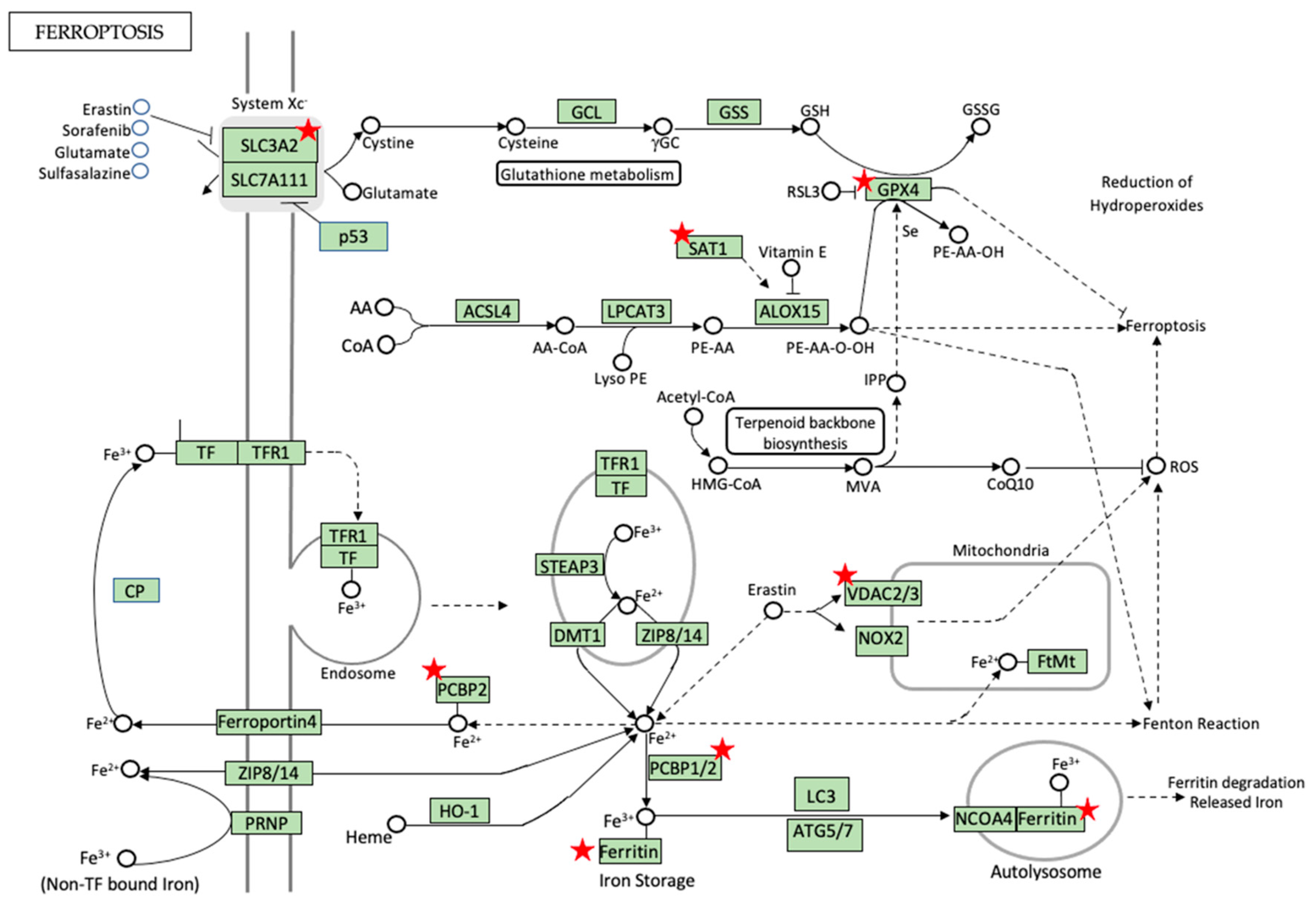
5.3. Iron Metabolism and Homeostasis—Can Ferroptosis Be the Game Changer?

The authors of a recent case-control study from our health care center in India on COVID-19 patients with and without mucormycosis compared the baseline iron parameters including iron, ferritin, total iron-binding capacity, and percentage transferrin saturation. However, these iron indices did not reveal any significant differences between CAM survivors and non-survivors [174], indicating that much needs to be understood regarding the complexity of iron biology during infectious pathologies such as CAM. Iron or iron-containing complexes, such as heme and iron–sulfur (Fe–S) clusters are integral to several biological processes such as oxygen transport (hemoglobin), oxygen storage (myoglobin), bioenergetic pathway intermediates (cytochrome-c), metabolic pathway intermediates (amino acid oxidases, fatty acid desaturases, and lipoxygenases), cellular detoxification (cytochrome P450), and cellular defense (nitric oxide synthase, NADPH oxidase, and myeloperoxidase). Notably, the cellular homeostatic machinery is highly reliant upon the cellular redox status, which is determined by the total cellular output of the ROS and antioxidant system. Iron availability is central in maintaining the cellular redox state because it is available in two highly interconvertible forms: reduced Fe2+ (soluble) and oxidized Fe3+ (insoluble). Iron cofactors such as heme, iron–sulfur clusters (Fe–S), and iron-oxo systems participate in a wide range of biological processes in electron transfer reactions and are central to metabolic pathways, mitochondrial function, cell cycle, and more [175,176,177,178,179,180,181]. The use of ferritin-bound iron is a cells’ protective mechanism of storing iron in macrophages and hepatic cells. Ferritin-bound iron release is a tightly regulated process known as ferritinophagy that involves the nuclear receptor coactivator 4 (NCOA4)-mediated transfer of ferritin to autolysosomes, as well as degradation and iron release for the biosynthetic pathways [182].
The overt production of ROS via chemically and reactive Fe2+ ions is detrimental to cellular membranes because it oxidizes membrane phospholipids and results in membrane rupture and release of DAMPs, which trigger sterile inflammation in iron-dependent regulated cell death known as ferroptosis. Ferroptosis comprises a rapidly evolving research field with enormous therapeutic potential, as it is becoming increasingly linked to inflammatory diseases, infectious diseases, and inflammation-associated immunosuppression. Ferroptosis can be induced by extrinsic (the inhibition of membrane cystine/glutamate antiporter system XC) or intrinsic pathways (the inhibition of anti-oxidant glutathione peroxidase/GPX4). Though the terminal effectors of ferroptosis are not clearly understood, it is a form of iron-dependent, peroxide-mediated regulated cell death (RCD) that is distinct from other RCD modes including apoptosis, necroptosis, and pyroptosis because it is independent of the activities of the caspases (apoptosis), MLKL (necroptosis), and gasdermin D (pyroptosis) molecular effectors [183,184,185,186,187,188,189,190,191,192,193,194,195,196].
Ferroptosis has been increasingly linked to the regulation of inflammatory networks in innate immune cells such as neutrophils and macrophages that exhibit graded polarities and differential sensitivities to ferroptosis [197], with pro-inflammatory M1 macrophages marked by the secretion of high iNOS, cytokines, and lipid mediators displaying ferroptotic-resistant phenotypes, as opposed to pro-ferroptotic propensity of pro-resolving alternatively activated M2 macrophages that secrete anti-inflammatory molecules. The intracellular redox regulation driven by metabolic iron, thiols, and lipid peroxides is emerging as a new mode of cellular reprograming under the term “redox-stress” that may have potential implications in the resetting of pro-inflammatory pathways driven by the ferroptotic mode of cell death, which is marked by the loss/rupture of the oxidation sensitive phospholipid plasma membranes followed by the release of intracellular DAMPs, resulting in the propagation of sterile inflammation. This is in striking contrast to the anti-inflammatory apoptotic mode of cell death that is marked by the preservation of cell membrane integrity and the containment of intracellular contents that are cleared by hydrolytic digestion and phagocytic engulfment by macrophages [198,199]. Iron-regulatory processes tend to have cell-specific mechanisms [200,201]. Iron-overload-triggered ferroptosis can be inhibited by iron chelators such as deferoxamine, deferiprone, and ciclopirox that decrease intracellular iron levels [202]. Ferroptosis has become increasingly linked to bacterial and yeast infections [203,204,205], but further detailed investigations are warranted in many contexts before targeting ferroptosis as a feasible strategy in infectious pathologies. Nevertheless, emerging opportunities for anti-ferroptotic agents in neurological disease settings are already in progress.

5.4. Pro-Ferroptotic Labile Iron Pool (LIP) and RNA-Binding Proteins (RBPs) in Regulation of Ferroptosis and Diabetes

The reactive iron-free radicals in the LIP can oxidatively damage the plasma membrane unsaturated lipids (PUFAs) [175,206]; therefore, the LIP is intrinsically under tight control by the action of iron-buffering compounds such as iron–glutathione (Fe–GSH) complexes (cytosolic GSH occurring at 2–10 mM and complexing Ferrous ions occurring at 1:1 stoichiometry) [207] and iron chaperones. Fe–GSH complexes are reported to be chaperoned by the poly(rC)-binding protein (PCBP) family that dictates intracellular iron distribution and transport [208]. PCBPs (1/2) chaperone iron (at low micromolar affinity) to and from iron transporters (DMT1, ferroportin 1), iron storage pools of ferritin, and iron-containing prolyl hydroxylase (PHD), deoxyhypusine hydroxylase (DOHH), heme-oxygenase (HO-1), and BolA2. PCBPs are RNA-binding proteins (RBPs) with intrinsically disordered regions (IDRs) composed of hnRNP-K-homology (KH) domains that preferably bind to Fe–GSH and facilitate iron transport to client proteins. As the affinities of Fe–GSH complexes and PCBP1 coordination lie in sub-micromolar ranges, it has been suggested that the majority of the cytosolic LIP exists in the PCBP1–Fe–GSH format [199,207]. Therefore, RBPs such as PCBPs intracellularly regulate the chemical reactivity and trafficking of pro-ferroptotic LIP. Ferritin sequesters iron in large amounts as inert ferric oxyhydroxides that can be mobilized by the cells in a process of ferritin degradation via lysosomes or ferritinophagy [209,210] (which is marked by the binding of ferritin to its cognate autophagic cargo receptor NCOA4), and the consequently liberated iron is transported back to the cytosol or mitochondria (erythroblasts) to begin heme synthesis; ferritinophagy and ferroptosis are accordingly implicated in the management of metabolic diseases [211]. PCBP1- and NCOA4-driven ferritinophagy could mobilize large pools of stored iron that may have pro-ferroptotic roles via the delivery of PCBP1-mediated iron to client ferroptotic proteins. PCBP1 chaperones can deliver ferrous ions to mono- or di-iron centers such as HIF-1α (pro-inflammatory macrophages are marked by mitochondrial ROS production and HIF-1α stabilization [212]), the regulating PHD2 enzyme (mono iron center), and the DOHH monooxygenase enzyme (di-iron center), which participates in the hydroxylation of the modified amino acid hypusine that is exclusively present in the eIF5a translational initiation factor. Deoxyhypusine synthase (DHPS)-driven eIF5A hypusination was recently shown to be a feature of proinflammatory macrophages, and DHPS deletion in macrophages resulted in improved insulin sensitivity and glucose homeostasis. Furthermore, hypusination is being increasingly implicated in metabolic diseases and microbial infections [213,214], hypusine polyamine precursors have been reported in intestinal epithelial renewal and M2 macrophage polarization [215], and dietary spermidine was recently shown to boost eIF5A hypusination and protect mitochondrial dysfunction during brain aging [216]. The polyamine pathway intermediate spermidine was recently reported to induce anti-inflammatory pathways following release from apoptotic cells [217]; it has protective effects in metabolic diseases and has emerged as a well-tolerable caloric restriction mimetic and provider of nitric oxide and arginine bioavailability [218]. Interestingly, the SAT1 (spermidine N1 acetyltransferase) gene was recently reported as a metabolic transcription target for p53 and a pro-ferroptotic molecule resulting in lipid peroxidation upon ROS stress [219]. Targeting the polyamine metabolic pathway was reported as a promising approach to control viral infections including SAR-CoV-2 [220]. SAT1 is a catabolic enzyme in the polyamine pathway that leads to the conversion of anti-inflammatory spermidine to N1 acetylspermidine. We recently observed the upregulation and downregulation of N1 acetylspermidine in COVID-19 and CAM plasma samples, respectively, via unbiased metabolomic profiling (unpublished data). Additionally, SAT1 and other ferroptotic pathway genes were differentially regulated in our in vitro culture model that recapitulated the disease modeling of COVID-19 pathways (Figure 6), thereby warranting detailed investigation to understand the role of metabolic ferroptotic and polyamine pathway intermediates in infectious diseases such as COVID-19 and CAM. Amino acid metabolism modifications, such as hypusination, require further investigations to understand their role in regulating infectious and metabolic diseases.
The authors of merging studies are trying to leverage post-transcriptional control of immune responses that is regulated by RBPs [130] such as tristetraprolin, Regnase-1, Roquin, and RNA methylases, which coordinate the inflammatory networks in immune cells and associated niches by modulating the mRNA pool of immune pathway genes [221]. Interestingly, PCBP1 RBP was recently implicated as an intracellular checkpoint for shaping T-cell responses by promoting T effector immune response as opposed to T regulatory functions [222]. Diabetic endotheliopathy (possibly driven by the HBP deregulation of endothelial glycocalyx) also posttranscriptionally alters endothelial gene expression via RBPs that are becoming increasingly linked to posttranscriptional immune regulation; RBPs generated during mRNA processing are emerging as frontiers in the regulation of pancreatic beta cell function [223,224,225].
We propose a posttranscriptional gene silencing (RNAi) regulation of TCR genes in COVID-19 pathology, which may intersect with tRNA biology pathway enzymes such as tRNA aminoacyl synthetases that are emerging as important players of immuno-metabolic dysregulation and linked to the intergenerational inheritance of metabolic traits in diabetes mellitus [226,227,228].

6. Routes of Infection and Current Perspectives in Clinical Presentation and Diagnosis

There is temporal variability, i.e., seasonal, etiological, geographical, and ecological, in the epidemiology of the transmission of Mucorales of human importance. Frequent hospital outbreaks of cutaneous mucormycosis involving contaminated surgical dressing, adhesive bandages, and tongue depressors have been reported. Furthermore, high spore counts in hospital environments (especially in the ongoing expansion and construction of premises) and contaminated air-conditioners together account for nearly 9% of nosocomial mucormycosis [229]. Regarding CAM, much speculation has been focused on ventilators, catheters, air humidifier bottles, industrial oxygen cylinders, steam inhalation practices, and the extensive usage of masks, which warrant systematic aero-mycological and surface mycological investigations. Furthermore, the Centers for Disease Control (CDC), Atlanta, GA, USA, published a systematic tool that aids in performing environmental assessments when investigating healthcare-associated disease outbreaks due to fungi including Mucorales [6,20,230].
The first step in the diagnosis of CAM is the knowledge of warning symptoms and signs, as well as a high index of clinical suspicion, that includes the identification of DM with or without DKA, the use of SC therapy; the use of immunosuppressants such as tocilizumab; identification of immunodeficiency; iatrogenic suppression using mechanical ventilation; and the identification of hemato–oncological patients, bone-marrow/solid organ transplantation recipients, iron overload, and obviously COVID-19 in the present context. The extent of damage in ROCM or PM needs a multidisciplinary investigation and management with swift decisions from a team of experts, e.g., those in the otorhinolaryngology, ophthalmology, pulmonology, radiology and imaging, internal medicine, infectious disease, neurology, neurosurgery, anesthesiology, maxillofacial surgery, mycology/microbiology, pathology, and clinical pharmacy fields [28,231,232].
In radiological studies using magnetic resonance imaging (MRI) of the paranasal sinuses, contrast-enhanced MRI has been used to identify the infection of vital tissue and assess cerebral invasion in ROCM, diagnostic nasal endoscopy, and color Doppler ultrasonography studies. The computed tomography (CT) of the thorax and the plain and high-resolution (HR) CT for PM have also been used [231,232]. Clinical and radiological findings have overlapped with COVID-19, so the diagnosis of disseminated and pulmonary mucormycosis is challenging. The reverse halo in peripheral lung locations that is suggestive of pulmonary mucormycosis in IC patient(s) and serves as a useful indicator for pre-emptive antifungal therapy is not an indicator for COVID-19, as it can be a commonly overlapping finding. Recently, EORTC and MSG updated the criteria for proven invasive fungal disease (IFD) [231], with recommendations for senso stricto among patients with cancer, transplant recipients, and other severely IC hosts. Even though the diagnosis of IFD via radiological findings remains the gold standard, helping in early detection and swift course of actions, it lacks sensitivity. The most significant pulmonary radiological features of IFD in non-cancer patients and among the trio of invasive pulmonary aspergillosis (IPA), mucormycosis, and COVID-19 overlap with “atypical” non-nodular pattern, consolidation, and ground-glass opacities [231].
The laboratory diagnosis and confirmation of mucormycosis is easy if the biopsy samples are available, as in the case of ROCM, whereas the diagnosis of disseminated or pulmonary mucormycosis is difficult due to the nonavailability of biopsy specimens due to various limitations. The microscopic examination of the tissues using KOH mount and Calcofluor white aids the rapid confirmation of diagnosis. Under microscopy, the Mucorales hyphae classically appears hyaline, broad (2–8 μm diameter), ribbon-like (often folded), pauci-septate, or aseptate with a right-angled branching. Unlike other molds, Mucorales grow faster within 24–48 h and appear cottony. However, the sensitivity of cultures is not more than 80%. The species-level identification of cultures can be made with conventional phenotypic, morphological, and physiological features such as growth rate and incubation temperatures on various media, but it needs expertise in mycological techniques and identification. The method of choice for the species identification of Mucorales is therefore ITS sequencing, which is mostly restricted to reference laboratories. Even though ITS barcoding has been a preferred molecular marker for the identification of various Mucorales species, it has a few limitations such as the failure to differentiate R. microsporus and R. azygosporus because both belong to the same species [233].
Therefore, attempts to directly diagnose mucormycosis from clinical specimens have been made by standardizing the Mucorales PCR using conventional, nested, RFLP, and real-time PCR (qPCR) formats on blood samples and other matrices such as bronchoalveolar lavage (BAL) or tissue specimens, and several in-house assays have shown good sensitivity. The commercially available multiplex, pan-Mucorales real-time (qRCR) kit-MicroGenius assay by PathoNostics readily detects several Mucorales species including Rhizopus, Lichtheimia, Rhizomucor, and Cunninghamella in sera and BAL, thus showing promising rapid diagnostic utility in present clinical setups. In this era of rapid diagnosis, lower turnaround-time (TAT), and laboratory quality initiatives, several technologies such as matrix-assisted laser desorption ionization–time-of-flight mass spectrometry (MALDI–TOF) have increasingly gained momentum and will start influencing the clinical microbiology laboratories in the future. The negative results of the serological markers such as (1→3)-β-D-glucan (BDG) and galactomannan (GM) (used in other IFD) can aid the diagnosis of invasive mucormycosis (IM) during strong suspicion of IFD. The evaluation of these surrogate markers in proven IM using an enzyme-linked immunospot (ELISpot) assay (leveraging Mucorales-specific T-cells with a derived diagnostic cut-off) has been reported, and elevated IL-10 and IL-4 levels in symptomatic patients and elevated Mucorales-specific T-cells with a higher IFN-γ levels in non-symptomatic patients were observed. These results indicate the assay’s promising utility in hematological patients and warrant rigorous investigation into its potential as a surrogate marker in IM and other patient populations [234].

Clinical Perspective of Rhino-Orbital Mucormycosis and Patient Management

Mucormycosis, primarily caused by Mucorales in immunocompromised or immune-competent hosts with diabetes/trauma, is a critical scenario for every clinician dealing with this condition. Rhino-orbital mucormycosis is the most common form of the disease; its fulminant nature, the extensive angio-invasion-induced ischemic necrosis-related inflammation of tissues, delays in suspicion in primary care, and lack of effective medical antifungal therapy have worsened diagnosis challenges.
Rhino-orbito-cerebral mucormycosis patients present with a history of facial heaviness, fever, purulent nasal discharge, headache, nasal congestion, and sinus pain. The disease can progress and present with the destruction of the turbinates and the nasal septum, paleness, blackening in the nasal and palatal mucosa, and ulcer formation in the palate with exposed underlying osteomyelitic bone. Peri-orbital swelling decreases vision, proptosis, and ophthalmoplegia with or without diplopia in patients of orbital involvement, and it alters neurological function and consciousness in cases of cerebral involvement, thus indicating ominous disease aggressiveness. Diagnosis primarily rests on microbiological analysis with KOH wet mounts of scrapping or tissue samples taken from pale, necrotic, and inflamed areas. Radiological imaging with CT/MRI and histological analysis of tissue samples aid diagnosis.
A diagnosis or high clinical suspicion of diagnosis leads to management that primarily involves the following steps:
  • Continued treatment of the primary immune deficiency condition.
  • Correction of biochemical parameters and management of associated diabetic status.
  • Aggressive debridement of the necrotic tissues of the rhino-orbital-facial region with the aim to clear all necrotic tissue and osteomyelitic bones to the maximum extent until the tissues bleed, with caution used in case of cerebral involvement to not to debride brain tissue.
  • Post-debridement wound and cavity local care and adjuvant medical management with amphotericin B and/or posaconazole and isavucanazole.
  • Continuation of cavity care after the completion of therapy with the regular clearance of crusts and saline irrigations for 3–6 months after treatment.
  • Prosthetic rehabilitation and/or reconstruction of the defect.
The morbidities and functional compromises associated with disease and management lead to significant losses of productivity and hinder the quality of life of patients, especially during the current COVID-19 pandemic, in which instances of mucormycosis have increased to overwhelming levels due to underlying immune dysregulation by COVID-19 and associated steroid therapies. Hence, high clinical suspicion and early referrals are key to early management and the preservation of the quality of life in cases of rhino-orbital mucormycosis.

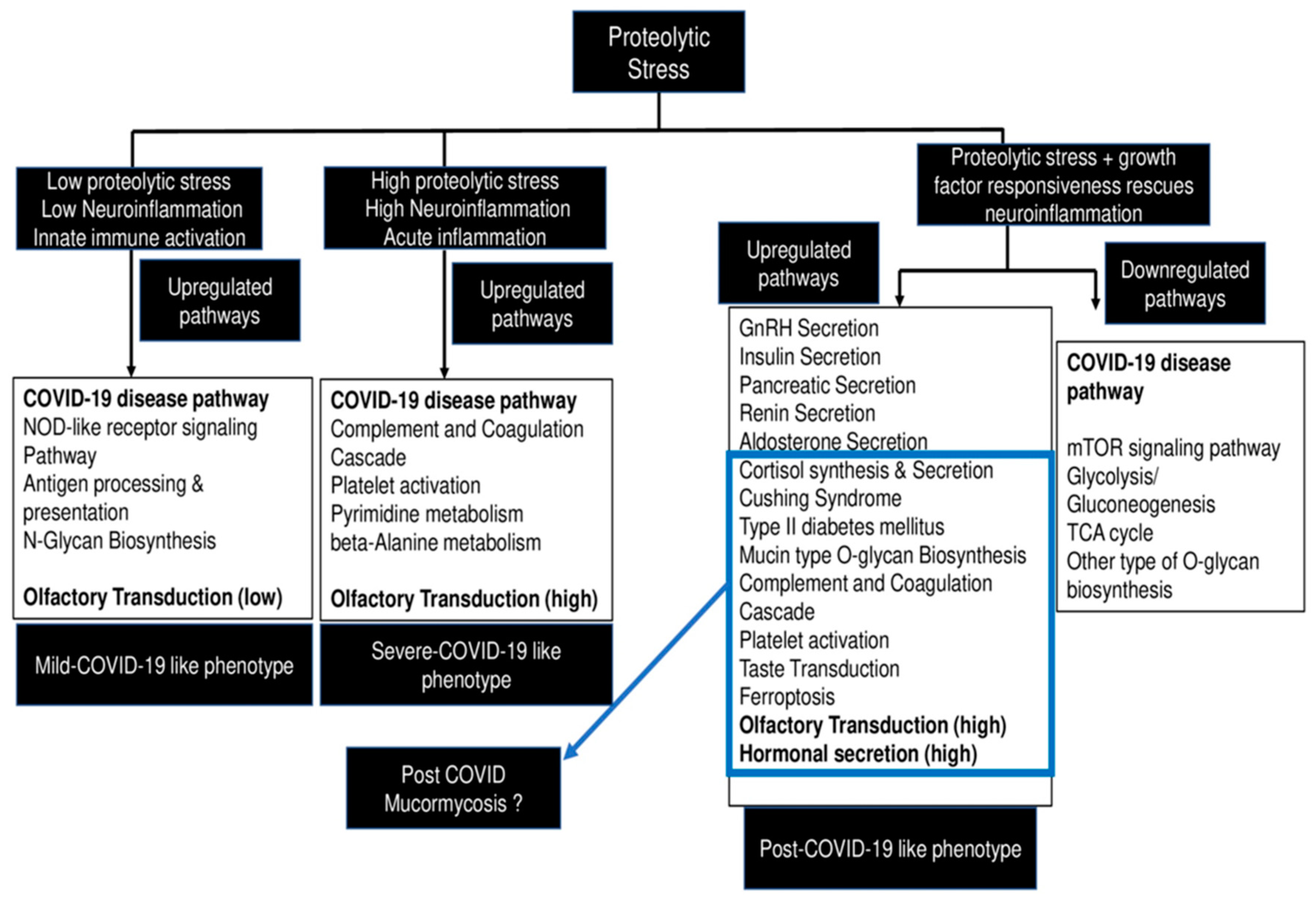
7. Future Perspective—A Working Hypothesis to Explain Development of COVID-19 and CAM in the Backdrop of Diabetes

Serine proteases can have cell-autonomous functions (besides SARS-CoV-2 activation) in olfactory microenvironment, in which they can activate and signal through cognate GPCR family of protease-activated receptors (PARs) that participate in transducing downstream inflammatory and calcium signaling pathways, resulting in the regulation of ion channels and transporters. In diabetic patients, endotheliopathy in the vascular olfactory niche can further add to the PAR-transducing protease repertoire by offering pre-activated TF-FVIIa and FXa serine proteases that could have Janus faces during inflammatory propagation. TF-FVIIa and FXa can activate PAR signaling concomitant to the complement coagulation pathway intensification that is central player in COVID-19 pathogenesis [235], and the resultant inflammatory loops can have pro-fibrogenic functions as the associated plasmin and uPAR–uPA signaling modulators are known to have growth factor-binding, TGF-beta-activating, integrin-signaling, and ECM-remodeling features. We report that protease stress is associated with concomitant metabolic deregulation and the differential expression of olfactory transduction pathway genes including the olfactory/odorant receptors (ORs). Importantly, the majority of the olfactory receptors are orphan receptors, and deorphanization is as a major challenge. Many fatty acid and lipid metabolites are potential activating and inhibitory ligands of ORs, and emerging studies have suggesting a broader expression pattern of ORs beyond the olfactory epithelium, including adipocytes, airways, kidneys, livers, lungs, and adipocytes. Extra-nasal ORs have been reported to perform seminal functions including blood pressure regulation via the renin–angiotensin–aldosterone pathway in kidneys and the hypoxia-sensing ORs in the glomus cells of the carotid body. Hypoxia metabolite lactate is a partial agonist of carotid body ORs. Hepatic ORs regulate hepatic lipid accumulation via the regulation of GLP-1, resulting in glucose and insulin tolerance. Airway smooth muscle cells express ORs that regulate bronchoconstriction through calcium signaling via the cAMP pathway. However, ligand-dependent contractility effects that are linked to inflammatory pathways of IL-8 and GMCSF have also been reported. The adipocytic effects of ORs are mediated by hepatic fatty acid oxidation and brown adipose tissue thermogenesis. Epicardial adipose tissue and diabetes mellitus patient plasma contain medium chain fatty acids that can activate cardiac muscle cells and regulate ionotropic effects [236,237]. The appreciable overlap of ORs expression pattern with affected organs in COVID-19 and the OR regulation of RAAS and hypoxia functions led us to propose their potential contribution in disease pathology and their use as therapeutic targets for diabetes, COVID-19 pathology, and CAM.
Importantly, neural-crest-originating astrocytes and pericytes are becoming increasingly linked to underlying neurovascular abnormalities in COVID-19 pathobiology. We also observed the upregulation of neural crest pathways in protease-treated monocytic cells. It is a possibility that developmental epithelial–mesenchymal transition (EMT) events initiated by active proteases during embryonic neural-crest induction could be recapitulated by providing exogenous proteolytic exposure to pathology competent monocytic cells in vitro, resulting in the upregulation of developmental multipotent neural-crest-like stem cells with ectomesodermal potential that capable of upregulating neurogenesis/gliogenesis (neurons and astrocyte) and vasculogenesis (microvascular pericyte), crucial cellular processes underlying neuroinflammatory diseases [Sharma et al., unpublished data]. The OR-expressing sustentacular cells in olfactory epithelia are glia-like, and the neuroendocrine cells have also been reported to be neural-crest-derived; their participation in the lung neuroinflammation in context of ARDS has been proposed with the pharmacological neuromodulation of the vagus nerve as a potential therapeutic approach to treat COVID-19 [238]. The hypothalamus–pituitary–adrenal gland axis (HPA), along with the vagal reflex and the possible involvement of the carotid body, has been proposed for the neural control of inflammation wherein the deregulated vagal anti-inflammatory reflex is central to inflammation and metabolic diseases including type 2 diabetes. The HPA axis under stress releases cortisol-releasing hormones, which induce the secretion of anti-inflammatory glucocorticoids from adrenal glands. Vagal anti-inflammatory reflex sense inflammation and the activation of efferent vagus nerve fibers results in dampened cytokine production following the binding of neurotransmitter acetylcholine to its receptor nicotinic α7nAChR on immune cells such as macrophages. Furthermore, the efferent sympathetic output to adrenal gland medulla chromaffin (neuroendocrine) cells triggers epinephrine release to activate specific receptors in immune cells to resolve inflammation [239]. Carotid body chemoreceptors have been proposed as afferent arms of the anti-inflammatory reflex and are becoming increasingly linked to inflammatory diseases such as sepsis and intermittent hypoxia [240]. The glomus cells of the carotid body that act as hypoxia sensors bear olfactory receptors and are neural-crest-derived, so we hypothesize carotid body chemosensors as central players in driving neuroinflammatory events in COVID-19/post-COVID neurological sequelae including anosmia.
Notably, small molecule neurotransmitters and neuromodulators generally belong to the class of amino acid metabolites including glutamate, sulfur-containing cysteine and methionine, proline, GABA, lysine, arginine, glycine, serine, alanine, aromatic amino acids, and branched-chain amino acids. These amino acid neurotransmitters recycle between neurons and astrocytes and conduct nerve impulse transmission through ionotropic channels, and a few of these amino acids participate in generating mitochondrial oxidative and nitrosative stress. The aromatic amino acid tyrosine is the precursor for the dopamine, norepinephrine, and epinephrine neurotransmitters that regulate neuroinflammation and are implicated in many neurocognitive diseases [241]. We hypothesize that the oxidative and nitrosative stress in macrophages due to metabolic deregulation during diabetes and COVID-19 can alter iron homeostasis and therefore lead to the lipid peroxidation and ferroptosis that compromise macrophage functionality and anti-Mucorales prowess. Vascular macrophages have been shown to upregulate ORs that bind to lipid metabolites and activate inflammasome and IL-1 production [242]. The parallel deregulation of the olfactory transduction pathway via metabolic-stress-triggered lipid and amino acid metabolites (proposed as potential ligands for ORs) and the activation of PARs due to serine proteases can drive the co-deregulation of olfactory transduction and complement coagulation pathways in macrophages (vascular immune) and olfactory epithelium (neurosensory) cells causing anosmia and neuroinflammation. The subsequent generation of DAMPs, as reported during inflammasome activation, can be passed onto the astrocytes and pericytes of the brain, which results in neurocognitive and neurodegenerative features. The post-viral effects of COVID-19 in the olfactory system and their neurological connections were described recently [69]. The pre-symptomatic or acute phase of COVID-19 is marked by high viral loads/replication in the nasal epithelium, which makes it a potential target to inhibit the intensification of infection by limiting the spread of the infection. Therefore, the prophylactic/therapeutic targeting of the olfactory epithelium by employing intranasal sprays could be a potential approach to prevent diabetes-associated COVID-19 and CAM (Figure 7).

8. Conclusions

The mechanistic insights of chemosensory/neurosensory deregulation in the olfactory epithelium and the attendant immune landscape reshaping during COVID-19 infection remain largely unexplored due to infection control concerns in disease-active patients. We leveraged the proteolytic predominance of olfactory microenvironment during acute phase of the disease to develop a novel virus-free cellular model to identify a potential host-associated serine protease-interactome that displays COVID-19-like disease signatures including maturity-onset diabetes, complement–coagulation, and olfactory transduction. Our data support the role of olfactory dysfunction in predicting neurological sequelae in CNS and suggest a new paradigm that could involve olfactory transduction-mediated neuroinflammation in the respiratory tract and peripheral tissues with resident macrophages expressing neuropeptide receptors. We also gathered appreciable patient data from COVID-19 and CAM plasma metabolomics and proteomics that suggest the beneficial effects of rescuing olfactory epithelium health with the potential repurposing of anti-metabolic drugs such as 2DG (2-deoxy-D-glucose) and nutraceutical azoles with anti-inflammatory and anti-oxidative effects. Our preliminary data demonstrated the anti-fungal activity of 2DG and L-carnosine (growth inhibition assay) against clinically relevant Mucorales species, a novel anti-fungal action (unpublished data). Both drugs have proven anti-diabetic and potential in-silico anti-COVID-19 effects, so our next work is directed towards the development of novel intranasal anti-diabetic sprays for controlling infectious airway diseases such as COVID-19 and CAM (Sharma et al., unpublished work).

Author Contributions

M.S. conceptualized the in vitro disease model, executed experiments, and conducted analysis. M.S., H.P.V., S.K.P. and P.R.K. wrote the manuscript. N.K.P., S.K.B., S.M.R. and M.P.S. provided intellectual input. R.A. assisted in presenting figures. All authors have read and agreed to the published version of the manuscript.

Funding

The research work discussed in this perspective was conducted in the Department of Otolaryngology and Head and Neck Surgery. The funding support was provided by Science and Engineering Research Board (SERB), Department of Science and Technology, Government of India under the following funding schemes SERB-CRG/2019/006745 and SERB-SPR/2019/001447.

Institutional Review Board Statement

The in vitro work was approved by Institute Ethics Committee of PGIMER, Chandigarh, India.

Informed Consent Statement

Not applicable.

Data Availability Statement

Data available in a publicly accessible repository. The data presented in this study are openly available in Zenodo at doi: 10.5281/zenodo.6321529.

Acknowledgments

M.S. thanks the funding agencies for the financial support and SERBCRG/2019/006745 for providing Junior Research Fellowship to Rhythm Arora. She would like to thank Satwant Kaur Gosal and Arunaloke Chakrabarti for teaching the basics of microbiology. The authors acknowledge Servier Medical Art (https://smart.servier.com, accessed on 21 December 2021) for providing cartoon components in the design of the hypothesis illustration.

Conflicts of Interest

Purushotham Reddy Koppula contributed to this article in his personal capacity, and the views expressed are his own and do not necessarily represent the views of Regeneron Phar-maceuticals Inc.

Abbreviations

ACE2Angiotensin-Converting Enzyme 2
AMPAdenosine Monophosphate
AMPKAMP-Activated Protein Kinase
BALBronchoalveolar Lavage
BDGβ-D-Glucan
BiPsBinding Proteins
CAMCOVID-19-Associated Mucormycosis
CAPACOVID-19-Associated Pulmonary Aspergillosis
CDCCenters for Disease Control
CotHCoat Protein Homolog
COVID-19Coronavirus Disease 2019
CSCell Surface
CTComputed Tomography
DAMPsDamage-Associated Molecular Pattern
2DG2-Deoxy-D-Glucose
DHPSDeoxyhypusine Synthase
DKADiabetic keto acidosis
DMDiabetes Mellitus
DMT1Divalent Metal Ion Transporter 1
DOHHDeoxyhypusine Hydroxylase
DPP4Dipeptidyl-peptidase 4
eIF5aEukaryotic Translation Initiation Factor 5 a
ELISpotEnzyme-Linked Immunospot
EMTEpithelial–Mesenchymal Transition
EORTCEuropean Organization for Research and Treatment of Cancer
FAKFocal Adhesion Kinase
Fe–GSHIron Glutathione
Fe–SIron Sulfur
FFAFree Fatty Acids
FISFFungal Infection Study Forum
GMGalactomannan
GnRHGonadotrophin-Releasing Hormone
GPIGlycosylphosphatidylinositol
GRP78Glucose-regulated proteins 78
HBPHexosamine Biosynthetic Pathway
HPSHigh Proteolytic Stress
HRHigh-Resolution
HSP70Heat Shock Protein 70
HSPGsHeparan Sulfate Proteoglycans
ICImmunocompromised
IDRsIntrinsically Disordered Regions
IFDInvasive Fungal Disease
IFN-γInterferon Gamma
IL-1Interleukin 1
IL-10Interleukin 10
IL-1βInterleukin 1 Beta
Il-6Interleukin 6
IL-8Interleukin 8
IL4Interleukin 4
IMInvasive Mucormycosis
IPAInvasive Pulmonary Aspergillosis
ISHAMInternational Society of Human and Animal Mycology
ISRIntegrated Stress Response
ITS SequencingInternal Transcribed Spacer
ITSInternal Transcribed Spacer
KOH MountPotassium Hydroxide Mount
LIPLabile Iron Pool
LMICLow- and Middle-Income Countries
LPSLow Proteolytic Stress
MADMucormycosis-Associated diabetes
MALDI–TOFMatrix-Assisted Laser Desorption Ionization–Time-of-Flight Mass Spectrometry
MLKLMixed Lineage Kinase Linked Domain
MPIAnti-Metabolic and Anti-Proteolytic Inhibitors
MRIMagnetic Resonance Imaging
MSAIMetabolic-Stress-associated Interactome
MSGEuropean Confederation of Medical Mycology (ECMM), and Mycoses Study Group
MTORMammalian Target of Rapamycin
NBTINon Transferrin Bound Iron
NCOA4Nuclear Receptor Coactivator 4
NONSNitric Oxide Nasal Spray
NRP1Neuropilin-1
OEOlfactory Epithelium
OSNsOlfactory Sensory Neuron
PAR-2Protease-Activated Receptor-2
PCBPPoly(rC) Binding Protein
PHDProlyl Hydroxylase
PITTRProtease-Induced Transcriptomic/Epi-Transcriptomic Reshaping
PMPulmonary Mucormycosis
PMNsPolymorphoneutrophils
PRRsPattern Recognizing Receptors
PRSS8Serine Protease-8
PUFAsPlasma Membrane Unsaturated Lipids
qPCRQuantitative Polymerase Chain Reaction
RBPReceptor Binding Protein
RBPsRNA-Binding Proteins
RCDRegulated Cell Death
RFLPRestriction Fragment Length Polymorphism
RNAiRNAInterference
ROCMRhino-Orbital-Cerebral-Mucormycosis
ROSReactive Oxygen Species
SARS-CoV-2Severe Acute Respiratory Syndrome Coronavirus 2
SAT1Spermidine N1 Acetyltransferase
SCSystemic Corticosteroids
SNCsSustentacular Cells
SPsSerine Proteases
T2DMType 2 Diabetes Mellitus
TATTurnaround-Time
TCRT-Cell Receptor
TLR4–IRF5Toll-Like Receptor 4–Activated Interferon Regulatory Factor 5
TLRsToll-Like-Receptors
TMPRSS2Transmembrane Serine Protease 2
TNF αTumor Necrosis Factor α
UDPUridine Diphosphate
VWFWillebrand Factor

References

  1. Spatafora, J.W.; Chang, Y.; Benny, G.L.; Lazarus, K.; Smith, M.E.; Berbee, M.L.; Bonito, G.; Corradi, N.; Grigoriev, I.; Gryganskyi, A.; et al. A phylum-level phylogenetic classification of zygomycete fungi based on genome-scale data. Mycologia 2016, 108, 1028–1046. [Google Scholar] [CrossRef] [PubMed] [Green Version]
  2. Prakash, H.; Chakrabarti, A. Global Epidemiology of Mucormycosis. J. Fungi 2019, 5, 26. [Google Scholar] [CrossRef] [PubMed] [Green Version]
  3. Morales-Franco, B.; Nava-Villalba, M.; Medina-Guerrero, E.O.; Sánchez-Nuño, Y.A.; Davila-Villa, P.; Anaya-Ambriz, E.J.; Charles-Niño, C.L. Host-Pathogen Molecular Factors Contribute to the Pathogenesis of Rhizopus spp. in Diabetes Mellitus. Curr. Trop. Med. Rep. 2021, 8, 6–17. [Google Scholar] [CrossRef] [PubMed]
  4. Ribes, J.A.; Vanover-Sams, C.L.; Baker, D.J. Zygomycetes in Human Disease. Clin. Microbiol. Rev. 2000, 13, 236–301. [Google Scholar] [CrossRef]
  5. Hallur, V.; Prakash, H.; Sable, M.; Preetam, C.; Purushotham, P.; Senapati, R.; Shankarnarayan, S.A.; Bag, N.D.; Rudramurthy, S.M. Cunninghamella arunalokei a New Species of Cunninghamella from India Causing Disease in an Immunocompetent Individual. J. Fungi 2021, 7, 670. [Google Scholar] [CrossRef]
  6. Muthu, V.; Rudramurthy, S.M.; Chakrabarti, A.; Agarwal, R. Epidemiology and Pathophysiology of COVID-19-Associated Mucormycosis: India versus the Rest of the World. Mycopathologia 2021, 186, 739–754. [Google Scholar] [CrossRef]
  7. Prakash, H.; Skiada, A.; Paul, R.; Chakrabarti, A.; Rudramurthy, S. Connecting the Dots: Interplay of Pathogenic Mechanisms between COVID-19 Disease and Mucormycosis. J. Fungi 2021, 7, 616. [Google Scholar] [CrossRef]
  8. Tandon, N.; Anjana, R.M.; Mohan, V.; Kaur, T.; Afshin, A.; Ong, K.; Mukhopadhyay, S.; Thomas, N.; Bhatia, E.; Krishnan, A.; et al. The increasing burden of diabetes and variations among the states of India: The Global Burden of Disease Study 1990–2016. Lancet Glob. Health 2018, 6, e1352–e1362. [Google Scholar] [CrossRef] [Green Version]
  9. Banerjee, M.; Pal, R.; Bhadada, S.K. Intercepting the deadly trinity of mucormycosis, diabetes and COVID-19 in India. Postgrad. Med. J. 2021. [Google Scholar] [CrossRef]
  10. Pal, R.; Singh, B.; Bhadada, S.K.; Banerjee, M.; Bhogal, R.S.; Hage, N.; Kumar, A. COVID-19-associated mucormycosis: An updated systematic review of literature. Mycoses 2021, 64, 1452–1459. [Google Scholar] [CrossRef]
  11. Gupta, R.; Kesavadev, J.; Krishnan, G.; Agarwal, S.; Saboo, B.; Shah, M.; Mittal, A.; Durani, S.; Luthra, A.; Singhal, A.; et al. COVID-19 associated mucormycosis: A Descriptive Multisite Study from India. Diabetes Metab. Syndr. Clin. Res. Rev. 2021, 15, 102322. [Google Scholar] [CrossRef] [PubMed]
  12. Rudramurthy, S.M.; Hoenigl, M.; Meis, J.F.; Cornely, O.A.; Muthu, V.; Gangneux, J.P.; Perfect, J.; Chakrabarti, A.; Isham, E.A. ECMM/ISHAM recommendations for clinical management of COVID-19 associated mucormycosis in low- and middle-income countries. Mycoses 2021, 64, 1028–1037. [Google Scholar] [CrossRef] [PubMed]
  13. Denova-Gutiérrez, E.; Lopez-Gatell, H.; Alomia-Zegarra, J.L.; López-Ridaura, R.; Zaragoza-Jimenez, C.A.; Dyer-Leal, D.D.; Cortés-Alcala, R.; Villa-Reyes, T.; Gutiérrez-Vargas, R.; Rodríguez-González, K.; et al. The Association of Obesity, Type 2 Diabetes, and Hypertension with Severe Coronavirus Disease 2019 on Admission Among Mexican Patients. Obesity 2020, 28, 1826–1832. [Google Scholar] [CrossRef] [PubMed]
  14. Heaney, A.I.; Griffin, G.D.; Simon, E.L. Newly diagnosed diabetes and diabetic ketoacidosis precipitated by COVID-19 infection. Am. J. Emerg. Med. 2020, 38, 2491.e3–2491.e4. [Google Scholar] [CrossRef] [PubMed]
  15. Alekseyev, K.; Didenko, L.; Chaudhry, B. Rhinocerebral Mucormycosis and COVID-19 Pneumonia. J. Med. Cases 2021, 12, 85–89. [Google Scholar] [CrossRef]
  16. John, T.; Jacob, C.; Kontoyiannis, D. When Uncontrolled Diabetes Mellitus and Severe COVID-19 Converge: The Perfect Storm for Mucormycosis. J. Fungi 2021, 7, 298. [Google Scholar] [CrossRef]
  17. Ayelign, B.; Negash, M.; Genetu, M.; Wondmagegn, T.; Shibabaw, T. Immunological Impacts of Diabetes on the Susceptibility of Mycobacterium tuberculosis. J. Immunol. Res. 2019, 2019, 6196532. [Google Scholar] [CrossRef]
  18. Affinati, A.H.; Wallia, A.; Gianchandani, R.Y. Severe hyperglycemia and insulin resistance in patients with SARS-CoV-2 infection: A report of two cases. Clin. Diabetes Endocrinol. 2021, 7, 8. [Google Scholar] [CrossRef]
  19. Prakash, H.; Chakrabarti, A. Epidemiology of Mucormycosis in India. Microorganisms 2021, 9, 523. [Google Scholar] [CrossRef]
  20. Garg, D.; Muthu, V.; Sehgal, I.S.; Ramachandran, R.; Kaur, H.; Bhalla, A.; Puri, G.D.; Chakrabarti, A.; Agarwal, R. Coronavirus Disease (COVID-19) Associated Mucormycosis (CAM): Case Report and Systematic Review of Literature. Mycopathologia 2021, 186, 289–298. [Google Scholar] [CrossRef]
  21. Alfishawy, M.; Elbendary, A.; Younes, A.; Negm, A.; Hassan, W.S.; Osman, S.H.; Nassar, M.; Elanany, M.G. Diabetes mellitus and Coronavirus Disease (COVID-19) Associated Mucormycosis (CAM): A wake-up call from Egypt. Diabetes Metab. Syndr. Clin. Res. Rev. 2021, 15, 102195. [Google Scholar] [CrossRef] [PubMed]
  22. Farias, L.A.B.G.; Damasceno, L.S.; Bandeira, S.P.; Barreto, F.K.D.A.; Leitão, T.D.M.J.S.; Cavalcanti, L.P.D.G. COVID-19 associated Mucormycosis (CAM): Should Brazil be on alert? Rev. Soc. Bras. Med. Trop. 2021, 54, e0410-2021. [Google Scholar] [CrossRef] [PubMed]
  23. Epidemiological Alert: COVID-19 Associated Mucormycosis (11 June 2021). Available online: https://iris.paho.org/handle/10665.2/54284 (accessed on 11 January 2022).
  24. Hoenigl, M.; Seidel, D.; Carvalho, A.; Rudramurthy, S.M.; Arastehfar, A.; Gangneux, J.P.; Nasir, N.; Bonifaz, A.; Araiza, J.; Klimko, N.; et al. The Emergence of COVID-19 Associated Mucormycosis: Analysis of Cases from 18 Countries. Lancet Microbe. 2022. [Google Scholar] [CrossRef]
  25. Patel, A.; Agarwal, R.; Rudramurthy, S.M.; Shevkani, M.; Xess, I.; Sharma, R.; Savio, J.; Sethuraman, N.; Madan, S.; Shastri, P.; et al. Multicenter Epidemiologic Study of Coronavirus Disease–Associated Mucormycosis, India. Emerg. Infect. Dis. 2021, 27, 2349–2359. [Google Scholar] [CrossRef]
  26. Banerjee, I.; Robinson, J.; Asim, M.; Sathian, B.; Banerjee, I. Mucormycosis and COVID-19 an epidemic in a pandemic? Nepal J. Epidemiol. 2021, 11, 1034–1039. [Google Scholar] [CrossRef]
  27. Statement from Health Minister, Government of India to Press. Available online: https://www.tribuneindia.com/news/nation/28-252-Black-fungus-cases-in-india-265262 (accessed on 11 January 2022).
  28. Cornely, O.A.; Alastruey-Izquierdo, A.; Arenz, D.; Chen, S.C.A.; Dannaoui, E.; Hochhegger, B.; Hoenigl, M.; Jensen, H.E.; Lagrou, K.; Lewis, R.E.; et al. Global guideline for the diagnosis and management of mucormycosis: An initiative of the European Confederation of Medical Mycology in cooperation with the Mycoses Study Group Education and Research Consortium. Lancet Infect. Dis. 2019, 19, e405–e421. [Google Scholar] [CrossRef]
  29. Farmakiotis, D.; Kontoyiannis, D.P. Mucormycoses. Infect. Dis. Clin. N. Am. 2016, 30, 143–163. [Google Scholar] [CrossRef]
  30. Sabirli, R.; Koseler, A.; Goren, T.; Turkcuer, I.; Kurt, O. High GRP78 levels in COVID-19 infection: A case-control study. Life Sci. 2021, 265, 118781. [Google Scholar] [CrossRef]
  31. Rayner, J.O.; Roberts, R.A.; Kim, J.; Poklepovic, A.; Roberts, J.L.; Booth, L.; Dent, P. AR12 (OSU-03012) suppresses GRP78 expression and inhibits SARS-CoV-2 replication. Biochem. Pharmacol. 2020, 182, 114227. [Google Scholar] [CrossRef]
  32. Bhattacharyya, A.; Sarma, P.; Sharma, D.J.; Das, K.K.; Kaur, H.; Prajapat, M.; Kumar, S.; Bansal, S.; Prakash, A.; Avti, P.; et al. Rhino-Orbital-Cerebral-Mucormycosis in COVID-19: A Systematic Review. Indian J. Pharmacol. 2021, 53, 317–327. [Google Scholar] [CrossRef]
  33. Devana, S.K.; Gupta, V.G.; Mavuduru, R.S.; Bora, G.S.; Sharma, A.P.; Parmar, K.M.; Kumar, S.; Mete, U.K.; Singh, S.K.; Mandal, A.K.; et al. Isolated Renal Mucormycosis in Immunocompetent Hosts: Clinical Spectrum and Management Approach. Am. J. Trop. Med. Hyg. 2019, 100, 791–797. [Google Scholar] [CrossRef] [PubMed] [Green Version]
  34. Reddy, N.V.S.; Natti, R.S.; Radha, T.; Sharma, M.; Chintham, M. Skull Base Mucormycosis in an Immunocompetent Patient: A Case Report and Literature Review. Indian J. Otolaryngol. Head Neck Surg. 2018, 71, 140–143. [Google Scholar] [CrossRef] [PubMed]
  35. Walther, G.; Wagner, L.; Kurzai, O. Updates on the Taxonomy of Mucorales with an Emphasis on Clinically Important Taxa. J. Fungi 2019, 5, 106. [Google Scholar] [CrossRef] [PubMed] [Green Version]
  36. Hosseini, S.M.S.; Borghei, P. Rhinocerebral mucormycosis: Pathways of spread. Eur. Arch. Oto-Rhino-Laryngol. Head Neck 2005, 262, 932–938. [Google Scholar] [CrossRef] [PubMed]
  37. Ni, M.; Zhang, Y.; Lee, A.S. Beyond the endoplasmic reticulum: Atypical GRP78 in cell viability, signalling and therapeutic targeting. Biochem. J. 2011, 434, 181–188. [Google Scholar] [CrossRef]
  38. Kwon, J.-W.; Jung, I.; Jee, D. Glucose-regulated protein 78 in the aqueous humor in diabetic macular edema patients. Medicine 2018, 97, e12757. [Google Scholar] [CrossRef]
  39. Liu, M.; Spellberg, B.; Phan, Q.T.; Fu, Y.; Fu, Y.; Lee, A.; Edwards, J.E.; Filler, S.G.; Ibrahim, A.S. The endothelial cell receptor GRP78 is required for mucormycosis pathogenesis in diabetic mice. J. Clin. Investig. 2010, 120, 1914–1924. [Google Scholar] [CrossRef] [Green Version]
  40. Ha, D.P.; Van Krieken, R.; Carlos, A.J.; Lee, A.S. The stress-inducible molecular chaperone GRP78 as potential therapeutic target for coronavirus infection. J. Infect. 2020, 81, 452–482. [Google Scholar] [CrossRef]
  41. Nasir, N.; Sayeed, M.A.; Jamil, B. Ralstonia pickettii Bacteremia: An Emerging Infection in a Tertiary Care Hospital Setting. Cureus 2019, 11, e5084. [Google Scholar] [CrossRef] [Green Version]
  42. Dolatabadi, S.; Scherlach, K.; Figge, M.; Hertweck, C.; Dijksterhuis, J.; Menken, S.B.; De Hoog, G.S. Food preparation with mucoralean fungi: A potential biosafety issue? Fungal Biol. 2016, 120, 393–401. [Google Scholar] [CrossRef]
  43. Rickerts, V.; Böhme, A.; Viertel, A.; Behrendt, G.; Jacobi, V.; Tintelnot, K.; Just-Nübling, G. Cluster of Pulmonary Infections Caused byCunninghamella bertholletiaein Immunocompromised Patients. Clin. Infect. Dis. 2000, 31, 910–913. [Google Scholar] [CrossRef] [PubMed] [Green Version]
  44. Lax, C.; Pérez-Arques, C.; Navarro-Mendoza, M.I.; Cánovas-Márquez, J.T.; Tahiri, G.; Pérez-Ruiz, J.A.; Osorio-Concepción, M.; Murcia-Flores, L.; Navarro, E.; Garre, V.; et al. Genes, Pathways, and Mechanisms Involved in the Virulence of Mucorales. Genes 2020, 11, 317. [Google Scholar] [CrossRef] [PubMed] [Green Version]
  45. Trieu, T.A.; Navarro-Mendoza, M.I.; Perez-Arques, C.; Sanchis, M.; Capilla, J.; Navarro-Rodríguez, P.; Lopez-Fernandez, L.; Torres-Martínez, S.; Garre, V.; Ruiz-Vázquez, R.M.; et al. RNAi-Based Functional Genomics Identifies New Virulence Determinants in Mucormycosis. PLoS Pathog. 2017, 13, e1006150. [Google Scholar] [CrossRef] [PubMed] [Green Version]
  46. Shirakawa, J. Pancreatic β-cell fate in subjects with COVID-19. J. Diabetes Investig. 2021, 12, 2126–2128. [Google Scholar] [CrossRef]
  47. Wu, C.-T.; Lidsky, P.V.; Xiao, Y.; Lee, I.T.; Cheng, R.; Nakayama, T.; Jiang, S.; Demeter, J.; Bevacqua, R.J.; Chang, C.A.; et al. SARS-CoV-2 infects human pancreatic β cells and elicits β cell impairment. Cell Metab. 2021, 33, 1565–1576.e5. [Google Scholar] [CrossRef]
  48. Tang, X.; Uhl, S.; Zhang, T.; Xue, D.; Li, B.; Vandana, J.J.; Acklin, J.A.; Bonnycastle, L.L.; Narisu, N.; Erdos, M.R.; et al. SARS-CoV-2 infection induces beta cell transdifferentiation. Cell Metab. 2021, 33, 1577–1591.e7. [Google Scholar] [CrossRef]
  49. Kikkert, M. Innate Immune Evasion by Human Respiratory RNA Viruses. J. Innate Immun. 2020, 12, 4–20. [Google Scholar] [CrossRef]
  50. Walls, A.C.; Park, Y.-J.; Tortorici, M.A.; Wall, A.; McGuire, A.T.; Veesler, D. Structure, Function, and Antigenicity of the SARS-CoV-2 Spike Glycoprotein. Cell 2020, 181, 281–292.e6. [Google Scholar] [CrossRef]
  51. Hoffmann, M.; Kleine-Weber, H.; Schroeder, S.; Krüger, N.; Herrler, T.; Erichsen, S.; Schiergens, T.S.; Herrler, G.; Wu, N.-H.; Nitsche, A.; et al. SARS-CoV-2 Cell Entry Depends on ACE2 and TMPRSS2 and Is Blocked by a Clinically Proven Protease Inhibitor. Cell 2020, 181, 271–280.e8. [Google Scholar] [CrossRef]
  52. Matsuyama, S.; Nao, N.; Shirato, K.; Kawase, M.; Saito, S.; Takayama, I.; Nagata, N.; Sekizuka, T.; Katoh, H.; Kato, F.; et al. Enhanced isolation of SARS-CoV-2 by TMPRSS2-expressing cells. Proc. Natl. Acad. Sci. USA 2020, 117, 7001–7003. [Google Scholar] [CrossRef] [Green Version]
  53. Menachery, V.D.; Dinnon, K.H.; Yount, B.L.; McAnarney, E.T.; Gralinski, L.E.; Hale, A.; Graham, R.L.; Scobey, T.; Anthony, S.J.; Wang, L.; et al. Trypsin Treatment Unlocks Barrier for Zoonotic Bat Coronavirus Infection. J. Virol. 2020, 94, e01774-19. [Google Scholar] [CrossRef] [PubMed] [Green Version]
  54. Letko, M.; Marzi, A.; Munster, V. Functional assessment of cell entry and receptor usage for SARS-CoV-2 and other lineage B betacoronaviruses. Nat. Microbiol. 2020, 5, 562–569. [Google Scholar] [CrossRef] [PubMed] [Green Version]
  55. Zang, R.; Gomez Castro, M.F.; McCune, B.T.; Zeng, Q.; Rothlauf, P.W.; Sonnek, N.M.; Liu, Z.; Brulois, K.F.; Wang, X.; Greenberg, H.B.; et al. TMPRSS2 and TMPRSS4 promote SARS-CoV-2 infection of human small intestinal enterocytes. Sci. Immunol. 2020, 5, eabc3582. [Google Scholar] [CrossRef]
  56. Daly, J.L.; Simonetti, B.; Klein, K.; Chen, K.-E.; Williamson, M.K.; Antón-Plágaro, C.; Shoemark, D.K.; Simón-Gracia, L.; Bauer, M.; Hollandi, R.; et al. Neuropilin-1 is a host factor for SARS-CoV-2 infection. Science 2020, 370, 861–865. [Google Scholar] [CrossRef] [PubMed]
  57. Cantuti-Castelvetri, L.; Ojha, R.; Pedro, L.D.; Djannatian, M.; Franz, J.; Kuivanen, S.; Van Der Meer, F.; Kallio, K.; Kaya, T.; Anastasina, M.; et al. Neuropilin-1 facilitates SARS-CoV-2 cell entry and infectivity. Science 2020, 370, 856–860. [Google Scholar] [CrossRef] [PubMed]
  58. Baggen, J.; Vanstreels, E.; Jansen, S.; Daelemans, D. Cellular host factors for SARS-CoV-2 infection. Nat. Microbiol. 2021, 6, 1219–1232. [Google Scholar] [CrossRef]
  59. Mlcochova, P.; Kemp, S.A.; Dhar, M.S.; Papa, G.; Meng, B.; Ferreira, I.A.T.M.; Datir, R.; Collier, D.A.; Albecka, A.; Singh, S.; et al. SARS-CoV-2 B.1.617.2 Delta variant replication and immune evasion. Nature 2021, 599, 114–119. [Google Scholar] [CrossRef]
  60. Garcia-Beltran, W.F.; St Denis, K.J.; Hoelzemer, A.; Lam, E.C.; Nitido, A.D.; Sheehan, M.L.; Berrios, C.; Ofoman, O.; Chang, C.C.; Hauser, B.M.; et al. mRNA-based COVID-19 vaccine boosters induce neutralizing immunity against SARS-CoV-2 Omicron variant. Cell 2022, 185, 457–466.e4. [Google Scholar] [CrossRef]
  61. Yang, C.; Zhao, H.; Tebbutt, S.J. Long-term effects on survivors with COVID-19. Lancet 2021, 398, 1872. [Google Scholar] [CrossRef]
  62. Khan, M.; Yoo, S.-J.; Clijsters, M.; Backaert, W.; Vanstapel, A.; Speleman, K.; Lietaer, C.; Choi, S.; Hether, T.D.; Marcelis, L.; et al. Visualizing in deceased COVID-19 patients how SARS-CoV-2 attacks the respiratory and olfactory mucosae but spares the olfactory bulb. Cell 2021, 184, 5932–5949.e15. [Google Scholar] [CrossRef]
  63. Meinhardt, J.; Radke, J.; Dittmayer, C.; Franz, J.; Thomas, C.; Mothes, R.; Laue, M.; Schneider, J.; Brünink, S.; Greuel, S.; et al. Olfactory transmucosal SARS-CoV-2 invasion as a port of central nervous system entry in individuals with COVID-19. Nat. Neurosci. 2021, 24, 168–175. [Google Scholar] [CrossRef] [PubMed]
  64. de Melo, G.D.; Lazarini, F.; Levallois, S.; Hautefort, C.; Michel, V.; Larrous, F.; Verillaud, B.; Aparicio, C.; Wagner, S.; Gheusi, G.; et al. COVID-19–related anosmia is associated with viral persistence and inflammation in human olfactory epithelium and brain infection in hamsters. Sci. Transl. Med. 2021, 13, eabf8396. [Google Scholar] [CrossRef] [PubMed]
  65. Wang, L.; Sievert, D.; Clark, A.E.; Lee, S.; Federman, H.; Gastfriend, B.D.; Shusta, E.V.; Palecek, S.P.; Carlin, A.F.; Gleeson, J.G. A human three-dimensional neural-perivascular ‘assembloid’ promotes astrocytic development and enables modeling of SARS-CoV-2 neuropathology. Nat. Med. 2021, 27, 1600–1606. [Google Scholar] [CrossRef] [PubMed]
  66. Ahn, J.H.; Kim, J.; Hong, S.P.; Choi, S.Y.; Yang, M.J.; Ju, Y.S.; Kim, Y.T.; Kim, H.M.; Rahman, T.; Chung, M.K.; et al. Nasal ciliated cells are primary targets for SARS-CoV-2 replication in the early stage of COVID-19. J. Clin. Investig. 2021, 131, e148517. [Google Scholar] [CrossRef]
  67. Lechien, J.R.; Chiesa-Estomba, C.M.; Beckers, E.; Mustin, V.; Ducarme, M.; Journe, F.; Marchant, A.; Jouffe, L.; Barillari, M.R.; Cammaroto, G.; et al. Prevalence and 6-month recovery of olfactory dysfunction: A multicentre study of 1363 COVID-19 patients. J. Intern. Med. 2021, 290, 451–461. [Google Scholar] [CrossRef]
  68. Vaira, L.A.; Deiana, G.; Lechien, J.R.; De Vito, A.; Cossu, A.; Dettori, M.; Del Rio, A.; Saussez, S.; Madeddu, G.; Babudieri, S.; et al. Correlations Between Olfactory Psychophysical Scores and SARS-CoV-2 Viral Load in COVID-19 Patients. Laryngoscope 2021, 131, 2312–2318. [Google Scholar] [CrossRef]
  69. Xydakis, M.S.; Albers, M.W.; Holbrook, E.H.; Lyon, D.M.; Shih, R.Y.; Frasnelli, J.A.; Pagenstecher, A.; Kupke, A.; Enquist, L.W.; Perlman, S. Post-viral effects of COVID-19 in the olfactory system and their implications. Lancet Neurol. 2021, 20, 753–761. [Google Scholar] [CrossRef]
  70. Butowt, R.; Meunier, N.; Bryche, B.; von Bartheld, C.S. The olfactory nerve is not a likely route to brain infection in COVID-19: A critical review of data from humans and animal models. Acta Neuropathol. 2021, 141, 809–822. [Google Scholar] [CrossRef]
  71. Brann, D.H.; Tsukahara, T.; Weinreb, C.; Lipovsek, M.; Van Den Berge, K.; Gong, B.; Chance, R.; Macaulay, I.C.; Chou, H.-J.; Fletcher, R.B.; et al. Non-neuronal expression of SARS-CoV-2 entry genes in the olfactory system suggests mechanisms underlying COVID-19-associated anosmia. Sci. Adv. 2020, 6, eabc5801. [Google Scholar] [CrossRef]
  72. Fodoulian, L.; Tuberosa, J.; Rossier, D.; Boillat, M.; Kan, C.; Pauli, V.; Egervari, K.; Lobrinus, J.A.; Landis, B.N.; Carleton, A.; et al. SARS-CoV-2 Receptor and Entry Genes Are Expressed by Sustentacular Cells in the Human Olfactory Neuroepithelium. bioRxiv 2020. [Google Scholar] [CrossRef] [Green Version]
  73. Cooper, K.; Brann, D.H.; Farruggia, M.C.; Bhutani, S.; Pellegrino, R.; Tsukahara, T.; Weinreb, C.; Joseph, P.V.; Larson, E.D.; Parma, V.; et al. COVID-19 and the Chemical Senses: Supporting Players Take Center Stage. Neuron 2020, 107, 219–233. [Google Scholar] [CrossRef] [PubMed]
  74. Acevedo, C.; Blanchard, K.; Bacigalupo, J.; Vergara, C. Possible ATP trafficking by ATP-shuttles in the olfactory cilia and glucose transfer across the olfactory mucosa. FEBS Lett. 2019, 593, 601–610. [Google Scholar] [CrossRef] [PubMed]
  75. Villar, P.S.; Delgado, R.; Vergara, C.; Reyes, J.G.; Bacigalupo, J. Energy Requirements of Odor Transduction in the Chemosensory Cilia of Olfactory Sensory Neurons Rely on Oxidative Phosphorylation and Glycolytic Processing of Extracellular Glucose. J. Neurosci. 2017, 37, 5736–5743. [Google Scholar] [CrossRef] [Green Version]
  76. Liang, F. Olfactory receptor neuronal dendrites become mostly intra-sustentacularly enwrapped upon maturity. J. Anat. 2018, 232, 674–685. [Google Scholar] [CrossRef] [PubMed]
  77. Liang, F.; Fengyi, L. Sustentacular Cell Enwrapment of Olfactory Receptor Neuronal Dendrites: An Update. Genes 2020, 11, 493. [Google Scholar] [CrossRef] [PubMed]
  78. Shelton, J.F.; Shastri, A.J.; Aslibekyan, S.; Auton, A. The UGT2A1/UGT2A2 Locus Is Associated with COVID-19-Related loss of smell or taste. Nat Genet. 2022, 54, 121–124. [Google Scholar] [CrossRef] [PubMed]
  79. Brazil, J.C.; Parkos, C.A. Finding the sweet spot: Glycosylation mediated regulation of intestinal inflammation. Mucosal Immunol. 2021, 15, 211–222. [Google Scholar] [CrossRef]
  80. Shulla, A.; Heald-Sargent, T.; Subramanya, G.; Zhao, J.; Perlman, S.; Gallagher, T. A Transmembrane Serine Protease Is Linked to the Severe Acute Respiratory Syndrome Coronavirus Receptor and Activates Virus Entry. J. Virol. 2010, 85, 873–882. [Google Scholar] [CrossRef] [Green Version]
  81. Yang, J.; Petitjean, S.J.L.; Koehler, M.; Zhang, Q.; Dumitru, A.C.; Chen, W.; Derclaye, S.; Vincent, S.P.; Soumillion, P.; Alsteens, D. Molecular interaction and inhibition of SARS-CoV-2 binding to the ACE2 receptor. Nat. Commun. 2020, 11, 4541. [Google Scholar] [CrossRef]
  82. Lamers, M.M.; Beumer, J.; van der Vaart, J.; Knoops, K.; Puschhof, J.; Breugem, T.I.; Ravelli, R.B.G.; van Schayck, J.P.; Mykytyn, A.Z.; Duimel, H.Q.; et al. SARS-CoV-2 productively infects human gut enterocytes. Science 2020, 369, 50–54. [Google Scholar] [CrossRef]
  83. Blanco-Melo, D.; Nilsson-Payant, B.E.; Liu, W.-C.; Uhl, S.; Hoagland, D.; Møller, R.; Jordan, T.X.; Oishi, K.; Panis, M.; Sachs, D.; et al. Imbalanced Host Response to SARS-CoV-2 Drives Development of COVID-19. Cell 2020, 181, 1036–1045.e9. [Google Scholar] [CrossRef] [PubMed]
  84. Zhang, H.; Kang, Z.; Gong, H.; Xu, D.; Wang, J.; Li, Z.; Li, Z.; Cui, X.; Xiao, J.; Zhan, J.; et al. Digestive system is a potential route of COVID-19: An analysis of single-cell coexpression pattern of key proteins in viral entry process. Gut 2020, 69, 1010–1018. [Google Scholar] [CrossRef]
  85. Fang, Y.; Liu, H.; Huang, H.; Li, H.; Saqi, A.; Qiang, L.; Que, J. Distinct stem/progenitor cells proliferate to regenerate the trachea, intrapulmonary airways and alveoli in COVID-19 patients. Cell Res. 2020, 30, 705–707. [Google Scholar] [CrossRef]
  86. Witkowski, M.; Tizian, C.; Ferreira-Gomes, M.; Niemeyer, D.; Jones, T.C.; Heinrich, F.; Frischbutter, S.; Angermair, S.; Hohnstein, T.; Mattiola, I.; et al. Untimely TGFβ responses in COVID-19 limit antiviral functions of NK cells. Nature 2021, 600, 295–301. [Google Scholar] [CrossRef]
  87. Lucas, C.; Wong, P.; Klein, J.; Castro, T.B.R.; Silva, J.; Sundaram, M.; Ellingson, M.K.; Mao, T.; Oh, J.E.; Israelow, B.; et al. Longitudinal analyses reveal immunological misfiring in severe COVID-19. Nature 2020, 584, 463–469. [Google Scholar] [CrossRef]
  88. Kawaguchi, M.; Yamamoto, K.; Kataoka, H.; Izumi, A.; Yamashita, F.; Kiwaki, T.; Nishida, T.; Camerer, E.; Fukushima, T. Protease-activated receptor-2 accelerates intestinal tumor formation through activation of nuclear factor-κB signaling and tumor angiogenesis in Apc Min/+ mice. Cancer Sci. 2020, 111, 1193–1202. [Google Scholar] [CrossRef] [Green Version]
  89. Antoniak, S.; Mackman, N. Multiple roles of the coagulation protease cascade during virus infection. Blood 2014, 123, 2605–2613. [Google Scholar] [CrossRef] [Green Version]
  90. Nhu, Q.M.; Shirey, K.; Teijaro, J.R.; Farber, D.; Netzel-Arnett, S.; Antalis, T.M.; Fasano, A.; Vogel, S.N. Novel signaling interactions between proteinase-activated receptor 2 and Toll-like receptors in vitro and in vivo. Mucosal Immunol. 2010, 3, 29–39. [Google Scholar] [CrossRef] [PubMed] [Green Version]
  91. Weithauser, A.; Rauch, U. Role of protease-activated receptors for the innate immune response of the heart. Trends Cardiovasc. Med. 2014, 24, 249–255. [Google Scholar] [CrossRef] [PubMed]
  92. Rallabhandi, P.; Awomoyi, A.; Thomas, K.E.; Phalipon, A.; Fujimoto, Y.; Fukase, K.; Kusumoto, S.; Qureshi, N.; Sztein, M.B.; Vogel, S.N. Differential Activation of Human TLR4 byEscherichia coliandShigella flexneri2a Lipopolysaccharide: Combined Effects of Lipid A Acylation State and TLR4 Polymorphisms on Signaling. J. Immunol. 2008, 180, 1139–1147. [Google Scholar] [CrossRef] [Green Version]
  93. Antoniak, S. The coagulation system in host defense. Res. Pract. Thromb. Haemost. 2018, 2, 549–557. [Google Scholar] [CrossRef] [PubMed]
  94. Wojtukiewicz, M.Z.; Hempel, D.; Sierko, E.; Tucker, S.C.; Honn, K.V. Protease-activated receptors (PARs)—biology and role in cancer invasion and metastasis. Cancer Metastasis Rev. 2015, 34, 775–796. [Google Scholar] [CrossRef] [PubMed] [Green Version]
  95. Kasthuri, R.S.; Taubman, M.B.; Mackman, N. Role of Tissue Factor in Cancer. J. Clin. Oncol. 2009, 27, 4834–4838. [Google Scholar] [CrossRef] [PubMed] [Green Version]
  96. Camerer, E.; Huang, W.; Coughlin, S.R. Tissue factor- and factor X-dependent activation of protease-activated receptor 2 by factor VIIa. Proc. Natl. Acad. Sci. USA 2000, 97, 5255–5260. [Google Scholar] [CrossRef] [Green Version]
  97. Posma, J.J.; Grover, S.; Hisada, Y.; Owens, A.P.; Antoniak, S.; Spronk, H.M.; Mackman, N. Roles of Coagulation Proteases and PARs (Protease-Activated Receptors) in Mouse Models of Inflammatory Diseases. Arter. Thromb. Vasc. Biol. 2019, 39, 13–24. [Google Scholar] [CrossRef] [PubMed]
  98. Bryzek, D.; Ciaston, I.; Dobosz, E.; Gasiorek, A.; Makarska, A.; Sarna, M.; Eick, S.; Puklo, M.; Lech, M.; Potempa, B.; et al. Triggering NETosis via protease-activated receptor (PAR)-2 signaling as a mechanism of hijacking neutrophils function for pathogen benefits. PLoS Pathog. 2019, 15, e1007773. [Google Scholar] [CrossRef]
  99. Sharma, M. Epithelial Cells Promote Fibroblast-Mediated Contraction of Collagen Gels by Secreting BFGF. In Proceedings of the Association for Research in Vision and Ophthalmology (ARVO) Annual Meeting, Seattle, WA, USA, 5–9 May 2013. [Google Scholar]
  100. Sharma, M. Establishment and Characterization of a Novel Serine Protease Induced Reprograming (SPIR) Method with Ap-plications in Ocular Tissue Regeneration. In Proceedings of the Association for Research in Vision and Ophthalmology (ARVO) Annual Meeting, Baltimore, MD, USA, 7–11 May 2017. [Google Scholar]
  101. Sharma, M.; Kumar, R.; Sharma, S.; Thomas, B.; Kapatia, G.; Singh, G.; Bal, A.; Ram, J.; Bhasin, M.; Guptasarma, P.; et al. Sus-tained Exposure to Trypsin Causes Cells to Transition into a State of Reversible Stemness That Is Amenable to Transdiffer-entiation. bioRxiv 2019, 679928. [Google Scholar] [CrossRef]
  102. Sharma, M.; Panda, N.K. Proteomic Profiling of Protease-Primed Virus-Permissive Caco-2 Cells Display Abor-tive-Interferon Pathway and Deregulated Thromboinflammatory SERPINS. Preprints 2020, 2020060206. [Google Scholar] [CrossRef]
  103. Humphries, F.; Shmuel-Galia, L.; Jiang, Z.; Wilson, R.; Landis, P.; Ng, S.-L.; Parsi, K.M.; Maehr, R.; Cruz, J.; Morales-Ramos, A.; et al. A diamidobenzimidazole STING agonist protects against SARS-CoV-2 infection. Sci. Immunol. 2021, 6, eabi9002. [Google Scholar] [CrossRef]
  104. Clinical Trails Arena. Available online: https://www.clinicaltrialsarena.com/news/sanotize-nasal-spray-reduces-covid-19-viral-load-uk-clinical-trail/ (accessed on 7 January 2022).
  105. Winchester, S.; John, S.; Jabbar, K.; John, I. Clinical efficacy of nitric oxide nasal spray (NONS) for the treatment of mild COVID-19 infection. J. Infect. 2021, 83, 237–279. [Google Scholar] [CrossRef]
  106. Singh, A.K.; Singh, R.; Joshi, S.R.; Misra, A. Mucormycosis in COVID-19: A systematic review of cases reported worldwide and in India. Diabetes Metab. Syndr. Clin. Res. Rev. 2021, 15, 102146. [Google Scholar] [CrossRef] [PubMed]
  107. Revannavar, S.M.; Supriya, P.; Samaga, L.; Vineeth, V. COVID-19 triggering mucormycosis in a susceptible patient: A new phenomenon in the developing world? BMJ Case Rep. 2021, 14, e241663. [Google Scholar] [CrossRef] [PubMed]
  108. Daryabor, G.; Atashzar, M.R.; Kabelitz, D.; Meri, S.; Kalantar, K. The Effects of Type 2 Diabetes Mellitus on Organ Metabolism and the Immune System. Front. Immunol. 2020, 11, 1582. [Google Scholar] [CrossRef] [PubMed]
  109. Lauterbach, M.A.; Saavedra, V.; Mangan, M.S.J.; Penno, A.; Thiele, C.; Latz, E.; Kuerschner, L. 1-Deoxysphingolipids cause autophagosome and lysosome accumulation and trigger NLRP3 inflammasome activation. Autophagy 2020, 17, 1947–1961. [Google Scholar] [CrossRef]
  110. Alzaid, F.; Julla, J.; Diedisheim, M.; Potier, C.; Potier, L.; Velho, G.; Gaborit, B.; Manivet, P.; Germain, S.; Vidal-Trecan, T.; et al. Monocytopenia, monocyte morphological anomalies and hyperinflammation characterise severe COVID-19 in type 2 diabetes. EMBO Mol. Med. 2020, 12, e13038. [Google Scholar] [CrossRef]
  111. Al-Rashed, F.; Sindhu, S.; Arefanian, H.; Al Madhoun, A.; Kochumon, S.; Thomas, R.; Al-Kandari, S.; Alghaith, A.; Jacob, T.; Al-Mulla, F.; et al. Repetitive Intermittent Hyperglycemia Drives the M1 Polarization and Inflammatory Responses in THP-1 Macrophages Through the Mechanism Involving the TLR4-IRF5 Pathway. Cells 2020, 9, 1892. [Google Scholar] [CrossRef]
  112. Zuo, Y.; Yalavarthi, S.; Shi, H.; Gockman, K.; Zuo, M.; Madison, J.A.; Blair, C.N.; Weber, A.; Barnes, B.J.; Egeblad, M.; et al. Neutrophil extracellular traps in COVID-19. JCI Insight 2020, 5, e138999. [Google Scholar] [CrossRef] [Green Version]
  113. Reusch, N.; De Domenico, E.; Bonaguro, L.; Schulte-Schrepping, J.; Baßler, K.; Schultze, J.L.; Aschenbrenner, A.C. Neutrophils in COVID-19. Front. Immunol. 2021, 12, 652470. [Google Scholar] [CrossRef]
  114. Szabo, P.A.; Dogra, P.; Gray, J.I.; Wells, S.B.; Connors, T.J.; Weisberg, S.P.; Krupska, I.; Matsumoto, R.; Poon, M.M.; Idzikowski, E.; et al. Longitudinal profiling of respiratory and systemic immune responses reveals myeloid cell-driven lung inflammation in severe COVID-19. Immunity 2021, 54, 797–814.e6. [Google Scholar] [CrossRef]
  115. Courjon, J.; Dufies, O.; Robert, A.; Bailly, L.; Torre, C.; Chirio, D.; Contenti, J.; Vitale, S.; Loubatier, C.; Doye, A.; et al. Heterogeneous NLRP3 inflammasome signature in circulating myeloid cells as a biomarker of COVID-19 severity. Blood Adv. 2021, 5, 1523–1534. [Google Scholar] [CrossRef]
  116. de Sá-Ferreira, C.O.; da Costa, C.H.M.; Guimarães, J.C.W.; Sampaio, N.S.; Silva, L.d.M.L.; de Mascarenhas, L.P.; Rodrigues, N.G.; dos Santos, T.L.; Campos, S.; Young, E.C. Diabetic ketoacidosis and COVID-19: What have we learned so far? Am. J. Physiol. Metab. 2022, 322, E44–E53. [Google Scholar] [CrossRef] [PubMed]
  117. Müller, J.A.; Groß, R.; Conzelmann, C.; Krüger, J.; Merle, U.; Steinhart, J.; Weil, T.; Koepke, L.; Bozzo, C.P.; Read, C.; et al. SARS-CoV-2 infects and replicates in cells of the human endocrine and exocrine pancreas. Nat. Metab. 2021, 3, 149–165. [Google Scholar] [CrossRef] [PubMed]
  118. Perico, L.; Benigni, A.; Casiraghi, F.; Ng, L.F.P.; Renia, L.; Remuzzi, G. Immunity, endothelial injury and complement-induced coagulopathy in COVID-19. Nat. Rev. Nephrol. 2020, 17, 46–64. [Google Scholar] [CrossRef]
  119. Silva, D.; Lima, C.; Magalhães, V.; Baltazar, L.; Peres, N.; Caligiorne, R.; Moura, A.; Fereguetti, T.; Martins, J.; Rabelo, L.; et al. Fungal and bacterial coinfections increase mortality of severely ill COVID-19 patients. J. Hosp. Infect. 2021, 113, 145–154. [Google Scholar] [CrossRef]
  120. Garcia-Vidal, C.; Moreno-García, E.; Hernández-Meneses, M.; Puerta-Alcalde, P.; Chumbita, M.; Garcia-Pouton, N.; Linares, L.; Rico, V.; Cardozo, C.; Martínez, J.A.; et al. Personalized Therapy Approach for Hospitalized Patients with Coronavirus Disease 2019. Clin. Infect. Dis. 2020, 74, 127–132. [Google Scholar] [CrossRef] [PubMed]
  121. Arastehfar, A.; Carvalho, A.; Van De Veerdonk, F.L.; Jenks, J.D.; Koehler, P.; Krause, R.; Cornely, O.A.; Perlin, D.S.; Lass-Flörl, C.; Hoenigl, M. COVID-19 Associated Pulmonary Aspergillosis (CAPA)—From Immunology to Treatment. J. Fungi 2020, 6, 91. [Google Scholar] [CrossRef] [PubMed]
  122. White, P.L.; Dhillon, R.; Cordey, A.; Hughes, H.; Faggian, F.; Soni, S.; Pandey, M.; Whitaker, H.; May, A.; Morgan, M.; et al. A National Strategy to Diagnose Coronavirus Disease 2019–Associated Invasive Fungal Disease in the Intensive Care Unit. Clin. Infect. Dis. 2020, 73, e1634–e1644. [Google Scholar] [CrossRef] [PubMed]
  123. Al-Hatmi, A.M.; Mohsin, J.; Al-Huraizi, A.; Khamis, F. COVID-19 associated invasive candidiasis. J. Infect. 2021, 82, e45–e46. [Google Scholar] [CrossRef]
  124. Antinori, S.; Galimberti, L.; Milazzo, L.; Ridolfo, A.L. Bacterial and Fungal Infections among Patients with SARS-CoV-2 Pneumonia. Le Infez. Med. 2020, 28, 29–36. [Google Scholar]
  125. Chowdhary, A.; Tarai, B.; Singh, A.; Sharma, A. Multidrug-Resistant Candida auris Infections in Critically Ill Coronavirus Disease Patients, India, April–July 2020. Emerg. Infect. Dis. 2020, 26, 2694–2696. [Google Scholar] [CrossRef]
  126. Mastrangelo, A.; Germinario, B.N.; Ferrante, M.; Frangi, C.; Voti, R.L.; Muccini, C.; Ripa, M.; Canetti, D.; Castiglioni, B.; Oltolini, C.; et al. Candidemia in Coronavirus Disease 2019 (COVID-19) Patients: Incidence and Characteristics in a Prospective Cohort Compared With Historical Non–COVID-19 Controls. Clin. Infect. Dis. 2020, 73, e2838–e2839. [Google Scholar] [CrossRef]
  127. Heard, K.L.; Hughes, S.; Mughal, N.; Moore, L. COVID-19 and fungal superinfection. Lancet Microbe 2020, 1, e107. [Google Scholar] [CrossRef]
  128. Moser, D.; Biere, K.; Han, B.; Hoerl, M.; Schelling, G.; Choukér, A.; Woehrle, T. COVID-19 Impairs Immune Response to Candida albicans. Front. Immunol. 2021, 12, 640644. [Google Scholar] [CrossRef]
  129. Singh, R.; Zogg, H.; Wei, L.; Bartlett, A.; Ghoshal, U.C.; Rajender, S.; Ro, S. Gut Microbial Dysbiosis in the Pathogenesis of Gastrointestinal Dysmotility and Metabolic Disorders. J. Neurogastroenterol. Motil. 2021, 27, 19–34. [Google Scholar] [CrossRef]
  130. Sharma, M.; Kaushal, K.; Rawat, S.S.; Muraleedharan, M.; Chhabra, S.; Verma, N.; Mittal, A.; Bahl, A.; Khullar, M.; Ramavat, A.; et al. The Cellular Stress Response Interactome and Extracellular Matirx Cross-Talk during Fibrosis: Stressed Extra-Matrix Affair. In Extracellular Matrix—Developments and Therapeutics; Madhurapantula, R.S., Orgel, J., Loewy, Z., Blumenberg, M., Eds.; IntechOpen: London, UK, 2021. [Google Scholar] [CrossRef]
  131. Beatson, R.; Graham, R.; Freile, F.G.; Cozzetto, D.; Kannambath, S.; Pfeifer, E.; Woodman, N.; Owen, J.; Nuamah, R.; Mandel, U.; et al. Cancer-associated hypersialylated MUC1 drives the differentiation of human monocytes into macrophages with a pathogenic phenotype. Commun. Biol. 2020, 3, 644. [Google Scholar] [CrossRef]
  132. Petiz, L.L.; Glaser, T.; Scharfstein, J.; Ratajczak, M.Z.; Ulrich, H. P2Y14 Receptor as a Target for Neutrophilia Attenuation in Severe COVID-19 Cases: From Hematopoietic Stem Cell Recruitment and Chemotaxis to Thrombo-inflammation. Stem Cell Rev. Rep. 2021, 17, 241–252. [Google Scholar] [CrossRef]
  133. Chatterjee, M.; Huang, L.Z.; Wang, C.; Mykytyn, A.Z.; Westendorp, B.; Wubbolts, R.W.; Bosch, B.-J.; Haagmans, B.L.; van Putten, J.P.; Strijbis, K. The Glycosylated Extracellular Domain of MUC1 Protects against SARS-CoV-2 Infection at the Respiratory Surface. bioRxiv 2021. [Google Scholar] [CrossRef]
  134. Schepler, H.; Wang, X.; Neufurth, M.; Wang, S.; Schröder, H.C.; Müller, W.E.G. The therapeutic potential of inorganic polyphosphate: A versatile physiological polymer to control coronavirus disease (COVID-19). Theranostics 2021, 11, 6193–6213. [Google Scholar] [CrossRef]
  135. Karcz, T.P.; Whitehead, G.S.; Nakano, K.; Nakano, H.; Grimm, S.A.; Williams, J.G.; Deterding, L.J.; Jacobson, K.A.; Cook, D.N. UDP-glucose and P2Y14 receptor amplify allergen-induced airway eosinophilia. J. Clin. Investig. 2021, 131, e140709. [Google Scholar] [CrossRef]
  136. Jain, S.; Pydi, S.P.; Jung, Y.-H.; Scortichini, M.; Kesner, E.L.; Karcz, T.P.; Cook, D.N.; Gavrilova, O.; Wess, J.; Jacobson, K.A. Adipocyte P2Y14 receptors play a key role in regulating whole-body glucose and lipid homeostasis. JCI Insight 2021, 6, e146577. [Google Scholar] [CrossRef]
  137. Zhang, X.; Yu, K.; Ma, L.; Qian, Z.; Tian, X.; Miao, Y.; Niu, Y.; Xu, X.; Guo, S.; Yang, Y.; et al. Endogenous glutamate determines ferroptosis sensitivity via ADCY10-dependent YAP suppression in lung adenocarcinoma. Theranostics 2021, 11, 5650–5674. [Google Scholar] [CrossRef]
  138. Taneri, P.E.; Gómez-Ochoa, S.A.; Llanaj, E.; Raguindin, P.F.; Rojas, L.Z.; Roa-Díaz, Z.M.; Salvador, D.; Groothof, D.; Minder, B.; Kopp-Heim, D. Anemia and iron metabolism in COVID-19: A systematic review and meta-analysis. Eur. J. Epidemiol. 2020, 35, 763–773. [Google Scholar] [CrossRef] [PubMed]
  139. Sonnweber, T.; Boehm, A.; Sahanic, S.; Pizzini, A.; Aichner, M.; Sonnweber, B.; Kurz, K.; Koppelstätter, S.; Haschka, D.; Petzer, V.; et al. Persisting alterations of iron homeostasis in COVID-19 are associated with non-resolving lung pathologies and poor patients’ performance: A prospective observational cohort study. Respir. Res. 2020, 21, 276. [Google Scholar] [CrossRef] [PubMed]
  140. Carota, G.; Ronsisvalle, S.; Panarello, F.; Tibullo, D.; Nicolosi, A.; Volti, G.L. Role of Iron Chelation and Protease Inhibition of Natural Products on COVID-19 Infection. J. Clin. Med. 2021, 10, 2306. [Google Scholar] [CrossRef]
  141. Maiti, B.K. Heme/Hemeoxygenase-1 System Is a Potential Therapeutic Intervention for COVID-19 Patients with Severe Complications. ACS Pharmacol. Transl. Sci. 2020, 3, 1032–1034. [Google Scholar] [CrossRef]
  142. Brodin, P. Immune determinants of COVID-19 disease presentation and severity. Nat. Med. 2021, 27, 28–33. [Google Scholar] [CrossRef]
  143. Combes, A.J.; Courau, T.; Kuhn, N.F.; Hu, K.H.; Ray, A.; Chen, W.S.; Cleary, S.J.; Chew, N.W.; Kushnoor, D.; Reeder, G.C.; et al. Global Absence and Targeting of Protective Immune States in Severe COVID-19. Nature 2021, 591, 124–130. [Google Scholar] [CrossRef]
  144. Lin, Z.; Long, F.; Yang, Y.; Chen, X.; Xu, L.; Yang, M. Serum ferritin as an independent risk factor for severity in COVID-19 patients. J. Infect. 2020, 81, 647–679. [Google Scholar] [CrossRef]
  145. Kusnadi, A.; Ramírez-Suástegui, C.; Fajardo, V.; Chee, S.J.; Meckiff, B.J.; Simon, H.; Pelosi, E.; Seumois, G.; Ay, F.; Vijayanand, P.; et al. Severely ill patients with COVID-19 display impaired exhaustion features in SARS-CoV-2–reactive CD8 + T cells. Sci. Immunol. 2021, 6, eabe4782. [Google Scholar] [CrossRef]
  146. Huang, C.; Huang, L.; Wang, Y.; Li, X.; Ren, L.; Gu, X.; Kang, L.; Guo, L.; Liu, M.; Zhou, X.; et al. 6-month consequences of COVID-19 in patients discharged from hospital: A cohort study. Lancet 2021, 397, 220–232. [Google Scholar] [CrossRef]
  147. Carvalho, T.; Krammer, F.; Iwasaki, A. The first 12 months of COVID-19: A timeline of immunological insights. Nat. Rev. Immunol. 2021, 21, 245–256. [Google Scholar] [CrossRef]
  148. Taylor, P.C.; Adams, A.C.; Hufford, M.M.; de la Torre, I.; Winthrop, K.; Gottlieb, R.L. Neutralizing monoclonal antibodies for treatment of COVID-19. Nat. Rev. Immunol. 2021, 21, 382–393. [Google Scholar] [CrossRef]
  149. Goel, R.R.; Apostolidis, S.A.; Painter, M.M.; Mathew, D.; Pattekar, A.; Kuthuru, O.; Gouma, S.; Hicks, P.; Meng, W.; Rosenfeld, A.M.; et al. Distinct antibody and memory B cell responses in SARS-CoV-2 naïve and recovered individuals after mRNA vaccination. Sci. Immunol. 2021, 6, eabi6950. [Google Scholar] [CrossRef]
  150. Zheng, M.; Gao, Y.; Wang, G.; Song, G.; Liu, S.; Sun, D.; Xu, Y.; Tian, Z. Functional exhaustion of antiviral lymphocytes in COVID-19 patients. Cell. Mol. Immunol. 2020, 17, 533–535. [Google Scholar] [CrossRef] [Green Version]
  151. Teijaro, J.R.; Farber, D.L. COVID-19 vaccines: Modes of immune activation and future challenges. Nat. Rev. Immunol. 2021, 21, 195–197. [Google Scholar] [CrossRef]
  152. Male, V. Are COVID-19 vaccines safe in pregnancy? Nat. Rev. Immunol. 2021, 21, 200–201. [Google Scholar] [CrossRef]
  153. Deinhardt-Emmer, S.; Wittschieber, D.; Sanft, J.; Kleemann, S.; Elschner, S.; Haupt, K.F.; Vau, V.; Häring, C.; Rödel, J.; Henke, A.; et al. Early postmortem mapping of SARS-CoV-2 RNA in patients with COVID-19 and the correlation with tissue damage. eLife 2021, 10, e60361. [Google Scholar] [CrossRef]
  154. Smeda, M.; Chlopicki, S. Endothelial barrier integrity in COVID-19-dependent hyperinflammation: Does the protective facet of platelet function matter? Cardiovasc. Res. 2020, 116, e118–e121. [Google Scholar] [CrossRef]
  155. Shah, M.; Sachdeva, M.; Dodiuk-Gad, R.P. COVID-19 and racial disparities. J. Am. Acad. Dermatol. 2020, 83, e35. [Google Scholar] [CrossRef]
  156. López-Reyes, A.; Martinez-Armenta, C.; Espinosa-Velázquez, R.; Vázquez-Cárdenas, P.; Cruz-Ramos, M.; Palacios-Gonzalez, B.; Gomez-Quiroz, L.E.; Martínez-Nava, G.A. NLRP3 Inflammasome: The Stormy Link Between Obesity and COVID-19. Front. Immunol. 2020, 11, 570251. [Google Scholar] [CrossRef]
  157. Gedefaw, L.; Ullah, S.; Leung, P.; Cai, Y.; Yip, S.-P.; Huang, C.-L. Inflammasome Activation-Induced Hypercoagulopathy: Impact on Cardiovascular Dysfunction Triggered in COVID-19 Patients. Cells 2021, 10, 916. [Google Scholar] [CrossRef]
  158. Karki, R.; Sharma, B.R.; Tuladhar, S.; Williams, E.P.; Zalduondo, L.; Samir, P.; Zheng, M.; Sundaram, B.; Banoth, B.; Malireddi, R.K.S.; et al. Synergism of TNF-α and IFN-γ Triggers Inflammatory Cell Death, Tissue Damage, and Mortality in SARS-CoV-2 Infection and Cytokine Shock Syndromes. Cell 2021, 184, 149–168.e17. [Google Scholar] [CrossRef]
  159. Hoagland, D.A.; Møller, R.; Uhl, S.A.; Oishi, K.; Frere, J.; Golynker, I.; Horiuchi, S.; Panis, M.; Blanco-Melo, D.; Sachs, D.; et al. Leveraging the antiviral type I interferon system as a first line of defense against SARS-CoV-2 pathogenicity. Immunity 2021, 54, 557–570.e5. [Google Scholar] [CrossRef]
  160. Yan, L.; Yang, Y.; Li, M.; Zhang, Y.; Zheng, L.; Ge, J.; Huang, Y.C.; Liu, Z.; Wang, T.; Gao, S.; et al. Coupling of N7-methyltransferase and 3′-5′ exoribonuclease with SARS-CoV-2 polymerase reveals mechanisms for capping and proofreading. Cell 2021, 184, 3474–3485.e11. [Google Scholar] [CrossRef]
  161. Balkhi, M.Y. Mechanistic understanding of innate and adaptive immune responses in SARS-CoV-2 infection. Mol. Immunol. 2021, 135, 268–275. [Google Scholar] [CrossRef]
  162. Cui, Y.; Zhang, Y.; Zhao, X.; Shao, L.; Liu, G.; Sun, C.; Xu, R.; Zhang, Z. ACSL4 exacerbates ischemic stroke by promoting ferroptosis-induced brain injury and neuroinflammation. Brain Behav. Immun. 2021, 93, 312–321. [Google Scholar] [CrossRef]
  163. Gómez-Henao, W.; Tenorio, E.P.; Sanchez, F.R.C.; Mendoza, M.C.; Ledezma, R.L.; Zenteno, E. Relevance of glycans in the interaction between T lymphocyte and the antigen presenting cell. Int. Rev. Immunol. 2020, 40, 274–288. [Google Scholar] [CrossRef]
  164. DeRosa, A.; Leftin, A. The Iron Curtain: Macrophages at the Interface of Systemic and Microenvironmental Iron Metabolism and Immune Response in Cancer. Front. Immunol. 2021, 12, 614294. [Google Scholar] [CrossRef]
  165. Winn, B.J. Is there a role for insulin-like growth factor inhibition in the treatment of COVID-19-related adult respiratory distress syndrome? Med. Hypotheses 2020, 144, 110167. [Google Scholar] [CrossRef]
  166. Cronin, S.J.F.; Woolf, C.J.; Weiss, G.; Penninger, J.M. The Role of Iron Regulation in Immunometabolism and Immune-Related Disease. Front. Mol. Biosci. 2019, 6, 116. [Google Scholar] [CrossRef] [Green Version]
  167. Weiss-Sadan, T.; Maimoun, D.; Oelschlagel, D.; Kaschani, F.; Misiak, D.; Gaikwad, H.; Ben-Nun, Y.; Merquiol, E.; Anaki, A.; Tsvirkun, D.; et al. Cathepsins Drive Anti-Inflammatory Activity by Regulating Autophagy and Mitochondrial Dynamics in Macrophage Foam Cells. Cell. Physiol. Biochem. 2019, 53, 550–572. [Google Scholar] [CrossRef] [Green Version]
  168. Nairz, M.; Weiss, G. Iron in infection and immunity. Mol. Asp. Med. 2020, 75, 100864. [Google Scholar] [CrossRef]
  169. Katsarou, A.; Pantopoulos, K. Basics and principles of cellular and systemic iron homeostasis. Mol. Asp. Med. 2020, 75, 100866. [Google Scholar] [CrossRef]
  170. Mochochoko, B.M.; Ezeokoli, O.T.; Sebolai, O.; Albertyn, J.; Pohl, C.H. Role of the high-affinity reductive iron acquisition pathway of Candida albicans in prostaglandin E2 production, virulence, and interaction with Pseudomonas aeruginosa. Med. Mycol. 2021, 59, 869–881. [Google Scholar] [CrossRef]
  171. Perea-García, A.; Borderia, D.A.; Vera-Sirera, F.; Pérez-Amador, M.A.; Puig, S.; Peñarrubia, L. Deregulated High Affinity Copper Transport Alters Iron Homeostasis in Arabidopsis. Front. Plant Sci. 2020, 11, 1106. [Google Scholar] [CrossRef]
  172. Stanford, F.; Matthies, N.; Cseresnyés, Z.; Figge, M.; Hassan, M.; Voigt, K. Expression Patterns in Reductive Iron Assimilation and Functional Consequences during Phagocytosis of Lichtheimia corymbifera, an Emerging Cause of Mucormycosis. J. Fungi 2021, 7, 272. [Google Scholar] [CrossRef]
  173. Jung, J.H.; Rha, M.-S.; Sa, M.; Choi, H.K.; Jeon, J.H.; Seok, H.; Park, D.W.; Park, S.-H.; Jeong, H.W.; Choi, W.S.; et al. SARS-CoV-2-specific T cell memory is sustained in COVID-19 convalescent patients for 10 months with successful development of stem cell-like memory T cells. Nat. Commun. 2021, 12, 4043. [Google Scholar] [CrossRef]
  174. Kumar, H.M.; Sharma, P.; Rudramurthy, S.M.; Sehgal, I.S.; Prasad, K.T.; Pannu, A.K.; Das, R.; Panda, N.K.; Sharma, N.; Chakrabarti, A. Serum iron indices in COVID-19-associated mucormycosis: A case–control study. Mycoses 2021, 65, 120–127. [Google Scholar] [CrossRef]
  175. Galaris, D.; Barbouti, A.; Pantopoulos, K. Iron homeostasis and oxidative stress: An intimate relationship. Biochim. Biophys. Acta 2019, 1866, 118535. [Google Scholar] [CrossRef]
  176. Maio, N.; Rouault, T.A. Outlining the Complex Pathway of Mammalian Fe-S Cluster Biogenesis. Trends Biochem. Sci. 2020, 45, 411–426. [Google Scholar] [CrossRef]
  177. Dickson-Murray, E.; Nedara, K.; Modjtahedi, N.; Tokatlidis, K. The Mia40/CHCHD4 Oxidative Folding System: Redox Regulation and Signaling in the Mitochondrial Intermembrane Space. Antioxidants 2021, 10, 592. [Google Scholar] [CrossRef]
  178. Berndt, N.; Kolbe, E.; Gajowski, R.; Eckstein, J.; Ott, F.; Meierhofer, D.; Holzhütter, H.; Matz-Soja, M. Functional Consequences of Metabolic Zonation in Murine Livers: Insights for an Old Story. Hepatology 2021, 73, 795–810. [Google Scholar] [CrossRef] [Green Version]
  179. Talib, E.A.; Outten, C.E. Iron-sulfur cluster biogenesis, trafficking, and signaling: Roles for CGFS glutaredoxins and BolA proteins. Biochim. Biophys. Acta 2021, 1868, 118847. [Google Scholar] [CrossRef]
  180. Braymer, J.J.; Freibert, S.A.; Rakwalska-Bange, M.; Lill, R. Mechanistic concepts of iron-sulfur protein biogenesis in Biology. Biochim. Biophys. Acta Mol. Cell Res. 2021, 1868, 118863. [Google Scholar] [CrossRef]
  181. Daniel, T.; Faruq, H.M.; Magdalena, J.L.; Manuela, G.; Horst, L.C. Role of GSH and Iron-Sulfur Glutaredoxins in Iron Metabolism—Review. Molecules 2020, 25, 3860. [Google Scholar] [CrossRef]
  182. Vogt, A.-C.; Arsiwala, T.; Mohsen, M.; Vogel, M.; Manolova, V.; Bachmann, M. On Iron Metabolism and Its Regulation. Int. J. Mol. Sci. 2021, 22, 4591. [Google Scholar] [CrossRef]
  183. Liu, T.; Pei, K.; Wang, Z.; Wang, Z.-L. Pivotal effects of external Fe2+ on remediation of arsenite by zero-valent iron/persulfate: Efficiencies and mechanism. Environ. Res. 2020, 189, 109922. [Google Scholar] [CrossRef]
  184. Han, V.X.; Jones, H.F.; Patel, S.; Mohammad, S.S.; Hofer, M.J.; Alshammery, S.; Maple-Brown, E.; Gold, W.; Brilot, F.; Dale, R.C. Emerging evidence of Toll-like receptors as a putative pathway linking maternal inflammation and neurodevelopmental disorders in human offspring: A systematic review. Brain, Behav. Immun. 2021, 99, 91–105. [Google Scholar] [CrossRef]
  185. Elgendy, S.M.; Alyammahi, S.K.; Alhamad, D.W.; Abdin, S.M.; Omar, H.A. Ferroptosis: An emerging approach for targeting cancer stem cells and drug resistance. Crit. Rev. Oncol. 2020, 155, 103095. [Google Scholar] [CrossRef]
  186. Anthonymuthu, T.S.; Tyurina, Y.Y.; Sun, W.-Y.; Mikulska-Ruminska, K.; Shrivastava, I.H.; Tyurin, V.A.; Cinemre, F.B.; Dar, H.H.; VanDemark, A.P.; Holman, T.R.; et al. Resolving the paradox of ferroptotic cell death: Ferrostatin-1 binds to 15LOX/PEBP1 complex, suppresses generation of peroxidized ETE-PE, and protects against ferroptosis. Redox Biol. 2020, 38, 101744. [Google Scholar] [CrossRef]
  187. Tang, D.; Kroemer, G. Ferroptosis. Curr. Biol. 2020, 30, R1292–R1297. [Google Scholar] [CrossRef] [PubMed]
  188. Stockwell, B.R.; Jiang, X.; Gu, W. Emerging Mechanisms and Disease Relevance of Ferroptosis. Trends Cell Biol. 2020, 30, 478–490. [Google Scholar] [CrossRef]
  189. Tang, R.; Xu, J.; Zhang, B.; Liu, J.; Liang, C.; Hua, J.; Meng, Q.; Yu, X.; Shi, S. Ferroptosis, necroptosis, and pyroptosis in anticancer immunity. J. Hematol. Oncol. 2020, 13, 110. [Google Scholar] [CrossRef] [PubMed]
  190. Yan, G.; Elbadawi, M.; Efferth, T. Multiple cell death modalities and their key features (Review). World Acad. Sci. J. 2020, 2, 39–48. [Google Scholar] [CrossRef] [Green Version]
  191. Tang, D.; Chen, X.; Kang, R.; Kroemer, G. Ferroptosis: Molecular mechanisms and health implications. Cell Res. 2021, 31, 107–125. [Google Scholar] [CrossRef]
  192. Dai, C.; Chen, X.; Li, J.; Comish, P.; Kang, R.; Tang, D. Transcription factors in ferroptotic cell death. Cancer Gene Ther. 2020, 27, 645–656. [Google Scholar] [CrossRef]
  193. Song, X.; Liu, J.; Kuang, F.; Chen, X.; Zeh, H.J.; Kang, R.; Kroemer, G.; Xie, Y.; Tang, D. PDK4 dictates metabolic resistance to ferroptosis by suppressing pyruvate oxidation and fatty acid synthesis. Cell Rep. 2021, 34, 108767. [Google Scholar] [CrossRef]
  194. Zeitler, L.; Fiore, A.; Meyer, C.; Russier, M.; Zanella, G.; Suppmann, S.; Gargaro, M.; Sidhu, S.S.; Seshagiri, S.; Ohnmacht, C.; et al. Anti-ferroptotic mechanism of IL4i1-mediated amino acid metabolism. eLife 2021, 10, e64806. [Google Scholar] [CrossRef]
  195. Lopes-Coelho, F.; Martins, F.; Hipólito, A.; Mendes, C.; Sequeira, C.O.; Pires, R.F.; Almeida, A.M.; Bonifácio, V.D.B.; Pereira, S.A.; Serpa, J. The Activation of Endothelial Cells Relies on a Ferroptosis-Like Mechanism: Novel Perspectives in Management of Angiogenesis and Cancer Therapy. Front. Oncol. 2021, 11, 656229. [Google Scholar] [CrossRef]
  196. Chen, L.; Hambright, W.S.; Na, R.; Ran, Q. Ablation of the Ferroptosis Inhibitor Glutathione Peroxidase 4 in Neurons Results in Rapid Motor Neuron Degeneration and Paralysis. J. Biol. Chem. 2015, 290, 28097–28106. [Google Scholar] [CrossRef] [Green Version]
  197. Kapralov, A.A.; Yang, Q.; Dar, H.H.; Tyurina, Y.Y.; Anthonymuthu, T.S.; Kim, R.; St Croix, C.M.; Mikulska-Ruminska, K.; Liu, B.; Shrivastava, I.H.; et al. Redox lipid reprogramming commands susceptibility of macrophages and microglia to ferroptotic death. Nat. Chem. Biol. 2020, 16, 278–290. [Google Scholar] [CrossRef] [PubMed]
  198. Tonnus, W.; Meyer, C.; Paliege, A.; Belavgeni, A.; Von Mässenhausen, A.; Bornstein, S.R.; Hugo, C.; Becker, J.U.; Linkermann, A. The pathological features of regulated necrosis. J. Pathol. 2019, 247, 697–707. [Google Scholar] [CrossRef]
  199. Bayır, H.; Anthonymuthu, T.S.; Tyurina, Y.; Patel, S.J.; Amoscato, A.A.; Lamade, A.M.; Yang, Q.; Vladimirov, G.K.; Philpott, C.C.; Kagan, V.E. Achieving Life through Death: Redox Biology of Lipid Peroxidation in Ferroptosis. Cell Chem. Biol. 2020, 27, 387–408. [Google Scholar] [CrossRef] [PubMed]
  200. Mleczko-Sanecka, K.; Silvestri, L. Cell-type-specific insights into iron regulatory processes. Am. J. Hematol. 2021, 96, 110–127. [Google Scholar] [CrossRef] [PubMed]
  201. Camaschella, C.; Nai, A.; Silvestri, L. Iron metabolism and iron disorders revisited in the hepcidin era. Haematologica 2020, 105, 260–272. [Google Scholar] [CrossRef] [Green Version]
  202. Stockwell, B.R.; Angeli, J.P.F.; Bayir, H.; Bush, A.I.; Conrad, M.; Dixon, S.J.; Fulda, S.; Gascón, S.; Hatzios, S.K.; Kagan, V.E.; et al. Ferroptosis: A Regulated Cell Death Nexus Linking Metabolism, Redox Biology, and Disease. Cell 2017, 171, 273–285. [Google Scholar] [CrossRef] [Green Version]
  203. Dar, M.A.; Hölscher, C. Arginase-1 Is Responsible for IL-13-Mediated Susceptibility to Trypanosoma cruzi Infection. Front. Immunol. 2018, 9, 2790. [Google Scholar] [CrossRef]
  204. Amaral, E.P.; Costa, D.L.; Namasivayam, S.; Riteau, N.; Kamenyeva, O.; Mittereder, L.; Mayer-Barber, K.D.; Andrade, B.B.; Sher, A. A major role for ferroptosis in Mycobacterium tuberculosis–induced cell death and tissue necrosis. J. Exp. Med. 2019, 216, 556–570. [Google Scholar] [CrossRef]
  205. Horwath, M.C.; Bell-Horwath, T.R.; Lescano, V.; Krishnan, K.; Merino, E.J.; Deepe, G.S. Antifungal Activity of the Lipophilic Antioxidant Ferrostatin-1. ChemBioChem 2017, 18, 2069–2078. [Google Scholar] [CrossRef]
  206. Stoyanovsky, D.; Tyurina, Y.; Shrivastava, I.; Bahar, I.; Tyurin, V.; Protchenko, O.; Jadhav, S.; Bolevich, S.; Kozlov, A.; Vladimirov, Y.; et al. Iron catalysis of lipid peroxidation in ferroptosis: Regulated enzymatic or random free radical reaction? Free Radic. Biol. Med. 2019, 133, 153–161. [Google Scholar] [CrossRef]
  207. Patel, S.J.; Frey, A.G.; Palenchar, D.J.; Achar, S.; Bullough, K.Z.; Vashisht, A.; Wohlschlegel, J.A.; Philpott, C.C. A PCBP1–BolA2 chaperone complex delivers iron for cytosolic [2Fe–2S] cluster assembly. Nat. Chem. Biol. 2019, 15, 872–881. [Google Scholar] [CrossRef] [PubMed]
  208. Philpott, C.C.; Jadhav, S. The ins and outs of iron: Escorting iron through the mammalian cytosol. Free Radic. Biol. Med. 2019, 133, 112–117. [Google Scholar] [CrossRef] [PubMed]
  209. Zhou, D.R.; Eid, R.; Miller, K.A.; Boucher, E.; Mandato, C.A.; Greenwood, M.T. Intracellular second messengers mediate stress inducible hormesis and Programmed Cell Death: A review. Biochim. Biophys. Acta 2019, 1866, 773–792. [Google Scholar] [CrossRef] [PubMed]
  210. Pierzynowska, K.; Rintz, E.; Gaffke, L.; Węgrzyn, G. Ferroptosis and Its Modulation by Autophagy in Light of the Pathogenesis of Lysosomal Storage Diseases. Cells 2021, 10, 365. [Google Scholar] [CrossRef]
  211. Ajoolabady, A.; Aslkhodapasandhokmabad, H.; Libby, P.; Tuomilehto, J.; Lip, G.Y.; Penninger, J.M.; Richardson, D.R.; Tang, D.; Zhou, H.; Wang, S.; et al. Ferritinophagy and ferroptosis in the management of metabolic diseases. Trends Endocrinol. Metab. 2021, 32, 444–462. [Google Scholar] [CrossRef]
  212. Willenborg, S.; Sanin, D.E.; Jais, A.; Ding, X.; Ulas, T.; Nüchel, J.; Popović, M.; MacVicar, T.; Langer, T.; Schultze, J.L.; et al. Mitochondrial metabolism coordinates stage-specific repair processes in macrophages during wound healing. Cell Metab. 2021, 33, 2398–2414.e9. [Google Scholar] [CrossRef]
  213. Cougnon, M.; Carcy, R.; Melis, N.; Rubera, I.; Duranton, C.; Dumas, K.; Tanti, J.-F.; Pons, C.; Soubeiran, N.; Shkreli, M.; et al. Inhibition of eIF5A hypusination reprogrammes metabolism and glucose handling in mouse kidney. Cell Death Dis. 2021, 12, 283. [Google Scholar] [CrossRef]
  214. Jeelani, G.; Nozaki, T. Eukaryotic translation initiation factor 5A and its posttranslational modifications play an important role in proliferation and potentially in differentiation of the human enteric protozoan parasite Entamoeba histolytica. PLoS Pathog. 2021, 17, e1008909. [Google Scholar] [CrossRef]
  215. Nakamura, A.; Kurihara, S.; Takahashi, D.; Ohashi, W.; Nakamura, Y.; Kimura, S.; Onuki, M.; Kume, A.; Sasazawa, Y.; Furusawa, Y.; et al. Symbiotic polyamine metabolism regulates epithelial proliferation and macrophage differentiation in the colon. Nat. Commun. 2021, 12, 2105. [Google Scholar] [CrossRef]
  216. Liang, Y.; Piao, C.; Beuschel, C.B.; Toppe, D.; Kollipara, L.; Bogdanow, B.; Maglione, M.; Lützkendorf, J.; See, J.C.K.; Huang, S.; et al. eIF5A hypusination, boosted by dietary spermidine, protects from premature brain aging and mitochondrial dysfunction. Cell Rep. 2021, 35, 108941. [Google Scholar] [CrossRef]
  217. Medina, C.B.; Mehrotra, P.; Arandjelovic, S.; Perry, J.S.A.; Guo, Y.; Morioka, S.; Barron, B.; Walk, S.F.; Ghesquière, B.; Krupnick, A.S.; et al. Metabolites released from apoptotic cells act as tissue messengers. Nature 2020, 580, 130–135. [Google Scholar] [CrossRef] [PubMed]
  218. Madeo, F.; Eisenberg, T.; Pietrocola, F.; Kroemer, G. Spermidine in health and disease. Science 2018, 359, eaan2788. [Google Scholar] [CrossRef] [Green Version]
  219. Ou, Y.; Wang, S.-J.; Li, D.; Chu, B.; Gu, W. Activation of SAT1 engages polyamine metabolism with p53-mediated ferroptotic responses. Proc. Natl. Acad. Sci. USA 2016, 113, E6806–E6812. [Google Scholar] [CrossRef] [PubMed] [Green Version]
  220. Huang, M.; Zhang, W.; Chen, H.; Zeng, J. Targeting Polyamine Metabolism for Control of Human Viral Diseases. Infect. Drug Resist. 2020, 13, 4335–4346. [Google Scholar] [CrossRef]
  221. Yoshinaga, M.; Takeuchi, O. RNA binding proteins in the control of autoimmune diseases. Immunol. Med. 2019, 42, 53–64. [Google Scholar] [CrossRef] [Green Version]
  222. Ansa-Addo, E.A.; Huang, H.-C.; Riesenberg, B.; Iamsawat, S.; Borucki, D.; Nelson, M.H.; Nam, J.H.; Chung, D.; Paulos, C.M.; Liu, B.; et al. RNA binding protein PCBP1 is an intracellular immune checkpoint for shaping T cell responses in cancer immunity. Sci. Adv. 2020, 6, eaaz3865. [Google Scholar] [CrossRef] [PubMed]
  223. Cornelius, V.A.; Yacoub, A.; Kelaini, S.; Margariti, A. Diabetic endotheliopathy: RNA-binding proteins as new therapeutic targets. Int. J. Biochem. Cell Biol. 2021, 131, 105907. [Google Scholar] [CrossRef]
  224. Moss, N.D.; Sussel, L. mRNA Processing: An Emerging Frontier in the Regulation of Pancreatic β Cell Function. Front. Genet. 2020, 11, 983. [Google Scholar] [CrossRef]
  225. Cao, J.Y.; Dixon, S.J. Mechanisms of ferroptosis. Cell. Mol. Life Sci. 2016, 73, 2195–2209. [Google Scholar] [CrossRef] [Green Version]
  226. Nie, A.; Sun, B.; Fu, Z.; Yu, D. Roles of aminoacyl-tRNA synthetases in immune regulation and immune diseases. Cell Death Dis. 2019, 10, 901. [Google Scholar] [CrossRef] [Green Version]
  227. Zhou, Z.; Sun, B.; Huang, S.; Jia, W.; Yu, D. The tRNA-associated dysregulation in diabetes mellitus. Metabolism 2019, 94, 9–17. [Google Scholar] [CrossRef] [PubMed]
  228. Arroyo, M.N.; Green, J.A.; Cnop, M.; Igoillo-Esteve, M. tRNA Biology in the Pathogenesis of Diabetes: Role of Genetic and Environmental Factors. Int. J. Mol. Sci. 2021, 22, 496. [Google Scholar] [CrossRef] [PubMed]
  229. Chakrabarti, A.; Chatterjee, S.; Das, A.; Panda, N.; Shivaprakash, M.; Kaur, A.; Varma, S.; Singhi, S.; Bhansali, A.; Sakhuja, V. Invasive zygomycosis in India: Experience in a tertiary care hospital. Postgrad. Med. J. 2009, 85, 573–581. [Google Scholar] [CrossRef] [PubMed]
  230. Targeted Environmental Investigation Checklist for Outbreaks of Invasive Infections Caused by Environmental Fungi (e.g., Aspergillus, Mucormycetes). Available online: https://www.cdc.gov/fungal/pdf/targeted-environmental-investigation-checklist-508.pdf (accessed on 12 January 2022).
  231. Bassetti, M.; Azoulay, E.; Kullberg, B.-J.; Ruhnke, M.; Shoham, S.; Vazquez, J.; Giacobbe, D.R.; Calandra, T. EORTC/MSGERC Definitions of Invasive Fungal Diseases: Summary of Activities of the Intensive Care Unit Working Group. Clin. Infect. Dis. 2021, 72, S121–S127. [Google Scholar] [CrossRef]
  232. Honavar, S. Code Mucor: Guidelines for the Diagnosis, Staging and Management of Rhino-Orbito-Cerebral Mucormycosis in the Setting of COVID-19. Indian J. Ophthalmol. 2021, 69, 1361–1365. [Google Scholar] [CrossRef]
  233. Dolatabadi, S.; Walther, G.; Ende, A.H.G.G.V.D.; de Hoog, G.S. Diversity and delimitation of Rhizopus microsporus. Fungal Divers. 2014, 64, 145–163. [Google Scholar] [CrossRef]
  234. Potenza, L.; Vallerini, D.; Barozzi, P.; Riva, G.; Gilioli, A.; Forghieri, F.; Candoni, A.; Cesaro, S.; Quadrelli, C.; Maertens, J.; et al. Mucorales-Specific T Cells in Patients with Hematologic Malignancies. PLoS ONE 2016, 11, e0149108. [Google Scholar] [CrossRef] [Green Version]
  235. Afzali, B.; Noris, M.; Lambrecht, B.N.; Kemper, C. The state of complement in COVID-19. Nat. Rev. Immunol. 2021, 22, 77–84. [Google Scholar] [CrossRef]
  236. Lee, S.-J.; Depoortere, I.; Hatt, H. Therapeutic potential of ectopic olfactory and taste receptors. Nat. Rev. Drug Discov. 2018, 18, 116–138. [Google Scholar] [CrossRef]
  237. Tong, T.; Wang, Y.; Kang, S.-G.; Huang, K. Ectopic Odorant Receptor Responding to Flavor Compounds: Versatile Roles in Health and Disease. Pharmaceutics 2021, 13, 1314. [Google Scholar] [CrossRef]
  238. De Virgiliis, F.; Di Giovanni, S. Lung innervation in the eye of a cytokine storm: Neuroimmune interactions and COVID-19. Nat. Rev. Neurol. 2020, 16, 645–652. [Google Scholar] [CrossRef] [PubMed]
  239. Conde, S.V.; Sacramento, J.F.; Martins, F.O. Immunity and the carotid body: Implications for metabolic diseases. Bioelectron. Med. 2020, 6, 24. [Google Scholar] [CrossRef] [PubMed]
  240. Iturriaga, R.; Del Rio, R.; Alcayaga, J. Carotid Body Inflammation: Role in Hypoxia and in the Anti-inflammatory Reflex. Physiology 2021. [Google Scholar] [CrossRef] [PubMed]
  241. Dalangin, R.; Kim, A.; Campbell, R.E. The Role of Amino Acids in Neurotransmission and Fluorescent Tools for Their Detection. Int. J. Mol. Sci. 2020, 21, 6197. [Google Scholar] [CrossRef] [PubMed]
  242. Orecchioni, M.; Kobiyama, K.; Winkels, H.; Ghosheh, Y.; McArdle, S.; Mikulski, Z.; Kiosses, W.B.; Fan, Z.; Wen, L.; Jung, Y.; et al. Olfactory receptor 2 in vascular macrophages drives atherosclerosis by NLRP3-dependent IL-1 production. Science 2022, 375, 214–221. [Google Scholar] [CrossRef]
  243. SMART—Servier Medical ART. Available online: https://smart.servier.com/ (accessed on 7 January 2022).
  244. Free COVID-19 (SARS-CoV-2) Illustrations. Available online: https://innovativegenomics.org/free-covid-19-illustrations/ (accessed on 7 January 2022).
Figure 1. GO plots of enrichment analysis Panel (A): COVID-19 pathways are upregulated following proteolytic stress. Transcriptomic profiling data of monocytic THP-1 cells treated with prototypic serine protease trypsin (proteolytic stress) at a low concentration are represented as low proteolytic stress (LPS) compared to control/C in Panel (A). GO plots of enrichment analysis Panel (B): Those treated with trypsin at escalating concentrations are represented as high proteolytic stress (HPS) compared to control/C in Panel (B). The LPS pathway network indicates upregulation of antigen processing and presentation, thyroid hormone synthesis, and COVID-19 pathway upregulation. The interferon response pathway genes were also found to be overexpressed under LPS conditions (data not shown), suggestive of a mild-COVID-19-like disease phenotype. The HPS condition was characterized by upregulation of COVID-19 pathway along with complement–coagulation, metabolic, neurodegenerative, and several bacterial and viral infectious pathways, thus indicating a hyper inflammatory or severe COVID-19-like disease pathway in vitro. HPS was found to result in concomitant upregulation of metabolic stress, which may have implications in pathophysiology of COVID-19 as a plethora of trypsin-like serine proteases including TMPRSS2, furin, DPP4, complement–coagulation serine proteases, neutrophil elastase, and trypsin are upregulated during the inception and course of the disease.
Diabetology 03 00013 g001aDiabetology 03 00013 g001b
Figure 2. Category Net plot: HPS treatment compared to control/C is marked by the upregulation of neurosensory olfactory transduction and complement coagulation pathways Panel (A); GSEA (Gene Set Enrichment Analysis) plot: acute inflammation Panel (B); and ridge plot for GO enrichment pathways: metabolic and maturity-onset diabetes pathways Panel (C). HPS was found to result in concomitant upregulation of metabolic stress, which may have implications in pathophysiology of COVID-19 because trypsin-like serine protease TMPRSS2 is upregulated in olfactory epithelium following SARS-CoV-2. This can induce metabolic stress, olfactory dysregulation (anosmia), and hyperinflammation.
Diabetology 03 00013 g002aDiabetology 03 00013 g002b
Figure 3. GO plot of enrichment analysis: HPS treatment compared to control/C is marked by upregulation of iron-ion binding Panel (A); dot plot for GO enrichment pathways: deregulation of neurosensory, acute inflammatory, and metabolic (diabetes) pathways Panel (B). This may have implications in pathophysiology of COVID-19-associated new onset diabetes and COVID-19-associated mucormycosis (CAM) because iron is an important nutrient for mucor growth and virulence.
Diabetology 03 00013 g003aDiabetology 03 00013 g003b
Figure 4. Ridge plot for GO enrichment pathways Panel (A): HPSGFC treatment is a unique condition that combines high proteolytic stress and growth factor levels and cytokine effects (for details, refer to the methodology section). Dot plot for GO enrichment pathways Panel (B): HPSGFC treatment compared to control/C is marked by the upregulation of neuroactive ligand receptor interaction pathways: the neurosensory (olfaction and taste), neuroendocrine (GnRH), and metabolic rebalancing (insulin secretion) pathways Panels (A) and (B): This treatment group resulted in downregulation of the COVID-19 pathway and associated carbon metabolism, spliceosome, oxidative phosphorylation, and RNA transport pathways. This has implications for COVID-19 and CAM because selective growth factor and cytokine responsiveness can help control COVID-19 severity, rescue metabolic health, and ameliorate neurosensory regeneration. The reversal of diabetes (insulin secretion) was linked to the downregulation of the COVID-19 pathway in our in vitro disease modelling. A regulated IL-1 beta/inflammasome activation pathway can have beneficial effects in controlling COVID-19.
Diabetology 03 00013 g004aDiabetology 03 00013 g004b
Figure 5. A working model of the development and course of COVID-19 based on our in vitro data. Transcriptomic profiling data of monocytic THP-1 cells treated with prototypic serine protease trypsin (proteolytic stress) at a low and escalating concentrations are represented as low proteolytic stress (LPS) and high proteolytic stress (HPS) compared to control/C, respectively. HPSGFC condition is depicted in the extreme right under the heading “proteolytic stress + growth factor responsiveness”. The addition of neuroregenerative growth factor to the high proteolytic environment is speculated to ameliorate COVID-19, but the upregulation of cortisol secretion can result in development of T2DM and post-COVID mucormycosis, even in the presence of the sufficient secretion of insulin. Olfactory transduction can be linked to both COVID-19 development and downregulation depending on the presence of growth factor responsiveness. We present the differentially regulated significant biological pathways under various treatment groups, data collected with the extended GO biological pathway enrichment analysis tool (data not shown). We use the term “neuroinflammation” to link co-expression of olfactory transduction and coagulation–complement pathways, and we hypothesize that neurosensory olfactory transduction can keep immune responses under check and limit hyperinflammation in presence of soluble growth factors and cytokines, as in HPSGFC conditions. Endocrine and neuroendocrine reshaping is also associated with growth factor and cytokine responsiveness. Therefore, olfactory dysfunction or anosmia alone may not be strong predictors of the course of COVID-19. Additional parameters such as growth factor assessment, proteolytic activity and neuroendocrine peptides in the plasma and nasopharyngeal swabs could help improve stratifications of disease severity.
Diabetology 03 00013 g005
Figure 6. Proteolytic stress coupled to growth factor responsiveness is marked by downregulation of ferroptotic pathway genes (highlighted in red), indicating that resistance to ferroptosis could have a beneficial effect in controlling diabetes-associated COVID-19 and CAM. Iron overload is associated with peroxidation of unsaturated fatty acids in plasma membrane, resulting in ferroptotic cell death, inflammation, production of DAMPs, and propagation of inflammatory pathways. Therefore, downregulation of pro-ferroptotic genes could be a strategy to handle iron-overload-mediated inflammatory deregulation in diabetes-associated COVID-19 severity and prevent the spread of CAM. The KEGG pathway was published with slight modifications with permission from Kanehisa laboratory.
Diabetology 03 00013 g006
Figure 7. A working hypothesis to explain the development of COVID-19 and CAM in the context of diabetes. We acknowledge Servier Medical Art Available online: https://smart.servier.com (accessed on 2 January 2022) [243] for providing the various cartoon components that comprise this illustration. We acknowledge SARS-CoV-2, ACE2, and TMPRSS2 receptor image credits as adapted from an image by Davian Ho for the Innovative Genomics Institute.” https://innovativegenomics.org/free-covid-19-illustrations/ (accessed on 2 January 2022) [244].
Diabetology 03 00013 g007
Publisher’s Note: MDPI stays neutral with regard to jurisdictional claims in published maps and institutional affiliations.

