Review

Microcrystal Delivery Using a Syringe and Syringe Pump Method for Serial Crystallography

College of General Education, Kookmin University, Seoul 02707, Republic of Korea
Analytica 2025, 6(1), 5; https://doi.org/10.3390/analytica6010005
Submission received: 30 January 2025 / Revised: 11 February 2025 / Accepted: 14 February 2025 / Published: 16 February 2025
(This article belongs to the Special Issue Feature Papers in Analytica)

Abstract

:
Serial crystallography (SX) determines the crystal structures of target molecules at room temperature with minimal radiation damage. During SX data collection, the stable delivery of many microcrystals to the X-ray interaction point is crucial for efficient sample consumption and effective beamtime usage. Most microcrystal delivery techniques for SX require sophisticated devices or specialized techniques, which can be challenging for data collection. This review introduces a straightforward method that delivers microcrystal samples in SX experiments using a commercially available syringe and syringe pump. This method does not require specialized skills for sample delivery and can be tested in the laboratory prior to SX data collection at the beamline. Advantages and disadvantages of this method are also discussed, along with various application cases. This straightforward sample delivery approach is concluded to facilitate efficient SX data collection.

1. Introduction

Macromolecular crystallography (MX) is a useful technique for studying biomolecule structures at atomic resolution [1,2]. This structural information provides insights into various molecular characteristics, including molecular architecture, molecular interactions, and enzymatic reactions [3,4,5]. In addition, the structural details revealed offer valuable insight for drug design [6,7,8], industrial enzyme engineering [9,10,11], and genome editing [12,13].
While MX has significantly advanced structural biology, biotechnology, and various industrial applications, it has limitations in providing accurate structural information, due to radiation damage and cryogenic environment during data collection [14,15]. X-ray-induced radiation damage not only disrupts crystal lattices, thereby reducing diffraction intensity, but can also provide biologically inaccurate structures [16,17,18]. For example, specific radiation damage can elongate or break disulfide bonds, or cause the decarboxylation of aspartic and glutamic acid residues [19,20,21]. Furthermore, crystal structures determined at cryogenic temperatures may provide biologically irrelevant information regarding molecular flexibility or amino acid conformations [22,23,24]. These limitations have raised concerns regarding the use of traditional crystal structures during structure analysis, drug design, and protein engineering [25].
These challenges can be addressed using emerging techniques, such as serial crystallography (SX) [26,27]. Serial femtosecond crystallography (SFX) using X-ray free electron lasers (XFEL) and serial synchrotron crystallography (SSX) using synchrotron X-rays both minimize radiation damage to target molecules [14,28]. In general, SX data collection is typically performed at room temperature, and therefore biologically relevant structural information is available, unlike traditional macromolecular crystallographic information, which is collected at cryogenic temperatures [29]. Notably, SX using pump–probe experiments involving optical lasers allows visualization of the time-resolved molecular dynamics of photoactive proteins [30,31,32]. Moreover, SX can be used in chemical mixing experiments using mix-and-inject systems to visualize time-resolved molecular reactions, such as enzymatic reactions or substrate/inhibitor binding [33,34,35]. Furthermore, SX data collection in the presence of target substrates offers deeper insight into reaction mechanisms [36,37]. Thus, SX has been shown to provide biologically accurate and detailed biomolecule structural information.
During SX data collection, many crystals are delivered to the X-ray interaction region (Figure 1), with each crystal exposed to the X-ray source only once [28]. Consequently, a large volume of diffraction data is required to determine a complete three-dimensional structure [38,39]. To deliver crystals to the X-ray interaction point in a serial and stable manner, a variety of crystal delivery systems have been developed, including injectors, injectors and viscous media, syringes and viscous media, fixed-target scanning, and hybrid sample delivery systems [40,41,42,43].
Most sample delivery systems require sophisticated sample delivery devices and the operation of complex equipment. Consequently, most SX experiments typically rely on professional sample delivery specialists to ensure successful data collection. However, depending on the characteristics and preparation of crystal samples, planned delivery methods may not perform as expected. Therefore, it is often essential for researchers to conduct specific predelivery tests. However, access to experimental equipment and the ability to operate it are often highly restricted.
Here, a straightforward sample delivery method based on syringes has been introduced. In addition, an experimental technique using commercially available syringes and syringe pumps is presented that enables researchers to play an active role in data collection. This review aims to provide insight into the functioning of this technique and to encourage researchers to independently conduct data collection during SX experiments.

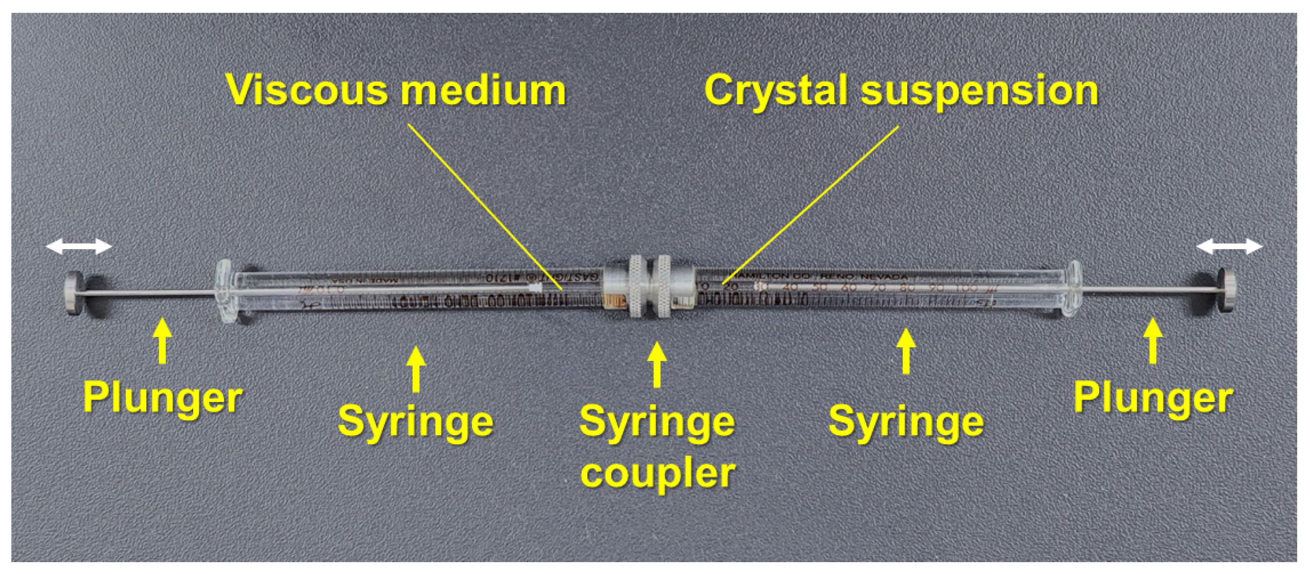
2. Syringe-Based Microcrystal Delivery Method

The advantage of using syringes for sample delivery is that they are commercially available, and therefore do not require the manufacturing of a specific sample delivery system. In contrast, many other injector systems commonly used in SX experiments require precise and professional manufacturing. Furthermore, syringe needles come in various inner diameters, a feature that allows researchers to select the most suitable option based on crystal size. The syringe-based sample delivery method generally involves embedding the crystals in a viscous medium and extruding them to the X-ray interaction point at a low flow rate (Figure 2). This approach helps reduce crystal consumption and facilitates data collection at XFEL facilities with low repetition rates or synchrotrons.
Sugahara et al. were the first to use syringes for crystal sample delivery during SFX experiments [44]. This study used syringes during the development of a grease-based viscous delivery medium that was used to reduce crystal sample consumption during SX experiments. Sample delivery was achieved using a syringe needle with an inner diameter of 110 μm. To demonstrate the applicability of the grease matrix, four different protein crystals—i.e., lysozyme, glucose isomerase, fatty acid-binding protein type 3, and thaumatin—were embedded in the grease matrix and delivered to the X-ray interaction point at a flow rate of 0.46–0.48 μL/min. Diffraction data were successfully collected for all proteins, and their crystal structures were determined. Moreover, to further reduce crystal consumption, the sample delivery was performed with a different experimental approach at lower flow rates. For example, a 110-μm inner diameter syringe needle was used to deliver grease-embedded lysozyme crystals at a flow rate of 0.12 μL/min while exposed to a 30 Hz X-ray source. This sample delivery condition showed no noticeable difference in the indexing rates or diffraction patterns when compared with the high-flow-rate sample delivery condition. In addition, when 7–10-μm lysozyme crystals embedded in grease were delivered using a 50-μm inner diameter syringe needle, only ~0.1 mg of crystals were consumed. In contrast, the sample consumption was around 1 mg when using a needle with a 110-μm inner diameter.
Berntsen et al. developed the “Lipidico” system that combines an EIGER X 16M detector with a high-viscosity injector developed in-house [45]. A commercially available syringe containing microcrystals embedded in a viscous medium was installed in the Lipidico system, and the crystal sample was extruded using a drive motor. When viscous material passes through the syringe needle, the injection stream can become unstable and curl near the needle tip. To address this issue, two approaches have been developed. First, a counteracting electrostatic charge was applied to a small piece of polystyrene (~5 mm) placed under the needle, which creates a downward-acting Coulomb force on the sample. Second, a weak suction force was applied using a Venturi funnel positioned beneath the syringe needle to stabilize the injection stream. Three different viscous media—i.e., petroleum jelly, monoolein, and silicone grease—were then tested using these two methods, showing that all combinations produced a stable and straight injection stream. Subsequently, the capability of the Lipidico system was demonstrated by collecting SSX data for lysozyme. Grease containing lysozyme crystals was extruded through a needle with an inner diameter of 110 μm at a velocity of 100–120 μm/s. Using approximately 20 μL of the lysozyme crystal-embedded grease mixture, the crystal structure of lysozyme was successfully determined, with no radiation damage to disulfide bonds observed.
Park and Nam conducted an SSX experiment at the Pohang Light Source II (PLS-II) [46]. For this experiment, lysozyme crystals embedded in lipidic cubic phase (LCP) and polyacrylamide (PAM) injection matrix were delivered using a syringe at extrusion flow rates of 100 nL/min and 200 nL/min, respectively. The observed thicknesses of the LCP and PAM injection streams were approximately 190 μm and 500 μm, respectively. This result indicates that the dimension of the injection stream can vary depending on the viscous material, even under identical experimental conditions. In addition, although the same batch of lysozyme crystals were analyzed under identical experimental settings, the diffraction limits differed between LCP and PAM injection streams, which yielded measurements of 1.56 Å and 1.76 Å, respectively. Taken together, these results indicate that the viscosity of the medium can affect crystal and data quality.
Park developed a syringe-based microliter volume (MLV) syringe injector for SFX data collection at PLA-XFEL [47]. The development of the MLV injector has been reported in detail, and experimental data have shown that this setup produces a straight and stable injection stream.
Figure 2. Syringe-based microcrystal delivery methods. (A) Photo of lysozyme crystals in the LCP injection medium. Original figures were obtained from a previous study [29]. (B) Photo of a lard injection stream containing lysozyme crystals. Original figures were obtained from a previous study [48].
Analytica 06 00005 g002

3. Sample Preparation

The syringe-based sample delivery method stably delivers microcrystals embedded in a viscous medium at a low flow rate to reduce sample consumption. However, occasionally an injection stream is not formed when delivering a crystal suspension, including solutions containing crystals, at a low flow rate [46]. In this case, crystal suspensions accumulate at the syringe needle tip and fall off once they reach a certain size. In this case, if the X-rays are exposed to these drops at the tip, crystals may be repeatedly exposed to X-rays for extended periods, thereby potentially leading to reduced sample quality due to radiation damage and X-ray heating. While increasing the flow rate at high speed can address this issue, this solution can result in excessive sample consumption. Therefore, to date sample delivery using a syringe is only useful for delivery of a viscous medium containing microcrystals, which produces a stable injection stream at low flow rate due to material viscosity.
Two primary methods are commonly used to embed crystal samples in a viscous medium: manual mixing with a spatula and mechanical mixing using a dual-syringe setup. In the manual mixing method, crystals and a viscous material, such as a mineral oil-based grease, are manually mixed on a plate with the spatula [44]. The resulting mixture is then transferred to a pipette tip, again using the spatula, and the tip of the pipette tip is sealed with parafilm. After centrifugation, the material is transferred into a syringe for sample extrusion [44]. Although this method has successfully embedded crystal samples in grease, thereby enabling the collection of diffraction data and facilitating structural determination, it carries the potential for significant sample loss during the preparation process [49].
The dual-syringe setup method involves transferring the crystal suspension and the viscous material into separate syringes, connecting these syringes with a syringe coupler, and pushing the syringe plungers gently to embed crystals into the viscous medium [50,51] (Figure 3). Once mixed, the material is then transferred into a single syringe, and a syringe needle is connected for sample extrusion. During mixing, the syringe plungers should be very gently pressed to minimize physical stress on crystal samples. Compared to the manual mixing method, this approach results in less sample loss during preparation and has therefore become the preferred method for embedding crystals into viscous media in recent years.
Meanwhile, in a viscous medium, the crystals are surrounded by the crystallization solution, creating a hydrated environment. If the crystallization solution does not surround the crystals, leading to a dehydrated environment within the viscous medium, the crystal lattices may become physically damaged.

4. Syringe Plunger Pushing for Sample Extrusion

Syringe-based sample delivery methods involve transporting crystal samples through a syringe needle to the X-ray interaction point by applying physical force to the syringe plunger. So far, systems used for extruding crystal samples from syringes in SX experiments can be broadly categorized as one of three types: in-house developed motor drive systems, commercially available syringe pumps, and high-pressure liquid chromatography (HPLC) pump systems. First, an in-house developed motor drive system was developed for syringe-based sample delivery experiments. Sugahara et al. controlled the flow rate for extruding crystal samples from a syringe using a stepper motor that pushes the syringe plunger [44]. In the Lipidico system, a drive device equipped with a 24 V DC motor, a 1:231 gearbox, and a screw with a 320 μm pitch was used to push the syringe plunger and extrude the crystal samples from the syringe [45].
Syringe pumps are widely used for sample delivery in SX experiments. For example, Stellato et al. delivered the crystals through a 100 µm inner-diameter fused silica fiber to a 100 µm inner-diameter glass capillary using a syringe pump [52]. Park and Nam delivered the viscous medium containing crystals from a syringe using a syringe pump [46]. Furthermore, syringe pumps are also extensively used in mix-and-inject experiments, in which chemicals are mixed with crystal samples to facilitate substrate binding or other chemical interactions [53].
HPLC systems are frequently used to build liquid jet injectors and LCP injectors [54,55]. For syringe-based microliter volume (MLV) injectors, an HPLC pump is used to push a syringe plunger [47]. In this setup, the syringe plunger is moved by water pressure supplied by the HPLC system, and the crystal sample is thereby transported via the syringe needle. Depending on the HPLC system, this setup can realize a maximum pressure of up to 40 MPa, and can provide flow rates that range from 0.0001 to 10.00 mL/min.

5. Advantages of Microcrystal Delivery Using Syringes and Syringe Pumps

Unlike the highly sophisticated and precise sample delivery methods typically required for general SX experiments, sample delivery methods that use only a syringe and syringe pump offer an accessible and straightforward alternative. These methods have several notable advantages from an application perspective.

5.1. Commercial Availability

Syringe pumps are widely used in various research and technological applications [56,57]. They are commercially available in a range of specifications and functionalities. In addition, their operation is straightforward, which eliminates the need for time and effort spent in developing and operating a custom driving device for pushing the syringe plunger. Furthermore, commercially available syringe pumps come with detailed operation manuals and programs, which offers users a quick understanding of their functions and operation. In contrast, in-house developed systems require significant time investments, both to manufacture the device and to understand its operational performance and functionality. Meanwhile, while HPLC systems are also commercially available, they are significantly more expensive than syringe pumps.

5.2. Preliminary Injection Tests

When using syringe-based sample delivery approaches, it is critical to produce a stable injection stream of the viscous medium [49]. If medium viscosity fluctuates and fails to generate a consistent injection stream, samples may end up being overexposed to X-rays, which can result in unreliable data, high levels of radiation damage, and low hit rate efficiency. To address these challenges, it is important to test the sample delivery system and evaluate injection stream stability before experimental data is collected. Preliminary injection tests for evaluating injection stream stability can be limited, since injection systems are typically installed at the beamline or developed by specialized research groups. This limitation can significantly reduce the overall efficiency of SX data collection. While it is possible to replicate the injector system used at the beamline in a laboratory, this requires considerable time, cost, and technical expertise. Alternatively, using commercially available syringes and syringe pumps allows researchers to prescreen conditions for generating stable injection streams, thereby enabling the identification of optimal parameters for stable sample delivery.

5.3. Portability

Despite successful selection of suitable viscous media and optimization of injection stream conditions in the laboratory, variations in syringe depression can lead to irreproducible injection streams during data collection at the beamline. To enhance reproducibility, it is important to maintain consistency between testing and data collection environments. Unlike other syringe pushing systems, syringe pumps are compact and portable, which allows them to be transported and used at the beamline. This ensures that the same syringe pump system tested in the laboratory can also be used during SX data collection, thereby improving reproducibility and enabling more efficient use of experimental time. To take advantage of this, there must be enough space at the beamline to install the syringe pump near the X-ray focal point.

5.4. Independent Operation

In typical SX experiments, sample delivery is performed by in-house sample delivery developers or by beamline managers. Consequently, researchers must undergo specialized training to understand and operate these devices, which can be time-consuming and inconvenient. In contrast, syringe pumps are simple to operate and enable researchers to independently implement syringe-based sample delivery for their experiments, streamlining the overall process and reducing reliance on external assistance.

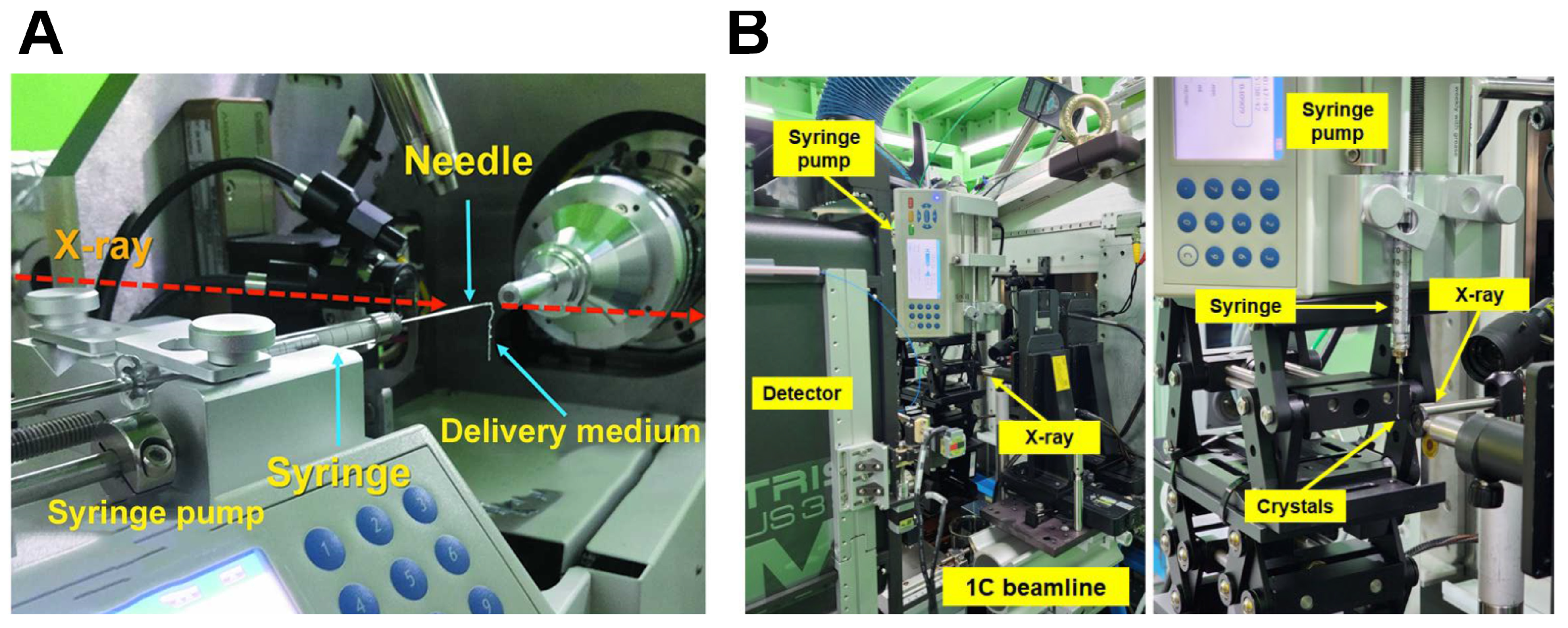
6. Applications of Syringe and Syringe Pump-Based Sample Delivery

Microcrystal delivery methods using syringes and syringe pumps have been applied to various SX experimental techniques (Table 1). Here, representative applications are introduced.

6.1. Demonstration of the SSX Experiment

Experimental protocols that use syringes and syringe pumps were first employed at the 11C beamline at PLS-II for monochromatic X-ray-based SSX [46]. Installation of a syringe and a syringe needle in the vertical direction is preferred for extruding a stable injection stream. However, when installing the syringe pump in the X-ray focusing region of the 11C beamline, interference with surrounding beamline equipment occurred, and there was no space to install the syringe pump vertically. Accordingly, the syringe was mounted horizontally, with a syringe pump depressing the plunger from the side (Figure 4A). Here the viscous medium containing the crystals delivered from the syringe needle did not form a vertical injection stream but rather a curved downward flow. During data collection, fluctuations in the position of the injection stream were observed on the monitor. However, since the thickness of the injection stream was wider than that of the X-ray beam, the viscous medium containing the crystals was continuously exposed to X-rays, thereby enabling the collection of diffraction data. Using LCP and PAM injection matrices, the room-temperature structures of lysozyme were determined at resolutions of 1.65 Å and 1.76 Å, respectively.
In another study at the PLS-II 1C beamline, a sample delivery method using a syringe and syringe pump was employed for pink-beam SSX [58]. The X-ray focal point at the 1C beamline had sufficient space to allow for the vertical installation of a syringe and syringe pump, resulting in a straight vertical injection stream (Figure 4B). In this experiment, crystals were embedded in a shortening injection matrix, which generated an injection stream with a thickness of approximately 300 µm. Exposing the X-ray beam to the edges of the injection stream improved the signal-to-noise ratio (SNR) of diffraction data. The room-temperature structures of lysozyme were then determined at a resolution of 1.80 Å. In conclusion, during both experiments, the syringe pump was appropriately adjusted to the sample position on the beamline, successfully collecting diffraction data from injection streams whether they flowed vertically or horizontally. However, for horizontal sample delivery, the downward angle of the injection stream can vary depending on the delivery material and/or flow rate, and this can potentially cause fluctuations in the sample position. Therefore, vertical sample delivery is preferred to ensure a stable injection stream, and syringes and syringe pumps should be installed accordingly.

6.2. Development of Viscous Media

Delivering crystal samples using viscous media provides a stable injection stream even at low flow rates, thereby reducing sample consumption in SX experiments using low-repetition-rate XFELs or synchrotrons [49]. However, medium viscosity can be affected by crystal sample properties, crystallization solutions, and environmental conditions, all of which can potentially destabilize the injection stream. Depending on the properties of the materials used in the injection stream, various physical impacts or interactions may also degrade crystal quality. To address these issues, it is crucial to identify viscous materials that can maintain the stability of the crystal samples [49].
The syringe and syringe pump method has been applied during the development of viscous media, such as a shortening injection matrix [59], a wheat starch injection matrix [60], alginate injection matrix [60], a lard injection matrix [48], and a beef tallow injection matrix [61]. Using viscous media, diffraction data were successfully collected, and the crystal structures of lysozyme and glucose isomerase were determined (Figure 5). However, each material exhibits unique physical properties, such as preferred injection flow rates, temperature stability, and chemical compatibility, and these findings highlight the need for material-specific optimization.

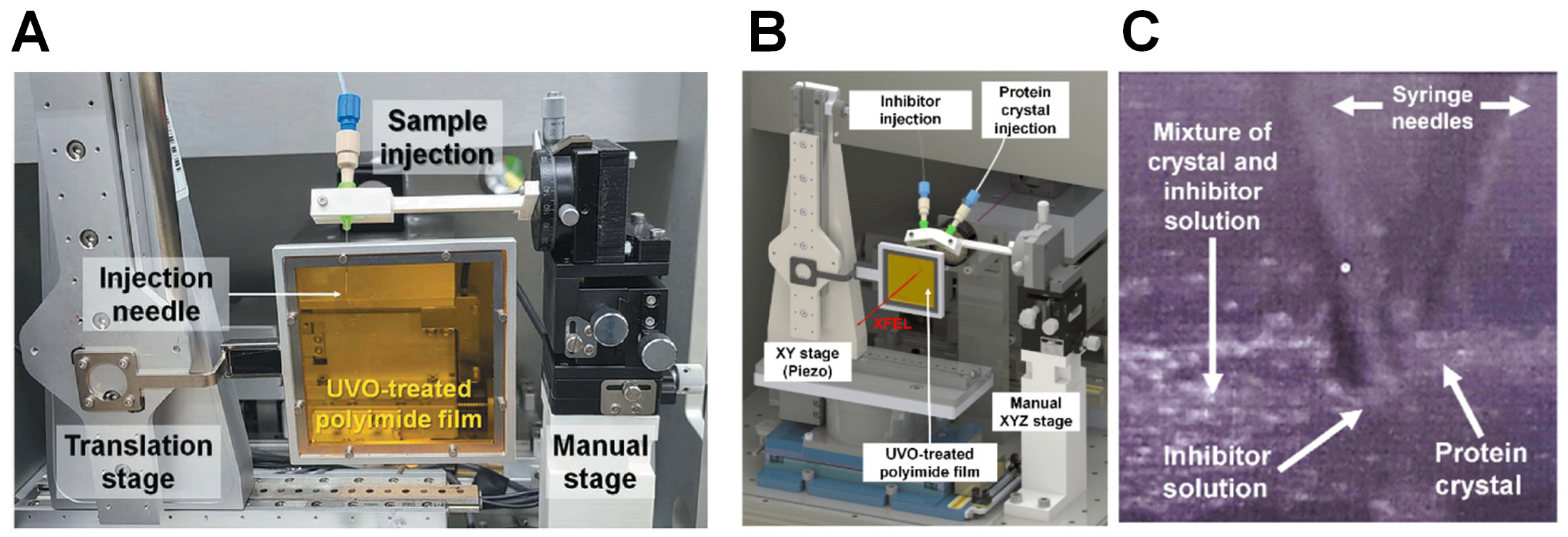
6.3. Hybrid Sample Delivery Methods

Delivery medium viscosity can also vary depending on sample environment, and this can also affect the stability of the injection stream [49]. To overcome this effect, the syringe and syringe pump method has been used to develop novel SX sample delivery techniques based on quartz capillaries [62] and polyimide-based single-channel microfluidic chips [63]. In general, capillaries and microfluidic chips are connected to a syringe needle, and this setup ensures that all of the viscous medium containing crystals passes through the inner space of the capillary and microfluidic chip. Moreover, since X-rays were aligned with the channel of the capillary and microfluidic chip, sample delivery via syringe pump pressure means that they are always exposed to X-rays regardless of the viscosity of the sample delivery medium.
In addition, the syringe and syringe pump method has also been used to develop a novel combination of the injection and transfer system (BITS) [64] (Figure 6A). Specifically, a viscous medium containing crystals was extruded from a syringe needle by syringe pump-mediated syringe depression; this resulted in the deposition onto UV/ozone (UVO)-treated polyimide film. The deposited viscous medium can then be transferred to the X-ray interaction point by the translation stage. This method has the advantage of using less sample consumption relative to traditional viscosity-based sample delivery methods and can also continuously provide fresh crystal samples to the X-rays.
Table 1. Reports of SX sample delivery using a syringe and syringe pump setup.
ApplicationCommentReference
Demonstration of the SSX experimentDemonstration of SSX with monochromatic X-ray beam using sample delivery using syringe and syringe pump[35]
Pink-beam serial synchrotron crystallography at the Pohang Light Source II[47]
Development of the viscous mediumShortening the injection matrix for serial crystallography.[48]
Polysaccharide-based injection matrix for serial crystallography[49]
Lard injection matrix[37]
Beef tallow matrix[50]
Hybrid sample delivery methodStable sample delivery in viscous media via a capillary for serial crystallography.[51]
Stable sample delivery in a viscous medium via a polyimide-based single-channel microfluidics chip for serial crystallography.[52]
Combination of an inject-and-transfer system for serial femtosecond crystallography[53]
Upgraded combined inject-and-transfer system for serial femtosecond crystallography[42]
Data processingApplication of Serial Crystallography for Merging Incomplete Macromolecular Crystallography Datasets[65]
Structure determinationRoom-temperature structure analysis[25]
A sample delivery technique using syringes and a syringe pump has also been applied to the development of an injection and diffusion technique [53] (Figure 6B). For this method, two syringes are connected to the syringe pump; one contains crystals in a viscous medium while the other contains an inhibitor solution that is mixed within the BITS system (Figure 6C). Although the structure of the protein-inhibitor complex was not determined in initial experiments and further improvements are needed, in theory this method may be useful for studying the slow reaction times of proteins and substrates in time-resolved studies.

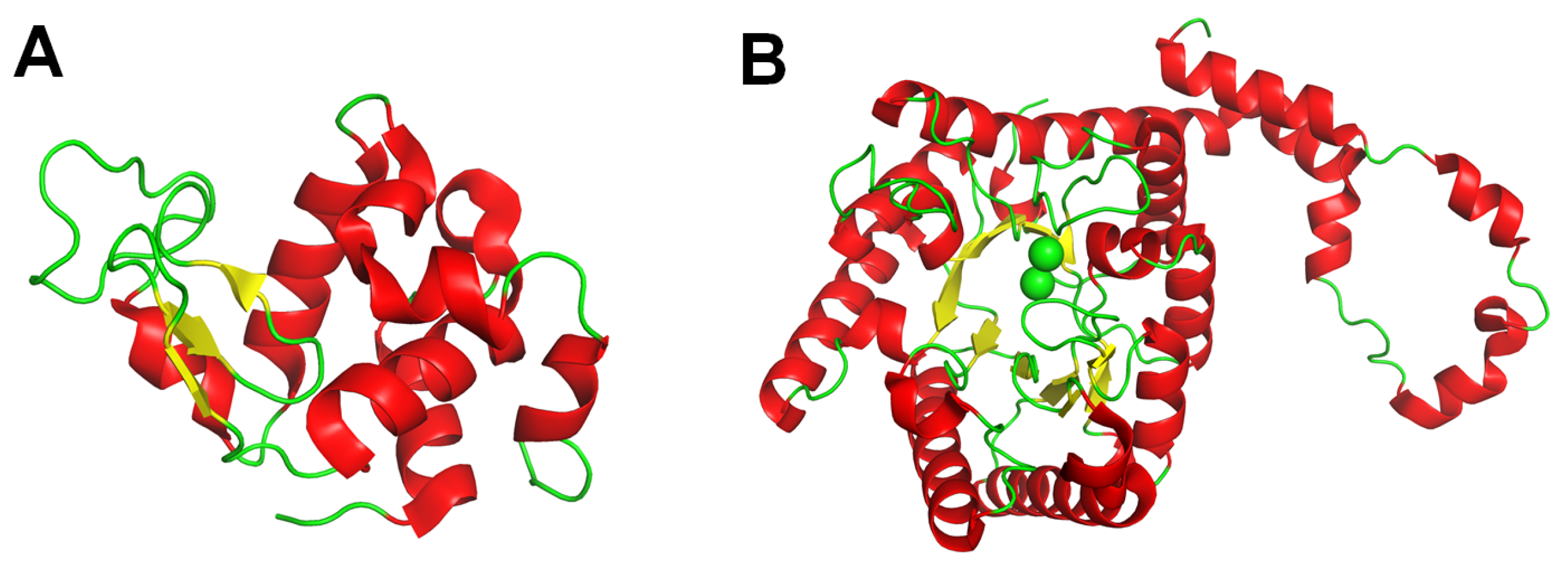
6.4. Room-Temperature Structure Determination

Using a syringe and syringe pump system, the crystal structures of lysozyme and glucose isomerase have been determined during the development of SX experimental techniques (Figure 7). These structures showed the room-temperature structure without significant radiation damage. These findings demonstrate that syringe and syringe pump-based techniques can be successfully applied to SX experiments.

7. Discussion

In SX experiments, successful data collection and efficient use of meantime depend on the stable and continuous delivery of crystal samples to the X-ray interaction point. Accordingly, various sample delivery methods have been developed for SX. However, these are generally tailored to specific sample types or experimental goals, and their development remains ongoing. Overall, these methods aim to optimize data collection in SX by reducing sample consumption and minimizing background scattering, thereby improving data quality. However, designing, manufacturing, and operating these delivery systems often require significant expertise and experience, a bottleneck that makes them accessible only to developers and skilled operators. Consequently, a system that provides greater convenience for typical SX researchers to test and operate samples independently would be valuable.
This review focuses on a sample delivery method that uses commercially available syringe and syringe pump for crystal sample delivery. For SX experiments, the required sample volumes vary depending on research objective and experiment type. Commercial syringes are available in various volumes, which allows researchers to select the appropriate size for their needs. Syringe needles are also available in a range of inner diameters and tip shapes, which enables customization to fit crystal samples and research requirements. In addition, a wide variety of syringe pumps are also commercially available, and these can safely deliver samples by controlling the syringe plunger’s movement at a desired flow rate. These pumps are generally easy to operate, do not require specialized technical skills, and are relatively portable. Portability is valuable since it facilitates testing in different environments and allows installation at beamline for SX data collection.
Syringe-based sample delivery methods are particularly suitable for embedding crystal samples in viscous materials. The injection stream extruding from a syringe needle typically has a larger diameter than the inner diameter of the syringe needle itself due to the cohesion and viscosity of the viscous medium extruded. While thicker injection streams offer greater stability, excessive stream thickness can result in unnecessary sample consumption if the width of the injection stream exceeds X-ray beam size. For example, if the injection stream is 200 μm thick and the beam size is 10 μm in size, only 5% of the stream is exposed to X-rays, meaning that approximately 95% of the sample is wasted. To minimize sample consumption, it is therefore crucial to reduce injection stream thickness by using needles with narrower inner diameters and by increasing the viscosity of the embedding material to prevent stream expansion upon extrusion. Importantly, the injection stream affects background scattering, which can lower the SNR and degrade diffraction data quality. Thus, a thinner injection stream is beneficial not only for sample consumption but also for enhancing data quality. If reducing the stream thickness is not feasible, the X-ray beam can be aligned with the edge rather than the center of the stream, since its cylindrical shape can help reduce background scattering [58].
The choice of syringe needle should also consider the dimensions of the cylinder relative to the size of the crystal being examined. Using a syringe needle with a diameter smaller than the crystal’s size can physically damage them during delivery. This strongly reduces diffraction quality and can cause clogging of the needle inlet.
To date, various viscous media, including Aliphatic LCP (e.g., monoolein [55,66]), hydrophilic organic compounds (e.g., agarose [51], hyaluronic acid [67], hydroxyethyl cellulose [68], sodium carboxymethyl cellulose [69], wheat starch [60], alginate [60], Pluronic F-127 [69], poly(ethylene oxide) [70], and polyacrylamide [71]), hydrophobic (grease [44,72], shortening [59], lard [48] and beef tallow [61]) have been successfully used for SX experiments. These compounds can therefore be used as crystal delivery media. However, the selected medium must facilitate the maintenance of crystal stability without causing physical stress or allowing chemical reactions with biomolecules. It is therefore essential to test crystal stability in the viscous medium prior to data collection. Even if crystals remain morphologically stable within a viscous medium, physical mixing or interactions between the crystals and the viscous material can degrade diffraction quality. Therefore, it is also crucial to prescreen crystal diffraction quality by exposing embedded crystals to X-rays in MX or SX experiments.
To incorporate syringe and syringe pumps in SX data collection, it is important to have sufficient space set aside for the installation of the syringe pump near the X-ray focusing region of the beamline. After connecting the syringe to the pump, the syringe needle tip must then be carefully aligned with the X-ray interaction point. This system must also allow for the precise alignment of the injection stream with the X-ray focal point to ensure the proper delivery of high-intensity X-rays to the sample. Meanwhile, maintaining the setup in a helium environment or using high-energy X-rays can be advantageous for reducing background scattering caused by differences in air density.
Syringes and syringe pumps have been employed in SX experiments using a wide variety of viscous delivery media, sample delivery methods, and inject-and-transfer systems. To date, only the structures of lysozyme and glucose isomerase have been determined, as these were used as model samples to demonstrate system development, resulting in a lack of diverse examples for structure determination. Since sample delivery methods using syringes and syringe pumps are based on the same principles as other syringe-based delivery systems, they can be broadly applied to SX-based structural determination of various biomolecules and chemical compounds.

8. Conclusions

This review introduces the sample delivery method using syringes and syringe pumps for SX experiments. The main advantage of this method is that it allows for straightforward sample delivery using commercially available syringe pumps and therefore does not require extensive training. This review is expected to contribute to the determination of crystal structures in future SX studies.

Funding

This work was funded by the National Research Foundation of Korea (NRF) (NRF-2021R1I1A1A01050838 to K.H.N.).

Data Availability Statement

Data sharing is not applicable.

Acknowledgments

I would like to thank Suk-Youl Park and Yongsam Kim at the Pohang Light Source II for their assistance during the demonstration of the sample delivery method. The author thanks the Global Science experimental Data Hub Center (GSDC) at the Korea Institute of Science and Technology Information (KISTI) for providing computing resources and technical support.

Conflicts of Interest

The author declares no conflicts of interest.

Abbreviations

The following abbreviations are used in this manuscript:
SXSerial crystallography
SFXSerial femtosecond crystallography
SSXSerial synchrotron crystallography
XFELX-ray free electron laser
LCPLipidic Cubic Phase
PALPohang Accelerator Laboratory
PLS-IIPohang Light Source II
BITSCombination of an inject-and-transfer system

References

  1. Duke, E.M.H.; Johnson, L.N. Macromolecular crystallography at synchrotron radiation sources: Current status and future developments. Proc. R. Soc. A Math. Phys. Eng. Sci. 2010, 466, 3421–3452. [Google Scholar] [CrossRef]
  2. Walter, R.L.; Thiel, D.J.; Barna, S.L.; Tate, M.W.; Wall, M.E.; Eikenberry, E.F.; Gruner, S.M.; Ealick, S.E. High-resolution macromolecular structure determination using CCD detectors and synchrotron radiation. Structure 1995, 3, 835–844. [Google Scholar] [CrossRef]
  3. Mulholland, A.J. Modelling enzyme reaction mechanisms, specificity and catalysis. Drug Discov. Today 2005, 10, 1393–1402. [Google Scholar] [CrossRef] [PubMed]
  4. Selvaraj, C.; Rudhra, O.; Alothaim, A.S.; Alkhanani, M.; Singh, S.K. Structure and chemistry of enzymatic active sites that play a role in the switch and conformation mechanism. In Protein Design and Structure; Advances in Protein Chemistry and Structural Biology; Elsevier: Amsterdam, The Netherlands, 2022; pp. 59–83. [Google Scholar]
  5. Schwalbe, H.; Audergon, P.; Haley, N.; Amaro, C.A.; Agirre, J.; Baldus, M.; Banci, L.; Baumeister, W.; Blackledge, M.; Carazo, J.M.; et al. The future of integrated structural biology. Structure 2024, 32, 1563–1580. [Google Scholar] [CrossRef]
  6. Blundell, T.L.; Jhoti, H.; Abell, C. High-throughput crystallography for lead discovery in drug design. Nat. Rev. Drug Discov. 2002, 1, 45–54. [Google Scholar] [CrossRef]
  7. Zheng, H.; Hou, J.; Zimmerman, M.D.; Wlodawer, A.; Minor, W. The future of crystallography in drug discovery. Expert Opin. Drug Discov. 2013, 9, 125–137. [Google Scholar] [CrossRef] [PubMed]
  8. Bajad, N.G.; Rayala, S.; Gutti, G.; Sharma, A.; Singh, M.; Kumar, A.; Singh, S.K. Systematic review on role of structure based drug design (SBDD) in the identification of anti-viral leads against SARS-Cov-2. Curr. Res. Pharmacol. Drug Discov. 2021, 2, 100026. [Google Scholar] [CrossRef] [PubMed]
  9. Wang, Y.; Yu, L.; Shao, J.; Zhu, Z.; Zhang, L. Structure-driven protein engineering for production of valuable natural products. Trends Plant Sci. 2023, 28, 460–470. [Google Scholar] [CrossRef] [PubMed]
  10. Wang, T.; Yang, W.-t.; Gong, Y.-m.; Zhang, Y.-k.; Fan, X.-x.; Wang, G.-c.; Lu, Z.-h.; Liu, F.; Liu, X.-h.; Zhu, Y.-s. Molecular engineering of PETase for efficient PET biodegradation. Ecotoxicol. Environ. Saf. 2024, 280, 116540. [Google Scholar] [CrossRef]
  11. Ndochinwa, G.O.; Wang, Q.-Y.; Okoro, N.O.; Amadi, O.C.; Nwagu, T.N.; Nnamchi, C.I.; Moneke, A.N.; Odiba, A.S. New advances in protein engineering for industrial applications: Key takeaways. Open Life Sci. 2024, 19, 20220856. [Google Scholar] [CrossRef] [PubMed]
  12. Nishimasu, H.; Shi, X.; Ishiguro, S.; Gao, L.; Hirano, S.; Okazaki, S.; Noda, T.; Abudayyeh, O.O.; Gootenberg, J.S.; Mori, H.; et al. Engineered CRISPR-Cas9 nuclease with expanded targeting space. Science 2018, 361, 1259–1262. [Google Scholar] [CrossRef] [PubMed]
  13. Hirano, S.; Nishimasu, H.; Ishitani, R.; Nureki, O. Structural Basis for the Altered PAM Specificities of Engineered CRISPR-Cas9. Mol. Cell 2016, 61, 886–894. [Google Scholar] [CrossRef]
  14. de la Mora, E.; Coquelle, N.; Bury, C.S.; Rosenthal, M.; Holton, J.M.; Carmichael, I.; Garman, E.F.; Burghammer, M.; Colletier, J.-P.; Weik, M. Radiation damage and dose limits in serial synchrotron crystallography at cryo- and room temperatures. Proc. Natl. Acad. Sci. USA 2020, 117, 4142–4151. [Google Scholar] [CrossRef] [PubMed]
  15. Shelley, K.L.; Garman, E.F. Quantifying and comparing radiation damage in the Protein Data Bank. Nat. Commun. 2022, 13, 1314. [Google Scholar] [CrossRef]
  16. Hattne, J.; Shi, D.; Glynn, C.; Zee, C.-T.; Gallagher-Jones, M.; Martynowycz, M.W.; Rodriguez, J.A.; Gonen, T. Analysis of Global and Site-Specific Radiation Damage in Cryo-EM. Structure 2018, 26, 759–766.e4. [Google Scholar] [CrossRef]
  17. Ravelli, R.B.G.; McSweeney, S.M. The ‘fingerprint’ that X-rays can leave on structures. Structure 2000, 8, 315–328. [Google Scholar] [CrossRef] [PubMed]
  18. Murray, J.; Garman, E. Investigation of possible free-radical scavengers and metrics for radiation damage in protein cryocrystallography. J. Synchrotron Radiat. 2002, 9, 347–354. [Google Scholar] [CrossRef] [PubMed]
  19. Banumathi, S.; Zwart, P.H.; Ramagopal, U.A.; Dauter, M.; Dauter, Z. Structural effects of radiation damage and its potential for phasing. Acta Crystallogr. D Biol. 2004, 60, 1085–1093. [Google Scholar] [CrossRef]
  20. Nam, K.H. Radiation Damage on Thaumatin: A Case Study of Crystals That Are Larger Than the Microfocusing X-ray Beam. Appl. Sci. 2023, 13, 1876. [Google Scholar] [CrossRef]
  21. Taberman, H. Radiation Damage in Macromolecular Crystallography—An Experimentalist’s View. Crystals 2018, 8, 157. [Google Scholar] [CrossRef]
  22. Nam, K.H. Temperature-dependent structural changes in xylanase II from Trichoderma longibrachiatum. Carbohydr. Res. 2024, 541, 109173. [Google Scholar] [CrossRef] [PubMed]
  23. Bradford, S.Y.C.; El Khoury, L.; Ge, Y.; Osato, M.; Mobley, D.L.; Fischer, M. Temperature artifacts in protein structures bias ligand-binding predictions. Chem. Sci. 2021, 12, 11275–11293. [Google Scholar] [CrossRef] [PubMed]
  24. Fraser, J.S.; van den Bedem, H.; Samelson, A.J.; Lang, P.T.; Holton, J.M.; Echols, N.; Alber, T. Accessing protein conformational ensembles using room-temperature X-ray crystallography. Proc. Natl. Acad. Sci. USA 2011, 108, 16247–16252. [Google Scholar] [CrossRef]
  25. Nam, K.H. Temperature-Dependent Structural Changes of the Active Site and Substrate-Binding Cleft in Hen Egg White Lysozyme. Crystals 2025, 15, 111. [Google Scholar] [CrossRef]
  26. Chapman, H.N.; Barty, A.; Bogan, M.J.; Boutet, S.; Frank, M.; Hau-Riege, S.P.; Marchesini, S.; Woods, B.W.; Bajt, S.; Benner, W.H.; et al. Femtosecond diffractive imaging with a soft-X-ray free-electron laser. Nat. Phys. 2006, 2, 839–843. [Google Scholar] [CrossRef]
  27. Chapman, H.N.; Caleman, C.; Timneanu, N. Diffraction before destruction. Philos. Trans. R. Soc. Lond. B Biol. Sci. 2014, 369, 20130313. [Google Scholar] [CrossRef] [PubMed]
  28. Boutet, S.; Lomb, L.; Williams, G.J.; Barends, T.R.M.; Aquila, A.; Doak, R.B.; Weierstall, U.; DePonte, D.P.; Steinbrener, J.; Shoeman, R.L.; et al. High-Resolution Protein Structure Determination by Serial Femtosecond Crystallography. Science 2012, 337, 362–364. [Google Scholar] [CrossRef] [PubMed]
  29. Nam, K.H. Guide to serial synchrotron crystallography. Curr. Res. Struct. Biol. 2024, 7, 100131. [Google Scholar] [CrossRef]
  30. Kupitz, C.; Basu, S.; Grotjohann, I.; Fromme, R.; Zatsepin, N.A.; Rendek, K.N.; Hunter, M.S.; Shoeman, R.L.; White, T.A.; Wang, D.; et al. Serial time-resolved crystallography of photosystem II using a femtosecond X-ray laser. Nature 2014, 513, 261–265. [Google Scholar] [CrossRef]
  31. Poddar, H.; Heyes, D.J.; Schirò, G.; Weik, M.; Leys, D.; Scrutton, N.S. A guide to time-resolved structural analysis of light-activated proteins. FEBS J. 2021, 289, 576–595. [Google Scholar] [CrossRef] [PubMed]
  32. Brändén, G.; Neutze, R. Advances and challenges in time-resolved macromolecular crystallography. Science 2021, 373, eaba0954. [Google Scholar] [CrossRef] [PubMed]
  33. Kupitz, C.; Olmos, J.L.; Holl, M.; Tremblay, L.; Pande, K.; Pandey, S.; Oberthür, D.; Hunter, M.; Liang, M.; Aquila, A.; et al. Structural enzymology using X-ray free electron lasers. Struct. Dyn. 2017, 4, 044003. [Google Scholar] [CrossRef] [PubMed]
  34. Worrall, J.A.R.; Hough, M.A. Serial femtosecond crystallography approaches to understanding catalysis in iron enzymes. Curr. Opin. Struc. Biol. 2022, 77, 102486. [Google Scholar] [CrossRef]
  35. Stagno, J.R.; Knoska, J.; Chapman, H.N.; Wang, Y.-X. Mix-and-Inject Serial Femtosecond Crystallography to Capture RNA Riboswitch Intermediates. In RNA Structure and Dynamics; Methods in Molecular Biology; Humana: New York, NY, USA, 2023; pp. 243–249. [Google Scholar]
  36. Kim, J.; Kim, Y.; Park, J.; Nam, K.H.; Cho, Y. Structural mechanism of Escherichia coli cyanase. Acta Crystallogr. D Biol. Crystallogr. 2023, 79, 1094–1108. [Google Scholar] [CrossRef] [PubMed]
  37. Kim, J.; Park, J.; Lee, K.; Chung, W.K.; Nam, K.H.; Cho, Y. Exploring the reaction dynamics of alanine racemase using serial femtosecond crystallography. Sci. Rep. 2024, 14, 31442. [Google Scholar] [CrossRef]
  38. White, T.A.; Kirian, R.A.; Martin, A.V.; Aquila, A.; Nass, K.; Barty, A.; Chapman, H.N. CrystFEL: A software suite for snapshot serial crystallography. J. Appl. Crystallogr. 2012, 45, 335–341. [Google Scholar] [CrossRef]
  39. White, T.A. Processing serial crystallography data with CrystFEL: A step-by-step guide. Acta Crystallogr. D Struct. Biol. 2019, 75, 219–233. [Google Scholar] [CrossRef] [PubMed]
  40. Grünbein, M.L.; Nass Kovacs, G. Sample delivery for serial crystallography at free-electron lasers and synchrotrons. Acta Crystallogr. D Biol. Crystallogr. 2019, 75, 178–191. [Google Scholar] [CrossRef]
  41. Martiel, I.; Muller-Werkmeister, H.M.; Cohen, A.E. Strategies for sample delivery for femtosecond crystallography. Acta Crystallogr. D Struct. Biol. 2019, 75, 160–177. [Google Scholar] [CrossRef]
  42. Zhao, F.Z.; Zhang, B.; Yan, E.K.; Sun, B.; Wang, Z.J.; He, J.H.; Yin, D.C. A guide to sample delivery systems for serial crystallography. FEBS J. 2019, 286, 4402–4417. [Google Scholar] [CrossRef]
  43. Aller, P.; Orville, A.M. Dynamic Structural Biology Experiments at XFEL or Synchrotron Sources. In Structural Proteomics; Methods in Molecular Biology; Humana: New York, NY, USA, 2021; pp. 203–228. [Google Scholar]
  44. Sugahara, M.; Mizohata, E.; Nango, E.; Suzuki, M.; Tanaka, T.; Masuda, T.; Tanaka, R.; Shimamura, T.; Tanaka, Y.; Suno, C.; et al. Grease matrix as a versatile carrier of proteins for serial crystallography. Nat. Methods 2015, 12, 61–63. [Google Scholar] [CrossRef] [PubMed]
  45. Berntsen, P.; Hadian Jazi, M.; Kusel, M.; Martin, A.V.; Ericsson, T.; Call, M.J.; Trenker, R.; Roque, F.G.; Darmanin, C.; Abbey, B. The serial millisecond crystallography instrument at the Australian Synchrotron incorporating the “Lipidico” injector. Rev. Sci. Instrum. 2019, 90, 085110. [Google Scholar] [CrossRef] [PubMed]
  46. Park, S.Y.; Nam, K.H. Sample delivery using viscous media, a syringe and a syringe pump for serial crystallography. J. Synchrotron Radiat. 2019, 26, 1815–1819. [Google Scholar] [CrossRef] [PubMed]
  47. Eom, I.; Chun, S.H.; Lee, J.H.; Nam, D.; Ma, R.; Park, J.; Park, S.; Park, S.H.; Yang, H.; Nam, I.; et al. Recent Progress of the PAL-XFEL. Appl. Sci. 2022, 12, 1010. [Google Scholar] [CrossRef]
  48. Nam, K.H. Lard injection matrix for serial crystallography. Int. J. Mol. Sci. 2020, 21, 5977. [Google Scholar] [CrossRef] [PubMed]
  49. Nam, K.H. Sample delivery media for serial crystallography. Int. J. Mol. Sci. 2019, 20, 1094. [Google Scholar] [CrossRef] [PubMed]
  50. Liu, W.; Ishchenko, A.; Cherezov, V. Preparation of microcrystals in lipidic cubic phase for serial femtosecond crystallography. Nat. Protoc. 2014, 9, 2123–2134. [Google Scholar] [CrossRef] [PubMed]
  51. Conrad, C.E.; Basu, S.; James, D.; Wang, D.; Schaffer, A.; Roy-Chowdhury, S.; Zatsepin, N.A.; Aquila, A.; Coe, J.; Gati, C.; et al. A novel inert crystal delivery medium for serial femtosecond crystallography. IUCrJ 2015, 2, 421–430. [Google Scholar] [CrossRef]
  52. Stellato, F.; Oberthür, D.; Liang, M.; Bean, R.; Gati, C.; Yefanov, O.; Barty, A.; Burkhardt, A.; Fischer, P.; Galli, L.; et al. Room-temperature macromolecular serial crystallography using synchrotron radiation. IUCrJ 2014, 1, 204–212. [Google Scholar] [CrossRef] [PubMed]
  53. Lee, K.; Lee, D.; Park, J.; Lee, J.-L.; Chung, W.K.; Cho, Y.; Nam, K.H. Upgraded Combined Inject-and-Transfer System for Serial Femtosecond Crystallography. Appl. Sci. 2022, 12, 9125. [Google Scholar] [CrossRef]
  54. DePonte, D.P.; Weierstall, U.; Schmidt, K.; Warner, J.; Starodub, D.; Spence, J.C.H.; Doak, R.B. Gas dynamic virtual nozzle for generation of microscopic droplet streams. J. Phys. D 2008, 41, 195505. [Google Scholar] [CrossRef]
  55. Weierstall, U.; James, D.; Wang, C.; White, T.A.; Wang, D.; Liu, W.; Spence, J.C.; Bruce Doak, R.; Nelson, G.; Fromme, P.; et al. Lipidic cubic phase injector facilitates membrane protein serial femtosecond crystallography. Nat. Commun. 2014, 5, 3309. [Google Scholar] [CrossRef] [PubMed]
  56. Zhang, X.; Xia, K.; Ji, A. A portable plug-and-play syringe pump using passive valves for microfluidic applications. Sens. Actuators B Chem. 2020, 304, 127331. [Google Scholar] [CrossRef]
  57. Bae, S.J.; Lee, S.J.; Heo, J.U.; Im, D.J. Syringe pump-embedded, on-demand sample supply and collection system for droplet 3D cell culture in digital microfluidics. Sens. Actuators B Chem. 2024, 419, 136439. [Google Scholar] [CrossRef]
  58. Kim, Y.; Nam, K.H. Pink-Beam Serial Synchrotron Crystallography at Pohang Light Source II. Crystals 2022, 12, 1637. [Google Scholar] [CrossRef]
  59. Nam, K.H. Shortening injection matrix for serial crystallography. Sci. Rep. 2020, 10, 107. [Google Scholar] [CrossRef]
  60. Nam, K.H. Polysaccharide-based injection matrix for serial crystallography. Int. J. Mol. Sci. 2020, 21, 3332. [Google Scholar] [CrossRef] [PubMed]
  61. Nam, K.H. Beef tallow injection matrix for serial crystallography. Sci. Rep. 2022, 12, 694. [Google Scholar] [CrossRef] [PubMed]
  62. Nam, K.H. Stable sample delivery in viscous media via a capillary for serial crystallography. J. Appl. Crystallogr. 2020, 53, 45–50. [Google Scholar] [CrossRef]
  63. Nam, K.H.; Cho, Y. Stable sample delivery in a viscous medium via a polyimide-based single-channel microfluidic chip for serial crystallography. J. Appl. Crystallogr. 2021, 54, 1081–1087. [Google Scholar] [CrossRef]
  64. Lee, K.; Kim, J.; Baek, S.; Park, J.; Park, S.; Lee, J.-L.; Chung, W.K.; Cho, Y.; Nam, K.H. Combination of an inject-and-transfer system for serial femtosecond crystallography. J. Appl. Crystallogr. 2022, 55, 813–822. [Google Scholar] [CrossRef] [PubMed]
  65. Nam, K.H. Application of Serial Crystallography for Merging Incomplete Macromolecular Crystallography Datasets. Crystals 2024, 14, 1012. [Google Scholar] [CrossRef]
  66. Fromme, R.; Ishchenko, A.; Metz, M.; Chowdhury, S.R.; Basu, S.; Boutet, S.; Fromme, P.; White, T.A.; Barty, A.; Spence, J.C.; et al. Serial femtosecond crystallography of soluble proteins in lipidic cubic phase. IUCrJ 2015, 2, 545–551. [Google Scholar] [CrossRef] [PubMed]
  67. Sugahara, M.; Song, C.; Suzuki, M.; Masuda, T.; Inoue, S.; Nakane, T.; Yumoto, F.; Nango, E.; Tanaka, R.; Tono, K.; et al. Oil-free hyaluronic acid matrix for serial femtosecond crystallography. Sci. Rep. 2016, 6, 24484. [Google Scholar] [CrossRef] [PubMed]
  68. Sugahara, M.; Nakane, T.; Masuda, T.; Suzuki, M.; Inoue, S.; Song, C.; Tanaka, R.; Nakatsu, T.; Mizohata, E.; Yumoto, F.; et al. Hydroxyethyl cellulose matrix applied to serial crystallography. Sci. Rep. 2017, 7, 703. [Google Scholar] [CrossRef] [PubMed]
  69. Kovacsova, G.; Grunbein, M.L.; Kloos, M.; Barends, T.R.M.; Schlesinger, R.; Heberle, J.; Kabsch, W.; Shoeman, R.L.; Doak, R.B.; Schlichting, I. Viscous hydrophilic injection matrices for serial crystallography. IUCrJ 2017, 4, 400–410. [Google Scholar] [CrossRef]
  70. Martin-Garcia, J.M.; Conrad, C.E.; Nelson, G.; Stander, N.; Zatsepin, N.A.; Zook, J.; Zhu, L.; Geiger, J.; Chun, E.; Kissick, D.; et al. Serial millisecond crystallography of membrane and soluble protein microcrystals using synchrotron radiation. IUCrJ 2017, 4, 439–454. [Google Scholar] [CrossRef] [PubMed]
  71. Park, J.; Park, S.; Kim, J.; Park, G.; Cho, Y.; Nam, K.H. Polyacrylamide injection matrix for serial femtosecond crystallography. Sci. Rep. 2019, 9, 2525. [Google Scholar] [CrossRef] [PubMed]
  72. Sugahara, M.; Motomura, K.; Suzuki, M.; Masuda, T.; Joti, Y.; Numata, K.; Tono, K.; Yabashi, M.; Ishikawa, T. Viscosity-adjustable grease matrices for serial nanocrystallography. Sci. Rep. 2020, 10, 1371. [Google Scholar] [CrossRef] [PubMed]
Figure 1. Schematic diagram of the experimental geometry of serial crystallography.
Analytica 06 00005 g001
Figure 3. Photo of the dual-syringe setup for mechanical mixing of crystals and a viscous medium.
Analytica 06 00005 g003
Figure 4. Photo of an experimental setup involving sample delivery using a syringe and syringe pump. (A) Horizontal syringe and syringe pump used for an SSX experiment at the 11C beamline at PLS-II. Reproduced with permission of the International Union of Crystallography [46]. (B) Experimental setup of a syringe and syringe pump installed vertically for a pink-beam SSX experiment at the 1C beamline at PLS-II. The original figures were obtained [58].
Analytica 06 00005 g004
Figure 5. Photo of injection streams for shortening (A), wheat starch (B), and beef tallow (C) injection media The original figures were obtained [59] for shortening, [60] for wheat starch, and [61] for beef tallow.
Analytica 06 00005 g005
Figure 6. Combination of Inject-and-Transfer Systems (BITS)-based sample delivery for SFX. (A) Photo of an SFX experimental setup using the BITS setup. Reproduced with permission of the International Union of Crystallography [64]. (B) Experimental setup of the BITS-based inject-and-diffuse system. (C) Close-up view of sample deposition using a BITS-based inject-and-diffuse system. The original figures were obtained from a previous study [53].
Analytica 06 00005 g006
Figure 7. Room-temperature structures of lysozyme and glucose isomerase as determined by SFX or SSX using the syringe and syringe pump method. SX structure of (A) lysozyme (PDB code: 7WUC) and (B) glucose isomerase (7E03) as determined by SFX and SSX, respectively.
Analytica 06 00005 g007
Disclaimer/Publisher’s Note: The statements, opinions and data contained in all publications are solely those of the individual author(s) and contributor(s) and not of MDPI and/or the editor(s). MDPI and/or the editor(s) disclaim responsibility for any injury to people or property resulting from any ideas, methods, instructions or products referred to in the content.

