Article

Chemical, Diagnostic, and Instrumental Analysis of an Ancient Roman Cippus funebris from the First Century AD

Department of Chemistry, University of Rome, “La Sapienza”, P.le A. Moro 5, 00185 Rome, Italy
*
Author to whom correspondence should be addressed.
Retired Senior Conservator-Restorer.
Analytica 2025, 6(1), 11; https://doi.org/10.3390/analytica6010011
Submission received: 11 February 2025 / Revised: 7 March 2025 / Accepted: 10 March 2025 / Published: 13 March 2025
(This article belongs to the Special Issue Feature Papers in Analytica)

Abstract

:
A diagnostic chemical analysis has been performed on a Roman Cippus funebris in precious white marble located close to an ancient Roman road. The Cippus was in good condition but almost completely covered by a black patina, requiring a conservative cleaning intervention. The restorer in charge of the restoration asked us to make a preliminary diagnosis, on the basis of which we could suggest the most appropriate intervention. The Cippus was dedicated to the young Quintus Cornelius Proclianus, who died at the age of 15, by his mother Valeria Calpurnia Scopele. It perfectly fits into the Roman funerary liturgy and also shows an Etruscan-type iconography that seems to confirm the Etruscan Gens of the family and its dating to the 1st century AD. Ion chromatography (IC) analyses were performed to determine anions and cations on solutions obtained from the extraction of salts from the four samples of the Cippus. pH, conductivity, and red-ox potential measures, as well as UV-visible spectra were carried out on the same solutions. A small fragment, spontaneously fallen from the Cippus’ surface, was also observed by optical microscopy (OM) and scanning electron microscopy (SEM) coupled with energy dispersive spectroscopy (EDS). From the analyses, the dark patina that covered the surface before cleaning turned out to be made of black crusts, that is, smog particles adsorbed on sulfates, but above all, by a layer of microflora. The results allowed us to suggest some conservative interventions.

Graphical Abstract

1. Introduction

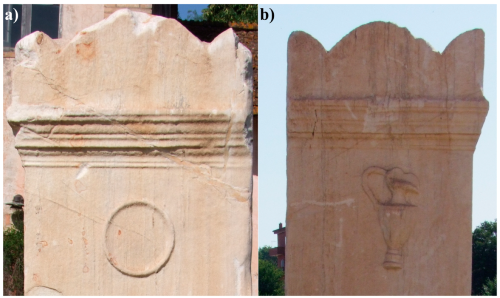
1.1. Roman Funebris Cippus

Several Cippi funebris from different periods have been found both in the Greek, where they undoubtedly originated, and in the Italian peninsula. Among the best known are those from Dolichas, near Vlachomandra [1], and two, located in the city of Thermum, dated between the 7th and 6th century BC with inscriptions in the northwestern Greek language, i.e., the Aetolian variant [2]. As it regards the Italian peninsula, we can cite, for example, the marble cippus from San Genesio [3] in the middle Arno valley, dating back to the first half of the third century BC and the numerous Greco–Roman cippi funebris, made of white marble and found in various locations along the coasts of the central Adriatic (some with Etruscan influence) and the upper Adriatic [4]. Hellenistic steles from the necropolis of Ancona have been dated between the second and third century BC and have been associated by archaeologists with the Greek workshops of Delos and/or with the local Adriatic ones [4].
This paper presents a study based on chemical–diagnostic analyses, as well as on the historical–artistic framework, of a Cippus funebris made of precious white marble located near an ancient roman road. It does not have any symbolic figure carved in bas-relief on the front, but only a votive inscription, which makes it quite different, for example, from the votive steles, mentioned above, of Hellenistic style in Ancona (third century BC) [5].
The Cippus was found in fairly good conditions of conservation but almost completely covered by a black patina; for this reason, a conservative cleaning intervention was recently carried out. Figure 1 shows the Cippus before and after the cleaning and restoration. Therefore, the aim of this study is to respond to the restorer’s request for a preliminary diagnosis on the basis of which to suggest also the most suitable intervention methodologies.

1.2. Historical–Artistic Information

The Cippus is a stele of white Lunense marble dedicated to the young Quintus Cornelius Proclianus, who died at the age of 15, by his mother, Valeria Calpurnia Scopele.
It was discovered in 1971, about 150 m north of its current location, in the agricultural estate and farmhouse called “Cascina”, located in Via Boccea 922, during a normal land reclamation work. The altar, after a cleaning/restoration intervention, was moved to a place more sheltered from the road (always within the same agricultural estate), with the permission of the competent authorities.
The data relating to the discovery have been recorded in an EDR file under the number EDR078389. The dimensions of the stele are 210 × 120 × 78 cm [6], and the weight is approximately 5400 kg.
The back side, now facing north, is completely smooth, suggesting an original position against a wall. The context of the discovery confirms the existence of an altar dedicated to Quintus Cornelius Proclianus, with an attached sarcophagus burial, the fragments of which can still be seen in the aforementioned farmhouse. He belonged to a well-known family of Roman nobility, owners of a large rustic villa, which in ancient times was connected to Rome by the “via Cornelia”, a road that much later, in the early Middle Ages, was replaced by the name “via Boccea”, probably due to the existence of a large boxwood forest [7]. It is, therefore, probable that it was not an isolated and monumental artifact, similar to those existing along the main consular roads (since the Cornelia was a secondary road), but rather an altar located on the private property of a noble family.
On the Cippus, the name of the deceased is engraved with a burin, following the Roman lapidary square capital letter, that is, using only capital letters, all of which can be inserted into a square.
The absence of the paternal dedication in the text suggests that the deceased youth was not a natural son but had been adopted by the nobleman Q. Cornelius Proclus after the marriage to his mother, Valeria Calpurnia Scopele, who belonged to the Gens Calpurnia [6]. This justifies the addition of the suffix “-ianus” in the surname, and since this rule was still in use at the end of the Republic, the epigraph can be dated between the end of the 1st century BC and the beginning of the 1st century AD.
In conclusion, this Cippus funebris fits perfectly into the Roman funerary liturgy, without bas-relief figures of Hellenistic type, but with only a votive inscription, it also shows an Etruscan type iconography with a “Patera” (220–225 mm—h = 20 mm) on the right side of the epigraph and a “Hydria” (170 × 350 mm—h = 20–30 mm) [6], on the left side (see Figure 2). More information on the Cippus can be found in the Supplementary Materials (S.M.), written in Italian by one of the authors.

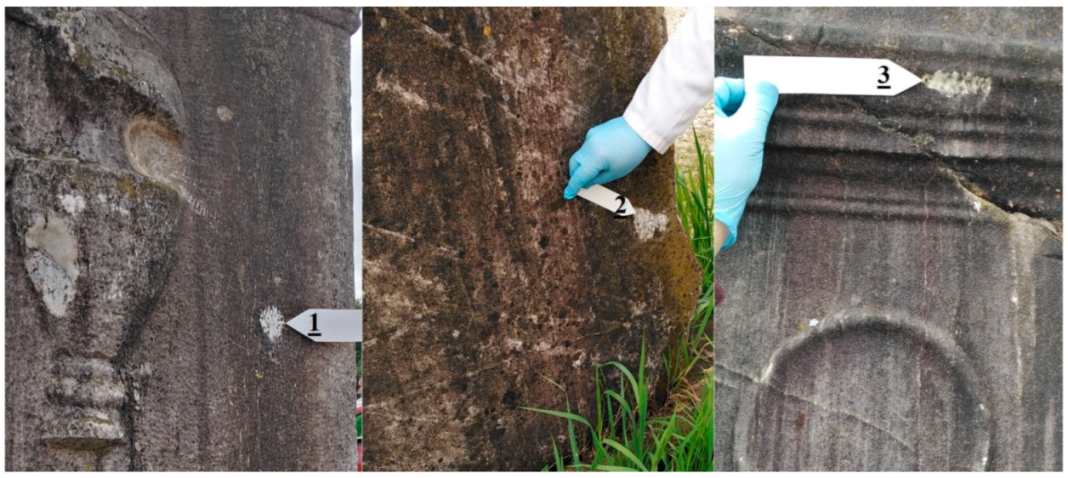
1.3. Conservation State

The restoration of the stele was necessary due to the presence of a black patina caused by both chemical (pollution) and biological agents (like fungi, lichens, or mosses) (see Supplementary Materials Figure S1) that colonized the surface with a synergetic effect. Marble, especially when, as in our case, is exposed outdoors, is particularly subject to degradation phenomena mainly due to acid rains, which transform the carbonate matrix into gypsum (sulfate) that traps particulate matter, forming black crusts and causing the material to fall, or other more soluble compounds which are washed away by rain but erode the surface. The compact black layer may be mainly constituted by black pigments such as melanin and melanoidin and related humic compounds produced by infesting organisms and microorganisms [8,9] and to their remains after death.
Dark patinas due to biological and vegetal colonization are difficult to remove [9,10,11] and remain hidden under the surface even after the application of biocytes (mainly quaternary ammonium compounds and isothiazolinones).
Currently, the best methods for the removal of biodeteriogens are the use of hydrogen peroxide (H2O2), which has a strong bactericidal and degrading effect even on weeds nested under the surface [12,13], and the use of high-temperature steam slightly alkalized with calcium carbonate, which penetrates even into fractures and leaves as residue calcium carbonate, which is the constituent of marble [8].
Even more effective is sodium percarbonate (CAS: 15630-89-4) and urea hydrogen peroxide (CAS: 124-43-6), also in mixtures with each other [13], as they release a great quantity of oxygen and leave as residues free water, oxygen, ammonia, and carbon dioxide, which are volatile products so that after the treatment washing will be required.
The combined action of the two methods, H2O2 and water steam, allows the complete removal of all organic material; it is up to the restorer to choose the number of applications to obtain the result without further damage to the artifact [14] and also the removal of soluble salts, even in depth.
On the contrary, treatment with sodium hypochlorite (NaClO), often used for its strong sterilizing action [12,15], needs a large number of washes to remove the sodium and chlorine, so it is used only in very difficult cases.
Other innovative chemical and physical methods should also be mentioned, such as the use of enzymes for cleaning [16] and photocatalysis, degradation by UV rays, laser-cleaning, cryogenic techniques, and the use of ultrasound to “cook” the biological matrix [17]. Many of these promising methods are still on a laboratory scale when applied to Cultural Heritage [11] and, in any case, are only applicable to small artifacts, certainly not to a cippus over 2 m high.

2. Materials and Methods

2.1. Optical Microscopy

Observations were made using a Zeiss Stemi-SR stereoscopic microscope (Oberkochen, Germany) equipped with Zeiss Axiocam 208 Color digital camera (Carl Zeiss Microscopy GmbH, Jena, Germany) [18,19,20].

2.2. Colorimetric Measurements

A Sikkens Color Collection 3031 swatch was used to measure the color.

2.3. Spectrophotometric Measurements

The UV-visible spectra of the solution, from 190 to 900 nm, were recorded using a Perkin Elmer Spectrophotometer Lambda 16 UV/Vis (Shelton, CT, USA), equipped with Helma QS 10 mm cuvettes.

2.4. Electrochemical Measurements

Electrochemical measurements were carried out using bench-top instruments.
pH: Amel 338 pH meter (Milan, Italy) equipped with Crison 52-02 electrode (Barcellona, Spain). pH 4.0 and pH 7.0 standard solutions from the XS instrument (Carpi, MO, Italy) were used for calibration.
Conductivity: An Amel 160 conductivity meter (Milan, Italy) equipped with an Amel 192/K1 conductivity cell and Amel TC100A temperature sensor (Milan, Italy); 84 μS and 1413 μS standard solutions from an XS instrument were used for calibration.
Redox Potential: Crison microPH 2002 (Barcelona, Spain) equipped with Orion 97-78-00 platinum electrode; 470 mV (HI7022) and 240 mV (HI7021) standard solutions by Hanna Instruments (Villafranca Padovana, Padova, Italy) were used for calibration.

2.5. Ion Chromatography Measurements

The anions content of the sample was determined by ion chromatography using a 761 Compact IC Chromatograph from Metrohm (Herisau, CH) equipped with a Dionex column AS14A 4 × 250 mm, a Rheodyne inline 1 µm filter (Sigma-Aldrich) as precolumn, a Metrohm Suppressor Module, and the IC NET 2.3 software interface. Eluent: 2.3 mM anhydrous sodium carbonate (99.9%) solution from Merck Certipur (Darmsdat, Germany), 2.2 mM anhydrous sodium bicarbonate (99.5%) solution from Fluka (Milan, Italy), and 1% v/v methanol (99.8%) from Sigma-Aldrich (Saint Louis, MO, USA), all ultrapure laboratory grade. Referenced multi-element solutions from Certipur Merck (Darmsdat, Germany) were used for the standard preparation. Ultrapure water (MilliQ, Millipore, Burlington, MA, USA; conductivity < 0.5 μS) was used for both standard and eluent preparation.
The cations content of the sample was also determined by ion chromatography using a 761 Compact IC chromatograph from Metrohm (Herisau, Switzerland) equipped with a CS12A 4 × 250 mm Dionex column (Sunnyvale, CA, USA), a 2 µm inline Supelco filter (Sigma, Bellefonte, PA, USA) as precolumn, and the IC NET 2.3 software interface. Eluent: 2 mM aqueous nitric acid solution from Carlo Erba (Milan, Italy), 4 mM anhydrous oxalic acid from Carlo Erba (Milan, Italy), and 1% v/v acetonitrile from MS Fluka (Milan, Italy), all ultrapure laboratory grade. Referenced solutions for AAS Sigma-Aldrich TraceCert cations (Saint Louis, MO, USA) were used for the standard preparation. Ultrapure water (MilliQ, Millipore, USA; conductivity < 0.5 μS) was used for both standard and eluent preparation.

2.6. SEM/EDS

A Zeiss LEO1450VP Scanning Electron Microscope (SEM) coupled with an Energy Dispersive X-ray Spectroscope (EDS) INCA300 from Oxford Instruments (Wycombe, UK) was used to investigate the structure, morphology, and elemental composition of some samples [21,22,23].

3. Results

3.1. Determination of the Soluble Salts Content

Soluble salts were determined in samples 1, 2, and 3, which were scratched from different areas of the Cippus (Figure 3), and in sample 4, which is the fragment shown on the right of Figure 4; on the left of the same figure is indicated the point from which the fragment spontaneously fell.
The analyses were performed according to the UNI-BBCC Norm [24,25]. Briefly, 100 mg of powdered sample was added to 100 mL of ultrapure water. The salts extraction from the samples was carried out with different steps. In a 45 KHz ultrasonic bath, for 8 min, and in a vortex for 1 min, for a total of 6 cycles. The resulting solution was left to decant but was not filtered, and preliminarily, pH, conductivity, and redox potential (ORP) were measured. The results are shown in Table 1.
Based on the experience of our research group [26], the data in Table 1 give us some information about the conservation state of the artwork.
  • The pH value of a solution coming from the solubilization of marble (calcium carbonate) would have been expected to be higher; the lowering of the pH value is probably due to biological contamination.
  • The conductivity values are quite low, indicating low concentrations of soluble salts. This is confirmed by ion chromatography data (see paragraph 3.3).
  • As expected, the solution obtained from sample 4 has higher pH and conductivity values (the latter almost double) than those of the three previous samples; in fact, samples scratched from the surface of the Cippus contain a high amount of biodeteriogens, while sample 4, being a fragment spontaneously fallen from the Cippus, has a smaller part of contaminated surface and a higher amount of marble.
  • The ORP values are also lower than expected. In fact, a marble exposed outdoors, in good conservation conditions, should have values around 300 mV. It is, therefore, very likely that the degradation due to biological contamination, in contact with the underlying marble, has created a very reducing environment.
  • The ORP measured for sample 4, which, as mentioned above, shows very little biological contamination, is more in line with what is expected from the dissolution of a carbonate rock.

3.2. Spectrophotometry UV-Vis

UV-visible spectrophotometry of solutions derived from treatments of samples of interest for Cultural Heritage is not provided for by any of the UNI-BBCC Norms, not so much because of the difficulty of execution (although, in fact, the zeroing of the instrument must be continuously repeated between one sample and the next), but perhaps because of the difficulties of interpretation.
However, UV-visible spectrophotometry is particularly useful for detecting the color of aqueous solutions, such as in rainfall, but also of organic substances that absorb in this region of the spectrum or of suspended solids [27].
Therefore, the UV-vis spectra of the solutions deriving from the 4 samples described above were analyzed. The spectrophotometric curves obtained are shown in Figure 5.
The spectrophotometric curves give the following information.
  • In the visible range, between 400 and 750 nm (Table 2), a diffuse brown color component can be seen, perceptible to the eye; it is almost absent in sample 4 (obtained from the “original” marble, that is, almost without patina), of low intensity in sample 1, but higher than in sample 1 and in samples 2 and 3, whose spectra almost overlap in this range. These data are congruent with the color measures explained later in the text
  • One of the most common indices of the presence of organic substances is an absorption in the UV, observable as a peak or as a shoulder, at about 254 nm (Table 2) [28,29]. Samples 2 and 3 show a similar and well-detectable absorption at this wavelength, while a lower intensity can be seen in the solution derived from sample 1. Unexpectedly, sample 4 also absorbs in this region but with an intensity about one-fifth of that of samples 2 and 3, which is certainly due to the fact that it does not contain a completely uncontaminated region (see Figure 4).
  • Considering the location of the Cippus, it is reasonable to assume the presence of humic acids produced by the degradation of decomposing plant species. In Supplementary Materials Figure S2, in order to facilitate the identification of such organic substances, spectra reported in the literature by other authors [28,29] have been collected where the relative peaks are better highlighted.
  • Finally, the peaks between 190 and 200 nm could be due to the presence of nitrates at very low concentrations [30], as confirmed by the ion chromatography results (see Section 3.3.1).

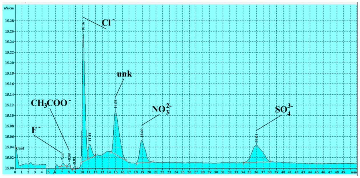
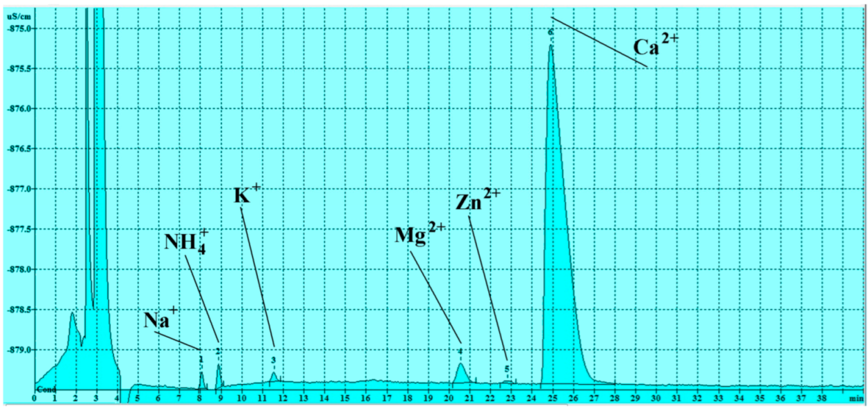
3.3. Soluble Salts Detected by Ion Chromatography

Ion chromatography allows the detection of at least 10 anions and as many cations, among those most commonly found in stone materials, such as marbles, but also in mortars, plasters, and stone artifacts of interest in the Cultural Heritage sector. Each sample chromatogram of both anions and cations was repeated at least three times.
The data show a very low content of soluble salts, which is consistent with the conductivity measurements. This may be due to the outdoor location, which allows them to be washed away by rain.

3.3.1. Anions

Figure 6 shows, as an example, one chromatogram relative to sample 1. Even if calibration graphs were obtained for the anions F, CH3COO, Cl, NO2, IO3, Br, NO3, SO4−2, C2O4−2, PO4−3, and COO, samples only contain the ones listed in Table 3. Unfortunately, although with a different area, an unidentified peak is also present in all samples at a retention time of about 15 min. It is indicated with the acronym “unk”, and since it elutes after chlorine, it could be cyanide, which is not identifiable in our experimental conditions; for certain identification, a more complex system would be necessary [31].
The values (average of at least 3 determinations) reported in Table 3 are relative only to the anions detected in at least 1 sample. With this instrumental setting, for anions, the limit of detection (LOD) varies from 0.01 mgL−1 for fluoride, up to 0.05 mgL−1 for phosphate. LOQs were determined according to the ISO 5725 regulations “accuracy measurements methodology” [32] and using referenced solutions [33] (see Section 2.5).
Table 3 shows the values expressed in ppm (i.e., in milligrams per 1 liter of solution). According to the UNI 11087:2003 Norm [24], instead starting from the dissolution of 1 g of sample in 1 liter of water, the concentration should be defined as mg g−1.

3.3.2. Cations

The standard “UNI 11087:2003-Cultural Heritage—Natural and artificial stone materials. Determination of soluble salt content”, which replaces the “Normal 13/83”, was withdrawn in 2016 without any replacement and provided for the determination of anions only. However, in the Cippus samples, cations were also determined using ion chromatography.
Figure 7 and Table 4 summarize the obtained data. Three repetitions were performed for each of the 4 samples. Even if calibration graphs were obtained for the cations Li+, Na+, NH4+, K+, Rb+, Fe+2, Mg+2, Mn+2, Zn+2, Ca+2, Sr+2, and Ba+2, samples only contain the ones listed in Table 4.
Table 4 shows the concentrations (average of at least 3 determinations, in ppm) of the cations detected in at least 1 sample. With this instrumental setting, the limit of detection (LOD) varies from 0.015 mgL−1 for Li, up to 0.08 mgL−1 for Mg, while LOQs can be estimated from 0.05 mgL−1 for Li up to 0.2 mgL−1 for Mg.
In this case, Table 4 shows the values expressed in ppm even if, according to the UNI 11087:2003 standard, the concentration should be defined as mg g−1 [24].

3.4. Color Measurement

In order to ascertain the color of the marble closest to the original, a series of measurements were performed by placing the Sikkens color swatch in close proximity to the marks on the sample on points 1–3 (see Figure 3). For the test on sample 4, the fragment was instead placed directly on the Sikkens swatch (see Figure 4).
Table 5 shows for each test point the color codes extracted from the Sikkens Color Collection 3031, the RGB and the CIELAB coordinates (where L represents the lightness that is the color between black and white, a* the position between red and green, and b* the position between yellow and blue); in the last column, a graphical representation of the corresponding color is also reported.
The conversion from Sikken 3031 to RGB was obtained using DTP studio 3.0 software (Grünteweg 31, D-26127 Oldenburg, Germany). The conversion from RGB to Lab was obtained using D65, 10°, 1964, in Robin D. Myers, Color Converter 2.3 software, at https://web.archive.org/web/20160309142102/http://rmimaging.com/tools.html (accessed on 9 March 2025).
The RGB coordinates show a slight prevalence of red, minimal in the fragment (sample 4). As expected, the color of the fragment is the most achromatic on the basis of the Lab coordinates (values of a* and b* closest to zero) (see Table 5).

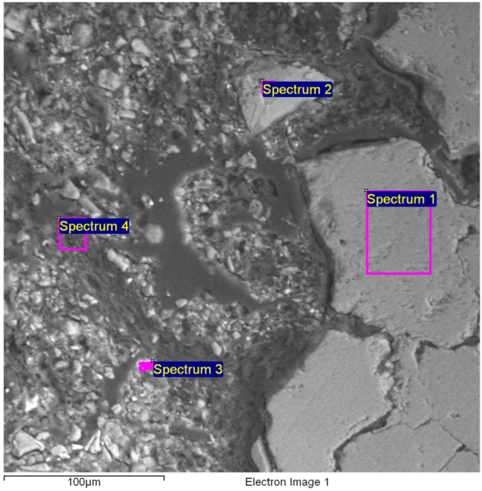
3.5. SEM Image and X-Ray Microanalysis by Energy Dispersive Spectroscopy (EDS)

SEM image (Figure 8) and X-ray microanalysis (by EDS, Table 6), relative to the fragment (sample 4), reveal that the disintegration mechanism of the marble occurs thanks to the infiltration of wet soil through the grain edges of the artifact. The infiltration is particularly evident in Figure 8 and in the EDS spectrum relating to point 4 and partly in the spectrum relating to point 3 for the significant presence of Si and Al in the composition that, in addition to that of Fe (spectrum 3), creates a microenvironment conducive to microflora colonization, resulting in an increase in inter-granular pressure.
Other SEM images of sample 4 (the detached fragment), and for some of them also the relative EDS graphs, have been reported, as shown in Supplementary Materials Figure S3.

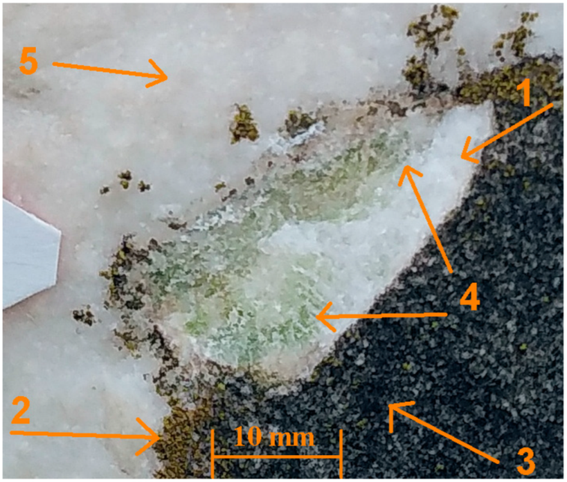
3.6. Macrophotography and Optical Microscopy (OM)

The macrophotograph of the lacuna left by the fragment (sample 4) that detached spontaneously (see Figure 9) provides evidence for the infiltration of microflora through the microfractures in the marble.
A small fragment of sample 4 was embedded in resin (Araldite 2020). A polished cross-section (Figure 10) was obtained by appropriately cutting the resin block and polishing it using Smirdex (Lefki, Xanthi, Greece) abrasive paper (from P1000 to P5000).
The polished cross-section of sample 4, observed by optical microscope under low magnification (Figure 10), highlights the granular structure of the marble with grains measuring approximately 150–200 µm and also the ongoing degradation phenomena. Starting from the exposed surface (top of the image), the following can be observed: the black patina, with a maximum thickness of approximately 0.64 mm (3), is mainly composed of dead microflora while in some points it is still fresh (2); a deep horizontal fracture, of approximately 0.9 mm, is already colonized by microflora (1); the internal part of the marble, homogeneous in color and structure, presents a crack which crosses it completely (4); on the internal side, recent colonization can be noted, which could have caused the detachment of the fragment itself (5).
At higher magnification (Figure 11), the colonized fracture marked with number 1 in Figure 10 and other smaller ones are more clearly visible.

4. Discussion of Results

Various types of analysis were carried out on different points of the Cippus surface and of the scratched patina that almost completely covered it before the conservative cleaning. We also found a spontaneously detached small flake that allowed us to carry out analyses on the original marble, although it was not completely free from the effects of environmental and microbiological pollution.
Macrophotographs and images from optical and electronic microscopy of polished cross-sections of this sample allowed us to establish that the patina was mainly constituted by dead and still living microflora and by a lower part of the black crust that is particulate adsorbed on sulfates.
The latter was formed by the attack of the carbonate material by atmospheric pollutants and by rain that resulted in acid due to their solubilization. The images also show the infiltration of soil between the marble grains (SEM) and the consequent growth of microflora (OM), with an overall effect of cracking and spalling. From an analytical point of view, the remarkable presence of microflora was highlighted, not only by the eyes and by the grey-blackish color of the artifact surface but also by the results of the analyses carried out on the solutions prepared to determine the content of soluble salts in the samples. In fact, the values of pH, conductivity, and redox potential are lower than expected in the case of the samples scratched from the surface of the Cippus (samples 1–3), while they are much more in line with those expected for sample 4, which contains a lower amount of microflora and a much higher content of marble. The conductivity values are consistent with the content of soluble salts obtained by IC data. It also can be noted that for samples 1–3, a higher content of bicarbonate and calcium results with respect to sample 4; this demonstrated that the marble substrate in the first case is more corroded, therefore having a higher surface for the solubilization, with respect to the last. The presence of acetates and nitrites, albeit in traces, also confirms significant contamination by microflora residues; more, the presence of NH4 in all samples suggests not only past, but also ongoing biological degradation. The presence of nitrates at unexpectedly low concentrations may be attributed to their high solubility, which greatly reduces their concentration when washed away by rain [34].

5. Conclusions

In conclusion, the data presented show that the major problem for the future conservation of the Cippus is not directly linked to problems of chemical nature, as an example, not due to the presence of soluble salts, but rather to the infestation of the exposed and cracked parts of the artifact by algae, mosses, lichens, microfungi, and perhaps also by some animal species (especially insects). The disinfestation–cleaning of the surface of the Cippus is then, in our opinion, the first requirement for the conservation of this important archaeological artifact of authentic Roman funerary style, taking care that the techniques and cleaning solution used do not damage it and are not too polluting or toxic [11].
On the basis of the available literature cited in the introduction, one could opt for the sterilization method, but considering that the Cippus shows both significant material losses (Figure 4) and overall microfractures visible by the OM (Figure 10 and Figure 11) and SEM (Figure 8), which will certainly provoke further materials losses, the subsequent consolidation is strongly recommended in order to restore the cohesion of the marble by reconstructing the crystalline matrix [35].
Modern restoration theories require the use of products that are compatible with the original matrix [36], in this case, an almost pure carbonate because the Cippus is Lunense marble [37].
Lime water combines consolidating, protective, and bactericidal properties and is also environmentally friendly [38]; however, its application requires great skill on the part of the restorer and mastery in its use [39] from the choice of the moment and the number of applications; manual skills that are being lost due to the race against time and the chronic lack of funds for restoration, which leads to the use of bottled products with nice labels.
The correct application of lime water is even more difficult because the Cippus is placed outdoors. The numerous applications must be carried out at the lowest possible temperature to avoid rapid evaporation, i.e., low penetration, and must be carried out wet-on-wet to keep the surface moist and facilitate deep absorption [39].
Given the low solubility of CaCO3 in water (13 mg L−1 at 25 °C), modern alternatives [39] such as nanolime [40] are available to increase the amount of carbonate absorbed and to simplify the operations [41]. The maximum consolidation effect with nanolime is obtained by working in environments with high relative humidity (~75% RH) [41], but the intervention has a high cost, which is not feasible for large objects that are not of the highest value, as in the case of the Cippus. Whether lime water or nanolime is used, the number of applications must be evaluated area by area to avoid bleaching effects, which can be controlled using spectrophotometric techniques, measuring both the reflectance spectrum and the L a* b* coordinates [42]. When the conservative purpose prevails over aesthetic criteria, some add a further application of consolidant that forms a sacrificial surface.

Supplementary Materials

The following supporting information can be downloaded at: https://www.mdpi.com/article/10.3390/analytica6010011/s1. Some supplementary files are available, collected in a single .ZIP file; it contains three files. The first, named “Supplementary-photo-and-chart.doc”, contains macrophotographs of the biodegraded marble surface, SEM photo and EDS results, and spectra of humic acids coming from literature; the second, named “Il-cippo-di-marmo-di-Quinto-Cornelio-Procliano.pdf”, is a text with the history of the Cippus written, in Italian, by Dr. Marco Castracane; the third, named “Via-Boccea-922-Cippo-di-Quinto-Cornelio-Procliano_autorizzazione.pdf”, is the letter from “Sovrintendenza di Roma” authorizing us to publish results.

Author Contributions

Conceptualization: M.T., E.D. and M.C. (Mauro Castrucci); methodology: G.V. and M.T.; software: G.V.; validation: G.V. and M.P.S.; formal analysis: M.P.S. and G.V.; investigation: M.C. (Marco Castracane); resources: M.C. (Marco Castracane); data curation: G.V. and M.C. (Mauro Castrucci); writing—original draft preparation: M.T. and E.D.; writing—review and editing: M.C. (Mauro Castrucci), G.V. and M.P.S.; visualization: E.D.; supervision: M.T.; All authors have read and agreed to the published version of the manuscript.

Funding

This research did not receive any external funding for diagnostic and chemical analysis.

Institutional Review Board Statement

Not applicable.

Informed Consent Statement

Not applicable.

Data Availability Statement

No data are available.

Acknowledgments

The authors would like to express their sincere gratitude to the architect Alfredo Pianella Di Cosimo for the kind permission to enter his private property where the Cippus is located and to the Cultural Association “Cornelia Antiqua” for generous funding for the restoration. Thanks also to Roberta Pardi and Daniela Porro of the “Soprintendenza Speciale Archeologia Belle Arti e Paesaggio” in Rome, whose permission was essential for the publication of the results.

Conflicts of Interest

The authors declare no conflicts of interest.

References

  1. Cavalli, E. Cippo funerario di Dolichas. Axon 2017, 1, 31–43. [Google Scholar] [CrossRef]
  2. Sowa, W. The Dialectology of Greek. In Handbook of Comparative and Historical Indo-European Linguistics; Klein, J., Joseph, B., Fritz, M., Eds.; De Gruyter Mouton: Berlin, Germany, 2017; Volume 1, pp. 710–716. [Google Scholar] [CrossRef]
  3. Bonamici, M. Cippo a clava con viaggio all’aldilà. In Studi Etruschi; Istituto Nazionale di Studi Etruschi ed Italici: Florence, Italy, 2019; Volume 82, pp. 87–97. ISBN 03917762StEtr82. [Google Scholar]
  4. Antonelli, F.; Lazzarini, L. The Use of White Marble in the Central and Upper Adriatic Between Greece and Rome: Hellenistic Stelae from the Necropolis of Ancona (Italy). Camb. Archaeol. J. 2013, 23, 149–162. [Google Scholar] [CrossRef]
  5. Haack, M.-L. Ritual and Cults, 250–89 BCE. In Etruscology; Naso, A., Ed.; De Gruyter: Berlin, Germany, 2017; pp. 1203–1214. [Google Scholar] [CrossRef]
  6. Steiner, A.P. Epitaph from Roma. Epigraphica 1981, 43, 213–216, ISBN-13: 9788875940478. Available online: https://edh.ub.uni-heidelberg.de/edh/inschrift/HD001255 (accessed on 9 March 2025).
  7. De Cristofaro, A. Da via Cornelia a Via di Boccea: Storia, Percorso e Paesaggi di una Strada Suburbana. In Atlante Tematico di Topografia Antica; L’Erma di Bretschneider: Rome, Italy, 2021; Volume 31, pp. 201–218. [Google Scholar] [CrossRef]
  8. Koch, T.; Fischer, C.; Schad, F.; Siegesmund, S. Quantification of surface changes and volume losses of selected rock types due to different cleaning processes. Environ. Earth Sci. 2023, 82, 132. [Google Scholar] [CrossRef]
  9. Diakumaku, E.; Gorbushina, A.A.; Krumbein, W.E.; Panina, L.; Soukharjevski, S. Black fungi in marble and limestones—An aesthetical, chemical and physical problem for the conservation of monuments. Sci. Total Environ. 1995, 167, 295–304. [Google Scholar] [CrossRef]
  10. Torralba, M.G.; Kuelbs, C.; Moncera, K.J.; Roby, R.; Nelson, K.E. Characterizing Microbial Signatures on Sculptures and Paintings of Similar Provenance. Microb. Ecol. 2021, 81, 1098–1105. [Google Scholar] [CrossRef]
  11. Romani, M.; Warscheid, T.; Nicole, L.; Marcon, L.; Di Martino, P.; Suzuki, M.T.; Lebaron, P.; Lami, R. Current and future chemical treatments to fight biodeterioration of outdoor building materials and associated biofilms: Moving away from ecotoxic and towards efficient, sustainable solutions. Sci. Total Environ. 2022, 802, 149846. [Google Scholar] [CrossRef]
  12. Lineback, C.B.; Nkemngong, C.A.; Tongyu Wu, S.; Li, X.; Teska, P.J.; Oliver, H.F. Hydrogen peroxide and sodium hypochlorite disinfectants are more effective against Staphylococcus aureus and Pseudomonas aeruginosa biofilms than quaternary ammonium compounds. Antimicrob. Resist. Infect. Control 2018, 7, 447. [Google Scholar] [CrossRef]
  13. Reale, R.; Campanella, L.; Devreux, G.; Dell’Aglio, E. Study of microbial melanin decolorization from carbonatic stones surfaces. J. Cult. Herit. 2021, 47, 21–27. [Google Scholar] [CrossRef]
  14. Zhang, H.; Sun, Q.; Geng, J.; Dai, J.; Ge, Z.; Wang, S. Cyclic heating of saturated steam changes the surface properties of sandstone. J. Therm. Anal. Calorim. 2023, 148, 5325–5334. [Google Scholar] [CrossRef]
  15. Barbut, F.; Menuet, D.; Verachten, M.; Girou, E. Comparison of the efficacy of a hydrogen peroxide dry-mist disinfection system and sodium hypochlorite solution for eradication of Clostridium Difficile spores. Infect. Control. Hosp. Epidemiol. 2009, 30, 507–514. [Google Scholar] [CrossRef] [PubMed]
  16. Konkol, N.; McNamara, C.; Sembrat, J.; Rabinowitz, M.; Mitchell, R. Enzymatic decolorization of bacterial pigments from culturally significant marble. J. Cult. Herit. 2009, 10, 362–366. [Google Scholar] [CrossRef]
  17. Crisci, G.M.; La Russa, M.F.; Macchione, M.; Malagodi, M.; Palermo, A.M.; Ruffolo, S.A. Study of archaeological underwater finds: Deterioration and conservation. Appl. Phys. A Mater. Sci. Process. 2010, 100, 855–863. [Google Scholar] [CrossRef]
  18. Marszałek, M. Application of Optical Microscopy and Scanning Electron Microscopy to the Study of Stone Weathering: A Cracow Case Study. Int. J. Archit. Herit. 2008, 2, 83–92. [Google Scholar] [CrossRef]
  19. Güleç, A.; Acun, S.; Ersen, A. A characterization method for the fifth-century traditional mortars in the land walls of Constantinople, Yedikule. Stud. Conserv. 2005, 50, 295–306. [Google Scholar] [CrossRef]
  20. Germinario, L.; Andrian, G.F.; Laviano, R. Petrography, mineralogy, chemical and technical properties of the building stone of Ostuni Cathedral (Italy): Inferences on diagnostics and conservation. Period Miner. 2014, 83, 379–400. [Google Scholar] [CrossRef]
  21. Astolfi, M.L. Advances in Analytical Strategies to Study Cultural Heritage Samples. Molecules 2023, 28, 6423. [Google Scholar] [CrossRef]
  22. Sanjurjo Sánchez, J.; Vidal Romaní, J.R.; Fernández Mosquera, D.; Alves, C.A. Study of origin and composition of coatings in a monument built with granitic rocks, by SEM, XRD, XRF and DTA-TGA. X-Ray Spectrom. Int. J. 2008, 37, 346–354. [Google Scholar] [CrossRef]
  23. Falcone, F.; Cinosi, A.; Siviero, G.; Rosatelli, G. Innovative methodological approach integrating SEM-EDS and TXRF microanalysis for characterization in materials science: A perspective from cultural heritage studies. Spectrochim. Acta Part B At. Spectrosc. 2024, 218, 106980. [Google Scholar] [CrossRef]
  24. UNI 11087:2003; Beni Culturali—Materiali Lapidei Naturali ed Artificiali—Determinazione del Con-Tenuto di Sali Solubili. Ente Nazionale Italiano di Unificazione: Rome, Italy, 2003.
  25. EN 16455:2014; Conservation of Cultural Heritage—Extraction and Determination of Soluble Salts in Natural Stone and Related Materials Used in and from Cultural Heritage. CEN: Brussels, Belgium, 2014.
  26. Tomassetti, M.; Castrucci, M.; Dell’Aglio, E.; Sammartino, M.P.; Visco, G.; Martini, E.; Innocenzi, F.; Reale, R.; Ronca, S. Analytical-petrographic Study of Bugnato Degradation of an Ancient Milan Building. Cur. Anal. Chem. 2024, 20, e130624231005. [Google Scholar] [CrossRef]
  27. Fontanella, L.U.; Tomassetti, M.; Visco, G.; Sammartino, M.P. Characterization of Rome’s rainwater in the early of 2018 aiming to find correlations between chemical-physical parameters and sources of pollution: A statistical study. J. Atmos. Chem. 2021, 78, 1–16. [Google Scholar] [CrossRef]
  28. Woldemichael, G.; Tulu, T.; Flechsig, G. Solar UV Photooxidation as Pretreatment for Stripping Voltammetric Trace Metal Analysis in River Water. Int. J. Electrochem. 2011, 2011, 481370. [Google Scholar] [CrossRef]
  29. Kiprop, A.K.; J-Coumon, M.C.; Pourtier, E.; Kimutai, S.; Kirui, S. Synthesis of Humic and Fulvic Acids and their Characterization using Optical Spectroscopy (ATR-FTIR and UV-Visible). Int. J. Appl. Sci. Technol. 2013, 3, 28–35. [Google Scholar]
  30. Tsadilas, C. Nitrate Handbook: Environmental, Agricultural, and Health Effects; CRC Press: Boca Raton, FL, USA, 2022; pp. 1–426. [Google Scholar]
  31. Caliamanis, A.; McCormick, M.J.; Carpente, P.D. Enhanced conductometric detection of cyanide in suppressed ion chromatography. J. Chromatogr. A 2000, 884, 75–80. [Google Scholar] [CrossRef]
  32. ISO 5725-1:2023; Accuracy (Trueness and Precision) of Measurement Methods and Results. ISO: Geneva, Switzerland, 2023.
  33. Ivanova, V.; Surleva, A.; Koleva, B. Validation of Ion Chromatographic Method for Determination of Standard Inorganic Anions in Treated and Untreated Drinking Water. IOP Conf. Ser. Mater. Sci. Eng. 2018, 374, 012053. [Google Scholar] [CrossRef]
  34. Lazzarini, L.; Laurenzi Tabasso, M. Il Restauro della Pietra; CEDAM: Padua, Italy, 1986; pp. 166–264. [Google Scholar]
  35. Ogino, T.; Suzuki, T.; Sawada, K. The formation and transformation mechanism of calcium carbonate in water. Geochim. Cosmochim. Acta 1987, 51, 2757–2767. [Google Scholar] [CrossRef]
  36. Prasad Gopi, S.; Subramanian, V.K. Polymorphism in CaCO3—Effect of temperature under the influence of EDTA (di sodium salt). Desalination 2012, 297, 38–47. [Google Scholar] [CrossRef]
  37. Amadori, M.L.; Santi, P.; Frapiccini, N.; Rinaldi Tufi, S.; Gorgoni, C.; Pallante, P. Roman White Marbles in Northern Marche Region (Italy): An Archaeometric Provenance Study. Geoheritage 2024, 16, 122. [Google Scholar] [CrossRef]
  38. Huesca-Tortosa, J.A.; Spairani-Berrio, Y.; Coviello, C.G.; Sabbà, M.F.; Rizzo, F.; Foti, D. Evaluation of Eco-Friendly Consolidating Treatments in Pugliese Tuff (Gravina Calcarenite) Used in Italian Heritage Buildings. Buildings 2024, 14, 940. [Google Scholar] [CrossRef]
  39. Cescon, M. Valutazione di Prodotti Inorganici per il Consolidamento di Materiali Lapidei Carbonatici. Master’s Thesis, Universita’ Ca’ Foscari, Venice, Italy, 2017. [Google Scholar]
  40. Hansen, E.; Doehne, E.; Fidler, J.; Larson, J.; Martin, B.; Matteini, M.; Rodriguez-Navarro, C.; Pardo, E.S.; Price, C.; de Tagle, A.; et al. A review of selected inorganic consolidants and protective treatments for porous calcareous materials. Stud. Conserv. 2003, 48 (Suppl. 1), 13–25. [Google Scholar] [CrossRef]
  41. Otero, J.; Carola, A.E.; Starinieri, V. Preliminary Investigations of Compatible Nanolime Treatments on Indiana Limestone and Weathered Marble Stone. Int. J. Archit. Herit. Conserv. Anal. Restor. 2022, 16, 394–404. [Google Scholar] [CrossRef]
  42. Pozo-Antonio, J.S.; Montojo, C.; López de Silanes, M.E.; de Rosario, I.; Rivas, T. In situ evaluation by colour spectrophotometry of cleaning and protective treatments in granitic Cultural Heritage. Int. Biodeterior. Biodegrad. 2017, 123, 251–261. [Google Scholar] [CrossRef]
Figure 1. The Cippus before (left) and after (right) cleaning.
Analytica 06 00011 g001
Figure 2. (a) Right side of the Cippus funebris where the figure of a “Patera” is sculpted in bas-relief; (b) left side of the Cippus funebris where a “Hydria” is depicted.
Analytica 06 00011 g002
Figure 3. Scratched areas from which samples 1, 2, and 3 were collected.
Analytica 06 00011 g003
Figure 4. On the left, the area from which the fragment (sample 4) spontaneously fell; on the right, sample 4, along with Sikken’s color swatch codes.
Analytica 06 00011 g004
Figure 5. UV-vis spectra of the solutions obtained by extraction of the soluble salts from the 4 cippus samples. The “cip” solutions 1, 2, 3, and 4 were analyzed as they were, while the concentration of the “csampl” solutions 1, 2, and 3 was doubled.
Analytica 06 00011 g005
Figure 6. Anionic chromatogram of sample 1 obtained in the experimental conditions detailed in Section 2.5 for the anions detection.
Analytica 06 00011 g006
Figure 7. Cationic chromatogram of sample 1.
Analytica 06 00011 g007
Figure 8. SEM image obtained by analyzing a cross-section of the flake that spontaneously detached from the Cippus surface. It also shows the 4 areas where the EDS analyses were performed.
Analytica 06 00011 g008
Figure 9. Macrophoto of the lacuna left by the detachment of sample 4. (1) Original marble, (2) still growing microflora, (3) dead, degraded, and decomposed microflora, (4) fresh microflora, still green from the presence of chlorophyll, (5) altered marble.
Analytica 06 00011 g009
Figure 10. Polished section of sample 4.
Analytica 06 00011 g010
Figure 11. Detail of the polished section shown in Figure 10.
Analytica 06 00011 g011
Table 1. Chemical measures for the Cippus funebris of Quintus Cornelius Proclianus.
Sample n.Dissolved (mg)pHORP (mV)Conductivity (µS)Temperature (°C)
n 1108.89.64152.643.122.5
n 2108.28.85144.029.021.7
n 3108.59.69149.834.422.2
n 4107.89.83310.681.822.1
Table 2. Spectrophotometric data relative to the 4 samples of the Cippus funebris (see Figure 3 and Figure 4).
Sample n.Abs at 200 nmAbs at 254 nmAbs at 440 nmAbs at 550 nmAbs at 664 nmAbs at 750 nm
n 10.24920.07180.02510.01260.00780.0039
n 20.29480.10510.03760.02040.01220.0068
n 30.23510.10200.03370.01780.00880.0044
n 40.11290.02660.01170.00660.00520.0041
Table 3. IC (anionic) data obtained for the Cippus funebris samples. 2.1 < SD% < 5.5.
Sample n.F
(ppm)
CH3COO
(ppm)
Cl
(ppm)
NO2
(ppm)
NO3
(ppm)
SO42−
(ppm)
HCO3
(ppm) *
n 10.0040.0570.359<LOD0.2440.28141.50
n 20.005<LOD0.174<LOD<LOD1.38146.10
n 30.011<LOD0.041<LOD<LOD0.04035.70
n 40.001<LOD 0.0070.0230.0160.05914.60
* It should be noted that the HCO3 concentration values were estimated through the ionic balance; therefore, it is affected by an error of at least 5.5%.
Table 4. Cations concentration by IC for Cippus samples 2.4 < SD% < 6.1.
Sample n.Na+
(ppm)
NH4+
(ppm)
K+
(ppm)
Mg+2
(ppm)
Zn+2
(ppm)
Ca+2
(ppm)
n 10.150.190.280.290.0413.0
n 20.100.070.170.29<LOD14.76
n 30.030.10<LOD0.23<LOD10.96
n 40.000.08<LOD0.080.094.55
Table 5. Data on color of the Cippus funebris samples. This table shows the color codes corresponding to the samples examined using the Sikkens color swatch, RGB coordinates and Cielab coordinates. At the end of the table, a graphical representation of the colors determined with the previous methods is also shown.
Test Point or SampleSikkens CodeRGBCIELAB
(L a* b*)
Color
test n.1F2.07.88R = 253 G = 241 B = 224L = 95.78 a* = 1.56 b* = 8.53
test n.2F2.05.85R = 245 G = 235 B = 220L = 93.64 a* = 1.21 b* = 7.41
test n.3FN.02.88R = 246 G = 240 B = 231L = 95.18 a* = 0.83 b* = 4.04
test n.4ON.00.90R = 242 G = 240 B = 237L = 95.03 a* = 0.74 b* = 0.90
Table 6. EDS data relative to the four areas evidenced in Figure 8.
SpectrumCOAlSiSKCaMnFeCuSnSbITotal
Spectrum 18.452.1 32.9 1.55.0 100.0
Spectrum 214.242.0 0.0 32.8 9.11.8100.0
Spectrum 3 42.12.96.9 2.02.242.90.9 100.0
Spectrum 4 68.76.114.32.21.34.6 2.7 100.0
Max.14.268.76.114.32.21.332.92.242.90.91.59.11.8
Min.8.442.02.90.02.21.32.02.22.70.91.55.01.8
Disclaimer/Publisher’s Note: The statements, opinions and data contained in all publications are solely those of the individual author(s) and contributor(s) and not of MDPI and/or the editor(s). MDPI and/or the editor(s) disclaim responsibility for any injury to people or property resulting from any ideas, methods, instructions or products referred to in the content.

