Article

Fish Gelatin Edible Films with Prebiotics and Structuring Polysaccharides for Probiotic Delivery: Physicochemical Properties, Viability, and In Vitro Gastrointestinal Release

by
Gabriel M. da Silva
1,
Newton Carlos Santos
2,*,
Luanna A. da Silva
3,
Thalis L. B. de Lima
2,
Mateus de Oliveira Leite
3,
Virgínia Mirtes de Alcântara Silva
4,
Liandra de S. Oliveira
2,
Victor Herbert de Alcântara Ribeiro
5,
Ariadne Soares Meira
6,
Poliana H. D. Felix
2,
Raquel Alves de Luna Dias
2,
Deyzi Gouveia
2,
Josivanda P. Gomes
2 and
Ana Paula T. Rocha
2
1
Núcleo de Pesquisa e Extensão-Laboratório de Combustíveis e Materiais (NPE–LACOM), Universidade Federal da Paraíba, Cidade Universitária s/n−Campus I, João Pessoa 58051-900, PB, Brazil
2
Centro de Tecnologia e Recursos Naturais, Federal University of Campina Grande, Campina Grande 58429-900, PB, Brazil
3
Federal Institute of Mato Grosso, Campo Novo do Parecis 78360-000, MT, Brazil
4
Faculdade de Engenharia de Alimentos, Universidade Estadual de Campinas, Campinas 13083-862, SP, Brazil
5
Centro de Ciências Humanas, Universidade Federal da Paraíba, Sociais e Agrárias, Bananeiras 58220-000, PB, Brazil
6
Centro de Ciências Agrárias, Federal University of Paraíba, Areia 58397-000, PB, Brazil
*
Author to whom correspondence should be addressed.
Polysaccharides 2025, 6(3), 79; https://doi.org/10.3390/polysaccharides6030079
Submission received: 8 July 2025 / Revised: 6 August 2025 / Accepted: 1 September 2025 / Published: 5 September 2025

Abstract

This study aimed to develop synbiotic edible films based on fish gelatin containing Lacticaseibacillus rhamnosus GG, evaluating the impact of different prebiotics (inulin and fructooligosaccharides, FOSs) and structuring polysaccharides (pectin and alginate) on their physical, mechanical, thermal properties, cell viability, and in vitro gastrointestinal behavior. Seven film formulations were prepared from fish gelatin solutions (3%, w/v) containing glycerol (30%, w/w, as plasticizer), with the addition of prebiotics (inulin or FOSs, 1:1 w/w to gelatin), either alone or in combination with pectin (1%, w/v) or alginate (0.5%, w/v). Specifically, F1 contained gelatin, glycerol, and L. rhamnosus GG (control); F2 and F5 included inulin or FOSs, respectively; F3 and F6 combined inulin or FOSs with pectin; and F4 and F7 combined inulin or FOSs with alginate. After incorporation of the probiotic, the solutions were cast and dried at 37 °C for 24 h. The incorporation of prebiotics and polysaccharides significantly influenced probiotic viability after film drying (p < 0.05). The control formulation (F1) showed the highest reduction (26.10%), while F4 (inulin + alginate) and F7 (FOS + alginate) exhibited the lowest losses of 10.41% and 10.98%, respectively. These films also demonstrated better performance during simulated digestion, with F7 showing the smallest reduction after 6 h (0.5 log), maintaining 7.0 colony-forming units per gram (CFU g−1), which is considered adequate for functional effects. Physically, the films varied in solubility (27.50% to 41.37%), thickness (0.085 to 0.095 mm), water vapor permeability (WVP) (8.17 to 11.75 g·mm/m2·d·kPa), and moisture content (13.47% to 17.50%). Mechanically, F4 showed the highest tensile strength (24.5 MPa), while F1 had the highest elongation at break (62%). During storage, F7 and F4 showed the lowest viability losses (29.8% and 29.4%, respectively) under refrigeration. Overall, the results indicate that the association of prebiotics with structuring polysaccharides improves stability, cellular protection, and functional performance of the films.

1. Introduction

In recent years, edible films have gained prominence as promising alternatives to traditional packaging, especially those derived from petroleum, which are not naturally degradable and remain in the environment for long periods, posing significant risks to human health [1,2]. These films consist of a thin layer formed from naturally edible substances such as proteins, polysaccharides, and lipids, which are non-toxic, biodegradable, and environmentally sustainable materials [3]. One of the most abundant, non-toxic, and popular biopolymers is gelatin. Gelatin has been widely used in protein-based biodegradable films. It is commercially produced from the partial hydrolysis or thermal degradation of the collagen found in the bones and skins of animals and fish [4].
Fish gelatin has been widely studied as a sustainable and functional alternative to bovine or porcine gelatin, as it exhibits good film-forming properties, lacks cultural and religious restrictions, and adds value to fish industry by-products [5,6]. Its combination with polysaccharides such as alginate and pectin can result in films with improved mechanical strength, lower solubility, and a more stable structure, due to electrostatic interactions between the positively charged groups of gelatin and the negatively charged groups of polysaccharides [7,8]. Recent advances in edible film and coating research have driven the development of functional biomaterials, which have proven especially effective in protecting perishable foods against chemical and microbiological deterioration, positioning themselves as viable alternatives to conventional packaging [2].
In addition to serving as a barrier against gases, moisture, and microorganisms, edible films have been widely investigated as matrices for the incorporation of bioactive compounds, such as antioxidants [3,9], antimicrobial agents [10], and probiotic microorganisms [11,12]. The use of edible films containing probiotics has gained attention as a promising strategy for fruit preservation, focusing on maintaining food quality and promoting health [13]. Therefore, like bioactive compounds, probiotics have also been incorporated into biopolymer matrices for the development of active food packaging. These packaging systems not only help control pathogenic microorganisms and improve food safety but also offer potential health benefits to consumers [12,14].
Probiotics are live microorganisms that have a positive impact on human health, particularly through improved intestinal function, when ingested in adequate amounts (107–109 CFU) [15]. According to Coimbra et al. [16], the use of edible films as a probiotic delivery system offers several advantages, such as the delivery of a high dose in a relatively compact volume; a reduction in viability loss during storage; and protection against the adverse conditions of the upper gastrointestinal tract [17].
Previous studies have demonstrated the potential of different film-forming matrices in maintaining probiotic viability. For example, Coimbra et al. [16] developed edible films using potato starch enriched with agro-industrial by-products such as grape pomace and apple peel, which are rich in phenolic compounds. They reported that these films were effective in incorporating Lactobacillus rhamnosus, maintaining viability above 7.0 log CFU g1 during 30 days of refrigerated storage. Complementarily, Sogut et al. [18] showed that the co-application of sodium caseinate and pullulan in the development of edible films improved the mechanical and barrier properties of the films and enhanced the viability of Lactobacillus plantarum and Bifidobacterium animalis, especially under gastrointestinal conditions. In addition, Ebrahimi et al. [19] reported that films based on carboxymethylcellulose and whey protein isolate preserved the viability of L. acidophilus and L. rhamnosus above 7 log CFU g1 after four weeks at 4 °C and also exhibited desirable water vapor permeability and tensile strength.
However, after the successful addition of probiotics, the critical parameter is to ensure the viability of beneficial microorganisms during processing and storage. As highlighted by Sogut et al. [18], films composed of synergistic combinations of polymers and probiotics require optimization to survive harsh gastrointestinal conditions, especially the acidic environment of the stomach. When probiotic microorganisms are incorporated into foods, they must be able to survive passage through the gastric juices of the digestive tract and then successfully proliferate in the intestine. An interesting option for probiotic delivery is to use an edible matrix for food packaging that protects the bacteria and supports their survival [19].
In this context, prebiotics such as inulin and fructooligosaccharides (FOSs) may exert a protective effect on probiotic cells, increasing their resistance to adverse conditions and promoting the selective growth of these beneficial bacteria in the colon [20,21]. Orozco-Parra et al. [22] developed bioactive symbiotic films based on cassava starch, inulin, and Lactobacillus casei, demonstrating that the incorporation of inulin not only exerted a plasticizing effect, improving the mechanical properties of the film, but also significantly increased the probiotic’s resistance to adverse conditions, such as gastric pH. The results indicated that inulin acted as a protective agent, maintaining the viability of L. casei above 6 log CFU g1 even after exposure to a simulated digestive system. These findings reinforce the potential of edible films enriched with prebiotics as an effective strategy for delivering beneficial microorganisms, combining functional and technological properties. Complementary studies, such as that by Soukoulis et al. [11], support these results by showing that inulin in polymeric matrices can improve both film quality and probiotic stability, opening up prospects for innovative applications in functional foods.
Although several studies have demonstrated the potential of different polymeric matrices for the incorporation and protection of probiotics, investigations involving fish gelatin-based edible films are still limited. Moreover, approaches that combine the presence of prebiotics (such as inulin or FOSs) with structuring polysaccharides (such as pectin or alginate) in synbiotic formulations are scarce, aiming to optimize microorganism stability and gastrointestinal release efficacy. Therefore, the present study proposes the development of synbiotic edible films based on fish gelatin containing Lacticaseibacillus rhamnosus GG, evaluating the impact of adding different prebiotics and polysaccharides on their physical, mechanical, and thermal properties, microbial viability, and simulated gastrointestinal release profile.

2. Materials and Methods

2.1. Materials

Cold-water fish gelatin (CAS No.: 9000-70-8), pectin from citrus fruit peel (galacturonic acid ≥ 74.0% on a dry basis, methyl ester of poly-D-galacturonic acid, CAS No.: 9000-69-5), and enzymes for in vitro digestion (α-amylase, porcine pancreas pancreatin, and porcine gastric mucosa pepsin) were obtained from Sigma Aldrich (St. Louis, MO, USA). Glycerol was obtained from ACS Científica (Sumaré, São Paulo, Brazil). Food-grade sodium alginate, inulin, and fructooligosaccharides (FOSs) were obtained from Adicel® (90% purity, São Paulo, Brazil). The probiotic culture of Lacticaseibacillus rhamnosus GG ATCC 53103 was obtained from BiotaPro® (São Paulo, Brazil). All remaining reagents were of analytical grade and were purchased from Sigma Aldrich (St. Louis, MO, USA), and the reagents were freshly prepared on the day of analysis.

2.2. Preparation of the Probiotic Suspension

The Lacticaseibacillus rhamnosus GG ATCC 53103 strain (Bi-otaPro®), in freeze-dried form with an initial concentration of approximately 10 log CFU g−1, was reactivated following a modified procedure based on Santos et al. [23]. The culture was first inoculated into Man Rogosa and Sharpe (MRS) broth and incubated at 37 °C for 48 h. After incubation, the cell suspension was centrifuged using a refrigerated centrifuge (NT815, Nova Técnica, Brazil) at 3663× g for 10 min at 8 °C. The supernatant was discarded, and the resulting pellet was washed twice with a commercially available sterile saline solution (0.85% NaCl, Farmax®, Brazil), followed by centrifugation under the same conditions. The final biomass, containing approximately 11 log CFU g−1, was determined by serial decimal dilutions in sterile saline and plating on MRS agar followed by incubation at 37 °C for 48 h. The probiotic suspension was transferred to sterile centrifuge tubes and stored at 4 °C until further use.

2.3. Film-Forming Solution Formulation

The film-forming solution was prepared using fish gelatin as the main biopolymer, combined with structuring polysaccharides and prebiotics according to Table 1. The concentrations of each component were defined based on preliminary tests and previous studies by Soukoulis et al. [11], Jafari et al. [4], and Morais et al. [24]. For this, fish gelatin (3%) was initially hydrated with 50.0 mL of distilled water for 30 min at a temperature of 50.0 °C, in the proportion defined by each experimental formulation (Table 1). Glycerol plasticizer was added at 30.0% (w/w, based on gelatin mass). Structuring polysaccharides, when present, were added in proportions of 1.0% for pectin and 0.50% for alginate, expressed as mass percentages relative to the total dry solids content of the formulation. Both pectin and alginate were previously dissolved in 50 mL of distilled water at 50 °C under magnetic stirring for 30 min to ensure complete dispersion before being incorporated into the film-forming solution.
To minimize the risk of microbial contamination during storage, all components, including distilled water, gelatin, prebiotic solutions, and polysaccharides, were sterilized before preparation. The gelatin solution and prebiotic mixture were subjected to heat treatment at 80 °C for 15 min to eliminate potential pathogens and ensure complete dissolution of gelatin. Following hydration and sterilization, gelatin was mixed with the prebiotic solutions (1:1 w/w), and the pH was adjusted to 7.0 using sterile 0.1 M sodium hydroxide solution. Structuring polysaccharides (citrus pectin (1.0%) or sodium alginate (0.50%)), previously sterilized and dispersed in distilled water at 50 °C, were added slowly under continuous stirring to guarantee thorough incorporation into the protein matrix. The prepared aliquots were then cooled to 40.0 °C and maintained isothermally to prevent gelatin solidification prior to probiotic inoculation [25].
Five pellets of L. rhamnosus GG culture were added to the film-forming solution, corresponding to a final concentration of 0.02 g/100 g of solution. The number of pellets was defined based on preliminary viability tests to ensure adequate probiotic incorporation without compromising film homogeneity. The pellets were previously dispersed in 15.0 mL of the prepared film-forming solution and stirred using a magnetic stirrer at 100 rpm for 10 min at room temperature (25.0 °C) to promote uniform dispersion. This pre-dispersed probiotic mixture was then incorporated into the remaining film-forming solution and gently stirred for an additional 5 min to ensure complete homogenization.
The mixtures were then subjected to degassing using a vacuum pump at 40 °C for 10 min. Subsequently, 30 mL of each solution was aseptically transferred using serological pipettes to sterile Petri dishes. The samples were dried at a controlled temperature of 37 °C for 15 h in a ventilated incubator. After this process, the probiotic films were carefully removed from the plates and stored under controlled relative humidity conditions (54.0%) in desiccators containing a saturated magnesium nitrate solution, maintained at room temperature (25.0 ± 1 °C) or under refrigeration (4.0 ± 1 °C), as described by Soukoulis et al. [11].
The films resulting from the conditions defined by the formulations can be seen in Figure 1. The photos were taken on a striped background to better visualize the transparency of the films. All the films are transparent, glossy, and flexible, with slight differences in thickness due to the concentration of the matrices used. Microbiological analyses were performed through total plate counts (TPCs), and no microbial growth was detected throughout, indicating the microbiological safety of edible films.

2.4. Viability of Probiotic Bacteria

The enumeration of viable probiotic cells was carried out both in the film-forming solution and in the dried film, based on procedures described by Soukoulis et al. [11] and Salimiraad et al. [26], with slight modifications. To evaluate the film-forming solution, 1.0 mL was diluted in sterile phosphate-buffered saline (PBS) and vortexed for 30 s to ensure homogenization. For the dried films, an approximately 1.0 g sample was placed in 9.0 mL of sterile PBS and allowed to hydrate and dissolve under continuous shaking in an orbital incubator at 37.0 °C for 1 h. Both types of samples were then subjected to serial tenfold dilutions using 0.10% peptone water. The diluted samples were plated on MRS agar and incubated at 37.0 °C for 72 h under microaerophilic or anaerobic conditions. The results were expressed as log colony-forming units per gram (log CFU g−1). All experiments were performed in triplicate.

2.5. Film Characterization

2.5.1. Water Solubility and Water Vapor Permeability (WVP)

The solubility of the films in water was assessed using a modified protocol based on the method described by Lima et al. [9]. The solubility percentage was calculated as the ratio of the dissolved portion to the initial dry weight of the sample. Water vapor permeability (WVP) was evaluated using a gravimetric approach, in accordance with the ASTM E96-95 standard [27]. For this test, film samples were securely sealed onto the openings of glass beakers (6.0 cm height × 3.30 cm diameter) containing a 5.0 cm layer of silica gel. These assemblies were then placed in a controlled environment chamber maintained at 56.0% relative humidity and 30.0 °C. The mass of each beaker was recorded every 2 h over a 12 h period to determine the rate of moisture uptake. All experiments were performed in triplicate. The WVP was calculated according to Equation (1).
W V P = ( C i / A ) × X )   ( P × ( R H 1 R H 2 )
where Ci is the slope coefficient of the line generated by the weight gain of the silica over time; X is the thickness (mm) and A is the area of the film (m2); P is the water vapor saturation pressure at 25 °C; RH1 is the relative humidity in the desiccator; and RH2 is the relative humidity inside the capsule. The result was expressed in g·mm. m−2·d−1·kPa−1.

2.5.2. Thickness and Moisture Content

The thickness of each film was measured at five different locations using a digital micrometer (Mitutoyo, Japan, Tóquio) with an accuracy of 0.001 mm. The moisture content of the films (0.50 g) was determined gravimetrically at the end of the drying process (105.0 °C) in a forced-air oven until constant weight was achieved [3]. All experiments were performed in triplicate.

2.5.3. Water Contact Angle (WCA)

The surface hydrophobicity of the films was assessed by measuring the water contact angle (WCA) using a goniometer (Kono Industries, Inc., New York, NY, USA) at ambient temperature. The procedure was adapted from Yin et al. [28], with minor adjustments. Film samples were affixed to microscope slides, and a 3.0 μL droplet of distilled water was carefully placed on the surface using a precision micro-syringe. The angles formed between the water droplet and the film surface were measured on both sides, and the average contact angle was calculated. Measurements were taken at three different points on each film to ensure consistency. All experiments were performed in triplicate.

2.5.4. Tensile Strength (TS) and Elongation at Break (EB)

The tensile strength (TS) and elongation at break (EB) of the films were determined using a texture analyzer (TA-XT plus, Stable Micro Systems, Surrey, UK), following the ASTM method D828-97 [29]. Before the test, strips measuring 60 mm × 20 mm were cut from each film and clamped in the texture analyzer with an initial grip distance of 40 mm and a test speed of 1 mm/s. All experiments were performed in triplicate.

2.6. Differential Scanning Calorimetry (DSC)

Differential scanning calorimetry (DSC) was performed using a Q2000 DSC (Mettler Toledo Corporation, Zurich, Switzerland) from 25.0 °C to 350.0 °C at a heating rate of 5 °C/min under a nitrogen atmosphere.

2.7. In Vitro Gastrointestinal Digestion

The simulated gastrointestinal digestion of the films was carried out following a standardized static digestion protocol adapted from Zhang et al. [30]. Simulated gastric fluid (SGF) and simulated intestinal fluid (SIF) were prepared and sterilized using 0.20 μm membrane filters. The SGF was composed of pepsin (3.30 g L−1) dissolved in phosphate-buffered saline (PBS, 10.0 mM, pH 2.50), while the SIF consisted of pancreatin (1.0 g L−1) and bile salts (3.0 g L−1) in PBS (10.0 mM, pH 8.0). For the digestion assay, 0.50 g of film was mixed with 2.70 mL of SGF and incubated at 37.0 °C for 2 h. After centrifugation at 4000× g for 5 min at 4.0 °C, the supernatant was removed, and the pellet was then treated with 2.70 mL of SIF, followed by another 2 h incubation under the same temperature. The full digestion process lasted 6 h. At each digestion stage, 1.0 mL aliquots were collected, diluted in sterile saline, and used for the enumeration of viable probiotic cells, following the protocol described in Section 2.4. All experiments were performed in triplicate.

2.8. Viability During Storage

The stability of L. rhamnosus GG within the film matrix was monitored throughout a 25-day storage period. The samples were kept in desiccators containing saturated magnesium nitrate solution to ensure a controlled environment with approximately 54.0% relative humidity. Two storage temperatures were tested, namely, ambient conditions (25.0 ± 1 °C) and refrigeration (4.0 ± 1 °C). At predetermined time points (0, 5, 10, 15, 20, and 25 days), the film samples were retrieved, and the viable probiotic cell count was determined following the procedure outlined in Section 2.4. All experiments were performed in triplicate.

2.9. Statistical Analysis

All experiments were carried out in triplicate, and the results are presented as the mean values accompanied by standard deviations. To evaluate differences among groups, one-way analysis of variance (ANOVA) was applied. The assumption of homogeneity of variances was tested using Levene’s test (p > 0.05). When statistically significant differences were observed (p < 0.05), Tukey’s multiple comparison test was used to identify specific group differences. Statistical processing was conducted using Assistant software version beta 7.7 (available at http://www.assistat.com accessed on 15 February 2025).

3. Results and Discussion

3.1. Cell Viability of the Film-Forming Solution and Films

The viability of probiotic microorganisms can be compromised during the processing and drying of films, making it essential to understand how the different components of the formulation influence cell survival [11]. Therefore, in this study, the viability of L. rhamnosus GG was evaluated in the film-forming solutions and after the drying of the films. The results obtained are presented in Figure 2.
In general, a significant reduction (p < 0.05) in the viability of L. rhamnosus GG was observed in all formulations after the drying process. However, as highlighted by Soukoulis et al. [31], viability losses attributed to thermal damage tend to be negligible when low drying temperatures are used, as in the present study. Therefore, it is assumed that the main cause of the observed reduction is related to osmotic stress generated during water removal from the matrix, rather than direct thermal effects. It is important to emphasize that the intensity of the loss varied significantly among formulations (p < 0.05), which highlights the fundamental role of the different components of the film-forming matrix in protecting probiotic cells against the adverse conditions of drying.
Notably, the F1 sample (control), composed only of gelatin and glycerol, showed the greatest loss of viability, with a reduction of 26.1% (p < 0.05). The absence of additional functional components, such as prebiotics (inulin and FOSs) or structuring polysaccharides (pectin and alginate), may have left the cells more exposed to heat- or osmotic-induced stress, resulting in greater microbial reduction. Although gelatin is a good film-forming agent with gel-forming properties, by itself, it offers limited protection to the cells, especially under low relative humidity conditions [32]. Furthermore, its protein structure does not have a high capacity to form stable three-dimensional networks around the cells, which leaves them more vulnerable to denaturation from dehydration and oxidation [33].
On the other hand, the introduction of prebiotics such as inulin (in formulations F2, F3, and F4) and FOSs (in formulations F5, F6, and F7) showed a positive impact on the viability of L. rhamnosus GG after drying, indicating a significant protective effect of these substances (p < 0.05). Compared to the control formulation (F1), all samples containing prebiotics exhibited significantly lower reductions in cell viability, highlighting the multifunctional role of these compounds (p < 0.05). In addition to their well-known cryoprotective and osmotically active properties, which contribute to moisture retention and stabilization of cell membranes during dehydration [22], prebiotics also act as selective fermentable substrates. Compounds such as inulin and FOSs are preferentially metabolized by probiotic strains like L. rhamnosus GG, enhancing their metabolic activity and boosting their survival during processing [31]. Furthermore, prebiotics serve as a viscoelastic matrix around the cells, reducing the formation of pores and cracks during drying and consequently limiting oxygen diffusion and mass loss [11].
Among the formulations containing inulin, F4 (inulin + alginate) showed the lowest loss of viability (10.41%), followed by F3 (inulin + pectin) (14.15%) and F2 (inulin alone) (16.75%) (p < 0.05). This behavior demonstrates that the combination of inulin with structuring polysaccharides contributed more effectively to the protection of probiotic cells during heat- or osmotic-induced stress. In particular, formulation F4, containing alginate, exhibited superior performance (lowest reduction of free cells), which can be attributed to the formation of a denser and more cohesive polymeric network, providing greater protection against osmotic stress.
Alginate is known for its ability to form highly hydrated three-dimensional gels through interactions with divalent cations, even at low concentrations [34]. Although calcium was not used for crosslinking in this study, the presence of alginate alone already promotes significant viscosity in the matrix and favors the formation of microenvironments around the cells. This microstructure acts as a physical barrier that limits oxygen diffusion and reduces damage caused by oxidation and dehydration [9]. Furthermore, alginate can interact with gelatin proteins through electrostatic bonds, promoting greater stability of the matrix network [8].
Similar results were observed in the formulations containing FOSs. The isolated presence of the prebiotic in F5 reduced the loss of viability by 16.51%, while its combination with pectin (F6) and, especially, with alginate (F7) provided additional protection, achieving even lower losses (F7: 10.98%). This reinforces the idea that the synergy between prebiotics and structuring polysaccharides significantly improves the properties of the matrix, making it more effective in encapsulating and protecting probiotics. The superiority of formulations F4 and F7 can be attributed to the presence of alginate, which is well known for its ability to form stable three-dimensional networks through interactions with calcium ions, providing greater rigidity and stability to the film [35].
The consistent performance of both F4 and F7 highlights the protective role of alginate in both inulin- and FOS-containing systems. F4 (with inulin + alginate) showed a loss of 10.41%, while F7 (with FOS + alginate) showed the lowest overall loss (10.98%), indicating the effectiveness of alginate in preserving probiotic viability regardless of the prebiotic type.
Romano et al. [36] developed edible methylcellulose films incorporating Lactobacillus delbrueckii subsp. bulgaricus and Lactobacillus plantarum, as well as FOSs as prebiotics at various concentrations in the film-forming solution. The drying step led to a significant decrease in cell viability in films without FOSs. Increasing the concentration of FOSs in the films had a strong protective effect, demonstrating that the addition of prebiotic components is a promising technology for effective probiotic protection.

3.2. Physical and Barrier Properties of the Films

The physical and barrier properties of edible films are essential to understanding their functionality as probiotic delivery matrices. The parameters of solubility, WVP, thickness, and moisture content were determined, and the results are shown in Table 2.
The solubility of the films varied significantly among the formulations (p < 0.05), with the control sample (F1) showing the highest solubility (41.37%), while F7 exhibited the lowest value (27.50%). The high solubility observed in F1 can be attributed to the absence of additional structuring compounds, such as polysaccharides or prebiotics, resulting in a more fragile and water-soluble matrix. On the other hand, the addition of inulin in F2, F3, and F4 and FOSs in F5, F6, and F7 significantly contributed to the reduction in solubility. This is because inulin and FOSs act as secondary structuring agents, promoting hydrogen bonding between their linear chains and the functional groups of the protein matrix, resulting in films that are more cohesive and less susceptible to dissolution in aqueous media [37].
However, this effect was even more pronounced in the formulations containing both prebiotics and structuring polysaccharides. Notably, F4 (inulin + alginate) and F7 (FOSs + alginate) exhibited the lowest solubility values (28.73% and 27.50%, respectively; p > 0.05), indicating that alginate played a central role in forming a more cohesive polymeric network with greater resistance to hydration. This is due to its ability to form ionic gels in the presence of cations, providing increased resistance to dissolution [34,35]. Salimiraad et al. [26], when producing a nanocellulose–nanochitosan–gelatin film containing probiotic bacteria (L. casei and Bacillus coagulans), reported solubility values ranging from 30.67% to 31.86%.
The WVP also showed statistically significant variation among the formulations (p < 0.05). The highest WVP values were observed in formulations F5 (11.75 g·mm/m2·d·kPa) and F1 (11.52 g·mm/m2·d·kPa), while the lowest values were obtained in F7 (8.17 g·mm/m2·d·kPa) and F4 (8.30 g·mm/m2·d·kPa). This behavior suggests that the control formulation (F1), composed only of gelatin and glycerol, despite forming a flexible film, produces a less dense, more porous matrix that is highly permeable to water vapor [32]. The addition of prebiotics such as FOSs and inulin in formulations F2, F3, and F6 resulted in a reduction in WVP, probably due to the increased cohesion conferred by these hydrophilic molecules. However, this effect was even more evident when the prebiotics were combined with structuring polysaccharides, such as pectin (F3, F6) and especially alginate (F4, F7).
Formulations F4 and F7 exhibited the lowest WVP values (p > 0.05), indicating the formation of a compact and less permeable matrix capable of acting as an effective barrier to moisture migration. This trend is consistent with the solubility results observed (Table 2), which were also lower for the formulations containing alginate (F4 and F7), suggesting a more cohesive and hydrophobized structure. The lower solubility, reflecting a reduced affinity for water, supports the hypothesis that these formulations form films with lower water vapor diffusion. The results of this study were consistent with those of Ebrahimi et al. [19], who reported similar findings for carboxymethylcellulose films containing L. casei, L. acidophilus, L. rhamnosus, and Bifidobacterium bifidum.
The thicknesses of the films were also measured, and the results are shown in Table 2. Significant variations were observed (p < 0.05), with values ranging from 0.085 mm (F1) to 0.095 mm (F4). The increase in thickness may be related to the presence of structuring polysaccharides (pectin and alginate), which increase the viscosity of the film-forming solution and promote greater solid retention during drying. This was evident in formulations F3, F4, F6, and F7. Similar results were reported by Sogut et al. [18] when developing edible films based on isolated whey protein and carrageenan as carriers for Lactobacillus spp., Lactococcus spp., and Bifidobacterium spp.
The moisture content of the films varied significantly among the formulations (p < 0.05), with the highest values observed in F1 (17.50%) and the lowest in F4 (13.47%) and F7 (13.66%). The high moisture content in F1 reflects the absence of components capable of structurally retaining water [38], resulting in more hygroscopic films. Notably, formulations containing inulin, FOSs, and especially polysaccharides such as alginate and pectin contributed to the reduction in moisture content. This reduction can be attributed to the ability of these components to form three-dimensional networks that retain water more stably, hindering its evaporation or migration [33]. Furthermore, the lower water vapor permeability in the formulations with alginate and pectin may have contributed to a more efficient water balance within the matrix. Films with lower moisture content tend to exhibit better physicochemical stability, reduced risk of microbial growth, and enhanced probiotic preservation [12]. Moisture contents in the range of 19.09–20.59% were observed by Ebrahimi et al. [19] for carboxymethylcellulose edible films containing L. casei, L. acidophilus, L. rhamnosus, and Bifidobacterium bifidum.

3.3. Water Contact Angle (WCA)

The water contact angle represents the angle formed where a liquid droplet meets the surface of the film, serving as an indicator of how easily the liquid spreads across the material. This measurement reflects the film’s surface wettability and reveals whether it exhibits more hydrophilic or hydrophobic behavior [39]. The WCA values obtained for the developed films are presented in Figure 3.
In the present study, the WCA values of the formulations ranged from 48.50° (F1) to 100.03° (F7) (p < 0.05), demonstrating that the composition of the polymer matrix had a significant influence on the wettability of the film surfaces. Although F1 showed the lowest WCA value among the control formulations with only gelatin and glycerol, this value (43.50°) does not classify the film as highly hydrophilic, but rather as moderately hydrophilic. According to Mukaila et al. [39], the WCA of a hydrophilic surface is generally less than 90°, while that of a hydrophobic surface is typically greater than 90°.
Interestingly, formulations F2 (41.14°) and F5 (50.34°) showed even lower WCA values than F1, indicating that the addition of prebiotics such as inulin (F2) and FOSs (F5) enhanced the hydrophilic character of the films (p < 0.05). This behavior is likely due to the increased availability of hydroxyl groups from the prebiotic molecules, which interact with water and increase surface wettability.
The incorporation of different polysaccharides and prebiotics significantly influenced the WCA values across all the formulations, demonstrating clear trends in surface wettability. Overall, the WCA values ranged from 41.14° (F2) to 100.03° (F7), indicating variations from moderately hydrophilic to distinctly hydrophobic behavior depending on the composition (p < 0.05). The formulations containing only gelatin and glycerol (F1) showed intermediate wettability (48.5°), while those incorporating prebiotics alone (F2 and F5) exhibited enhanced hydrophilicity, with even lower WCA values (41.14° and 50.34°, respectively). This suggests that prebiotics contribute to increased surface polarity, likely due to their hydroxyl-rich structures.
In contrast, the films containing structuring polysaccharides such as alginate (F4 and F7) and pectin (F3 and F6) exhibited significantly higher WCA values, especially when combined with prebiotics. F4 and F7 reached 90.14° and 100.03°, respectively, while F6 showed 93.65°, clearly classifying these surfaces as hydrophobic. This indicates that the presence of polysaccharides, particularly in combination with prebiotics, promotes the formation of more compact hydrogel networks, reducing the exposure of hydrophilic groups and increasing surface roughness. According to Zhai et al. [40], higher WCA values are associated with reduced wettability and greater resistance to moisture, which is essential for minimizing the leaching of active compounds. Therefore, the collective behavior of the formulations suggests that by modulating the type and combination of added biopolymers, it is possible to tailor the surface properties of the films to meet specific functional demands, such as moisture barrier performance in intelligent packaging applications.
Interestingly, although FOSs and alginate are inherently hydrophilic biopolymers, their combined incorporation into the gelatin matrix may have promoted microstructural rearrangements or phase separation phenomena, leading to surface roughness that increases apparent hydrophobicity. Therefore, we attribute this increase to interfacial compatibility and the spatial orientation of hydrophilic groups [41]. Overall, the enhanced hydrophobicity observed in F4, F6, and especially in F7 confirms their potential suitability for intelligent packaging systems designed for products with a high moisture content.

3.4. Mechanical and Thermal Properties

Tensile strength (TS) is an important indicator of the mechanical resistance of films, while elongation at break (EB) reflects their flexibility [9]. As shown in Figure 4, the TS (Figure 4A) and EB (Figure 4B) of the edible films were significantly influenced by the film composition (p < 0.05).
As shown in Figure 4A, the tensile strength (TS) of the films ranged from 16.20 MPa (F1) to 24.50 MPa (F4) (p < 0.05). The control formulation (F1), composed only of gelatin and glycerol, exhibited the lowest TS value, reflecting the low structural cohesion of the isolated protein matrix and the plasticizing effect of glycerol, which, although it increases the film’s flexibility, compromises its mechanical strength. The incorporation of prebiotics such as inulin (F2) and FOSs (F5) was not sufficient to significantly reinforce the matrix, suggesting that these compounds alone do not promote meaningful structural interactions. In contrast, the addition of structuring polysaccharides, such as pectin (F3, F6) and especially alginate (F4, F7), provided considerable mechanical reinforcement to the gelatin matrix, possibly due to the formation of denser, more cohesive, and intertwined polymer networks with greater capacity to absorb and redistribute stress.
The highlight was formulation F4, which presented the highest TS value (24.50 MPa), indicating strong interactions between polymer chains, possibly mediated by hydrogen bonding and ionic crosslinking between the anionic groups of alginate and the amino groups of gelatin. Bertan et al. [42] reported a TS of 22.60 MPa for gelatin films. These results support previous findings showing that the presence of anionic polysaccharides can intensify intermolecular interactions and thereby significantly improve the mechanical integrity of films [43].
Regarding elongation at break (EB), Figure 4B shows values ranging from 51.56% (F4) to 62.00% (F1) (p < 0.05). The control formulation (F1), composed only of gelatin and glycerol, presented the highest EB value, highlighting the high flexibility of the matrix, which is expected due to the absence of structural reinforcements. However, the high EB of F1 contrasts with its low tensile strength (16.20 MPa), indicating the typical behavior of ductile materials that deform easily under stress but do not withstand high mechanical loads. In contrast, the formulations that incorporated structuring polysaccharides, such as alginate (F4, F7) and pectin (F3, F6), showed reduced EB values (between 51.56% and 54.30%) while demonstrating higher TS values, especially in F4 (24.50 MPa).
This inverse relationship between EB and TS is common in polymeric systems: the mechanical reinforcement promoted by structuring polysaccharides tends to increase the rigidity and cohesion of the matrix but at the expense of flexibility, resulting in films that are stronger yet less elastic [24]. Park et al. [44], in their studies using dialdehyde alginate, explained that the reason for the improvement in mechanical strength is that gelatin chains are crosslinked by dialdehyde alginate to form a more robust and compact network structure. Overall, the results obtained in this study indicate that formulations F4, F6, and F7 are mechanically superior and are potentially applicable in active packaging systems where greater structural integrity is required in high-humidity environments.
The differential scanning calorimetry (DSC) curves of all the films were determined and are shown in Figure 5. All the films exhibited endothermic thermal events distributed over different temperature ranges, mainly associated with the loss of residual water, the transition of glass (Tg), and the melting (Tm) or partial thermal degradation of polymer chains, as described by Bertan et al. [42]. It is important to highlight that all the observed events occurred at temperatures above 37 °C, indicating that all the evaluated formulations have adequate thermal stability and remain in a solid state under physiological conditions, such as body temperature. This characteristic is essential for applications in oral delivery systems, ensuring the structural integrity of the films until they reach the gastrointestinal tract.
The films containing pectin (F3 and F6) exhibited broad endothermic peaks around 83.47–100.75 °C, with associated enthalpy values of ΔH = 0.65 and 1.22 J g−1, respectively, attributed to the release of physically adsorbed water and water bound by hydrogen bonds to hydrophilic groups in the polymer matrix, mainly –OH and –COOH groups from pectin and gelatin. This behavior was previously described by Tessaro et al. [45], who reported similar peaks in gelatin films containing polysaccharides. In film F6, a secondary endothermic peak was observed at Tm = 162.14 °C (ΔH = 1.31 J g−1), corresponding to the melting transition phase of pectin chains, both native and functionalized with phenolic compounds, as discussed by Karaki et al. [46]. This peak may indicate melting or relaxation events of partially organized crystalline regions promoted by the interaction between pectin, FOSs, and gelatin.
The films containing alginate (F4 and F7) exhibited two well-defined endothermic events. For F4, transitions were recorded at 100.75 °C (ΔH = 1.20 J g−1) and 170.22 °C (ΔH = 2.41 J g−1), while F7 presented transitions at 141.24 °C (ΔH = 1.34 J g−1) and 183.66 °C (ΔH = 1.25 J g−1). These events are consistent with partial dissociation of the alginate ionic network, followed by moderate thermal degradation, as previously described by Zinina et al. [47]. The higher temperature transitions in F7 suggest greater thermal resistance, possibly due to stronger intermolecular associations.
The control film (F1) displayed a single Tg-like event at 70.0 °C, with ΔH = 0.67 J g−1, while F2 and F5, which contain prebiotics (FOSs and inulin, respectively), showed slightly shifted events: (F2) Tg = 78.06 °C, ΔH = 0.55 J g−1 and (F5) Tg = 133.14 °C, ΔH = 1.13 J g−1. These shifts indicate that even in the absence of polysaccharides, prebiotic additives can modulate the thermal behavior of the films. This effect is likely due to their ability to retain moisture and form hydrogen bonds with gelatin chains, thereby reducing polymer chain mobility.
Overall, the shift of the endothermic peaks to higher temperatures in F4, F6, and F7 compared to the control film (F1, with events below 100 °C) suggests that the addition of polysaccharides promoted greater thermal stability of the gelatin matrix. This can be explained by the formation of additional intermolecular interactions (physical and chemical crosslinking) that restrict the mobility of polymer chains, requiring greater energy input for the thermal transition.
Furthermore, the formulations with prebiotics FOSs or inulin, even in the absence of polysaccharides (F2 and F5), also showed shifts in thermal events, which may be attributed to moisture retention and the formation of additional hydrogen bonds with gelatin, influencing the thermal behavior of the network. These results demonstrate that the thermal stability of the films can be efficiently modulated through the addition of polysaccharides and prebiotics, making this parameter essential for applications involving storage at different temperature ranges or thermal processing, as occurs in active food packaging.

3.5. Probiotic Viability During Simulated Gastrointestinal Digestion

Figure 6 shows the viability values of L. rhamnosus GG in the different edible film formulations throughout the in vitro gastrointestinal simulation (0 h, 2 h, and 6 h).
Overall, it was observed that the cell count progressively and significantly decreased (p < 0.05) as the digestion time advanced, with the greatest reductions occurring in the final stages (6 h), particularly in the formulations with lower structural complexity. The results indicate that the control formulation (F1), composed only of gelatin and glycerol, showed the highest loss of viability throughout digestion, with a reduction from 7.65 to 4.90 log CFU g−1, equivalent to a ~36% decrease by the end of digestion. This result highlights the limitation of the isolated protein matrix in providing effective protection against the acidic and enzymatic stress of the digestive tract. This vulnerability is attributed to the chemical nature of gelatin, which undergoes rapid hydrolysis by digestive proteases, such as pepsin in SGF and trypsin and chymotrypsin in SIF. These enzymes cleave the peptide bonds of gelatin, leading to the progressive breakdown of the protein network and subsequent exposure of encapsulated probiotic cells to harsh conditions. In addition, the acidic environment (pH ~1.2) of SGF promotes protonation of amino acid residues and unfolding of the protein matrix, which increases its susceptibility to enzymatic attack. Glycerol, though beneficial for flexibility, does not offer chemical resistance against hydrolysis or acid degradation [48,49,50].
On the other hand, the addition of prebiotics such as inulin (F2, F3, F4) and FOSs (F5, F6, F7) contributed to significantly greater protection of the probiotic cells (p < 0.05). Formulations F2 and F6 showed final reductions of only 1.0 log and 0.85 log, respectively, representing survival rates of 25.10% (F2) and 31.60% (F6). F4 and F7, which combined prebiotics with alginate, demonstrated the best performance: F4 maintained 6.6 log CFU g−1 (31.60% survival), and F7 showed virtually no loss by the end of the simulation (from 7.50 to 7.0 log, a reduction of only 0.50 log and 66.80% survival). Formulation F3, containing inulin and pectin, also showed high efficacy (only a 0.90 log reduction), indicating that the structuring polysaccharides (pectin and alginate) act as physical barriers, delaying the diffusion of H+ ions and enzymes and providing extra protection to the probiotic cell membrane.
Chemically, alginate and pectin form stable hydrogel networks through ionic crosslinking (alginate with Ca2+, pectin via calcium bridges or hydrogen bonding), which are less susceptible to enzymatic degradation under gastric conditions. These polysaccharides are not significantly hydrolyzed in the stomach, and their resistance to SGF allows the film to remain intact during the initial digestion phase. In SIF, although some partial degradation can occur due to the activity of pancreatic enzymes and bile salts, the gel matrix remains partially preserved, allowing for a more controlled and delayed release of the probiotic cells [50]. Moreover, the presence of prebiotics such as inulin and FOSs contributes to the formation of a dense, hydrophilic matrix through hydrogen bonding, which not only limits enzyme penetration but also provides a chemical buffer effect, protecting against pH shifts [11].
These findings reinforce that the composition of the film-forming matrix is a critical factor in the protection and functional delivery of probiotics. The presence of prebiotics not only exerts a cryoprotective and stabilizing effect during drying but also serves as an energy substrate during digestion, favoring the maintenance of cell viability. Additionally, the incorporation of alginate or pectin results in the formation of gels resistant to gastric pH, offering a more controlled release matrix targeted to the intestine. Overall, among all the formulations, F7 was the most promising, maintaining viability above 7 log CFU g−1 until the end of digestion, a value considered adequate for functional effects in the host [15]. Previous studies have already demonstrated that the presence of prebiotics in edible films significantly increases the resistance of microorganisms to the simulated gastrointestinal environment, favoring their viable arrival in the colon and enhancing their beneficial health effects [11,22].
Thus, in addition to the physical protection offered by the film matrix, the chemical interactions and stability of the film components in gastrointestinal fluids played a decisive role in determining the survival of probiotics during digestion. This mechanistic understanding strengthens the potential of these biopolymeric systems as functional delivery platforms.

3.6. Cell Viability During Storage

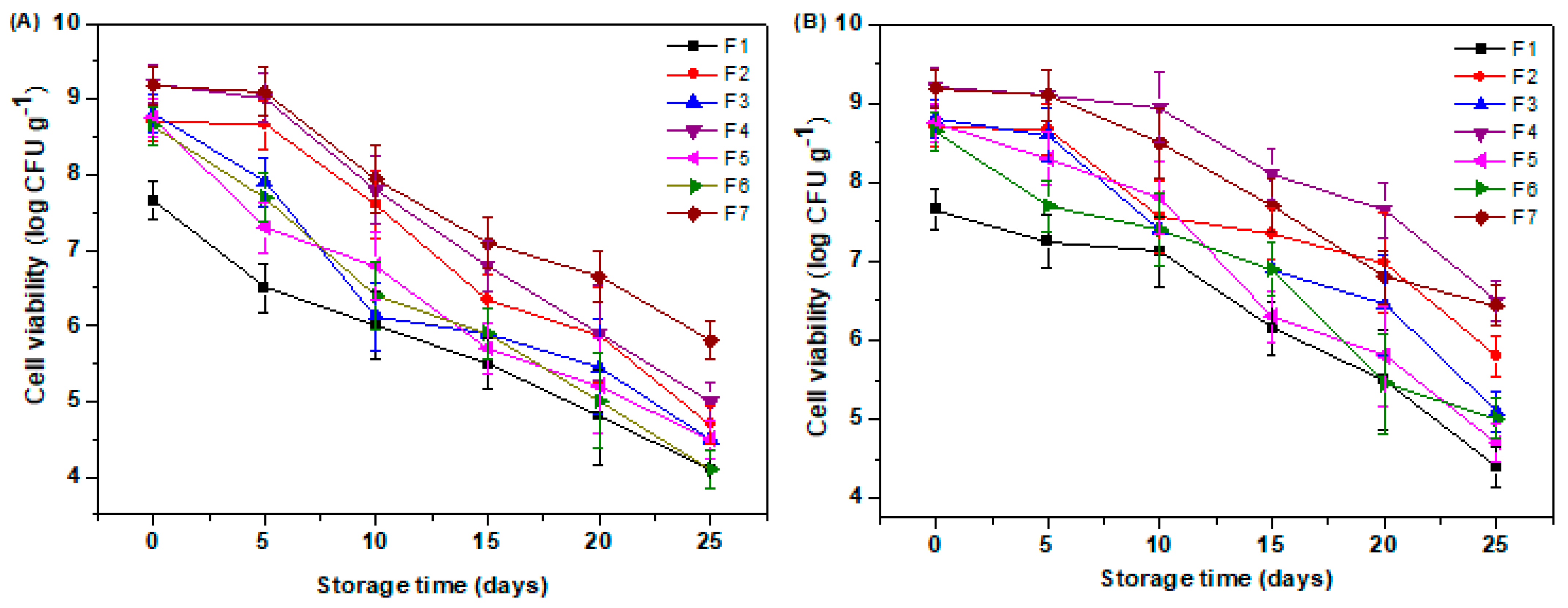
Evaluating the stability of probiotics during storage is a crucial step in the development of functional delivery systems, as the effectiveness of these products is directly related to their ability to maintain a viable number of microorganisms over time. However, among the most critical factors affecting probiotic survival are temperature and storage conditions [23]. In this context, the present study assessed the viability of probiotic bacteria in the different edible film formulations over 25 days under two storage conditions. Figure 7 presents the viability data of L. rhamnosus GG in the different edible film formulations during 25 days of storage at two temperatures: ambient (25.0 ± 1 °C) (Figure 7A) and refrigeration (4.0 ± 1 °C) (Figure 7B).
The results revealed a clear and consistent impact of temperature on the preservation of cell viability. In all the formulations, refrigerated storage resulted in significantly lower reductions in viable cell counts compared to storage at room temperature. This behavior is associated with the lower metabolic activity of microorganisms and the slowdown of degradation reactions in the polymer matrix at lower temperatures [26].
Regarding the composition of the formulations, it was observed that those incorporating gelatin combined with FOSs or inulin and alginate (F7 and F4, respectively) offered the best protection for probiotic cells, especially under refrigeration. After 25 days, these formulations showed reductions of only 2.74 log (29.8%) and 2.71 log (29.40%), respectively. The presence of alginate was particularly relevant due to its ability to form a dense and homogeneous hydrogel matrix with high water retention and low gas permeability, providing an effective physical barrier against environmental stress factors such as oxygen and temperature fluctuations [17].
In contrast, the control formulation (F1), composed only of gelatin and glycerol, showed one of the poorest performances, especially at room temperature, with a reduction of 3.55 log (46.40%) by the end of the storage period. This result reinforces the limitation of isolated protein matrices, which, although capable of forming films with good network-forming capacity, tend to exhibit a porous and less cohesive structure that facilitates the diffusion of moisture, oxygen, and heat [49]. The absence of structurally reinforcing and/or prebiotic components compromises the integrity of the matrix and significantly reduces its efficiency as a probiotic protective carrier. These findings are consistent with previous studies conducted by Soukoulis et al. [11], who developed edible films based on whey protein isolate and carrageenan as carriers for Lactobacillus spp., Lactococcus spp., and Bifidobacterium spp.
Therefore, the data obtained in this study demonstrate that optimizing the polymer matrix, especially through the inclusion of alginate and prebiotics, combined with refrigeration, is an essential strategy to increase stability and extend the shelf life of probiotics incorporated into edible films.

4. Conclusions

This study demonstrated that the rational selection and combination of biopolymers, specifically gelatin, inulin, FOSs, pectin, and alginate, can significantly influence the physicochemical, mechanical, and functional properties of edible films designed for probiotic delivery. The formulation composed of gelatin, FOSs, and alginate (F2) exhibited the best overall performance, providing the highest cell viability after drying (92.70%), simulated gastrointestinal digestion (76.10%), and storage at 25 °C for 30 days (68.40%). These results highlight a synergistic effect where FOSs enhanced prebiotic function and moisture retention, while alginate contributed to film structuring and protection under acidic and enzymatic conditions. In addition, thermal analysis showed that the incorporation of polysaccharides increased the thermal stability of the films (ΔH up to 1.50 J g−1), and water vapor permeability values ranged from 0.18 to 0.32 g·mm/kPa·m2·day, indicating potential for active packaging applications. The modulation of film hydrophobicity, mechanical resistance, and barrier properties by varying polysaccharide composition supports the feasibility of tailoring films to specific application demands.
These findings have broader implications for the design of sustainable, clean-label delivery systems not only for probiotics but also for other thermosensitive or bioactive compounds. Future studies should explore in vivo performance, sensory acceptance in real food matrices, and the incorporation of additional bioactives to expand functionality. Moreover, a systematic study of molecular interactions within these polymeric networks may further enhance predictive design strategies for functional edible films.

Author Contributions

Conceptualization, G.M.d.S., N.C.S., A.S.M., and P.H.D.F.; data curation, T.L.B.d.L., M.d.O.L., L.d.S.O., and P.H.D.F.; formal analysis, G.M.d.S., L.A.d.S., T.L.B.d.L., L.d.S.O., and P.H.D.F.; funding acquisition, R.A.d.L.D., D.G., J.P.G., and A.P.T.R.; investigation, L.A.d.S., M.d.O.L., V.M.d.A.S., V.H.d.A.R., and A.S.M.; methodology, G.M.d.S., N.C.S., M.d.O.L., V.M.d.A.S., V.H.d.A.R., and D.G.; project administration, J.P.G., and A.P.T.R.; resources, J.P.G., and A.P.T.R.; software, L.A.d.S.; supervision, R.A.d.L.D., J.P.G., and A.P.T.R.; validation, V.H.d.A.R.; visualization, A.S.M.; writing—original draft, G.M.d.S., N.C.S., and V.M.d.A.S.; writing—review and editing, N.C.S., R.A.d.L.D., D.G., J.P.G., and A.P.T.R. All authors have read and agreed to the published version of the manuscript.

Funding

This research received no external funding.

Institutional Review Board Statement

Not applicable.

Informed Consent Statement

Not applicable.

Data Availability Statement

The original contributions presented in this study are included in this article; further inquiries can be directed to the corresponding author.

Acknowledgments

The authors acknowledge Fundação de Apoio à Pesquisa do Estado da Paraíba (FAPESQ-PB) and Conselho Nacional de Desenvolvimento Científico e Tecnológico (CNPq). The authors are grateful to the Federal University of Campina Grande (UFCG) and Laboratory GM® for technical support.

Conflicts of Interest

The authors declare no conflicts of interest.

References

  1. Huda, A.N.; Asfar, M.; Bastian, F.; Syarifuddin, A. Extraction of fish oil from Sardinella longiceps and pectin from Dillenia serrata fruit peel and theirs usage in gum Arabic edible film. Food Chem. Adv. 2025, 7, 101026. [Google Scholar] [CrossRef]
  2. Khalili, E.; Khaniki, G.J.; Shojaee-Aliabadi, S.; Shariatifar, N.; Aslani, R.; Mirmoghtadaie, L. Development and characterization of amaranth protein-based edible films incorporating Satureja khuzestanica essential oil. Appl. Food Res. 2025, 5, 100967. [Google Scholar] [CrossRef]
  3. Santos, N.C.; Almeida, R.L.J.; Albuquerque, J.C.; de Lima, T.L.B.; de Sousa, F.M.; de Alcântara Silva, V.M.; de Sousa, Á.B.B. Effect of Ultrasound and Freeze-Drying to Enhance the Extraction of Phenolic Compounds in Dragon Fruit Peels and Apply Them in Edible Starch-Based Films. Packag. Technol. Sci. 2024, 37, 901–916. [Google Scholar] [CrossRef]
  4. Jafari, M.; Afkhami, R.; Sedaghat, N. Preparation and characterization of active Cirish fructans–fish gelatin film: Physicochemical, antioxidant, and antimicrobial properties. Food Sci. Nutr. 2023, 11, 709–721. [Google Scholar] [CrossRef] [PubMed]
  5. Gómez-Estaca, J.; Montero, P.; Fernández-Martín, F.; Gómez-Guillén, M.C. Physico-chemical and film-forming properties of bovine-hide and tuna-skin gelatin: A comparative study. J. Food Eng. 2009, 90, 480–486. [Google Scholar] [CrossRef]
  6. Hosseini, S.F.; Rezaei, M.; Zandi, M.; Farahmandghavi, F. Development of bioactive fish gelatin/chitosan nanoparticles composite films with antimicrobial properties. Food Chem. 2016, 194, 1266–1274. [Google Scholar] [CrossRef]
  7. Sáenz, C.; Tapia, M.S.; Chávez, J.; Robert, P. Microencapsulation by spray drying of bioactive compounds from cactus pear (Opuntia ficus-indica). Food Chem. 2009, 114, 616–622. [Google Scholar] [CrossRef]
  8. Ortega-Toro, R.; Jiménez, A.; Talens, P.; Chiralt, A. Properties of starch–hydroxypropyl methylcellulose based films obtained by compression molding. Carbohydr. Polym. 2014, 109, 155–165. [Google Scholar] [CrossRef]
  9. Lima, A.T.; Silva, H.A.; Rodrigues, T.J.A.; Santos, N.C.; Rocha, A.P.T.; de Araújo, G.T. Effect of chitosan concentration, glycerol and cashew peel extract on the physical, mechanical and morphological properties of starch-based films. Packag. Technol. Sci. 2024, 37, 1003–1015. [Google Scholar] [CrossRef]
  10. Costa, E.S.; dos Santos Moreira, I.; Gomes, J.P.; do Socorro Rocha Bastos, M.; Mattos, A.L.A.; Santos, N.C.; Matsui, K.N. Barrier, mechanical and antimicrobial properties of yam starch film with Cássia cinnamon essential oil and its application in strawberry storage. Packag. Technol. Sci. 2025, 38, 681–697. [Google Scholar] [CrossRef]
  11. Soukoulis, C.; Behboudi-Jobbehdar, S.; Yonekura, L.; Parmenter, C.; Fisk, I.D. Stability of Lactobacillus rhamnosus GG in prebiotic edible films. Food Chem. 2014, 159, 302–308. [Google Scholar] [CrossRef]
  12. Lu, J.; Lu, Y.; Chang, C.; Wu, J. Novel probiotic fermented edible film based on gum arabic/whey protein isolate/isomalt/glycerol incorporated with Lactobacillus rhamnosus: Physical, antibacterial, and fresh pork preservation properties. Food Hydrocoll. 2025, 161, 110875. [Google Scholar] [CrossRef]
  13. Nguyen, N.H.K.; Bach, G.L.; Tran, T.T. Effects of film-forming components on the viability of probiotics and the application of synbiotic pectin film in preserving Da Xanh pomelo and Thai jackfruit fresh-cut. Food Sci. Biotechnol. 2024, 33, 3093–3104. [Google Scholar] [CrossRef] [PubMed]
  14. Todhanakasem, T.; Boonchuai, P.; Itsarangkoon Na Ayutthaya, P.; Suwapanich, R.; Hararak, B.; Wu, B.; Young, B.M. Development of bioactive Opuntia ficus-indica edible films containing probiotics as a coating for fresh-cut fruit. Polymers 2022, 14, 5018. [Google Scholar] [CrossRef]
  15. Monteiro, S.S.; Almeida, R.L.; Santos, N.C.; Pereira, E.M.; Silva, A.P.; Oliveira, H.M.L.; Pasquali, M.A.D.B. New functional foods with cactus components: Sustainable perspectives and future trends. Foods 2023, 12, 2494. [Google Scholar] [CrossRef]
  16. Coimbra, P.; Alarico, S.; Empadinhas, N.; Braga, M.E.; Gaspar, M.C. Sustainable starch-based edible films with agrifood residues as potential carriers for the probiotic Lactobacillus rhamnosus. Innov. Food Sci. Emerg. Technol. 2023, 88, 103452. [Google Scholar] [CrossRef]
  17. Kalkan, S.; Çopur, Ş.Y.; Akben, S.B.; Otağ, M.R.; Engin, M.S. Effect of Incorporation of Mix Probiotic Culture in Sodium Alginate Film Characteristics: Antimicrobial, Physiochemical, Mechanical, and Barrier Properties. Probiotics Antimicrob. Proteins 2025, 1–16. [Google Scholar] [CrossRef]
  18. Sogut, E.; Filiz, B.E.; Seydim, A.C. Whey protein isolate-and carrageenan-based edible films as carriers of different probiotic bacteria. J. Dairy Sci. 2022, 105, 4829–4842. [Google Scholar] [CrossRef]
  19. Ebrahimi, B.; Mohammadi, R.; Rouhi, M.; Mortazavian, A.M.; Shojaee-Aliabadi, S.; Koushki, M.R. Survival of probiotic bacteria in carboxymethyl cellulose-based edible film and assessment of quality parameters. LWT 2018, 87, 54–60. [Google Scholar] [CrossRef]
  20. Lian, W.C.; Hsiao, H.C.; Chou, C.C. Viability of microencapsulated bifidobacteria in simulated gastric juice and bile solution. Int. J. Food Microbiol. 2003, 86, 293–301. [Google Scholar] [CrossRef]
  21. Kailasapathy, K.; Chin, J. Survival and therapeutic potential of probiotic organisms with reference to Lactobacillus acidophilus and Bifidobacterium spp. Immunol. Cell Biol. 2000, 78, 80–88. [Google Scholar] [CrossRef]
  22. Orozco-Parra, J.; Mejía, C.M.; Villa, C.C. Development of a Bioactive Synbiotic Edible Film Based on Cassava Starch, Inulin, and Lactobacillus casei. Food Hydrocoll. 2020, 104, 105754. [Google Scholar] [CrossRef]
  23. Santos, N.C.; Almeida, R.L.J.; Monteiro, S.S.; de Lima, T.L.B.; da Silva Lúcio, A.; da Silva Nogueira, L.P.; Rocha, A.P.T. Microencapsulating Lacticaseibacillus rhamnosus GG by spray drying using pea protein, pectin, and tapioca flour: Probiotic viability, digestibility and thermal stability. Food Bioprod. Process. 2025, 150, 207–2016. [Google Scholar] [CrossRef]
  24. Morais, S.K.Q.; Felipe, K.H.; de Souza Silva, J.D.; Santos, N.C.; de Farias Araújo, M.S.; Vieira, P.P.F.; Gouveia, D.S. Development of Nile tilapia gelatin films with grape pomace extract for fish fillet storage: Hydrophobic, functional properties, and biodegradability. Food Biosci. 2025, 68, 106675. [Google Scholar] [CrossRef]
  25. Krishna, M.; Nindo, C.I.; Min, S.C. Development of fish gelatin edible films using extrusion and compression molding. J. Food Eng. 2012, 108, 337–344. [Google Scholar] [CrossRef]
  26. Salimiraad, S.; Safaeian, S.; Basti, A.A.; Khanjari, A.; Nadoushan, R.M. Characterization of novel probiotic nanocomposite films based on nano chitosan/nano cellulose/gelatin for the preservation of fresh chicken fillets. LWT 2022, 162, 113429. [Google Scholar] [CrossRef]
  27. ASTM E96/E96M-16 Standard Test; Methods for Water Vapor Transmission of Materials. ASTM-American Society for Testing and Materials: West Conshohocken, PA, USA, 2016. [CrossRef]
  28. Yin, Y.C.; Yin, S.W.; Yang, X.Q.; Tang, C.H.; Wen, S.H.; Chen, Z.; Xiao, B.J.; Wu, L.Y. Surface modification of sodium caseinate films by zein coatings. Food Hydrocoll. 2014, 36, 1–8. [Google Scholar] [CrossRef]
  29. ASTM Designation D 828–97; Standard Test Method for Tensile Properties of Paper and Paperboard Using Constant-Rate-of-Elongation Apparatus. American Society for Testing and Materials: West Conshohocken, PA, USA, 2002.
  30. Zhang, Y.; Zhao, K.; Guo, Y.; Teng, A.; Li, S.; Ma, Y.; Wang, W. Effects of physical crosslinking methods on digestibility in vitro and safety of edible packaging: A primary study on collagen films. Food Mater. Res. 2023, 3, 1–7. [Google Scholar] [CrossRef]
  31. Soukoulis, C.; Behboudi-Jobbehdar, S.; Macnaughtan, W.; Parmenter, C.; Fisk, I.D. Stability of Lactobacillus rhamnosus GG incorporated in edible films: Impact of anionic biopolymers and whey protein concentrate. Food Hydrocoll. 2017, 70, 345–355. [Google Scholar] [CrossRef]
  32. Khodaei, D.; Hamidi-Esfahani, Z.; Lacroix, M. Gelatin and low methoxyl pectin films containing probiotics: Film characterization and cell viability. Food Biosci. 2020, 36, 100660. [Google Scholar] [CrossRef]
  33. Bello, A.B.; Kim, D.; Kim, D.; Park, H.; Lee, S.H. Engineering and functionalization of gelatin biomaterials: From cell culture to medical applications. Tissue Eng. Part B Rev. 2020, 26, 164–180. [Google Scholar] [CrossRef] [PubMed]
  34. Dou, L.; Li, B.; Zhang, K.; Chu, X.; Hou, H. Physical properties and antioxidant activity of gelatin-sodium alginate edible films with tea polyphenols. Int. J. Biol. Macromol. 2018, 118, 1377–1383. [Google Scholar] [CrossRef] [PubMed]
  35. Romano, N.; Tavera-Quiroz, M.J.; Bertola, N.; Mobili, P.; Pinotti, A.; Gómez-Zavaglia, A. Edible methylcellulose-based films containing fructo-oligosaccharides as vehicles for lactic acid bacteria. Food Res. Int. 2014, 64, 560–566. [Google Scholar] [CrossRef]
  36. Russo, R.; Malinconico, M.; Santagata, G. Effect of cross-linking with calcium ions on the physical properties of alginate films. Biomacromolecules 2007, 8, 3193–3197. [Google Scholar] [CrossRef]
  37. Correa, A.C.; Lopes, M.S.; Perna, R.F.; Silva, E.K. Fructan-type prebiotic dietary fibers: Clinical studies reporting health impacts and recent advances in their technological application in bakery, dairy, meat products and beverages. Carbohydr. Polym. 2024, 323, 121396. [Google Scholar] [CrossRef]
  38. Santos, N.C.; Almeida, R.L.J.; Brito, A.C.D.O.; Silva, V.M.D.A.; Albuquerque, J.C.; Saraiva, M.M.T.; Mota, M.M.D.A. Effect of pulse electric field (PEF) intensity combined with drying temperature on mass transfer, functional properties, and in vitro digestibility of dehydrated mango peels. J. Food Meas. Charact. 2023, 17, 5219–5233. [Google Scholar] [CrossRef]
  39. Mukaila, T.; Adeniyi, A.; Bello, I.; Sarker, N.C.; Monono, E.; Hammed, A. Optimizing film mechanical and water contact angle properties via PLA/starch/lecithin concentrations. Clean. Circular Bioecon. 2024, 8, 100095. [Google Scholar] [CrossRef]
  40. Zhai, X.; Ma, X.; Sun, Y.; Xue, Y.; Ban, W.; Song, W.; Shen, T.; Li, Z.; Huang, X.; Sun, Q.; et al. A Hydrophobic Ratiometric Fluorescent Indicator Film Using Electrospinning for Visual Monitoring of Meat Freshness. Foods 2025, 14, 2200. [Google Scholar] [CrossRef]
  41. Silva, K.C.G.; Sato, A.C.K. Biopolymer gels containing fructooligosaccharides. Food Res. Int. 2017, 101, 88–95. [Google Scholar] [CrossRef] [PubMed]
  42. Bertan, D.W.; da Silva Lima, L.R.; Benoso, P.; Lourenço, R.V.; Bittante, A.M.Q.B.; Gomes, A.; do Amaral Sobral, P.J. Impact of hydroethanolic extracts from Guaco leaves (Mikania glomerata Sprengel) on mechanical properties and bioactivities of gelatin-based bioactive films. Food Bioprod. Process. 2025, 151, 355–371. [Google Scholar] [CrossRef]
  43. Romo, I.; Abugoch, L.; Tapia, C. Soluble complexes between chenopodins and alginate/chitosan: Intermolecular interactions and structural-physicochemical properties. Carbohydr. Polym. 2020, 227, 115334. [Google Scholar] [CrossRef] [PubMed]
  44. Park, J.; Nam, J.; Yun, H.; Jin, H.J.; Kwak, H.W. Aquatic polymer-based edible films of fish gelatin crosslinked with alginate dialdehyde having enhanced physicochemical properties. Carbohydr. Polym. 2021, 254, 117317. [Google Scholar] [CrossRef] [PubMed]
  45. Tessaro, L.; Pereira, A.G.R.; Martelli-Tosi, M.; Sobral, P.J.D.A. Improving the Properties of Gelatin-Based Films by Incorporation of “Pitanga” Leaf Extract and Crystalline Nanocellulose. Foods 2024, 13, 1480. [Google Scholar] [CrossRef]
  46. Karaki, N.; Aljawish, A.; Muniglia, L.; Humeau, C.; Jasniewski, J. Physicochemical characterization of pectin grafted with exogenous phenols. Food Hydrocoll. 2016, 60, 486–493. [Google Scholar] [CrossRef]
  47. Zinina, O.; Merenkova, S.; Galimov, D. Development of biodegradable alginate-based films with bioactive properties and optimal structural characteristics with incorporation of protein hydrolysates. Sustainability 2023, 15, 15086. [Google Scholar] [CrossRef]
  48. Kozlov, P.V.; Burdygina, G.I. The structure and properties of solid gelatin and the principles of their modification. Polymer 1983, 24, 651–666. [Google Scholar] [CrossRef]
  49. Segtnan, V.H.; Isaksson, T. Temperature, sample and time dependent structural characteristics of gelatine gels studied by near infrared spectroscopy. Food Hydrocoll. 2004, 18, 1–11. [Google Scholar] [CrossRef]
  50. Long, T.; Tan, W.; Tian, X.; Tang, Z.; Hu, K.; Ge, L.; Li, D. Gelatin/alginate-based microspheres with sphere-in-capsule structure for spatiotemporal manipulative drug release in gastrointestinal tract. Int. J. Biol. Macromol. 2023, 226, 485–495. [Google Scholar] [CrossRef]
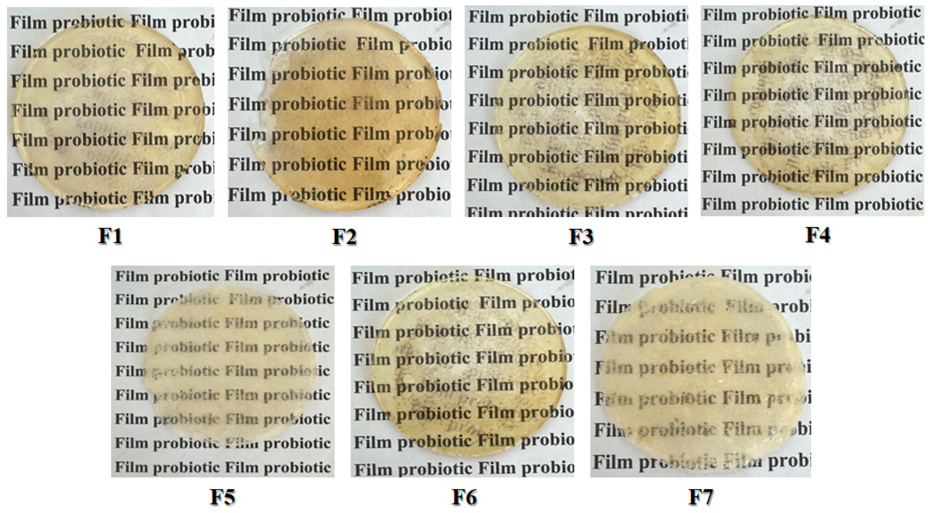
Figure 1. Developed edible films containing fish gelatin, plasticizer, prebiotics, structuring polysaccharides, and L. rhamnosus GG. Caption: F1: film with gelatin, glycerol, and L. rhamnosus GG; F2: film with gelatin, glycerol, inulin, and L. rhamnosus GG; F3: film with gelatin, glycerol, inulin, L. rhamnosus GG, and pectin; F4: film with gelatin, glycerol, inulin, L. rhamnosus GG, and alginate; F5: film with gelatin, glycerol, FOSs, and L. rhamnosus GG; F6: film with gelatin, glycerol, FOSs, L. rhamnosus GG, and pectin; F7: film with gelatin, glycerol, FOSs, L. rhamnosus GG, and alginate.
Polysaccharides 06 00079 g001
Figure 2. Viable cell counts of the film-forming solution (before drying) and the edible films (after drying). Caption: F1: film with gelatin, glycerol, and L. rhamnosus GG; F2: film with gelatin, glycerol, inulin, and L. rhamnosus GG; F3: film with gelatin, glycerol, inulin, L. rhamnosus GG, and pectin; F4: film with gelatin, glycerol, inulin, L. rhamnosus GG, and alginate; F5: film with gelatin, glycerol, FOSs, and L. rhamnosus GG; F6: film with gelatin, glycerol, FOSs, L. rhamnosus GG, and pectin; F7: film with gelatin, glycerol, FOSs, L. rhamnosus GG, and alginate. Different letters (a–c) indicate significant differences among samples by Tukey’s test (p < 0.05). Different letters (A–B) indicate significant differences between the film-forming solution and edible film by Tukey’s test (p < 0.05).
Polysaccharides 06 00079 g002
Figure 3. Images of the water contact angle (WCA) and significance analysis of the probiotic edible films. Caption: F1: film with gelatin, glycerol, and L. rhamnosus GG; F2: film with gelatin, glycerol, inulin, and L. rhamnosus GG; F3: film with gelatin, glycerol, inulin, L. rhamnosus GG, and pectin; F4: film with gelatin, glycerol, inulin, L. rhamnosus GG, and alginate; F5: film with gelatin, glycerol, FOSs, and L. rhamnosus GG; F6: film with gelatin, glycerol, FOSs, L. rhamnosus GG, and pectin; F7: film with gelatin, glycerol, FOSs, L. rhamnosus GG, and alginate. Different letters (a–e) indicate significant differences among samples by Tukey’s test (p < 0.05).
Polysaccharides 06 00079 g003
Figure 4. Tensile strength (A) and elongation at break (B) of edible films containing Lacticaseibacillus rhamnosus GG. Caption: F1: film with gelatin, glycerol, and L. rhamnosus GG; F2: film with gelatin, glycerol, inulin, and L. rhamnosus GG; F3: film with gelatin, glycerol, inulin, L. rhamnosus GG, and pectin; F4: film with gelatin, glycerol, inulin, L. rhamnosus GG, and alginate; F5: film with gelatin, glycerol, FOSs, and L. rhamnosus GG; F6: film with gelatin, glycerol, FOSs, L. rhamnosus GG, and pectin; F7: film with gelatin, glycerol, FOSs, L. rhamnosus GG, and alginate. Different letters (a–f) indicate significant differences among samples by Tukey’s test (p < 0.05).
Polysaccharides 06 00079 g004
Figure 5. Differential scanning calorimetry (DSC) thermograms of edible films containing Lacticaseibacillus rhamnosus GG. Caption: F1: film with gelatin, glycerol, and L. rhamnosus GG; F2: film with gelatin, glycerol, inulin, and L. rhamnosus GG; F3: film with gelatin, glycerol, inulin, L. rhamnosus GG, and pectin; F4: film with gelatin, glycerol, inulin, L. rhamnosus GG, and alginate; F5: film with gelatin, glycerol, FOSs, and L. rhamnosus GG; F6: film with gelatin, glycerol, FOSs, L. rhamnosus GG, and pectin; F7: film with gelatin, glycerol, FOSs, L. rhamnosus GG, and alginate.
Polysaccharides 06 00079 g005
Figure 6. Probiotic viability of edible films containing Lacticaseibacillus rhamnosus GG subjected to in vitro digestion. Caption: Digestion 0 h, SGF-2 h, and SIF-4 h represent probiotic viability at the beginning of digestion, after 2 h in simulated gastric fluid, and after 4 h in simulated intestinal fluid, respectively; F1: film with gelatin, glycerol, and L. rhamnosus GG; F2:film with gelatin, glycerol, inulin, and L. rhamnosus GG; F3: film with gelatin, glycerol, inulin, L. rhamnosus GG, and pectin; F4: film with gelatin, glycerol, inulin, L. rhamnosus GG, and alginate; F5: film with gelatin, glycerol, FOSs, and L. rhamnosus GG; F6: film with gelatin, glycerol, FOSs, L. rhamnosus GG, and pectin; F7: film with gelatin, glycerol, FOSs, L. rhamnosus GG, and alginate. Uppercase letters indicate statistically significant differences (p < 0.05) between the digestion phases. Lowercase letters indicate significant differences (p < 0.05) between the formulations.
Polysaccharides 06 00079 g006
Figure 7. Probiotic viability of edible films containing Lacticaseibacillus rhamnosus GG during storage at (A) room temperature (25.0 ± 1 °C) and (B) refrigeration (4.0 ± 1 °C). Caption: F1: film with gelatin, glycerol, and L. rhamnosus GG; F2: film with gelatin, glycerol, inulin, and L. rhamnosus GG; F3: film with gelatin, glycerol, inulin, L. rhamnosus GG, and pectin; F4: film with gelatin, glycerol, inulin, L. rhamnosus GG, and alginate; F5: film with gelatin, glycerol, FOSs, and L. rhamnosus GG; F6: film with gelatin, glycerol, FOSs, L. rhamnosus GG, and pectin; F7: film with gelatin, glycerol, FOSs, L. rhamnosus GG, and alginate.
Polysaccharides 06 00079 g007
Table 1. Formulations of edible films containing fish gelatin, plasticizer, prebiotics, structuring polysaccharides, and L. rhamnosus GG.
FormulationsGelatin
(% m/m)
Glycerol
(% w/w)
Prebiotic
(2.0 g.100 g−1)
Probiotic (0.02 g.100 g−1)Polysaccharide (% m/m)
F13.030NoYes No
F23.030InulinYesNo
F33.030InulinYesPectin (1.0%)
F43.030InulinYesAlginate (0.50%)
F53.030FOSsYesNo
F63.030FOSsYesPectin (1.0%)
F73.030FOSsYesAlginate (0.50%)
Caption: FOSs, fructooligosaccharides.
Table 2. Solubility, water vapor permeability (WVP), thickness, and moisture content of probiotic edible films.
FilmsSolubility (%)WVP
(g·mm/m2·d·kPa)
Thickness (mm)Moisture Content (%)
F141.37 ± 0.22 a11.52 ± 0.91 a0.085 ± 0.003 c17.50 ± 0.22 a
F238.20 ± 0.75 b10.24 ± 0.76 b0.091 ± 0.002 a,b15.23 ± 0.31 b
F332.51 ± 0.38 c,d9.65 ± 0.82 b,c0.088 ± 0.004 b15.10 ± 0.27 b
F428.73 ± 0.78 e8.30 ± 0.57 d0.095 ± 0.003 a13.47 ± 0.19 c
F534.08 ± 0.44 c11.75 ± 0.36 a0.089 ± 0.001 a,b15.36 ± 0.50 b
F631.88 ± 0.11 d9.08 ± 0.13 c0.090 ± 0.002 a,b14.72 ± 0.17 b
F727.50 ±0.27 e8.17 ± 0.28 d0.088 ± 0.003 b13.66 ± 0.24 c
Caption: mean ±standard deviation; WVP: water vapor permeability; F1: film with gelatin, glycerol, and L. rhamnosus GG; F2: film with gelatin, glycerol, inulin, and L. rhamnosus GG; F3: film with gelatin, glycerol, inulin, L. rhamnosus GG, and pectin; F4: film with gelatin, glycerol, inulin, L. rhamnosus GG, and alginate; F5: film with gelatin, glycerol, FOSs, and L. rhamnosus GG; F6: film with gelatin, glycerol, FOSs, L. rhamnosus GG, and pectin; F7: film with gelatin, glycerol, FOSs, L. rhamnosus GG, and alginate. Different letters (a–e) indicate significant differences among samples by Tukey’s test (p < 0.05).
Disclaimer/Publisher’s Note: The statements, opinions and data contained in all publications are solely those of the individual author(s) and contributor(s) and not of MDPI and/or the editor(s). MDPI and/or the editor(s) disclaim responsibility for any injury to people or property resulting from any ideas, methods, instructions or products referred to in the content.