Technical Note

Ruminal Lipid A Analysis by Matrix-Assisted Laser Desorption/Ionization Time-of-Flight Mass Spectrometry

1
Department of Animal Sciences, University of Florida, Gainesville, FL 32611, USA
2
Department of Infectious Diseases and Immunology, College of Veterinary Medicine, University of Florida, Gainesville, FL 32611, USA
*
Author to whom correspondence should be addressed.
Polysaccharides 2021, 2(4), 817-824; https://doi.org/10.3390/polysaccharides2040049
Submission received: 22 September 2021 / Revised: 13 October 2021 / Accepted: 14 October 2021 / Published: 1 November 2021

Abstract

:
Lipopolysaccharides (LPS) are cell wall components from Gram-negative bacteria and are composed of three covalently linked regions: the O-antigen, the core oligosaccharide, and the lipid A moiety, which carries most of their endotoxic activity. The objective of this study was to isolate and compare the lipid A structures from ruminal LPS derived from total mixed ration (TMR)- and pasture-fed cows, by using matrix-assisted laser desorption/ionization time-of-flight mass spectrometry (MALDI-TOF MS). Ruminal bacteria were collected from two rumen-cannulated Holstein cows; one fed a TMR (60:40, forage–concentrate) and the other pasture fed. The representativeness of each sample was validated by comparing the rumen microbiome from the cows in our study to the core rumen microbiome from the previous literature. Lipopolysaccharides from each respective sample were extracted with a phenol–water extraction procedure and purified via ultracentrifugation. To isolate lipid A from the core and O-antigen, pure ruminal LPS samples were hydrolyzed with acetic acid. Lipid A derived from the TMR-fed cow potentially exhibited a tetra-acylated structure, whereas lipid A derived from the pasture-fed cow potentially exhibited a penta-acylated lipid A structure. Both samples were quantified using limulus amebocyte lysate (LAL) assay and exhibited low endotoxic activity, consistent with the MALDI-TOF MS observations. Results indicate that the lipid A acylation pattern differs between diets, and that ruminal bacteria express solely under-acylated lipid A structures contrary to hexa-acylated lipid A, typically expressed by bacteria such as E. coli.

1. Introduction

Lipopolysaccharide (LPS) is a component of the outer membrane from the Gram-negative bacterial cell wall and is comprised of three covalently linked regions: the O-antigen, the core oligosaccharide, and the endotoxin, lipid A moiety, which carries most of the endotoxic activity of LPS [1,2]. Among Gram-negative bacteria species, there is structural diversity on the lipid A region [3,4]. Specifically, the number of acyl chains on the lipid A moiety is directly correlated with its ability to induce cytokine production where the hexa-acylated forms are usually the most immunostimulant, contrary to the under-acylated forms (penta- or tetra-acylated) that result in weak host inflammatory responses [5,6].
Ruminal bacteria are predominantly Gram-negative and are the major source of LPS in the rumen [7]. The most predominant phylum in ruminal fluid is Bacteroidetes, refs. [8,9] and, from previous reports, LPS from species of that phylum have shown reduced endotoxicity [10,11], as well as the expression of under-acylated lipid A moieties contrary to the widely used hexa-acylated lipid A expressed from strains such as Escherichia coli (E. coli) [12,13,14].
Although E. coli LPS are the most studied LPS in ruminants, it is unclear if ruminal bacteria LPS exhibit a similar chemical composition to E. coli LPS. Moreover, there are evidences that environmental factors such as diet can alter LPS composition in cows [15]; however, to our knowledge, this has never been documented in ruminal bacteria. For this reason, we hypothesized that the endotoxin derived from ruminal Gram-negative bacteria would exhibit penta-acylated and/or tetra-acylated lipid A structures. Therefore, the objective of this preliminary study was to use matrix-assisted laser desorption/ionization time-of-flight mass spectrometry (MALDI-TOF MS) to determine the different lipid A patterns present in ruminal LPS and compare the acylation pattern of lipid A derived from the ruminal contents of total mixed ration (TMR)- and pasture-fed cows.

2. Materials and Methods

2.1. Sampling

The University of Florida Institutional Animal Care and Use Committee approved all the procedures for animal care and handling required for this experiment. Ruminal fluid was obtained from two adult rumen-cannulated Holstein cows (same age and similar body weight and body condition score). Ruminal microbial composition of each animal was analyzed by 16S rRNA sequencing to assure that they were representative of the general population. Cows were fed either a total mixed ration (TMR) or pasture. TMR had 60% whole plant corn silage, 12.5% ground corn, 13% citrus pulp, 12% soybean meal, and 2.5% mineral and vitamin mix (Dry Matter basis; DM); the diet nutrient composition was: 16% crude protein (CP), 35% neutral detergent fiber (NDF), and 4.3% water soluble carbohydrates. Pasture was 100% bahiagrass (12.5% CP, 70% NDF, 4% WSC, 60% in vitro digestibility) and mineral premix. Mineral premix contained (DM basis) 52.2% wheat middlings, 18.7% rumen protected choline (Balchem, New Hampton, NY, USA), 16.0% magnesium sulfate heptahydrate, 4.8% magnesium oxide, 4.5% sodium chloride, 0.685% of a mixture containing vitamins A, D, and E and iodine, 0.45% Sel-Plex 2000 (Alltech Biotechnology, Nicholasville, KY, USA), 0.40% Rumensin 90 (Elanco Animal Health, Indianapolis, IN, USA), 0.27% IntelliBond Vital4 (Micronutrients, Indianapolis, IN, USA), and 2.0% ClariFly Larvicide (Central Life Sciences, Dallas, TX, USA). Cows were fed ad libitum; TMR-fed cow ate approximately 23 kg of DM per day, while pasture-fed cow ate approximately 10 kg/d (DM). The cows were adapted to their respective diets 3 weeks before ruminal content collection. Approximately 1 h after morning feeding, ruminal contents were manually collected (approximately 7 L) from each cow and strained through four layers of cheesecloth, into a pre-warmed thermos, and immediately transported to the lab. The material was strained again through two-layers of cheesecloth, transferred into beakers, and put on ice for 15 min. The preparation was centrifuged (Sorvall RC-5B Refrigerated Superspeed Centrifuge, DuPont Instruments®, Kingston, ON, USA) at 1000× g for 10 min at 4 °C and the supernatant was collected and centrifuged at 11,250× g for 20 min at 4 °C to obtain the bacterial pellet that was resuspended in Milli-Q water and centrifuged at 16,250× g for 20 min at 4 °C [16]. Finally, bacterial pellets from each cow were transported in pyrogen-free tubes, homogenized into 15 mL of Milli-Q water and stored in −80 °C for later ruminal LPS and DNA extraction.

2.2. Ruminal LPS Extraction

A modified hot-phenol extraction was utilized to extract the LPS from the isolated ruminal bacteria derived from both cows. This was conducted essentially as described by [10,17] with a few modifications; to isolate total LPS, the bacteria pellets were boiled at 100–110 °C in a heat block for 30 min and, after the addition of 50 mL Milli-Q water, the bacteria suspension was treated with 50 mL of 90% phenol at 68 °C for 30 min. The preparation was placed in −20 °C for 30 min, centrifuged at 5000× g for 10 min at 4 °C and the aqueous (top), middle, and phenol (bottom) layers were collected in separate tubes and tested for LPS presence with silver stain (Thermo Scientific™ Pierce™ Silver Stain Kit, Thermo Fisher Scientific, Waltham, MA, USA). The aqueous layer exhibited the greatest concentration of LPS and was transported into regenerated cellulose dialysis membrane (Fisherbrand™, Loughborough, UK) for further dialysis against Milli-Q at 4 °C until phenol was not detectable at 260 nm in Milli-Q. Dialyzed samples were treated with 5 mM MgCl2, followed by 20 μg/mL DNase I (M0303s, New England Biolabs, Ipswich, MA, USA) for 2 h at 37 °C, to degrade contaminating DNA, 20 μg/mL RNase H (T3018, New England Biolabs, Ipswich, MA, USA) for 2 h at 37 °C, to degrade contaminating RNA and 30 mg/mL Proteinase K (Fisher BioReagents™ Proteinase K, Catalog No.BP1700-100, Loughborough, UK), to degrade contaminating protein. If protein contamination was suspected, then the concentration and length of incubation with Proteinase K was doubled. The preparation was freeze-dried and crude LPS mass was determined by weighing the sample. After lyophilization, dry samples were resuspended into 15 mL of Milli-Q water and centrifuged at 1110× g for 10 min at 4 °C to remove any solid contaminants. The supernatant was treated with 0.15 mL of 50 mM acetic acid, 95% ethanol and transferred with glass Pasteur pipette into ultracentrifuge tubes (Quick-Seal® Round-Top Polypropylene Tube, Indianapolis, IN, USA) and then spun for 8 h at 4 °C and 105,000× g in an ultracentrifuge (Optima XE, Beckman Coulter Life Sciences, Indianapolis, IN, USA). The supernatant was removed and LPS gels were resuspended in 2 mL of endotoxin-free water and lyophilized to determine the dry weight of pure LPS. To confirm the quality of ruminal-derived LPS, the aqueous, middle, and bottom layers, as well as the pure LPS, were visualized with the Pierce™ Silver Stain Kit (Thermo Scientific™, Waltham, MA, USA) in accordance to the manufacturer’s instructions. In all cases, the method indicated a purity similar to that of LPS from pure bacterial isolates. Ruminal LPS presence and activity was determined with chromogenic kinetic Limulus amebocyte lysate assay (LAL: Thermo Scientific™ Pierce™ Chromogenic Endotoxin Quant Kit, Waltham, MA, USA) in a 96-well microplate using an incubating microplate spectrophotometer (Spectra Max 340 PC, Molecular Devices Corp., Sunnyvale, CA, USA).

2.3. Ruminal Lipid A Isolation

To isolate lipid A, 20 mg of pure ruminal LPS extracted from the TMR-fed cow (TMR lipid A) was resuspended in 0.5 mL of 1M acetic acid and heated to 100 °C for 2 h to hydrolyze the lipid A from the core and O-antigen. The sample was dried under vacuum and resuspended in 100 μL Milli-Q water and then sonicated for 10 min in a bath sonicator. In total, 500 μL of acidified ethanol was added to the sample and briefly vortexed. The lipid A was then pelleted by centrifugation at 10,000× g at 4 °C for 10 min. The pellet was sonicated, resuspended in nonacidified 95% ethanol, and centrifuged for washing. Washing with nonacidified ethanol was repeated a total of three times. The pellet was dried under vacuum. The same exact procedure was followed to isolate lipid A from pure LPS extracted from ruminal fluid of pasture-fed cow (Pasture lipid A).

2.4. MALDI-TOF MS Analysis

For MALDI-TOF MS analysis, 2 mg from each sample, respectively, were solubilized in 100 μL of a chloroform–methanol solution (2:1, vol/vol), and then dilutions of 1:10, 1:100, and 1:1000 were prepared. The standard and samples were spotted using the sandwich method, in which 1 μL of sample was sandwiched between 1 μL of Trihydroxyacetophenone (THAP) matrix (saturated matrix prepared in 1:2 acetonitrile–water + 0.1% trifluoroacetic acid (TFA)) above and below the sample spot. (i.e., 1 μL THAP/1 μL sample/1 μL THAP) [18]. Mass spectrometry data were acquired in a model Bruker autoflex maX MALDI Time-of-Flight analyzer (Billerica, MA, USA). The instrument was operated in the negative ion reflectron mode across the mass ranges indicated (800–5000 Da). Acquired spectra were analyzed on the open source mMass software. Lipid A substituent percentages were calculated by dividing the relative peak intensity by the sum of relative peak intensities in the sample as measured by MALDI-TOF MS and then multiplying by 100. Commercially available lipid A (E. coli F583, Sigma Aldrich, Milwaukee, MO, USA) was used as a standard. Analysis of the lipid A from ruminal Gram-negative bacteria were evaluated by MALDI-TOF MS analysis in the 1300–1900 Da range for masses coinciding to under- and hexa-acylated lipid A molecules.

2.5. DNA Extraction, PCR Amplification, and 16S rRNA Sequencing
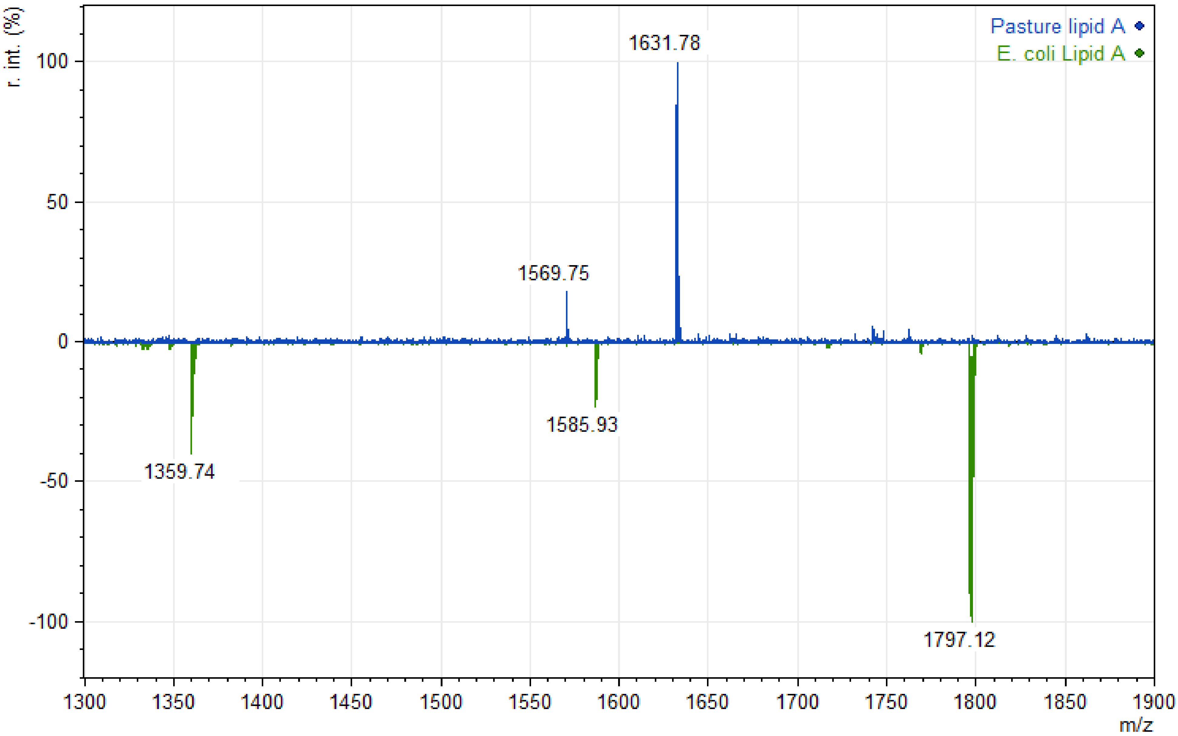
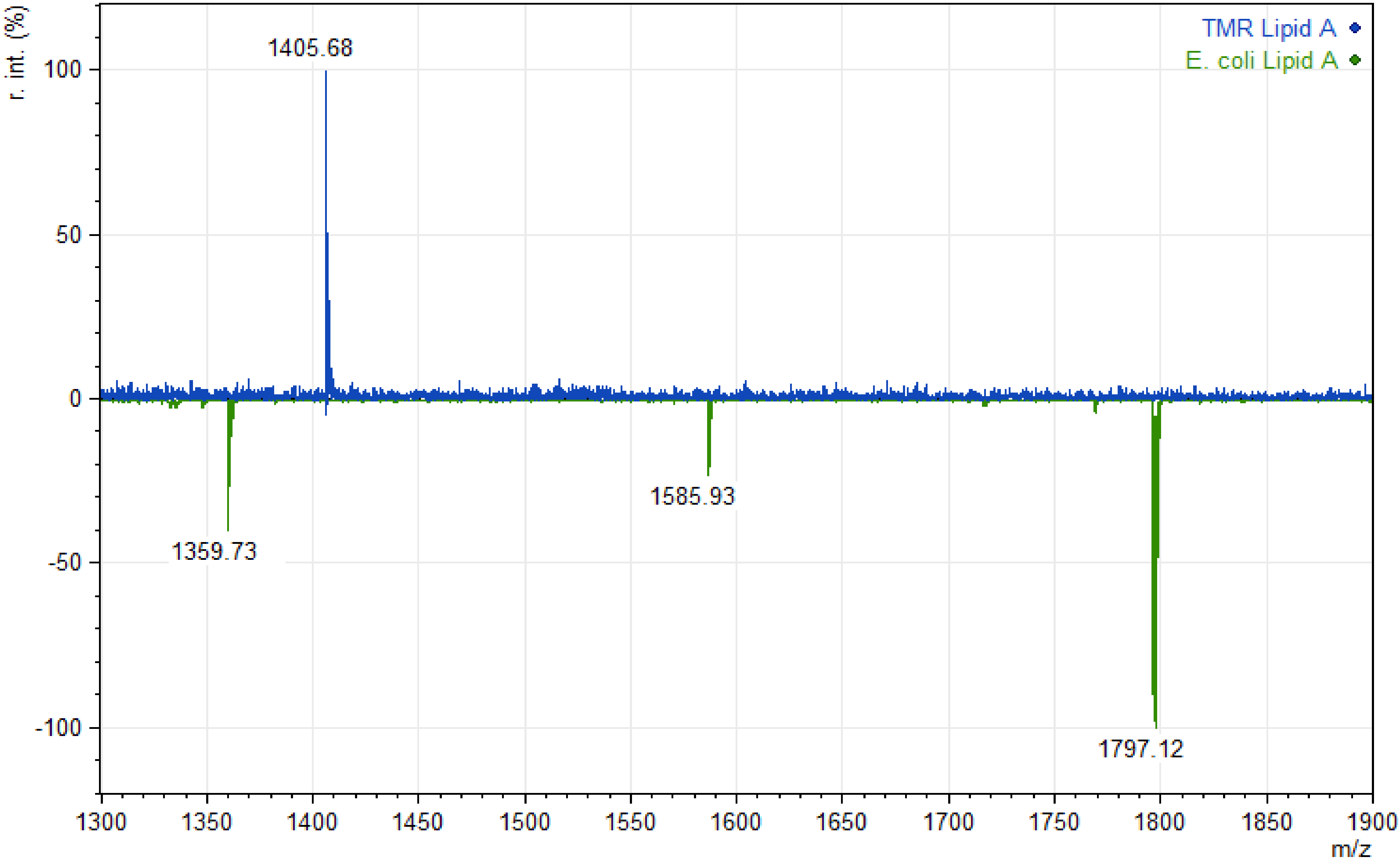
A phenol–chloroform extraction was utilized to extract the DNA from the ruminal bacteria derived from both cows. This was conducted as described previously [19,20]. Briefly, DNA was extracted from ruminal bacteria by using the phenol–chloroform extraction method and quantified by Qubit Fluorometer (Invitrogen, San Diego, CA, USA), the preparation was stored at −80 °C for later DNA sequencing. All procedures for DNA sequencing were performed according to Kozich et al. (2013) [21]. Briefly, the V4 region of the 16S rRNA gene was amplified by dual-index (forward and reverse) universal bacterial primers by using polymerase chain reaction (PCR) in a Bio-Rad C1000 TouchTM Thermal Cycler (BIO-RAD, Hercules, CA, USA). The primers used for TMR-fed cow were forward: 5′-CAAGCAGAAGACGGCATACGAGATGTCGTAGTAGTCAGTCAGCC GGACTACHVGGGTWTCTAAT-3′; reverse; 5′-AATGATACGGCGACCACCGAGATCTACACCGTGAGTGTATGGTAATTGTGTGCCAGCMGCCGCGGTAA-3′ and the primers used for pasture-fed cow were forward: 5′-CAAGCAGAAGACGGCATACGAGATTAGCAG ACAGTCAGTCAGCCGGACTACHVGGGTWTCTAAT-3′, reverse: 5′-AATGATACGGCGA C CACCGAGATCTACACCGTGAGTGTATGGTAATTGTGTGCCAGCMGCCGCGGTAA-3′. To confirm the success of amplification, preparations were run on a 1% agarose gel. Then the amplicons were normalized by using a SequalPrep plate kit (Invitrogen, Waltham, MA, USA) and the DNA concentration was quantified in the Qubit® Fluorometer. For DNA library construction, an equal pool of all the amplicons was made and validated by RT-PCR. Samples from each donor cow were sequenced by using a MiSeq reagent kit V2 (2 × 250 cycles run; Illumina, San Diego, CA, USA) in an Illumina MiSeq platform (Illumina, San Diego, CA, USA).
Sequenced amplicons were processed using the DADA2 pipeline [22] at Nephele (https://nephele.niaid.nih.gov/, accessed date; 4 March 2021), a web-based platform for microbiome data analysis. Quality control of the reads were carried out and reads with average quality of <25 were removed. Paired-end reads were merged and taxonomy assigned using the 16S rRNA SILVA v132 database [23]. DADA2 generated Amplicon Sequence Variants (ASVs) to allow distinguishing ASVs by a single nucleotide change. All sequences from this project have been deposited in the National Center for Biotechnology Information and the Short Read Archives are available under project accession PRJNA707102. The relative abundances were calculated using the Microbiome Analyst (https://www.microbiomeanalyst.ca, accessed date; 4 March 2021), a web-based platform for comprehensive statistical, visual, and meta-analysis of microbiome data.

3. Results

For LPS extraction, we extracted LPS from the ruminal bacteria of two cows fed a TMR or pasture. We collected approximately 7 L from each cow and the yield of lyophilized LPS before ultracentrifugation was 34 mg from the TMR-fed cow and 30 mg from the pasture-fed cow, the yield of lyophilized LPS after ultracentrifugation was 8 mg from the TMR-fed cow and 6 mg from the pasture-fed cow, and after the hydrolyzation of purified LPS the yield of purified lipid A was approximately 2 mg from each sample.
Analysis of both TMR-derived lipid A and pasture-derived lipid A revealed ion signals with a mass/charge ratio (m/z) below 1700 Da. TMR-derived lipid A spectra (Figure 1) conceivably exhibited a dominant peak at 1405.68 m/z, and pasture-derived lipid A spectra (Figure 2) conceivably exhibited a dominant peak at 1631.78 m/z. The complex structure of ruminal lipid A does not allow for definite conclusions about the acylation pattern; however, a potential acylation pattern that can be proposed for TMR-derived lipid A would be tetra-acylated due to the range where its dominant peak was observed. On the other hand, a potential acylation pattern that can be proposed for pasture-derived lipid A would be penta-acylated due to the range where its dominant peak was observed. Further investigation is needed at species level to confirm our preliminary findings. In our study, E. coli lipid A exhibited, as expected from previous studies, an ion at 1797.12 Da that represents hexa-acylated lipid A structure [24]. More specifically, the difference between ions at 1797.12 m/z and 1585.93 m/z was 211.19, which potentially corresponds to a fatty acid residue with 14 carbons, consistent with the previous literature [6,24]. The difference between the ions at 1585.93 m/z and 1359.74 m/z indicates that it may correspond to a fatty acid residue with 14 carbons and 3-OH groups (14:0(3-OH)), which is also consistent with the previous literature [6,24]. Furthermore, our preliminary study focused on the analysis of lipid As by MALDI-TOF MS, focusing on the major peaks, as well as, the differences between them. Therefore, further research is needed on the remaining structure of lipid A such as the number of phosphate groups in ruminal lipid A.
From LAL assay, both TMR lipid A and pasture lipid A exhibited lower endotoxic activity compared with E. coli standard. From the previous literature, penta-acylated as well as tetra-acylated forms of lipid A are associated with low endotoxicity and weak host immune response [6].
The microbial composition of each sample (Table 1) was analyzed by 16S rRNA sequencing, which revealed an overall bacterial composition that was consistent with the core microbiome from the previous literature [25], indicating that these samples were representative of the general population of TMR- and pasture-fed cows. More specifically, in both samples, Bacteroidetes and Firmicutes were the two most abundant phyla.

4. Discussion

Our results are consistent with the hypothesis of the potential presence of under-acylated and the absence of hexa-acylated lipid A structures in ruminal LPS [26]. Similarly to our study, previously published data reported the presence of penta-acylated forms of lipid A to be conserved across the Bacteroidetes phylum [11,14], which is the most predominant phylum in the rumen fluid, even under subacute ruminal acidosis conditions [8,26], which further corroborates our findings that ruminal lipid A exhibits under-acylated lipid A structures.
In our report, Bacteroidetes were in higher abundance in the TMR-fed cow; however, the lipid A from the TMR-fed cow presumably exhibited a tetra-acylated lipid A pattern. Despite this finding being paradoxical, it suggests a potential factor that would affect the ruminal lipid A structure. More specifically, the abundance at the species level would differ among TMR- and pasture-fed cows, similarly as it has been previously reported for the temperature-regulated structural modifications of lipid A [27]. Thus, in the TMR-fed cow, it is possible that the dominant species of Bacteroidetes express lipid As with four acyl chains, thus with lower endotoxicity. This is normal because TMR-fed cows usually receive feed additives such as ionophores [28] that reduce the microbial load, thus higher endotoxic strains may not be able to survive. For this reason, future research should focus on characterizing the lipid A from species of Bacteroidetes phylum.
Our preliminary report indicates that under-acylated lipid A structures seem to be conserved across the Gram-negative bacteria in the rumen, and hexa-acylated structures, expressed by commercially available LPS such as from E. coli, do not seem to be expressed by ruminal bacteria. Previous studies, in humans, showed that the nuclear factor NF-κB pathway, an important proinflammatory signaling pathway, which is commonly triggered by pathogenic bacteria upon interaction with the host, is repressed by the penta-acylated lipid A expressed by gut microbiome bacteria, supporting the idea that commensal microbes contribute to the immune tolerance in support of homeostasis [11]. Thus, potential application of our proposed study would be the investigation of the effects of ruminal LPS to trigger host immune responses in rumen epithelial tissue and investigate potential antagonistic interactions between ruminal and E. coli LPS.

5. Conclusions

To our knowledge, this preliminary study is the first to determine the specific composition of ruminal lipid A from TMR- and pasture-fed cows. Our findings indicate that ruminal LPS exhibit under-acylated lipid A structures contrary to hexa-acylated lipid A typically expressed by commercially available LPS, such as from E. coli. However, we cannot ignore the fact that the presence of single or double phosphate groups, as well as the structure of disaccharide backbone, play important roles in LPS activity. For this reason, further study is needed to elucidate the structural diversity of fatty acid chains, phosphate groups, and disaccharide backbone in ruminal Gram-negative bacteria. Furthermore, LPS derived from the TMR-fed cow potentially exhibited tetra-acylated lipid A, whereas LPS derived from the pasture-fed cow exhibited penta-acylated lipid A. Further research is needed to investigate the potential effect of diet on the expression of different lipid A acylation patterns of ruminal LPS, as well as determine if structural diversity differs under non-healthy conditions such as under ruminal acidosis.

Author Contributions

Conceptualization, A.F. and E.S.; methodology, E.S.; writing—review and editing, E.S., J.R.V., M.S.K., T.J., A.R., R.R.L., P.F., K.C.J. and A.T.; project administration, A.F.; funding acquisition, A.F. All authors have read and agreed to the published version of the manuscript.

Funding

This project was funded by startup funds from the Faciola Lab at the University of Florida.

Institutional Review Board Statement

Not applicable.

Informed Consent Statement

Not applicable.

Data Availability Statement

The sequences data presented in this study are available on request from the corresponding author.

Acknowledgments

The authors thank Diwakar Vyas and John Driver for helpful discussion.

Conflicts of Interest

The authors declare no conflict of interest.

References

  1. Caroff, M.; Karibian, D. Structure of bacterial lipopolysaccharides. Carbohydr. Res. 2003, 338, 2431–2447. [Google Scholar] [CrossRef]
  2. Erridge, C.; Bennett-Guerrero, E.; Poxton, I.R. Structure and function of lipopolysaccharides. Microbes Infect. 2002, 4, 837–851. [Google Scholar] [CrossRef]
  3. Lodowska, J.; Wolny, D.; Weglarz, L.; Dzierzewicz, Z. The structural diversity of lipid A from gram-negative bacteria. Postep. Hig. Med. Dosw. 2007, 61, 106–121. [Google Scholar]
  4. Coats, S.R.; Pham, T.-T.T.; Bainbridge, B.W.; Reife, R.A.; Darveau, R.P. MD-2 Mediates the ability of tetra-acylated and penta-acylated lipopolysaccharides to antagonize Escherichia coli lipopolysaccharide at the TLR4 signaling complex. J. Immunol. 2005, 175, 4490–4498. [Google Scholar] [CrossRef] [Green Version]
  5. Munford, R.S.; Varley, A.W. Shield as signal: Lipopolysaccharides and the evolution of immunity to gram-negative bacteria. PLoS Pathog. 2006, 2, e67. [Google Scholar] [CrossRef]
  6. Steimle, A.; Autenrieth, I.B.; Frick, J.-S. Structure and function: Lipid A modifications in commensals and pathogens. Int. J. Med. Microbiol. 2016, 306, 290–301. [Google Scholar] [CrossRef] [Green Version]
  7. Nagaraja, T.G.; Lechtenberg, K.F. Acidosis in feedlot cattle. Vet. Clin. N. Am. Food Anim. Pract. 2007, 23, 333–350. [Google Scholar] [CrossRef]
  8. Plaizier, J.C.; Li, S.; Danscher, A.M.; Derakshani, H.; Andersen, P.H.; Khafipour, E. Changes in microbiota in rumen digesta and feces due to a grain-based subacute ruminal acidosis (SARA) challenge. Microb. Ecol. 2017, 74, 485–495. [Google Scholar] [CrossRef]
  9. Khafipour, E.; Li, S.; Plaizier, J.C.; Krause, D.O. Rumen microbiome composition determined using two nutritional models of subacute ruminal acidosis. Appl. Environ. Microbiol. 2009, 75, 7115–7124. [Google Scholar] [CrossRef] [Green Version]
  10. Nagaraja, T.G.; Bartley, E.E.; Fina, L.R.; Anthony, H.D.; Bechtle, R.M. Evidence of endotoxins in the rumen bacteria of cattle fed hay or grain. J. Anim. Sci. 1978, 47, 226–234. [Google Scholar] [CrossRef]
  11. D’Hennezel, E.; Abubucker, S.; Murphy, L.O.; Cullen, T.W. Total lipopolysaccharide from the human gut microbiome silences toll-like receptor signaling. mSystems 2017, 2, e00046-17. [Google Scholar] [CrossRef] [Green Version]
  12. Sonesson, H.R.A.; Zähringer, U.; Grimmecke, H.D.; Westphal, O.R.E. Bacterial endotox. In Chemical Structure, Biological, Activity; Bringham, K., Ed.; Marcel Dekker Inc.: New York, NY, USA, 1994. [Google Scholar]
  13. Berezow, A.B.; Ernst, R.K.; Coats, S.R.; Braham, P.H.; Karimi-Naser, L.M.; Darveau, R.P. The structurally similar, penta-acylated lipopolysaccharides of Porphyromonas gingivalis and Bacteroides elicit strikingly different innate immune responses. Microb. Pathog. 2009, 47, 68–77. [Google Scholar] [CrossRef] [Green Version]
  14. Jacobson, A.N.; Choudhury, B.P.; Fischbach, M.A. The biosynthesis of lipooligosaccharide from Bacteroides thetaiotaomicron. mBio 2018, 9, e02289-17. [Google Scholar] [CrossRef] [Green Version]
  15. Bilal, M.S.; Abaker, J.A.; Ul Aabdin, Z.; Xu, T.; Dai, H.; Zhang, K.; Liu, X.; Shen, X. Lipopolysaccharide derived from the digestive tract triggers an inflammatory response in the uterus of mid-lactating dairy cows during SARA. BMC Vet. Res. 2016, 12, 284. [Google Scholar] [CrossRef] [PubMed] [Green Version]
  16. Krizsan, S.J.; Ahvenjärvi, S.; Volden, H.; Broderick, G.A. Estimation of rumen outflow in dairy cows fed grass silage-based diets by use of reticular sampling as an alternative to sampling from the omasal canal1. J. Dairy Sci. 2010, 93, 1138–1147. [Google Scholar] [CrossRef] [PubMed] [Green Version]
  17. Lam, J.S.; Anderson, E.M.; Hao, Y. LPS quantitation procedures. Methods Mol. Biol. 2014, 1149, 375–402. [Google Scholar] [CrossRef]
  18. Zhou, P.; Altman, E.; Perry, M.B.; Li, J. Study of matrix additives for sensitive analysis of lipid A by matrix-assisted laser desorption ionization mass spectrometry. Appl. Environ. Microbiol. 2010, 76, 3437–3443. [Google Scholar] [CrossRef] [PubMed] [Green Version]
  19. Stevenson, D.M.; Weimer, P.J. Dominance of Prevotella and low abundance of classical ruminal bacterial species in the bovine rumen revealed by relative quantification real-time PCR. Appl. Microbiol. Biotechnol. 2007, 75, 165–174. [Google Scholar] [CrossRef] [PubMed]
  20. Dai, X.; Paula, E.M.; Lelis, A.L.J.; Silva, L.G.; Brandao, V.L.N.; Monteiro, H.F.; Fan, P.; Poulson, S.R.; Jeong, K.C.; Faciola, A.P. Effects of lipopolysaccharide dosing on bacterial community composition and fermentation in a dual-flow continuous culture system. J. Dairy Sci. 2019, 102, 334–350. [Google Scholar] [CrossRef] [Green Version]
  21. Kozich, J.J.; Westcott, S.L.; Baxter, N.T.; Highlander, S.K.; Schloss, P.D. Development of a Dual-Index Sequencing Strategy and Curation Pipeline for Analyzing Amplicon Sequence Data on the MiSeq Illumina Sequencing Platform. Appl. Environ. Microbiol. 2013, 79, 5112–5120. [Google Scholar] [CrossRef] [Green Version]
  22. Callahan, B.J.; McMurdie, P.J.; Rosen, M.J.; Han, A.W.; Johnson, A.J.A.; Holmes, S.P. DADA2: High-resolution sample inference from Illumina amplicon data. Nat. Methods 2016, 13, 581–583. [Google Scholar] [CrossRef] [PubMed] [Green Version]
  23. Pruesse, E.; Quast, C.; Knittel, K.; Fuchs, B.M.; Ludwig, W.; Peplies, J.; Glöckner, F.O. SILVA: A comprehensive online resource for quality checked and aligned ribosomal RNA sequence data compatible with ARB. Nucleic Acids Res. 2007, 35, 7188–7196. [Google Scholar] [CrossRef] [Green Version]
  24. Jones, J.W.; Shaffer, S.A.; Ernst, R.K.; Goodlett, D.R.; Turecek, F. Determination of pyrophosphorylated forms of lipid A in gram-negative bacteria using a multivaried mass spectrometric approach. Proc. Nat. Acad. Sci. USA 2008, 105, 12742–12747. [Google Scholar] [CrossRef] [Green Version]
  25. De Menezes, A.B.; Lewis, E.; O’Donovan, M.; O’Neill, B.F.; Clipson, N.; Doyle, E.M. Microbiome analysis of dairy cows fed pasture or total mixed ration diets. FEMS Microbiol. Ecol. 2011, 78, 256–265. [Google Scholar] [CrossRef] [Green Version]
  26. Sarmikasoglou, E.; Faciola, A.P. Ruminal lipopolysaccharides analysis: Uncharted waters with promising signs. Animals 2021, 11, 195. [Google Scholar] [CrossRef]
  27. Li, Y.; Powell, D.A.; Shaffer, S.A.; Rasko, D.A.; Pelletier, M.R.; Leszyk, J.D.; Scott, A.J.; Masoudi, A.; Goodlett, D.R.; Wang, X.; et al. LPS remodeling is an evolved survival strategy for bacteria. Proc. Nat. Acad. Sci. USA 2012, 109, 8716–8721. [Google Scholar] [CrossRef] [PubMed] [Green Version]
  28. McGuffey, R.K.; Richardson, L.F.; Wilkinson, J.I.D. Ionophores for dairy cattle: Current status and future outlook. J. Dairy Sci. 2001, 84, E194–E203. [Google Scholar] [CrossRef]
Figure 1. Comparison of MALDI-TOF MS profiles of TMR and E. coli lipid A. In comparison to lipid A from E. coli standard (green scan), lipid A from TMR-fed cow (blue scan) exhibits tetra-acylated structure. m/z = mass-to-charge ratio.
Polysaccharides 02 00049 g001
Figure 2. Comparison of MALDI-TOF MS profiles of pasture and E. coli lipid A. In comparison to lipid A from E. coli standard (green scan), lipid A from pasture-fed cow (blue scan) exhibits penta-acylated structure. m/z = mass-to-charge ratio.
Polysaccharides 02 00049 g002
Table 1. Relative abundance of phyla (above 0.1% of community) in ruminal content of dairy cows fed a total mixed ration or pasture diet.
Phyla in Ruminal ContentProportion of Sequences
Total Mixed Ration 1Pasture
Bacteroidetes63.3228.46
Firmicutes23.8234.29
Spirochaetes5.7912.26
Proteobacteria2.7523.78
Fibrobacteres1.840.00
Euryarchaeota1.100.16
Synergistetes0.650.00
Planctomycetes0.430.19
Cyanobacteria0.300.50
Kiritimatiellaeotanot detected0.35
1 Total mixed ration with 60% whole plant corn silage, 12.5% ground corn, 13% citrus pulp, 12% soybean meal, and 2.5% mineral and vitamin mix (DM basis).
Publisher’s Note: MDPI stays neutral with regard to jurisdictional claims in published maps and institutional affiliations.

