Article

Effects of Combining Shockwaves or Radiofrequency with Aerobic Exercise on Subcutaneous Adipose Tissue and Lipid Mobilization: A Randomized Controlled Trial

1
Department of Physiotherapy, School of Health, Polytechnic of Porto, Rua Dr. António Bernardino de Almeida, 400, 4200-072 Porto, Portugal
2
FP-I3ID, FP-BHS, Fernando Pessoa Health School, Rua Delfim Maia, 334, 4200-253 Porto, Portugal
3
Center for Rehabilitation Research (CIR), School of Health, Polytechnic of Porto, Rua Dr. António Bernardino de Almeida, 400, 4200-072 Porto, Portugal
4
REQUIMTE/LAQV, School of Health, Polytechnic of Porto, Rua Dr António Bernardino Almeida, 400, 4249-015 Porto, Portugal
*
Author to whom correspondence should be addressed.
Obesities 2025, 5(2), 31; https://doi.org/10.3390/obesities5020031
Submission received: 24 January 2025 / Revised: 7 April 2025 / Accepted: 29 April 2025 / Published: 1 May 2025

Abstract

:
Reducing abdominal subcutaneous fat is a common concern among women, with evidence suggesting that combining aerobic exercise with external shock waves or radiofrequency may enhance fat reduction. This study aimed to assess the effects of six sessions of external shock wave therapy or radiofrequency combined with an aerobic exercise program on abdominal subcutaneous fat and lipid mobilization, compared to the effects of an aerobic exercise program alone. Thirty-one women (aged 18–60) were randomly assigned to three groups: EG1 (shockwave therapy + aerobic exercise), EG2 (radiofrequency + aerobic exercise), and CG (aerobic exercise only). Body composition measures, mean temperature, adipose tissue thickness, lipid profile, and glycerol and interleukin-6 levels were assessed before and after intervention. A significant decrease in the EG groups compared to the CG was observed in the subcutaneous abdominal thickness (p < 0.001, effect size of η2p = 0.446) and waist–hip ratio (p ≤ 0.001, effect size of η2p = 0.408). No significant changes were verified in the levels of lipolytic activity, lipid profile, and interleukine-6. Six sessions of shockwave or radiofrequency therapy combined with aerobic exercise reduced subcutaneous fat thickness and improved hip–waist ratio more effectively than aerobic exercise alone, without affecting lipid mobilization by changes in lipid profile, lipolytic activity, or interleukin-6 levels.

1. Introduction

Obesity and overweight are caused by an imbalance between caloric intake and energy expenditure, resulting in the expansion of subcutaneous white adipose tissue (SAT) through the hypertrophy and hyperplasia of the adipocytes, which store energy in the form of triglycerides (TGs) [1,2,3,4].
In obese and non-obese individuals, localized fat increases the risk for cardiometabolic diseases [5,6,7,8,9]. Its accumulation in the abdomen can also lead to body image dissatisfaction, low self-esteem, social anxiety, and eating disorders [7,10].
Aerobic exercise is essential for treating localized fat, promoting fat mass reduction, improving lipid metabolism, and reducing the risk of cardiometabolic diseases [11,12]. In addition, non-invasive procedures have been increasingly used, namely external shock wave therapy (ESWT) and radiofrequency (RF), which are capable of stimulating lipolysis (hydrolysis of TG into 3 free fatty acids and 1 glycerol), mobilizing localized fat [11,12,13].
ESWT using pressure waves may induce several biological reactions in the SAT through mechanotransduction and cavitation [14,15]. On the other hand, RF using high-frequency electromagnetic energy promotes impedance-dependent heating of tissues, resulting in changes on SAT [16,17]. Depending on the treatment protocols applied with these technologies and the morphophysiological characteristics of the tissues, some effects can be verified, ranging from increased cellular metabolism leading to adipocyte hypotrophy, to irreversible cellular damage inducing an elimination of adipocytes and their lipid content by phagocytosis [12,14,17].
Thus, a combined approach of aerobic exercise with ESWT or RF aims to make the intervention safer because the fatty acids resulting from lipolysis are used as an energy source for muscle activity, preventing the TG storage and their conversion into low-density lipoproteins by the liver [18,19].
Very few studies have verified the isolated effect of ESWT on localized fat in the abdominal region, and none have verified its effects associated with aerobic exercise on lipolytic activity and interleukin-6 (IL-6) behavior. The existence of a study that evaluates the behavior of IL-6 in an application protocol of RF associated with aerobic exercise is also unknown.
Thus, the main objective of this study is to analyze the effect of six sessions of ESWT or RF on the abdominal SAT associated with aerobic exercise in lipid mobilization through changes in lipolytic activity, lipid profile, and interleukin-6, compared to the effects of an aerobic exercise program alone.

2. Materials and Methods
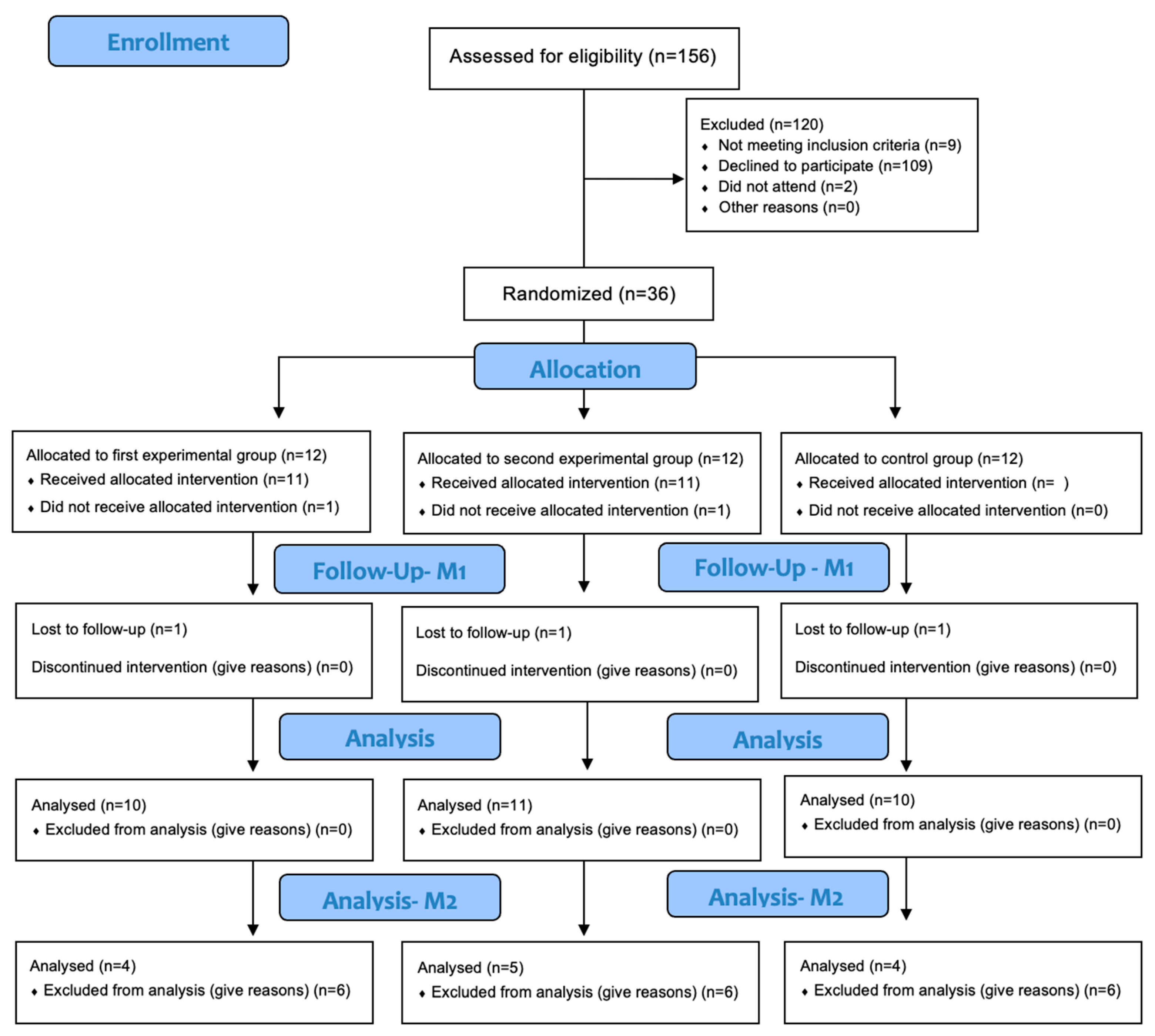
2.1. Study Design

This study was classified as a randomized controlled clinical trial, according to the CONSORT criteria [20], with blinded investigators, with three groups of 14 women each–Experimental group 1 (EG1): shockwave therapy + aerobic exercise; Experimental group 2 (EG2): radiofrequency + aerobic exercise; and control group (CG): aerobic exercise only—with an allocation ratio of 1:1:1.
The study was approved by the Ethics Committee of the School of Health Polytechnic of Porto (registration number E0088/2019). The participants were duly informed and clarified about the research project (rationale, procedures, and associated risks). In cases where there was no objection from the participants, they signed an informed consent form according to the Declaration of Helsinki. Anonymity and confidentiality of the participants were ensured at all stages of the study. This study has been registered on ClinicalTrial.gov with the protocol ID AN-009.

2.2. Sample and Eligibiliy Criteria

The target population were female patients of the Clinic Dr. Ana Sousa and Health School of the Polytechnic Institute of Porto teachers, aged between 18 and 60 years, who volunteered to participate in the study. Invitations to participate were sent by email, including the link to access the questionnaire for sample characterization and selection. Individuals who met the eligibility criteria were invited to participate in this study.
Participants were considered eligible if they met the following criteria: individuals with a body mass index (BMI) between 18.5 kg/m2 (normal) and 29.9 kg/m2 (overweight) with complaints of localized abdominal fat. Participants were excluded if they were smokers, alcoholics, athletes, individuals with orthopedic dysfunction (that prevented exercise), dietary restrictions or dieting for weight loss in the past 3 months or at the beginning of the study, changed hormonal treatment in the past 6 months, pregnant or in the postpartum period of less than one year, breastfeeding, or intending to become pregnant during the study period, individuals with metabolic, hematologic, renal, dermatologic (in the area under study), cardiovascular, respiratory, digestive, rheumatologic, and oncologic diseases, individuals with sensitivity disorders, or taking medications (anticoagulants, corticosteroids up to 6 weeks before, non-steroidal anti-inflammatory drugs, antihistamines, diuretics, supplements or drugs) with effects on cardiovascular and thermoregulatory functions up to 2 months before.
Each participant was assigned a numerical code to randomize each group. The distribution of participants was stratified according to their score on the International Physical Activity Questionnaire (IPAQ) scale and their age. Thus, participants were homogeneously and randomly assigned to groups. The evaluators were blind to the allocation of participants.

2.3. Instruments and Variables

2.3.1. Questionnaires

The sociodemographic questionnaire was sent by e-mail and completed before the first assessment (M0) to verify eligibility for the study and to collect sociodemographic data.
The short version of the IPAQ was used to determine the physical activity level of the participants. It has been validated for the Portuguese population with a concurrent validity of 0.49, reliability with σ of Spearman of 0.77, and a coefficient of reproducibility of 0.83. IPAQ allowed the calculation of MET-minutes/week according to the guidelines [21].
The Food Frequency Questionnaire (FFQ) was used to assess the characteristics of the participants’ food intake in the last 12 months. This questionnaire allowed us to identify consumption patterns and is validated for the Portuguese population with average values of correlations with food records of daily rates of 0.54. The reproducibility of the questionnaire has a mean correlation value of 0.57 for the 22 nutrients [22].
The IPAQ and the FFQ were completed on a computer provided by the researchers at M0.

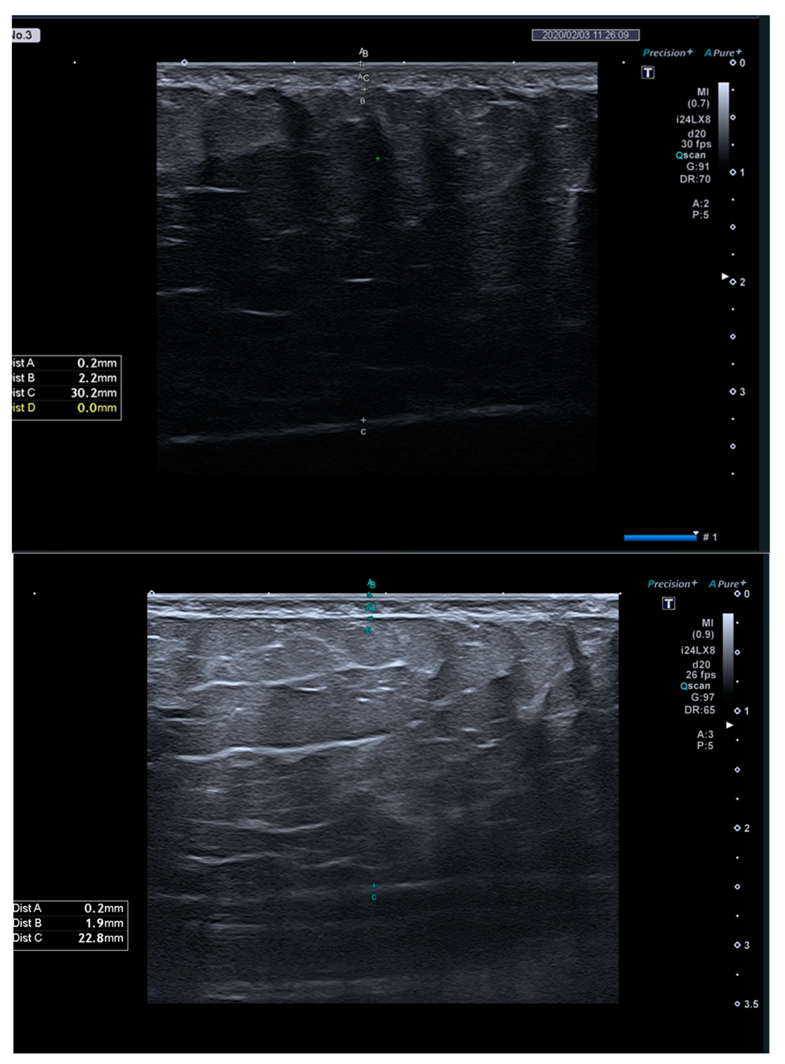
2.3.2. Anthropometric Measurements and Body Composition

Height, waist, and hip circumference were measured to calculate the waist-to-hip ratio using an inelastic and flexible tape measure (COMED SAS, Strasbourg, France).
Body mass, total muscle mass, percentage of total fat mass, percentage of trunk fat mass, visceral fat, and BMI were determined using the Tanita model BC-545 Inner Scan TM (Tanita Corporation, Amsterdam, The Netherlands) scale with a maximum capacity of 150 kg and an accuracy of 0.1 kg per kg. The correlation coefficient with dual-energy X-ray absorptiometry (DEXA) varied between 0.88 and 0.89 [23].
To measure the thickness of SAT, the Canon Aplio i800 model echograph (Canon Medical Systems Corporation, Otawara, Tochigi, Japan) with a PLI-2004BX model matrix linear probe (multi-frequency up to 24 MHz) was used. Measurements were taken at the mid-abdomen. Each subject assumed the dorsal decubitus position with knees bent. Measurements were taken 2 cm from the umbilicus, on the right side, with the probe placed perpendicular to the skin surface and oriented parallel to the linea alba. The thickness of SAT was determined by direct measurements through the echograph measurement option in the frozen images, using the skin and muscle as landmarks for the localization of SAT at the end of the expiratory phase in apnea (Figure 1). Two measurements were taken, and their average was used.

2.3.3. Clinical Analyses

For glycerol, interleukin-6, and lipid profile analysis, venous blood was collected by a BD Vacutainer® vacuum system using BD Vacutainer® SST® II Advance® tubes (Becton, Dickinson and Company, Franklin Lakes, NJ, USA) by a professional licensed in Clinical Analyses and Public Health. Sigma® 3K15 Centrifuge (Sigma-Aldrich Corporation, St Louis, MO, USA) was used to centrifuge the samples, and Prestige 24i autoanalyzer (PZ Cormay S.A., Motycz, Poland) was used to determine the lipid profile values.
To determine the values of glycerol and interleukin-6, Randox reagents were used (Randox Laboratories, Crumlin, UK). Prestige 24i reagents were used to calculate the values of the lipid profile.

2.3.4. Shockwave Protocol

To perform ESWT, the BTL-6000 X-Wave Optimal device (BTL Industries Ltd., Hertfordshire, UK) with a 20 mm multifocal transmitter was used. The following parameters were selected: 2.6 to 3.6 bar pressure, 15 Hz frequency, and 3000 pulses per 10 × 15 cm area. Participants remained in the supine position, and ultrasound gel was used as the contact medium. ESWT was applied to the abdominal area (defined by the square junction of the anterosuperior iliac spine and the last rib, bilaterally). Pain and paresthesia were defined as stop criteria.

2.3.5. Radiofrequency Protocol

For the RF protocol the BTL-6000 TR-Therapy Elite device (BTL Industries Ltd., Hertfordshire, UK) was used. The capacitive mode was selected and applied through a 70 mm electrode. The following parameters were chosen: frequency of 500 kHz and the power percentage was adjusted according to the temperature obtained [16,24].
The temperature was monitored through the combination Schliephak Scale (qualitative monitoring of the participant’s perception of temperature) and the FLIR E6 (Boston) thermograph, which has a frequency of 9 Hz and a margin of error of less than 0.06 °C.
The application time was defined as 1.5 min per area of the applicator’s head, seeking to reach and maintain an epidermal temperature of 40 to 42 °C [16,24].
The RF application was performed in the same area as described in the ESWT protocol, and a water-based cream was used as the contact medium.

2.3.6. Aerobic Exercise Protocol

Aerobic exercise was performed on a Monark lower-extremity cycloergometer and monitored with a heart rate monitor (Polar FT7 heart rate monitor with an accuracy of +/− 1 beat per minute (bpm)) and the Borg Perceived Exertion Scale [25].

2.4. Data Collection

All data collection procedures were performed in the same room and under the same conditions at both evaluation times. M0 was performed before the intervention cycle, and M1, the last evaluation, was performed 48 to 72 h after the last intervention. At both assessments, height, waist and hip circumference, body composition (body mass, total muscle mass, percentage of total fat mass, percentage of trunk fat mass, visceral fat, and BMI), SAT thickness, and blood samples were obtained.
The intervention consisted of a total of six sessions. Two weekly sessions were performed, completing a 3-week intervention cycle with a minimum interval of 48 h. In each session, participants assigned to EG1 underwent the ESWT protocol at the level of the abdominal region, followed by aerobic exercise. EG2 performed the aerobic exercise after the RF protocol. CG performed only the aerobic exercise.
For aerobic exercise performance, for each participant, the theoretical maximum heart rate (HRmax) was determined based on the Tanaka equation [26]:
HRmax = 208 − (0.7 × age).
Based on these values, and using the Karvonen equation, the target heart rate (THR) was calculated [27]:
THR= HRrest + (% intensity × HRreserve),
where HRrest is the heart at rest for each participant and HRreserve is the heart rate reserve. The HRrest was obtained after 5–10 min of rest in the sitting position using a heart rate monitor. HRreserve was calculated as follows:
HRreserve = HRmax − Hrrest.
Each aerobic exercise session lasted 40 min, with a 5 min warm-up, 30 min during which each participant was asked to maintain a heart rate in the THR range (45 to 55%) [27], and the last 5 min during which speed was progressively reduced.

2.5. Sample Size Calculation and Data Analysis

The G-Power 3.0.10 (Universität Düsseldorf) software was used to calculate the number of participants required in each group, for a power of 95% and α = 0.05. This calculation was based on the most similar article, in which anthropometric and body composition measurements and SAT thickness were also assessed by ultrasound [24]. A minimum of 10 participants in each group was necessary, based on the values of the referred study.
Statistical Package for Social Sciences (IBM SPSS®) software version 27.0 (IBM Corporation, Armonk, New York, NY, USA) was used for statistical analysis and interpretation of the data at a 5% significance level (p < 0.05).
To assess the normality of the variables, the Shapiro–Wilk test was used. Descriptive statistics were performed using measures of central tendency (mean and median) and dispersion (SD and percentiles 25–75). To compare the results among groups, ANOVA test was performed, followed by Tukey’s post hoc test, to determine the existence of significant differences among the groups. To analyze the results between moments, the t-test for 2 paired samples was used.
The effect size was measured using the partial eta squared (η2p), where 0.01 was determined as small, 0.06 as medium, and 0.14 as large [28].
Non-parametric tests, Kruskal–Wallis and Wilcoxon with Bonferroni correction, were used for the IL-6 variable.

3. Results

Thirty-one participants completed this study, performing the two assessment moments and completing the six intervention sessions (Figure 2).
No significant differences between the groups were observed for any variables at M0 (p > 0.05). The mean BMI values for all groups were considered normal, and the mean MET values on the IPAQ were considered moderate (Table 1).
Table 2 shows the results of each group between moments and between groups. The difference variable (M1–M0) showed significant differences at the level of thickness of the SAT (p < 0.001) and waist-to-hip ratio (p = 0.001). It was observed that both experimental groups showed greater differences between moments than the CG, both regarding SAT thickness (GE1 < GC p = 0.001; GE2 < GC p = 0.001) and waist-to-hip ratio (GE1 < GC p = 0.002; GE2 < GC p = 0.002). The comparison between the moments observed showed that both experimental groups significantly reduced the thickness of the SAT (p < 0.001), as well as the waist-to-hip ratio (p < 0.001). In this last variable, the CG also decreased significantly (p = 0.004) (Table 2).
The variables collected by clinical analysis (Table 3 and Table 4) did not show a significant difference between the groups. However, an increase in GE2 was observed for the level of TG from M0 to M1 (p = 0.038).
No side effects were observed or reported during the interventions.

4. Discussion

In the current clinical practice of healthcare professionals, there are numerous technologies for body contouring using different types of energies (electromagnetic, mechanical, thermal, and electric) such as cryolipolysis, HIFU, shockwaves, RF, etc. However, it remains unclear how to set appropriate guidelines for selecting and using them, mainly due to the wide variety of technologies studied, non-standardized protocol parameters, and unavailability of the precise cellular-level mechanism of action in SAT, and the pace of their study does not keep up with the speed of the emergence of new supposedly more effective equipment. Also, long-term effects are needed to clarify its safety and efficacy in humans.
The main objective of this study was to analyze the effect of six sessions of ESWT or RF on the abdominal SAT associated with aerobic exercise. SAT thickness was significantly reduced in the experimental groups treated with ESWT or RF along with aerobic exercise. This reduction can be explained by the reversible and/or irreversible effects of these technologies. Reversible effects refer to the hypotrophy of adipocytes by the local stimulation of lipolysis, which involves the hydrolysis of TG to fatty acids and glycerol, preserving functions and cellular integrity without causing an inflammatory process. Irreversible effects involve permanent damage to the adipocytes, a process known as adipocytolysis [3,12,29]. These results are consistent with the findings of Adatto et al. (2011), Nassar et al. (2015), Ferraro et al. (2012), and Hexsel et al. (2017), who found a significant reduction in SAT thickness using radial ESWT [5,30,31,32]. Radial ESWT is generated by accelerating a projectile within a transducer through a pneumatic or electromagnetic mechanism. This projectile collides with an applicator, transforming kinetic energy into a pressure wave that expands radially in the tissues. The energy deposition primarily affects superficial tissue layers [14,15,33]. These waves exhibit rapid pressure rise, high peak pressure, low tensile amplitude, and a fast cycle, transferring mechanical energy directly into the tissues and indirectly through the generation of cavitation bubbles. It seems that mechanotransduction is the most consensual pathway activating a series of cellular events, such as migration, proliferation, differentiation, and apoptosis [15,34], in a dose-dependent manner (pressure, frequency, and number of impulses) and specific to different types of mechanosensitive cells [15,34,35]. However, the exact mechanism by which ESWT and RF influence localized fat reduction remains unclear [5,14,32,36]. The literature suggests that reversible effects include the release of nitric oxide, which improves microcirculation [30,31,32,37], increases membrane permeability, and promotes substance exchange and the reorganization of the extracellular matrix [36,38]. Other effects include reduced oxidative stress [38] and angiogenesis [30,31,39,40], which collectively favor lipolysis, leading to adipocyte hypotrophy [30,32,36,38,41]. Irreversible effects, including apoptosis, occur at higher ESWT energy levels, causing damage to cellular membranes, endoplasmic reticulum, cytoskeleton, and intercellular junctions, alongside increased cellular metabolism [5,14,31,37]. An additional explanation for reduced SAT thickness involves volumetric changes due to neocollagenesis and neoelastogenesis stimulated by fibroblast activity [14,36,38].
The reduction in SAT thickness observed with RF is consistent with the findings reported in various studies from 2016 to 2020 [41,42,43,44]. In this study, the technology used is called CRET (capacitive-resistive electric transfer), which allows electrical current to penetrate tissues between an active electrode and a dispersive plate placed away from the application area [45,46,47], generating heat in the SAT. By applying these electrodes that transfer high-frequency energy to the body and because adipose tissue has a higher resistance to electrical flow, it generates an increased temperature on this type of tissue [17,48]. The increased temperature activates the Autonomic Nervous System, triggering the release of catecholamines (adrenaline and noradrenaline), which stimulate lipolysis. Lipolysis refers to the hydrolysis of lipids into their metabolites: fatty acids and glycerol. This process occurs within adipocytes and is regulated by the enzymatic activity of hormone-sensitive lipase (HSL) and lipoprotein lipase. HSL is expressed intracellularly in adipocytes, and its activation depends on catecholamines, natriuretic peptides, growth hormones, glucocorticoids, and tumor necrosis factor-α (TNF-α). As a physiological process governed by multiple signaling pathways, lipolysis serves as the foundation for non-surgical fat reduction methods, which are categorized into two types, the Noncytolytic Methods and the Cytolytic Methods (Adipocytolysis). The Noncytolytic Methods stimulate lipid mobilization in adipocytes without compromising their function or structural integrity. They act as temporary metabolic enhancers, promoting the breakdown of lipids into fatty acids and glycerol, which are then released from adipocytes and metabolized by the liver. However, their effects on adipose tissue and body contouring are short-lived as they rely on natural physiological pathways. The Cytolytic Methods break down or dissolve lipids by partially or completely destroying adipocytes, disrupting their plasma membrane [18]. Chemical or mechanical ablation is used to induce permanent changes in fat cells, leading to long-lasting reductions in body fat and improvements in body contouring. Vasodilation improves blood perfusion, enhancing hormone circulation and lipolysis, leading to adipocyte hypotrophy [49,50,51]. These effects appear to be related to hyperthermia around 40–42 °C, the therapeutic temperatures range used in this study [52]. The irreversible effects of RF on SAT, including apoptosis or necrosis [50,53] appear to be related to higher temperatures (50–110 °C) within short durations [52]. Another mechanism for reduced SAT thickness involves volumetric changes due to neocollagenesis and neoelastogenesis triggered by heat [11,46].
In view of these findings, the combination of ESWT or RF with aerobic exercise is important to enhance the effects of these technologies by promoting lipolysis. On the other hand, it allows the oxidation of the fatty acids released into the blood circulation, preventing them from being reconverted into TG and re-stored in the adipose tissue or ectopically accumulated in the liver, overloading this organ and leading to the production of LDL cholesterol. Thus, the aerobic exercise adopted in this study was intended to allow lipid oxidation in a global manner [54]. Because of this intervention, there was a significant decrease in the difference variable of the waist-to-hip ratio in the experimental groups, with all groups showing a significant decrease in their values between moments, which agrees with Vale et al. (2020) [24].
No significant changes in total body mass, fat mass, muscle mass, visceral fat, or BMI were observed after the intervention. This lack of change may be due to an insufficient number of aerobic exercise sessions or an insufficient duration of the sessions [54,55].
The study also aimed to assess the effect of ESWT and RF on lipid mobilization in abdominal SAT by examining variations in lipolytic activity, lipid profiles, and adipocytokine IL-6 levels. Regarding glycerol, no significant differences were observed among groups after the intervention, consistent with findings by Noites et al. (2020) using RF [56]. ESWT, RF, and exercise appear to be sufficient to activate a global lipolytic cascade [14,41]. However, since the time of assessment of M1 in our study was between 48 and 72 h, this finding cannot be verified by evaluation of plasma levels.
Similarly, TG levels were not significantly different between groups, consistent with studies by Ferraro et al. (2012) on ESWT, as well as Levenberg et al. (2010) and Noites et al. (2020) on RF [31,51,56]. However, in this study, GE2 showed a significant increase in plasma TG levels between moments. This finding may be explained by the fact that it was not possible to control dietary and physical activity habits during the study, which may have conditioned this result. On the other hand, the increase in plasma TG levels in EG2 may be explained by the fact that this group had also received RF treatment, possibly leading to an increase in the availability of circulating fatty acids that could be converted into TG in the liver and released back into the bloodstream as very low-density lipoprotein cholesterol [57]. Despite these variations in TG levels observed in all groups throughout the study, all groups had mean values below 150 mL/dL, which is within the levels recommended by the European Guidelines on Cardiovascular Disease [58].
Other lipid variables, including total cholesterol, HDL, and LDL, showed no significant changes, consistent with studies by Levenberg et al. (2010) and Noites et al. (2020) in the RF application, as well as Ferraro et al. (2012) in the ESWT application [31,51,56].
Similarly, IL-6 levels were not significantly different, although the literature suggests an association between increased IL-6 secretion and adipocytolytic effects [3]. This result may indicate the absence of such effects or their localized expression within the first hour after the intervention.
Finally, regarding the methodology, in future studies, participants should be stratified based on VF levels so that investigators can use more accurate instruments for visceral fat measurement, such as dual-energy X-ray absorptiometry (DEXA) and magnetic resonance imaging (MRI), it would be appropriate to verify the clinical analyses at the end of the six intervention sessions and to evaluate the long-term effects of the intervention after 3 and 6 months. It is also recommended to include a control group that does not perform aerobic exercise to understand its isolated effect.

5. Conclusions

In this study, ESWT or RF on the abdominal SAT combined with aerobic exercise had beneficial effects on reducing SAT thickness and the waist-to-hip ratio compared to the effects of an aerobic exercise program alone. However, no changes in lipolytic activity, lipid profile, and IL-6 levels were observed.

Author Contributions

Conceptualization, L.M., M.M.A. and A.N.; methodology, L.M., J.N., A.P., A.N. and S.T.; software, L.M.; validation, M.M.A. and A.N.; formal analysis, R.V., M.M.A. and A.N.; investigation, L.M., J.N., A.P., A.S. and S.T.; resources, M.M.A. and A.N.; data curation, L.M., M.M.A. and A.N.; writing—original draft preparation, L.M.; writing—review and editing, R.V., M.M.A. and A.N.; visualization, R.V.; supervision, M.M.A. and A.N.; project administration, A.N. All authors have read and agreed to the published version of the manuscript.

Funding

This research was supported by the Fundação para a Ciência e Tecnologia (FCT) through R&D Units funding [UIDB/05210/2020].

Institutional Review Board Statement

This study was conducted in accordance with the Declaration of Helsinki and approved by the Ethics Committee of School of Health-Polytechnic of Porto (number E0088/201, 21 January 2019). This study has been registered on ClinicalTrial.gov with the protocol ID AN-009.

Informed Consent Statement

Informed consent was obtained from all subjects involved in the study.

Data Availability Statement

The data are available upon request from the corresponding author.

Acknowledgments

This study was carried out with the support of BTL Portugal and Canon Medical Systems, which provided the equipment for the intervention protocol (radiofrequency and shock wave) and the ultrasound scanner required, respectively.

Conflicts of Interest

The authors declare no conflicts of interest.

Abbreviations

The following abbreviations are used in this manuscript:
SATSubcutaneous adipose tissue
TGTriglycerides
ESWTExternal shock wave therapy
RFRadiofrequency
IL-6Interleukin-6
BMIBody mass index
IPAQInternational physical activity questionnaire
METEquivalent metabolic
FFQFood frequency questionnaire
HRHeart rate

References

  1. Duncan, D.I.; Kim, T.H.M.; Temaat, R. Quantification of Adipose Volume Reduction with a Prospective Study Analyzing the Application of External Radiofrequency Energy and High Voltage Ultrashort Pulse Duration Electrical Fields. J. Cosmet. Laser Ther. 2016, 18, 323–329. [Google Scholar] [CrossRef] [PubMed]
  2. Crewe, C.; An, Y.A.; Scherer, P.E. The Ominous Triad of Adipose Tissue Dysfunction: Inflammation, Fibrosis, and Impaired Angiogenesis. J. Clin. Investig. 2017, 127, 74–82. [Google Scholar] [CrossRef]
  3. Maillard, F.; Pereira, B.; Boisseau, N. Effect of High-Intensity Interval Training on Total, Abdominal and Visceral Fat Mass: A Meta-Analysis. Sports Med. 2018, 48, 269–288. [Google Scholar] [CrossRef]
  4. Romero, M.; Zorzano, A. Role of Autophagy in the Regulation of Adipose Tissue Biology. Cell Cycle 2019, 18, 1435–1445. [Google Scholar] [CrossRef]
  5. Nassar, A.H.; Dorizas, A.S.; Shafai, A.; Sadick, N.S. A Randomized, Controlled Clinical Study to Investigate the Safety and Efficacy of Acoustic Wave Therapy in Body Contouring. Dermatol. Surg. 2015, 41, 366–370. [Google Scholar] [CrossRef]
  6. van Baak, M.A.; Mariman, E.C.M. Mechanisms of Weight Regain After Weight Loss—The Role of Adipose Tissue. Nat. Rev. Endocrinol. 2019, 15, 274–287. [Google Scholar] [CrossRef] [PubMed]
  7. Friedmann, D.P. A Review of the Aesthetic Treatment of Abdominal Subcutaneous Adipose Tissue: Background, Implications, and Therapeutic Options. Dermatol. Surg. 2015, 41, 18–34. [Google Scholar] [CrossRef] [PubMed]
  8. Thomaz, F.S.; John, O.D.; Sinha, P.; Shafie, S.R.; Worrall, S. The Metabolic Syndrome: An Overview and Proposed Mechanisms. Obesities 2024, 4, 226–255. [Google Scholar] [CrossRef]
  9. Leopold, J.A.; Antman, E.M. Obesity and Ideal Cardiovascular Health: Results from the My Research Legacy Study. Obesities 2021, 1, 36–48. [Google Scholar] [CrossRef]
  10. Kouidrat, Y.; Louhou, R.; Mondot, C.; Daami, I.; Amad, A.; Diouf, M. Quality of Life in Patients with Obesity: The Role of Multidisciplinary Rehabilitation Medicine. Obesities 2024, 4, 160–168. [Google Scholar] [CrossRef]
  11. Mazzoni, D.; Lin, M.J.; Dubin, D.P.; Khorasani, H. Review of Non-Invasive Body Contouring Devices for Fat Reduction, Skin Tightening and Muscle Definition. Australas. J. Dermatol. 2019, 60, 278–283. [Google Scholar] [CrossRef] [PubMed]
  12. Pinto, H. Local Fat Treatments: Classification Proposal. Adipocyte 2016, 5, 22–26. [Google Scholar] [CrossRef]
  13. Alizadeh, Z.; Halabchi, F.; Mazaheri, R.; Abolhasani, M.; Tabesh, M. Review of the Mechanisms and Effects of Noninvasive Body Contouring Devices on Cellulite and Subcutaneous Fat. Int. J. Endocrinol. Metab. 2016, 14, e36727. [Google Scholar] [CrossRef]
  14. Modena, D.A.O.; da Silva, C.N.; Grecco, C.; Guidi, R.M.; Moreira, R.G.; Coelho, A.A.; Sant’Ana, E.; de Souza, J.R. Extracorporeal shockwave: Mechanisms of Action and Physiological Aspects for Cellulite, Body Shaping, and Localized Fat—Systematic Review. J. Cosmet. Laser Ther. 2017, 19, 314–319. [Google Scholar] [CrossRef] [PubMed]
  15. Simplicio, C.L.; Purita, J.; Murrell, W.; Santos, G.S.; dos Santos, R.G.; Lana, J.F.S.D. Extracorporeal Shock Wave Therapy Mechanisms in Musculoskeletal Regenerative Medicine. J. Clin. Orthop. Trauma 2020, 11, S309–S318. [Google Scholar] [CrossRef]
  16. Franco, W.; Kothare, A.; Goldberg, D.J. Controlled Volumetric Heating of Subcutaneous Adipose Tissue Using a Novel Radiofrequency Technology. Lasers Surg. Med. 2009, 41, 745–750. [Google Scholar] [CrossRef] [PubMed]
  17. Vale, A.L.; Pereira, A.S.; Morais, A.; Noites, A.; Mendonça, A.C.; Martins Pinto, J.; Vilarinho, R.; Carvalho, P. Effects of Radiofrequency on Adipose Tissue: A Systematic Review with Meta-Analysis. J. Cosmet. Dermatol. 2018, 17, 703–711. [Google Scholar] [CrossRef]
  18. Ahmadian, M.; Duncan, R.E.; Sul, H.S. The Skinny on Fat: Lipolysis and Fatty Acid Utilization in Adipocytes. Trends Endocrinol. Metab. 2009, 20, 424–428. [Google Scholar] [CrossRef]
  19. Mika, A.; Macaluso, F.; Barone, R.; Di Felice, V.; Sledzinski, T. Effect of Exercise on Fatty Acid Metabolism and Adipokine Secretion in Adipose Tissue. Front. Physiol. 2019, 10, 26. [Google Scholar] [CrossRef]
  20. Hopewell, S.; Boutron, I.; Chan, A.-W.; Collins, G.S.; de Beyer, J.A.; Hróbjartsson, A.; Nejstgaard, C.H.; Østengaard, L.; Schulz, K.F.; Tunn, R.; et al. An Update to SPIRIT and CONSORT Reporting Guidelines to Enhance Transparency in Randomized Trials. Nat. Med. 2022, 28, 1740–1743. [Google Scholar] [CrossRef]
  21. Craig, C.L.; Marshall, A.L.; Sjöström, M.; Bauman, A.E.; Booth, M.L.; Ainsworth, B.E.; Pratt, M.; Ekelund, U.; Yngve, A.; Sallis, J.F.; et al. International Physical Activity Questionnaire: 12-Country Reliability and Validity. Med. Sci. Sports Exerc. 2003, 35, 1381–1395. [Google Scholar] [CrossRef] [PubMed]
  22. Lopes, C.; Aro, A.; Azevedo, A.; Ramos, E.; Barros, H. Intake and Adipose Tissue Composition of Fatty Acids and Risk of Myocardial Infarction in a Male Portuguese Community Sample. J. Am. Diet. Assoc. 2007, 107, 276–286. [Google Scholar] [CrossRef]
  23. Bolanowski, M.; Nilsson, B.E. Assessment of Human Body Composition Using Dual-Energy X-Ray Absorptiometry and Bioelectrical Impedance Analysis. Med. Sci. Monit. 2001, 7, 1029–1033. [Google Scholar]
  24. Vale, A.L.; Pereira, A.S.; Morais, A.; de Carvalho, P.; Vilarinho, R.; Mendonça, A.; Noites, A. Effect of Four Sessions of Aerobic Exercise with Abdominal Radiofrequency in Adipose Tissue in Healthy Women: Randomized Control Trial. J. Cosmet. Dermatol. 2020, 19, 359–367. [Google Scholar] [CrossRef] [PubMed]
  25. Borg, G.A.V. Psychophysical Bases of Perceived Exertion. Med. Sci. Sports Exerc. 1982, 14, 377–381. [Google Scholar] [CrossRef] [PubMed]
  26. Tanaka, H.; Monahan, K.D.; Seals, D.R. Age-Predicted Maximal Heart Rate Revisited. J. Am. Coll. Cardiol. 2001, 37, 153–156. [Google Scholar] [CrossRef]
  27. ACSM. American College of Sports Medicine’s Exercise Testing and Prescription; Lippincott Williams & Wilkins: Philadelphia, PA, USA, 2023. [Google Scholar]
  28. Maher, J.M.; Markey, J.C.; Ebert-May, D. The Other Half of the Story: Effect Size Analysis in Quantitative Research. CBE—Life Sci. Educ. 2013, 12, 345–351. [Google Scholar] [CrossRef]
  29. Duncan, D.I.; Kim, T.H.M.; Temaat, R. A Prospective Study Analyzing the Application of Radiofrequency Energy and High-Voltage, Ultrashort Pulse Duration Electrical Fields on the Quantitative Reduction of Adipose Tissue. J. Cosmet. Laser Ther. 2016, 18, 257–267. [Google Scholar] [CrossRef]
  30. Adatto, M.A.; Adatto-Neilson, R.; Novak, P.; Krotz, A.; Haller, G. Body Shaping with Acoustic Wave Therapy AWT®/EPAT®: Randomized, Controlled Study on 14 Subjects. J. Cosmet. Laser Ther. 2011, 13, 291–296. [Google Scholar] [CrossRef]
  31. Ferraro, G.A.; De Francesco, F.; Cataldo, C.; Rossano, F.; Nicoletti, G.; D’Andrea, F. Synergistic Effects of Cryolipolysis and Shock Waves for Noninvasive Body Contouring. Aesthetic Plast. Surg. 2012, 36, 666–679. [Google Scholar] [CrossRef]
  32. Hexsel, D.; Camozzato, F.O.; Silva, A.F.; Siega, C. Acoustic Wave Therapy for Cellulite, Body Shaping and Fat Reduction. J. Cosmet. Laser Ther. 2017, 19, 165–173. [Google Scholar] [CrossRef] [PubMed]
  33. Liu, Y.; Chen, X.; Guo, A.; Liu, S.; Hu, G. Quantitative Assessments of Mechanical Responses Upon Radial Extracorporeal Shock Wave Therapy. Adv. Sci. 2018, 5, 1700797. [Google Scholar] [CrossRef]
  34. Sukubo, N.G.; Tibalt, E.; Respizzi, S.; Locati, M.; d’Agostino, M.C. Effect of Shock Waves on Macrophages: A Possible Role in Tissue Regeneration and Remodeling. Int. J. Surg. 2015, 24, 124–130. [Google Scholar] [CrossRef]
  35. Shoham, N.; Gefen, A. Mechanotransduction in Adipocytes. J. Biomech. 2012, 45, 1–8. [Google Scholar] [CrossRef] [PubMed]
  36. Kuhn, C.; Angehrn, F.; Sonnabend, O.; Voss, A. Impact of Extracorporeal Shock Waves on the Human Skin with Cellulite: A Case Study of an Unique Instance. Clin. Interv. Aging 2008, 3, 201–210. [Google Scholar] [CrossRef] [PubMed]
  37. Schlaudraff, K.U.; Kiessling, M.C.; Császár, N.B.; Schmitz, C. Predictability of the Individual Clinical Outcome of Extracorporeal Shock Wave Therapy for Cellulite. Clin. Cosmet. Investig. Dermatol. 2014, 7, 171–183. [Google Scholar] [CrossRef]
  38. Christ, C.; Brenke, R.; Sattler, G.; Siems, W.; Novak, P.; Daser, A. Improvement in Skin Elasticity in the Treatment of Cellulite and Connective Tissue Weakness by Means of Extracorporeal Pulse Activation Therapy. Aesthetic Surg. J. 2008, 28, 538–544. [Google Scholar] [CrossRef]
  39. Angehrn, F.; Kuhn, C.; Voss, A. Can Cellulite Be Treated with Low-Energy Extracorporeal Shock Wave Therapy? Clin. Interv. Aging 2007, 2, 623–630. [Google Scholar] [CrossRef]
  40. Mariotto, S.; de Prati, A.C.; Cavalieri, E.; Amelio, E.; Marlinghaus, E.; Suzuki, H. Extracorporeal Shock Wave Therapy in Inflammatory Diseases: Molecular Mechanism That Triggers Anti-Inflammatory Action. Curr. Med. Chem. 2009, 16, 2366–2372. [Google Scholar] [CrossRef]
  41. van Hall, G. The Physiological Regulation of Skeletal Muscle Fatty Acid Supply and Oxidation During Moderate-Intensity Exercise. Sports Med. 2015, 45, 23–32. [Google Scholar] [CrossRef]
  42. Hayre, N.; Palm, M.; Jenkin, P. A Clinical Evaluation of a Next Generation, Non-Invasive, Selective Radiofrequency, Hands-Free, Body-Shaping Device. J. Drugs Dermatol. 2016, 15, 1557–1561. [Google Scholar] [PubMed]
  43. Mlosek, R.K.; Woźniak, W.; Malinowska, S.; Lewandowski, M.; Nowicki, A. The Effectiveness of Anticellulite Treatment Using Tripolar Radiofrequency Monitored by Classic and High-Frequency Ultrasound. J. Eur. Acad. Dermatol. Venereol. 2012, 26, 696–703. [Google Scholar] [CrossRef] [PubMed]
  44. Suh, D.H.; Kim, C.M.; Lee, S.J.; Kim, H.; Yeom, S.K.; Ryu, H.J. Safety and Efficacy of a Non-Contact Radiofrequency Device for Body Contouring in Asians. J. Cosmet. Laser Ther. 2017, 19, 89–92. [Google Scholar] [CrossRef]
  45. Wanitphakdeedecha, R.; Sathaworawong, A.; Manuskiatti, W.; Sadick, N.S. Efficacy of Multipolar Radiofrequency with Pulsed Magnetic Field Therapy for the Treatment of Abdominal Cellulite. J. Cosmet. Laser Ther. 2017, 19, 205–209. [Google Scholar] [CrossRef]
  46. Levy, A.S.; Grant, R.T.; Rothaus, K.O. Radiofrequency Physics for Minimally Invasive Aesthetic Surgery. Clin. Plast. Surg. 2016, 43, 551–556. [Google Scholar] [CrossRef]
  47. Sadick, N.; Rothaus, K.O. Aesthetic Applications of Radiofrequency Devices. Clin. Plast. Surg. 2016, 43, 557–565. [Google Scholar] [CrossRef]
  48. Albornoz-Cabello, M.; Ibáñez-Vera, A.J.; De la Cruz-Torres, B. Efficacy of Monopolar Dielectric Transmission Radio Frequency in Panniculus Adiposus and Cellulite Reduction. J. Cosmet. Laser Ther. 2017, 19, 422–426. [Google Scholar] [CrossRef] [PubMed]
  49. Boisnic, S.; Branchet, M.C.; Birnstiel, O.; Beilin, G. Clinical and Histopathological Study of the TriPollar Home-Use Device for Body Treatments. Eur. J. Dermatol. 2010, 20, 367–372. [Google Scholar] [CrossRef]
  50. Kaplan, H.; Gat, A. Clinical and Histopathological Results Following TriPollar™ Radiofrequency Skin Treatments. J. Cosmet. Laser Ther. 2009, 11, 78–84. [Google Scholar] [CrossRef]
  51. Levenberg, A. Clinical Experience with a TriPollar Radiofrequency System for Facial and Body Aesthetic Treatments. Eur. J. Dermatol. 2010, 20, 615–619. [Google Scholar]
  52. Rossmann, C.; Haemmerich, D. Review of Temperature Dependence of Thermal Properties, Dielectric Properties, and Perfusion of Biological Tissues at Hyperthermic and Ablation Temperatures. Crit. Rev. Biomed. Eng. 2014, 42, 467–492. [Google Scholar] [CrossRef] [PubMed]
  53. Franco, W.; Kothare, A.; Ronan, S.J.; Grekin, R.C.; McCalmont, T.H. Hyperthermic Injury to Adipocyte Cells by Selective Heating of Subcutaneous Fat with a Novel Radiofrequency Device: Feasibility Studies. Lasers Surg. Med. 2010, 42, 361–370. [Google Scholar] [CrossRef] [PubMed]
  54. Purdom, T.; Kravitz, L.; Dokladny, K.; Mermier, C. Understanding the Factors that Effect Maximal Fat Oxidation. J. Int. Soc. Sports Nutr. 2018, 15, 3. [Google Scholar] [CrossRef] [PubMed]
  55. Swift, D.L.; McGee, J.E.; Earnest, C.P.; Carlisle, E.; Nygard, M.; Johannsen, N.M. The Effects of Exercise and Physical Activity on Weight Loss and Maintenance. Prog. Cardiovasc. Dis. 2018, 61, 206–213. [Google Scholar] [CrossRef]
  56. Noites, A.; Vale, A.L.; Pereira, A.S.; Morais, A.; Vilarinho, R.; Carvalho, P.; Amorim, M.; Moreira, T.; Mendonça, A. Effect of an Aerobic Exercise Session Combined with Abdominal Radiofrequency on Lipolytic Activity in Women: Randomized Control Trial. J. Cosmet. Dermatol. 2020, 19, 638–645. [Google Scholar] [CrossRef]
  57. Alves-Bezerra, M.; Cohen, D.E. Triglyceride Metabolism in the Liver. Compr. Physiol. 2017, 8, 1–22. [Google Scholar] [CrossRef]
  58. Perk, J.; De Backer, G.; Gohlke, H.; Graham, I.; Reiner, Z.; Verschuren, M.; Albus, C.; Benlian, P.; Boysen, G.; Cifkova, R.; et al. European Guidelines on Cardiovascular Disease Prevention in Clinical Practice (Version 2012). The Fifth Joint Task Force of the European Society of Cardiology and Other Societies on Cardiovascular Disease Prevention in Clinical Practice (constituted by Representatives of Nine Societies and by Invited Experts). Eur. Heart J. 2012, 33, 1635–1701. [Google Scholar] [CrossRef]
Figure 1. Subcutaneous adipose tissue (SAT) thickness measured directly using ultrasound imaging.
Obesities 05 00031 g001
Figure 2. Flow diagram of the study.
Obesities 05 00031 g002
Table 1. Characteristics of participants.
EG 1
(n = 10)
EG2
(n = 11)
CG
(n = 10)
Differences Between Groups, ANOVA
(p Value)
Age, years36.50 ± 9.338.27 ± 10.635.40 ± 11.10.816
Height, m1.62 ± 0.651.61 ± 0.651.63 ± 5.040.724
Body mass (kg)64.11 ± 6.1861.66 ± 6.5363.09 ± 10.880.786
BMI, kg/m224.21 ± 1.8123.68 ± 2.0523.67 ± 3.990.882
Waist/hip ratio0.84 ± 0.050.84 ± 0.040.83 ± 0.040.761
Muscle mass, kg42.94 ± 3.6642.01 ± 4.6942.89 ± 4.600.858
Fat mass, %29.28 ± 4.1927.91 ± 5.1127.43 ± 7.450.756
IPAQ-METs, minutes/week1622.10 ± 1520.911381.27 ± 858.401100.60 ± 741.670.569
FFQ
Energy, (kcal/day)2071.04 ± 789.191999.24 ± 580.041901.06 ± 469.170.831
Proteins, (g/day)92.80 ± 37.36108.05 ± 28.7992.66 ± 21.100.403
Carbohydrates, (g/day)206.37 ± 95.91215.31 ± 56.02212.88 ± 62.820.960
Total fat, (g/day)102.0 ± 47.3280.32 ± 29.0978.16 ± 23.790.249
Saturated fat, (g/day)23.26 ± 7.3421.91 ± 7.0319.56 ± 5.690.471
Monounsaturated fat, (g/day)48.09 ± 26.4836.30 ± 15.7337.47 ± 12.840.324
Polyunsaturated fat, (g/day)23.27 ± 17.6615.13 ± 6.0414.66 ± 4.570.157
Cholesterol, (mg/day)307.91 ± 67.45361.84 ± 119.47366.26 ± 153.970.480
Sugars, (g/day)102.62 ± 59.5589.01 ± 24.4991.95 ± 34.450.740
Data are expressed as mean ± standard deviation. Legend: IPAQ, International Physical Activity Questionnaire; FFQ, Food Frequency Questionnaire.
Table 2. Effects of the intervention protocol on anthropometric and body composition measurements, subcutaneous fat thickness, and mean temperature.
EG 1
(n = 10)
EG2
(n = 11)
CG
(n = 10)
Differences Between Groups, ANOVA
(p Value)
Effect Size
2p)
Difference Between Moments, t Test
(p Value)
Body Mass, kg
M064.11 ± 6.1861.66 ± 6.5363.09 ± 10.880.786 EG1: 1.000
M164.11 ± 6.4261.59 ± 6.6763.18 ± 10.460.768 EG2: 0.718
M1-M0
(95% CI)
0 ± 0.89
(−0.64; 0.63)
−0.07 ± 0.65
(−0.51; 0.36)
0.09 ± 0.92
(−0.57; 0.75)
0.9030.007CG: 0.765
Fat mass, %
M029.28 ± 4.1927.91 ± 5.1127.43 ± 7.450.756 EG1: 0.680
M129.44 ± 3.7828.47 ± 4.7427.45 ± 6.610.693 EG2: 0.089
M1-M0
(95% CI)
0.16 ± 1.19
(−0.69; 1.00)
0.56 ± 0.99
(−0.10; 1.23)
0.02 ± 1.05
(−0.73; 0.77)
0.4920.049CG: 0.954
Muscle mass, kg
M042.94 ± 3.6642.01 ± 4.6942.89 ± 4.600.858 EG1: 0.702
M142.86 ± 3.6941.9 ± 4.6143.06 ± 4.760.810 EG2: 0.649
M1-M0
(95% CI)
−0.08 ± 0.64
(−0.53; 0.38)
−0.11 ± 0.77
(−0.63; 0.41)
0.17 ± 0.54
(−0.21; 0.55)
0.5820.001CG: 0.342
Trunk fat mass, %
M024.09 ± 5.2023.14 ± 5.2721.7 ± 9.140.730 EG1: 0.760
M123.87 ± 4.6722.78 ± 5.8421.76 ± 7.520.745 EG2: 0.653
M1-M0
(95% CI)
−0.22 ± 2.21
(−1.80; 1.36)
−0.35 ± 2.54
(−2.06; 1.35)
0.06 ± 1.83
(−1.25; 1.37)
0.9110.007CG: 0.920
Visceral fat index
M04.6 ± 2.594.27 ± 1.903.8 ± 2.100.720 EG1: 0.357
M14.0 ± 1.563.91 ± 1.973.7 ± 2.00.934 EG2: 0.221
M1-M0
(95% CI)
−0.6 ± 1.96
(−2.0; 0.80)
−0.36 ± 0.92
(−0.98; 0.26)
−0.1 ± 0.32
(−0.33; 0.13)
0.6740.030CG: 0.343
BMI, kg/m2
M024.21 ± 1.8123.68 ± 2.0523.67 ± 3.990.882 EG1: 0.769
M124.18 ± 1.8723.65 ± 2.0223.71 ± 3.810.890 EG2: 0.740
M1-M0
(95% CI)
−0.03 ± 0.31
(−0.25; 0.19)
−0.03 ± 0.26
(−0.21; 1.15)
0.04 ± 0.36
(−0.22; 0.29)
0.8520.011CG: 0.735
SAT thickness, mm
M021.93 ± 6.6617.42 ± 5.3619.13 ± 10.300.410 EG1: <0.001
M118.86 ± 7.0114.29 ± 4.4318.81 ± 10.070.282 EG2: <0.001
M1-M0
(95% CI)
−3.07 ± 1.25
(−3.97; −2.17)
−3.13 ± 1.92
(−4.41; −1.84)
−0.32 ± 1.27
(−1.22; 0.59)
<0.001 a0.446CG: 0.449
Waist-to-hip ratio
M00.84 ± 0.050.84 ± 0.040.83 ± 0.040.761 EG1: <0.001
M10.82 ± 0.050.82 ± 0.040.82 ± 0.040.987 EG2: <0.001
M1-M0
(95% CI)
−0.02 ± 0.01
(−0.02; −0.01)
−0.02 ± 0.01
(−0.03; −0.01)
−0.01 ± 0.01
(−0.01; −0.003)
0.001 b0.408CG: 0.004
Mean Temperature, °C
M028.99 ± 3.1930.4 ± 1.8830.23 ± 1.790.350 EG1: 0.744
M129.33 ± 0.8230.02 ± 1.5630.51 ± 1.850.218 EG2: 0.536
M1-M0
(95% CI)
0.34 ± 3.19
(−1.94; 2.62)
−0.38 ± 1.98
(−1.71; 0.95)
0.28 ± 2.32
(−0.84; 0.97)
0.7680.019CG: 0.712
Data are expressed as mean ± standard deviation. Legend: BMI, Body mass index; SAT, subcutaneous adipose tissue. a EG1 < CG (p = 0.001); EG2 < CG (p = 0.001). b EG1 < CG (p = 0.002); EG2 < CG (p = 0.002).
Table 3. Effects of the intervention protocol on glycerol and lipid profile.
EG 1
(n = 10)
EG2
(n = 11)
CG
(n = 10)
Differences Between Groups, ANOVA
(p Value)
Effect Size
2p)
Difference Between Moments, t Test
(p Value)
Glycerol, mmol/L
M00.06 ± 0.03 0.05 ± 0.020.07 ± 0.040.255 EG1: 0.169
M10.05 ± 0.020.04 ± 0.030.04 ± 0.010.760 EG2: 0.855
M1-M0
(95% CI)
−0.01 ± 0.03
(−0.03; 0.01)
0.00 ± 0.02
(−0.01; 0.01)
−0.03 ± 0.03
(−0.05; −0.002)
0.1270.142CG: 0.080
Total cholesterol, mg/dL
M0167.89 ± 41.87149.25 ± 30.57138.39 ± 28.10.176 EG1: 0.657
M1163.61 ± 26.66162.55 ± 45.77150.1 ± 28.670.646 EG2: 0.086
M1-M0
(95% CI)
−4.28 ± 27.82
(−25.66; 17.11)
13.31 ± 23.19
(−2.27; 28.98)
11.71 ± 19.12
(−1.97; 25.34)
0.2140.108CG: 0.085
Triglycerides, mg/dL
M069.72 ± 31.1854.74 ± 20.4247.39 ± 12.800.105 EG1: 0.370
M175.58 ± 26.6067.85 ± 34.4450.72 ± 13.750.131 EG2: 0.038
M1-M0
(95% CI)
5.86 ± 18.50
(−8.37; 20.07)
13.11 ± 18.25
(0.85; 25.37)
3.33 ± 5.23
(−0.41; 7.07)
0.3280.079CG: 0.075
HDL cholesterol, mg/dL
M052.81 ± 16.7155.03 ± 9.0650.11 ± 10.200.656 EG1: 0.993
M152.79 ± 12.6658.41 ± 13.2752.8 ± 11.430.504 EG2: 0.288
M1-M0
(95% CI)
−0.02 ± 7.77
(−5.99; 5.94)
3.38 ± 9.99
(−3.33; 10.09)
2.69 ± 9.03
(−3.77; 9.14)
0.6890.027CG: 0.371
LDL cholesterol, mg/dL
M0101.13 ± 29.2783.27 ± 25.4578.80 ± 20.910.148 EG1: 0.445
M195.71 ± 20.2890.58 ± 32.5387.16 ± 22.780.777 EG2: 0.078
M1-M0
(95% CI)
−5.43 ± 20.28
(−21.01; 10.16)
7.31 ± 12.34
(−0.98; 15.60)
8.35 ± 12.30
(−0.44; 17.16)
0.1080.152CG: 0.060
Data are expressed as mean ± standard deviation. Legend: HDL, high-density lipoprotein; LDL, low-density lipoprotein.
Table 4. Effects of the intervention protocol on interleukin-6.
EG 1
(n = 10)
EG2
(n = 11)
CG
(n = 10)
Differences Between Groups,
Kruskal–Wallis
(p Value)
Difference Between Moments,
Wilcoxon
(p Value)
IL-6, pg/mL
M05.25
(3.28; 5.25)
4.57
(3.23; 6.84)
3.67
(1.15; 7.36)
0.714EG1: 0.250
M12.89
(1.92; 2.89)
4.56
(3.53; 5.95)
2.70
(1.45; 7.15)
0.584EG2: 0.844
M1–M0−2.36
(−2.62; −2.36)
−0.50
(−3.07; 2.72)
−0.57
(−3.58; 3.46)
0.780CG: 1.000
Data are expressed as median (percentiles 25–75). Legend: IL-6, interleukin-6.
Disclaimer/Publisher’s Note: The statements, opinions and data contained in all publications are solely those of the individual author(s) and contributor(s) and not of MDPI and/or the editor(s). MDPI and/or the editor(s) disclaim responsibility for any injury to people or property resulting from any ideas, methods, instructions or products referred to in the content.

