Review

The Endocrine–Metabolic Axis Regulation in Offspring Exposed to Maternal Obesity—Cause or Consequence in Metabolic Disease Programming?

1
CNC—Center for Neuroscience and Cell Biology, CIBB—Centre for Innovative Biomedicine and Biotechnology, University of Coimbra, 3004-531 Coimbra, Portugal
2
Ph.D. Programme in Experimental Biology and Biomedicine (PDBEB), Institute for Interdisciplinary Research (IIIUC), University of Coimbra, 3004-531 Coimbra, Portugal
3
Obstetrics Department, Centro Hospitalar e Universitário de Coimbra, 3004-561 Coimbra, Portugal
4
Coimbra Institute for Clinical and Biomedical Research (iCBR), Faculty of Medicine, University of Coimbra, 3000-548 Coimbra, Portugal
5
Centre of Investigation in Environment-Genetics and Oncobiology (CI-MAGO), 3000-548 Coimbra, Portugal
6
Laboratory of Metabolism and Exercise (LametEx), Research Centre in Physical Activity, Health and Leisure (CIAFEL), Laboratory for Integrative and Translational Research in Population Health (ITR), Faculty of Sport, University of Porto, 4099-002 Porto, Portugal
*
Author to whom correspondence should be addressed.
Obesities 2022, 2(3), 236-255; https://doi.org/10.3390/obesities2030019
Submission received: 16 June 2022 / Revised: 10 July 2022 / Accepted: 13 July 2022 / Published: 19 July 2022

Abstract

:
Obesity incidence is rising worldwide, including women of reproductive age, contributing to increased gestations in which Maternal Obesity (MO) occurs. Offspring born to obese mothers present an increased predisposition to develop metabolic (e.g., obesity, diabetes) and cardiovascular disease (CVD). The developmental programming of the metabolic dysfunction in MO offspring can initiate in utero. The different availability of metabolic substrates, namely glucose, can modulate cellular growth, proliferation, and differentiation, resulting in different levels of tissue maturation and function. We defined the remodelling of these early processes as the first hit of metabolic disease programming. Among these, adipocyte early differentiation and gut dysbiosis are initial repercussions occurring in MO offspring, contributing to -tissue-specific dysfunction. The second hit of disease programming can be related to the endocrine–metabolic axis dysregulation. The endocrine–metabolic axis consists of multi-organ communication through the release of factors that are able to regulate the metabolic fate of cells of organs involved in physiological metabolic homeostasis. Upon adipose tissue and gut early dysregulation, these organs’ endocrine function can be programmed to the disrupted release of multiple factors (e.g., adiponectin, leptin, glucagon-like peptide). This can be perceived as a natural mechanism to overcome metabolic frailty in an attempt to prevent or postpone organ-specific disease. However, the action of these hormones on other tissues may potentiate metabolic dysfunction or even trigger disease in organs (liver, pancreas, heart) that were also programmed in utero for early disease. A second phase of the endocrine–metabolic dysregulation happens when the affected organs (e.g., liver and pancreas) self-produce an endocrine response, affecting all of the involved tissues and resulting in a new balance of the endocrine–metabolic axis. Altogether, the second hit exacerbates the organ-specific susceptibility to disease due to the new metabolic environment. The developmental programming of the endocrine–metabolic axis can start a vicious cycle of metabolic adaptations due to the release of factors, leading to an endocrine response that can jeopardize the organism’s function. Diseases programmed by MO can be boosted by endocrine dysregulation, namely Non-Alcoholic Fatty Liver Disease, Non-Alcoholic Fatty Pancreas Disease, and the aggravation of the adipose tissue and gut dysfunction. Chronic metabolic dysregulation can also predispose MO offspring to CVD through the modulation of the endocrine environment and/or the metabolic status. To cease the vicious cycle of MO disease transmission among generations and-provide preventive and specialized prenatal and postnatal care to MO offspring, it is necessary to understand the molecular mechanisms underlying the MO-related disease development. In this review, we summarize most of the developmental programming molecular events of the endocrine–metabolic axis described on the offspring exposed to MO, providing a brief overview of the potential mechanisms that predispose MO offspring to metabolic disease, and discuss the programming of the endocrine–metabolic axis as a plausible mechanism for metabolic disease predisposition in MO offspring.

1. Introduction

Obesity has been rapidly increasing worldwide, especially among women of childbearing age. In Europe, it is estimated that 26.8–54% of reproductive-aged women are either overweight or obese [1]. More than an imbalance between energy intake and expenditure, obesity results from the interaction between genetic, behavioral, endocrine, physiologic, and other factors [2].
Being overweight or obese before pregnancy represents an increased risk for excessive gestational weight gain (GWG), whose prevalence reaches up to 40% of pregnancies [3]. Maternal obesity (MO) includes a high pre-gestational body mass index and/or excessive GWG. MO is associated with short- and long-term adverse outcomes for both the mother and the offspring [4]. In the mothers, MO is related to increased odd-ratios for the long-term development of obesity, metabolic syndrome, type 2 diabetes mellitus (T2DM), and cardiovascular disease (CVD) [5]. During pregnancy, MO is associated with a higher risk of developing pregnancy-related disorders such as gestational diabetes mellitus (GDM), hypertensive disorders (e.g., gestational hypertension, pre-eclampsia), venous thromboembolism, and wound infection [6,7,8,9,10]. MO is also associated with increased risks of complications during delivery: cesarean delivery risk is increased 1.41-fold in overweight and 1.75-fold in obese women, usually associated with protracted labor and cephalopelvic disproportion that result from fetal macrosomia [11]. In addition, fetuses of overweight women present an elevated risk of incidence of structural birth defects(e.g., neural tube defects), prematurity, macrosomia, hypoglycemia, birth injury from shoulder dystocia, and stillbirth [6].
According to the Developmental Origins of Health and Disease (DOHaD) hypothesis, in response to the intrauterine environment, a single genotype may give origin to an array of fetal physiological and/or morphological state alterations [12]. It is now accepted that maternal lifestyle before and during pregnancy influences lifelong offspring’s metabolism and responses to metabolic demands, predisposing them to non-communicable diseases (NCDs), including obesity, T2DM, and CVD [13]. Offspring’s early consequences of MO generally include fetal macrosomia. MO offspring can present low gestational weight instead if MO is concomitant with hypertension and compromised placental vasculature. Offspring from MO also have a higher occurrence of obesity and diabetes in childhood as well as CVD in young adulthood [14,15,16]. The clinical evidence reports that newborns delivered by obese mothers present a three-fold increased risk of developing obesity [3], a higher tendency to be large for gestational age (LGA) [17], an adverse lipid profile [3], increased body-adiposity [18], and an abnormal body fat mass distribution [1]. Moreover, adults born to obese mothers present an increased body mass index (BMI) in comparison with adults born to lean mothers in Australian [19], Israeli [20], and Finnish [21] populations. Offspring born to mothers with excessive GWG present increased adiposity, especially for higher GWG at early pregnancy, and increased high-density lipoprotein, triglycerides, and inflammatory biomarkers in mid-pregnancy excessive GWG [18]. Clinical studies have reported sexual dimorphism in response to MO. MO-offspring 6-year-old males presented greater adiposity than males born to lean mothers, whereas no differences for females were detected [22].
These data highlight that, beyond genetic predisposition and postnatal environment, NCDs transmission can be intergenerationally (direct impact on first-generation health or impact on the second generation through the programming of first-generation germline cells) or even transgenerationally programmed (vicious cycle of obesity transmission across generations without genetic explanation) [23]. To prevent the vicious cycle of transmission among generations and provide more specialized prenatal and postnatal care, it is necessary to understand the molecular mechanisms underlying the development of NCDs in MO offspring. Here, we provide a focused overview of the potential molecular mechanisms that predispose MO offspring to metabolic disease and discuss whether the programming of the metabolic–endocrine axis is an implicated mechanism for metabolic disease predisposition in MO offspring or a consequence of other underlying mechanisms.

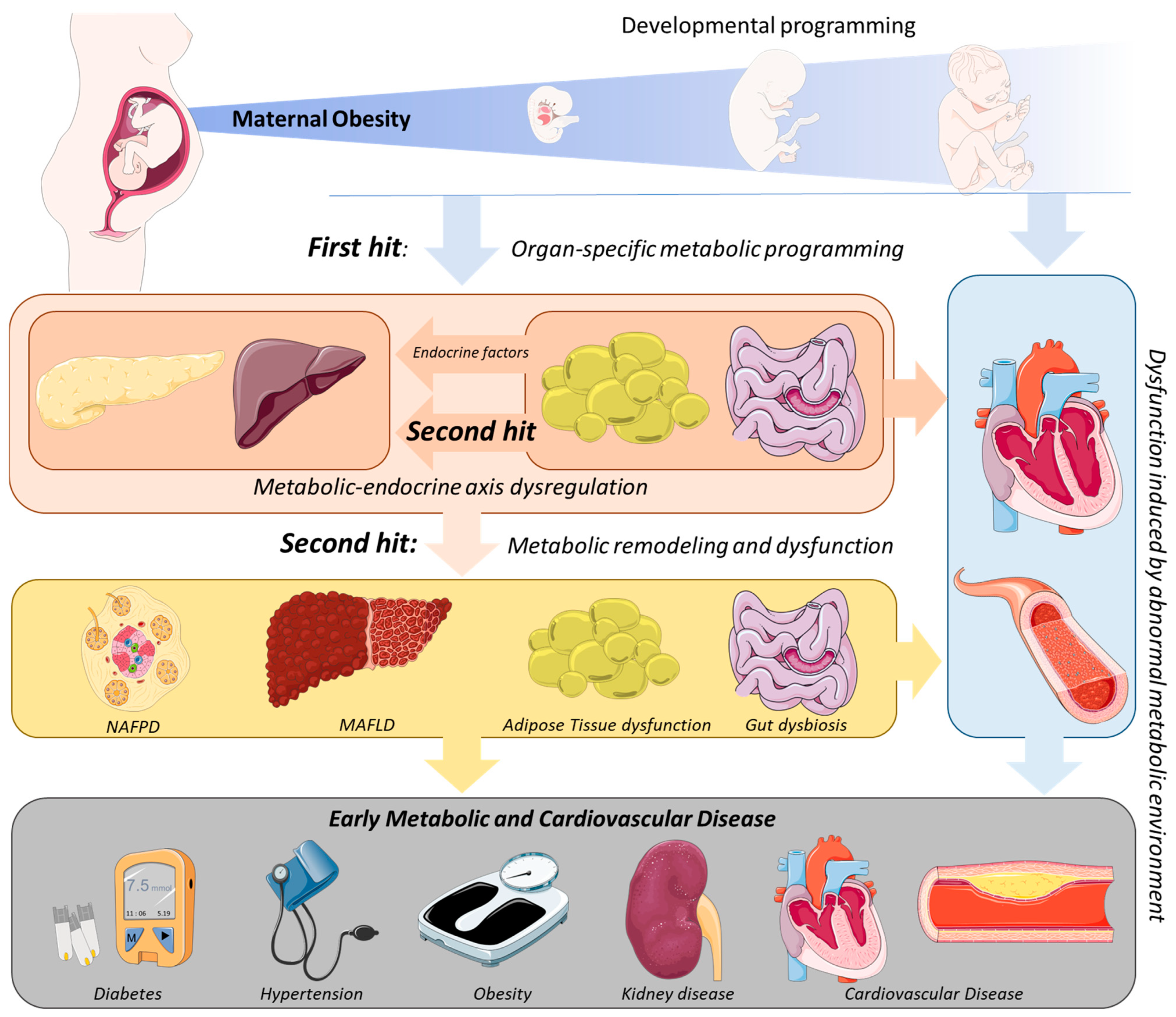
2. Mechanistic Links between MO and the Development of Metabolic Disease in the Offspring

The evidence in clinical studies of increased disease incidence in MO offspring has made it necessary to explore the mechanistic links between offspring metabolic disease development and MO. For ethical reasons, animal studies are critical to examining the cellular and molecular mechanisms associated with the phenotypes of the human offspring born to MO. Several animal models have been used in an attempt to elucidate these mechanisms. Studies in rats, sheep, and non-human primates have consistently proven MO’s deleterious effects on offspring, displaying similar phenotypes to those described in humans [24]. Increased birth- and postnatal-body weight [25,26,27], excessive body fat percentage [28,29], adiposity [29,30] and hyperinsulinemia [28,30] are frequent in rodent, ovine, and non-human primate studies ranging from MO offspring’s early ages until adulthood. These outcomes are often accompanied by an abnormal lipidic profile (increased cholesterol and triglycerides levels) [28] and endocrine dysregulation, evidenced by impaired adipokine levels [15,21,31].
The mechanisms associated with these outcomes and, subsequently, increased predisposition to metabolic disease for MO progeny seem to be multifactorial. In this focused manuscript, we explore (a) metabolic programming and oxidative stress, (b) epigenomic alterations, and (c) endocrine-stress response dysregulation as the main potential mechanisms involved in offspring’s metabolic disease programming by MO. We define the first two as the first hit of offspring adaptations to MO, i.e., structural and molecular alterations already observed at developmental stages that are maintained throughout offspring life. The first hit of developmental programming is tissue-specific and a direct result of the intrauterine environment on each organ cell’s maturation, growth, and proliferation.
MO influences fetal nutrient availability, namely glucose and lipids, due to the different blood maternal metabolome and the ability of each molecule to cross through the placenta [32]. For example, glucose is only partially buffered by the placenta, meaning that if a higher glucose concentration exists in maternal blood, it will also be increased in fetal circulation [33]. This leads to fetal hyperinsulinemia, which potentiates insulin resistance, metabolic dysregulation, and oxidative stress; these are thought to be involved in offspring’s metabolic disease programming [16]. Accordingly, insulin resistance in white adipose, hepatic, and cardiac tissues of MO offspring has been reported [16]. The hepatic and cardiac ratio between phosphorylated insulin receptor substrate 1 (IRS-1) at Ser307 residue and total IRS-1 is increased for 3- [34] and 4-month-old [26] MO mice offspring. In addition, many studies have shown mitochondrial dysfunction in individuals with insulin resistance (IR) [35]. MO offspring present sex-specific cardiac mitochondrial function modulation [36]. Newborn female MO rat offspring present increased expression of mitochondrial fusion proteins, and males show increased levels of proteins involved in mitophagy [36]. Four-month-old MO mice offspring present altered cardiac mitochondrial ultrastructure, with swollen and disrupted mitochondria, reduced mitochondrial membrane potential and increased ROS production [26]. Cardiac oxidative stress was determined by higher malondialdehyde levels in newborn rat MO offspring [37]. Hepatic oxidative stress in non-human primates [31] and 20-week-old male mice MO offspring [38] was marked by increased levels of 8-hydroxy-2-deoxyguanosine. Increased pancreatic glutathione peroxidase mRNA levels were detected in 20-week-old male mice MO offspring [38], potentially as a way to counteract oxidative stress.
Although previously hypothesized, it remains unknown whether MO-induced metabolic programming in the offspring has an effect on the long-term regulation of gene expression [16]. In spite of this, the literature has pointed to epigenetic dysregulation as a possible disease-programming mechanism due to the fetal epigenome responsiveness to maternal dietary patterns [39]. MO-induced microRNA (miRNA) expression alterations, DNA methylation, and post-translational histone modifications are the main mechanisms that lead to the epigenetic modulation of gene expression in offspring [40]. Hepatic miRNAs that regulate the insulin-signaling pathway are decreased in 4-month-old MO sheep offspring [39]. The inhibition of IRS-1 translation in white adipose tissue (WAT) is provoked by elevated miRNA-126 in 2-month-old MO mice offspring [41]. Genome-scale DNA methylation is altered by MO in 21-day-old rat male offspring [42]. Differential DNA methylation patterns and post-translational histone modifications (acetylation/methylation) were also found for genes encoding for adipocytokine, adiponectin, and leptin in MO mice offspring [43].
The programming of hypothalamic appetite-regulating centers is another mechanism potentially involved in MO programming of offspring’s metabolic disease [44]. MO induces endocrine stress-response dysregulation, leading to offspring’s hyperphagia and obesity [16]. The orexigenic drive (i.e., increased food intake, weight gain, the ratio between orexigenic and anorexigenic neuronal number/peptide expression and signaling) is increased in MO offspring [45]. Some studies have suggested that, in rodents, the regulation of the central nervous system (CNS) occurs postnatally; however, recent research has indicated that this might occur in utero [43]. In the hypothalamus of newborn MO rat offspring, neuropeptide Y mRNA expression and protein levels are increased [45]. In contrast, in another study, 1-day-old MO offspring’s hypothalamus presented decreased mRNA expression of the leptin receptor, signal transducer/activator of transcription (STAT)-3, and hypothalamic neuropeptide Y [46], demonstrating that appetite-regulating centers are likely modulated by the MO-induced intrauterine environment. Nevertheless, current research fails to explain this contradictory data. Further research is needed to unravel this issue and whether MO-induced endocrine dysregulation in the hypothalamus extends to other organs and remains in adulthood.
In addition to biochemical end-point alterations, evidence supports deeper alterations, with the fetal tissues and organs adapting their function to an adverse intrauterine environment caused by MO [47], leading to organ dysfunction that could persist over the offspring’s life course and lead to metabolic disease development (Figure 1). For a detailed review of the mechanisms associated with the first hit of developmental programming by MO, consult the reviews [16,35].

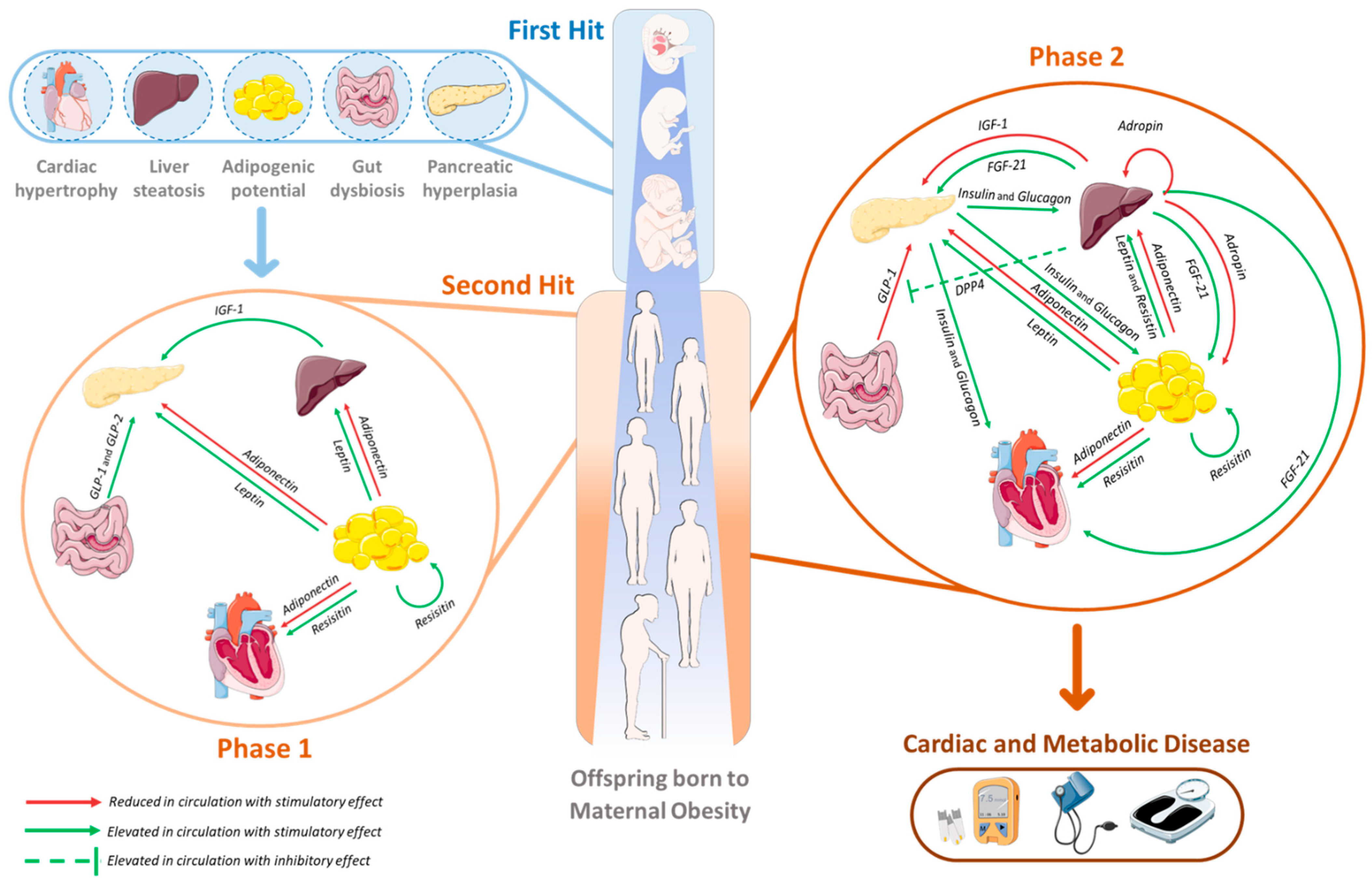
3. The Second Hit of Developmental Programming by MO: The Endocrine–Metabolic Axis

The endocrine–metabolic axis consists of the synchronized regulation and communication between multiple organs to a coordinated response to any metabolic stimuli through the release of molecules that are able to influence other cells’ functions. This communication is critical to avoid unnecessary energy expenditure and to the synchronization of the metabolic mechanisms according to the physiological demands across several organs, such as the liver, pancreas, gut, and adipose tissue (AT) [48,49]. This process occurs multiple times a day and is critical, for example, in the preprandial to the postprandial transition [50]. For inter-organ communication, the release of molecules able to produce a response in the other tissues is crucial. Among these molecules (hormones) exist peptides (e.g., insulin, leptin) or enzymes (e.g., Dipeptidyl peptidase-4 (DPP4)) [48,51].
Dysfunction in adipose tissue and gut is observed in the early life stages of MO offspring as a consequence of the first hit of disease programming in MO offspring (see Section 3.1 and Section 3.3). Along with these alterations, endocrine factors (e.g., adipokines, glucagon-like peptide (GLP) 1 and 2) are released in different concentrations due to MO [2,25,52,53]. We propose that this dysfunction (disrupted release of endocrine factors) represents a second hit (i.e., challenge) to the organs involved in body metabolic homeostasis (e.g., liver, pancreas). The exclusive modulation of the hormonal landscape originating in adipose tissue and gut represents phase one of the endocrine–metabolic axis programming by MO (i.e., second hit) (Figure 1 and Figure 2).
Later in MO offspring’s life, a second phase of the second hit (Figure 2) is observed when the other tissues (e.g., liver and pancreas) manipulate their own secretome [25,51] as a consequence of (1) organ-specific developmental programming (i.e., first hit) and (2) release of endocrine molecules from adipose tissue and gut (i.e., phase one of the second hit). Since MO offspring are more susceptible to disease, these alterations can start a vicious cycle of metabolic adaptations that challenge the metabolic homeostasis and predispose the offspring to metabolic disease (Figure 1 and Figure 2).
For a comprehensive understanding, next we describe the longitudinal modulation of each organ secretome due to MO, the consequences of the inter-organ metabolic (dys)regulation, and in particular the relationship with the development of CVD. Understanding the temporal coordination of these events is an important step to better assess health status and evaluate susceptibility to disease at each point of the MO offspring’s life course.

3.1. Adipose Tissue: An Endocrine Organ Involved in MO-Offspring’s Programming

Adipose tissue is now recognized as a highly metabolic organ involved in the regulation of vital physiological functions [54,55]. AT dysfunction has been implicated in systemic metabolic disorders both as a cause and a consequence. However, fat distribution is a more powerful indicator of obesity risk and associated comorbidities than whole-adiposity [54].
Three AT types exert complementary functions: (i) WAT, mainly adapted for fat storage; (ii) brown AT (BAT), more abundant in newborns, with thermogenic function, given the high mitochondrial content and expression of uncoupling protein 1 (UCP-1); and (iii) beige AT, normally developed upon sympathetic stimulation (e.g., exercise practice, prolonged cold exposure) [55]. Two types are distinguished within the WAT: subcutaneous (SAT) and visceral AT (VAT) [55]. While SAT confers protective effects for energy homeostasis, VAT is associated with IR, high triglyceride content, high blood pressure, and increased metabolic risk [2,56]. Obesity, IR, glucose intolerance, and dyslipidemia, often combined, belong to the cluster of factors involved in metabolic syndrome development [54].
Moreover, AT functions as triglyceride storage, with a significant role in thermoregulation and mechanical protection, also constituting an endocrine organ that secretes bioactive peptides known as adipokines [56]. Adipokines regulate physiological processes, such as appetite, food intake, immune and inflammatory function, and glucose and lipid metabolism [57]. More than 600 adipokines were already reported, including leptin, adiponectin, resistin, and visfatin [54,55]. Adipokines secreted by ectopic lipid depots, which result from TG storage near internal organs, such as the liver, skeletal muscle, heart, and pancreas, drain to the portal vein, directly impacting liver tissue and cellular function [55].
In lean individuals, the relation of adipokines with insulin signaling and metabolic homeostasis has already been observed in the AT [16,37,38]. Obesity is associated with impaired adipocyte remodeling. Upon surpassing TG storage capacity, the AT size (hyperplasia) and volume (hypertrophy) increases, culminating in AT dysfunction and consequent dysfunctional adipokine secretion [56,57].
MO increases the adipogenic potential of offspring’s adipocyte precursor cells [42], which represents the first hit of developmental programming on adipose tissue (Figure 1). Other adaptations include WAT insulin receptor-β, IRS-1, PI3K, and AKT1/2 expression levels, which were reduced in 2-month-old female MO rat offspring [42], suggesting an IR event in AT and potentially AT dysfunction. In GDM-portraying mothers, lower irisin levels were observed in colostrum and transitional milk, which might induce an increased WAT/BAT ratio in the offspring and promote fat storage [45,58]. After weaning, male MO offspring show a significant increase in the percentage of fat composition [59]. Greater fat mass was also observed in male and female human adults (between 20 and 30 years old) born to obese mothers [60]. Increased expression of pro-adipogenic genes (Pparc, Fabp4) and transcription factors (Pparγ, C/ebpα) and the reduction of the anti-adipogenic gene (Pref-1) for adult MO mice offspring were observed for WAT [42].
Similar to non-pregnant obese individuals, MO results in adipokine secretion alteration with implications for metabolic homeostasis [61]. Obese pregnant women have decreased adiponectin and elevated leptin plasma levels, which positively correlate with maternal BMI, fat mass, IR, glucose production, and fetal growth [61,62]. In the placenta, decreased adiponectin levels, in the context of MO, counterbalance adiponectin’s physiologic role [61]. Hypomethylation of the adiponectin promoter was observed in the maternal side of third-trimester MO placentas [52]. These observations are clear indications that adipokines could be involved in MO pathophysiology.
Adiponectin inhibits insulin signaling at IRS-1 via proliferator-activated receptor-alpha (PPARα) activation and ceramide synthesis [63]. Coupled with MO-derived decreased activated AMP-activated protein kinase (AMPK), MO adiponectin levels contribute to placental mTOR activation, which may prompt increased nutrient delivery to the fetus [61]. The dysregulation of adipokines has been reported in the human fetal umbilical cord blood. MO human offspring newborns present increased concentration of adiponectin, leptin [64], and TNF-α [65] in the fetal cord blood.
Early alteration in adipokine release results in the first phase of the second hit of the endocrine–metabolic axis developmental programming by MO (Figure 2). Due to adipocyte’s dysfunctions and in utero early differentiation, adipokine secretion will promote a different metabolic state in MO offspring through the regulation of multi-organ metabolic functions, including in the liver, pancreas, skeletal muscle, and heart (Figure 2).
Leptin secretion is proportional to adipocyte volume [57]. The activation of mitogen-activated protein kinase (MAPK) and STAT signaling cascades by leptin induction resulted in AT hyperplasia and hypertrophy in male Sprague-Dawley adipocyte primary cultures [66], demonstrating a cumulative effect. Upon leptin treatment, C57BL/6J mice adipose-derived stem cell (ASC) fat depots revealed increased expression of peroxisome proliferator-activated receptor-gamma (PPARγ) and proinflammatory cytokines (IL-6, IL-10, and TNFα), which resulted in enhanced lipid droplet formation via mTOR signaling [67]. Plasma hyperleptinemia and, consequently, hyperphagia have been identified both in rodents and non-human primates MO offspring [28,29,68]. Postnatally, leptin plasma concentrations are stable between 4-day-old and 3-week-old rats [69]. A high-fat high-sucrose (HFHS)-diet-induced MO stimulated an increase in offspring’s circulating leptin levels after weaning in Sprague–Dawley rats [70,71] and in 11-week-old offspring of a high-fat-induced MO [59]. Serum leptin levels were found to be increased in obese children [58]. However, a significant decrease in circulating leptin was observed in both sexes of human MO offspring between 20 and 30 years old [60]. While leptin stimulates the expression of proinflammatory cytokines, these molecules inhibit adiponectin secretion [72].
Adiponectin concentration in MO offspring cord blood was increased in humans [64]; nevertheless, a significant decrease in circulating adiponectin was observed in male and female young adult humans born to MO [60]. The expression of both adiponectin receptors (AdipoR1 and AdipoR2) was decreased in the AT of high-fat diet (HFD)–induced obese male mice [63].
Resistin treatment in 3T3-L1 adipocytes negatively impacted insulin signaling through lower expression and activation of proteins involved in the insulin signaling pathway and increased gene expression of suppressor of cytokine signaling 3 (SOCS-3), whose inhibition prevented the effects on insulin signaling [73]. Resistin circulating concentrations of maternal overweight offspring were increased at 3 weeks of age in Sprague–Dawley rats [71].
Visfatin is essentially released from VAT [54], but its effects have been strongly observed in the hepatic tissue. Visfatin affected the Janus kinase (JAK)/STAT3 and IkappaB kinase (IKK)/factor nuclear kappa (NF-kB) pathways in HepG2 cells, contributing to increased expression of proinflammatory cytokines and reduced levels of proteins involved in insulin signaling [74]. Treatment with inhibitors of each pathway prevented the observed effects, suggesting the involvement of visfatin regulating these signaling cascades within hepatocytes [74]. Visfatin circulating concentrations are usually greater in obese children, correlating well with resistin concentrations [75,76].
Overall, in an attempt to overcome AT impairment, cells modulate the release of adipokines and other AT-related hormones that are able to regulate other organs’ metabolism via endocrine pathways. The observation of early-AT dysfunction in MO offspring suggests that this could be one of the first hits in metabolic disease programming, followed by other organs’ metabolic dysfunction. Research has shown evidence of MO-induced pancreatic, hepatic, and cardiovascular dysregulation in offspring across different life stages [16]. The next section discusses the impact of adipokines specifically on the liver and on the predisposition to hepatic disease.

3.2. Impact of MO Programming of Endocrine–Metabolic Axis Dysregulation in Offspring’s Hepatic Disease Development

Dysregulation of the endocrine–metabolic axis can be programmed by various mechanisms in MO offspring despite the non-instantaneous development of obesity, diabetes, or other metabolic diseases. Overall, children born to abnormal maternal GWG present an increased risk of developing obesity [77]. Excessive GWG and MO are positively correlated with offspring’s IR and T2DM increased risk during childhood and adulthood [20] [78,79]. Across human organs, the relation between IR and tissue metabolic dysregulation is intricate and bidirectional, which induces a temporal propagation of metabolic disease (Figure 2).
Even though some evidence suggests that endocrine–metabolic dysregulation is programmed in utero (i.e., epigenetic dysregulation), the natural role of aging is likely to represent a challenge, triggering an exacerbation of metabolic disease. The development of cellular IR, also observed in normative aging, in the liver, AT, skeletal muscle, and heart precedes the impairment of insulin release from the pancreas in healthy offspring from MO [80,81]. The age-related IR development also produces a challenge to the liver in order to achieve physiological glucose homeostasis [81,82].
Insulin resistance is a widely described mechanism that plays a role in organ-specific pathophysiology, namely in Metabolic dysfunction-Associated Fatty Liver Disease (MAFLD) [83]. The origin of hepatic IR includes a multi-organ contribution, promoting a changed inflammatory environment (i.e., pro-inflammatory cytokine IL-6 and TNF-α), impairment of gut microbiota, lipotoxicity, and/or adiponectin signaling [84].
Adiponectin levels are usually lower in Non-Alcoholic Fatty Liver Disease (NAFLD) patients and correlated inversely with IR [85], suggesting a minor adiponectin contribution in enhancing fatty acid (FA) and glucose oxidation and a FA synthesis suppression via AMPK phosphorylation at residue threonine 172 of the α subunit (activation) [86]. Blockage of AMPK activation inhibits adiponectin’s effects on glucose and lipid metabolism, resulting in insulin insensitivity [86]. Even small intrahepatic fat accumulation (1.5%) can promote hepatic IR [87] by overactivating PKC epsilon via hepatocytes’ diacylglycerol (DAG) increased levels, consequently impairing IRS-1 and IRS-2 and the downstream PI3K pathway [88]. Adiponectin treatment, in animal and in in vitro studies, ameliorated hepatic steatosis [89,90]. Babies born to obese mothers present increased body adiposity [18] and consequent early lipotoxicity and dysfunctional adipokine release [70]. The impairment of AT is likely to shift the hepatic metabolism and lead to a premature MAFLD-like phenotype in the MO offspring liver. Impaired hepatic insulin signaling [91], triglycerides, and increased expression of gluconeogenic enzymes have been reported for MO non-human primate offspring [31]. In addition to adiponectin, serum leptin concentrations are increased in NAFLD subjects [92], suggesting a potential AT dysfunction prior to hepatic fat accumulation, which results in new metabolic homeostasis stimulated by adipokines’ levels. HepG2 leptin treatment led to decreased expression of low-density lipoprotein (LDL) receptors and decreased LDL uptake, with augmented proprotein convertase subtilisin/kexin type 9 (PCSK9), which proved essential for the observed response [93].
Adropin, mainly expressed in the liver, increases skeletal muscle insulin signaling [94], improves AT and liver glucose homeostasis, and reduces FA synthesis [95]. Obese subjects present lower adropin in circulation [96,97], including obese children [58]. Colostrum from women with GDM also presents reduced adropin levels [98], which exposes the offspring to reduced adropin, associated with higher body fat mass [99].
Fetuin-A, a regulator of insulin sensitivity, is also correlated with IR and NAFLD and suggests involvement in excessive fetal growth [100,101]. Although no alterations in cord blood Fetuin-A levels were observed, insulin-like growth factor (IGF)-1 and IGF-2 were significantly increased in GDM [102]. Circulating levels of Fibroblast Growth Factor (FGF) 21, a regulator of the systemic lipid metabolism, are increased in NAFLD [103] and GDM offspring during adolescence [104], and it was described that FGF21 gene expression was dysregulated in murine offspring of MO [105].
Another protein secreted by the liver with a critical role in the endocrine–metabolic axis is DPP4. DPP4 is able to regulate gut–pancreas communication by inactivating GLP-1 and Gastric Inhibitory Peptide (GIP), regulating body glucose homeostasis (see Section 3.3 and Figure 2) [51]. DPP4-bound syncytiotrophoblast-derived extracellular vesicles were found to be increased eight-fold in the circulation of GDM women, being considerably higher in the uterine than in paired peripheral blood [106]. In fact, maternal BMI significantly correlated with plasma DPP4 activity both in maternal and fetal cord blood [107]. Elevated DPP4 activity was also observed in individuals presenting IR and NAFLD [108].
It is clear that the hepatic secretome (e.g., hepatokines) changes to interact with other cell types and promote inflammation and IR as a response to hepatic metabolic alterations and steatosis. NAFLD patients present a specific hepatokine profile that modulates metabolism and IR in AT, skeletal muscle, and pancreas [51]. Accordingly, myocytes’ insulin sensitivity impairs at low degrees of hepatic fat accumulation (up to 6%) [87]. However, little is known about how MO programs the hepatic endocrine function.
Among the mentioned proteins, many others, including part of the liver secretome, are dysregulated during NAFLD and potentially play a role in MO-related offspring hepatic disease programming through endocrine regulation [51]. It is relevant that the modulation of both adipokines and hepatokines in offspring born to MO share a similar profile to the NAFLD and MAFLD patients. This highlights the susceptibility of MO offspring to develop hepatic disease early in life as well as the role of the endocrine–metabolic axis in its origins. As discussed, some of these proteins are able to regulate other organs’ metabolism and even modulate the endocrine communication among them, such as the gut–pancreas axis (Figure 2).

3.3. The Contribution of the Gut–Pancreas Axis to the Endocrine–Metabolic Imbalance in MO Offspring

Gut microbiota plays an important role in fat and fat-soluble vitamin absorption, digestion of complex carbohydrates, bile maintenance acid-related metabolism, preservation of intestinal epithelial barrier, and permeability [109]. The composition and diversity of gut microbiota change throughout pregnancy and are different during obesity development [70,110,111]. Even though for many years it was proposed that the gut microbiota was developed at birth and early life stages, recent studies found bacterial DNA on the placenta, amniotic fluid, umbilical cord, and infant meconium, contradicting the idea of fetal development in a sterile womb [112,113,114].
Multiple studies have associated MO with offspring gut dysbiosis, intestinal permeability, reduced macrophage phagocytosis, and dampened cytokine production [109]. Lower α-diversity of the fecal microbial community was detected in mice offspring exposed to HFD [115]. As well, lower diversity (including Firmicutes phylum, which is critical in maintaining the integrity of the intestinal epithelial barrier) of the gut microbiota in HFD-fed dams weaned mice offspring [116,117]. Reduced bacteroides in the human neonatal intestinal microbiota from HFD mothers during gestation persisted at least until 6 weeks of age [118]. Furthermore, the gut microbiota of children born to obese mothers also showed higher numbers of Parabacteroides spp. and Oscillibacter spp. and lower numbers of Blautia spp. (associated with obesity) and Eubacterium spp. [119].
Altogether, these profile changes in the endocrine function of intestinal K- and ileum L-cells from the intestine, responsible for releasing incretins into circulation, cause variations namely in GIP and GLP-1/GLP-2, respectively [120,121]. Both hormones are closely related to pancreatic endocrine function. While GIP is responsible for promoting glucagon release to circulation, GLP-1 stimulates postprandial insulin secretion [121,122].
GLP-1 and GLP-2 circulating concentrations after the weaning period are increased in offspring born to HFHS diet-induced MO [70]. However, in young adults, GLP-1 fasting plasma levels were reduced in human offspring exposed to GDM or type 1 diabetes mellitus (T1DM) during pregnancy [53]. This is likely a consequence of liver steatosis and metabolic dysfunction, which increases hepatic secretion of DPP4, as described in the previous section (Section 3.2). Similar secretory behavior is found with aging, obesity, and diabetes [121].
Although leptin, GLP-1, and GLP-2 levels were increased, no differences were found in circulating insulin levels of the offspring after the weaning of an HFHS diet-induced MO in Sprague–Dawley rats [70], suggesting AT dysfunction and gut adaptations as initial second hits of systemic metabolic MO programming, occurring prior to altered pancreatic insulin release. Indeed, circulating insulin concentration, fasting insulin secretion, and total insulin secretion were significantly increased in human male young adults exposed to MO, and lower insulin sensitivity was described in both male and female MO offspring [60].
The repercussions of dysfunctional adipokine release from ectopic fat also extend to pancreatic cells. G protein-coupled receptors (GPCRs) have been reported as probable binding sites for adipokines in response to obesity in humans [123]. The implication of apelin, chemerin, and other adipokines has been suggested to be involved in the adipokine-induced β-cell response to regulate insulin secretion through interaction with the islets GPCRs [123]. In the rat insulinoma cell line INS-1, a well-established model to study pancreatic β-cell function, leptin signaling activated protein kinase A (PKA), even while AMPK was inhibited and influenced F-actin organization, resulting in increased surface ATP-sensitive potassium channels [124], which is associated with the inhibition of insulin secretion. Indeed, adiponectin can stimulate insulin secretion and increase membrane capacitance in isolated pancreatic islets of 16-week-old C57BL/6 mice [125].
Increased β-cell mass has been detected in fetal sheep and 3-month-old mice MO offspring [25,126]. In young adults, higher levels of glucagon during the oral glucose tolerance test (OGTT) were detected in human offspring of overweight women, suggesting a reduced postprandial suppression of glucagon concomitant with impairment in GLP-1 levels [53], potentially regulated by the hepatokine DPP4.
In more severe cases, pancreatic cells’ dysregulation results in cell death, compromising the endocrine function of the pancreas in 8-week-old male rat MO offspring [127]. Later, dead cells might be replaced by adipocytes, leading to Non-Alcoholic Fatty Pancreas Disease (NAFPD). At this point, newly formed adipocytes start releasing, in site, adipokines and inflammatory cytokines that end up exacerbating pancreatic cells’ dysfunction [128].
Overall, these data support the idea that the offspring’s pancreas undergoes structural adaptations due to MO as the first hit of developmental programming of metabolic disease. However, a second hit is required to produce alterations in the pancreatic endocrine function. This second hit can be the complex endocrine environment resulting from the intricate relationship between gut dysbiosis and AT dysfunction. The involvement of pancreatic and hepatic dysfunction in MO offspring leads to a metabolic shift in the endocrine–metabolic axis, which is able to affect the function of organs highly dependent on the physiological energetic status, such as skeletal muscle and heart.

3.4. Relation of MO Programmed Metabolic Dysfunction with Cardiovascular Disease Development in the Offspring

Cardiovascular disease is the main cause of death worldwide, with metabolic syndrome, IR, and NAFLD being major risk factors [129]. Most of the endocrine dysregulations described in MO offspring have been proposed as part of or correlated with higher CVD incidence, including alterations in adiponectin [130], leptin [131], insulin [132], glucagon [133], IGF-1 [134], GLP-1 [135], among others. Some of these factors have a direct impact on cardiomyocytes’ function, regulating cardiac metabolism. However, cardiac dysfunction can be also caused by the altered metabolic environment and nutrient availability (concentration and variety of FAs, glucose, ketone bodies, etc.) for the cardiac metabolism [35]. Increased cardiac energy production through the stimulation of glycolysis instead of FA oxidation and loss of metabolic flexibility are common features in CVD (e.g., cardiomyopathy, heart failure) [136]. It is clear in the literature the contribution of MO to offspring’s increased risk of CVD, which potentially represents the final consequence of the endocrine–metabolic axis dysregulation [16,137] (Figure 2).
Regarding the impact of MO on the offspring’s cardiovascular system, animal models provided evidence of increased endothelial dysfunction at 3-months-old and systolic blood pressure at 6-months-old in female MO mice offspring [138]. Myocardial fibrosis and inflammation [139,140] increased cardiac inflammation markers [140] and decreased cardiac contractile function [141] for ovine MO fetal and adult offspring. Impaired cardiac physiology, thickening of the left ventricle wall [142], increased mass, and increased myocardial lipid accumulation were described for MO mice adult offspring [35].
In human cohorts, increased GWG/MO induced offspring’s higher adiposity and adverse metabolic profile (i.e., elevated VLDL, triglycerides, and saturated FA), enhance the risk of offspring’s CVD development [143]. Human cohort studies have demonstrated increased systolic blood pressure in 6- and 21-year-old MO offspring [18,19]. Indeed, a human epidemiological follow-up study in the UK disclosed that MO offspring aged between 32 and 62 years old are more likely to be admitted to the hospital due to cardiovascular events and have a higher risk of premature death than those of lean mothers [144]. This accumulating body of clinical evidence suggests that the sudden worldwide rise in NCDs among the young-adult population might be attributable, at least in part, to the increased incidence of MO, and that NCDs can be “dictated” in utero, even before birth [13].
It has been recently suggested that cardiometabolic disease development could be prompted due to an imbalance between the release of anti- and proinflammatory adipokines [145]. Clinical research has suggested that impaired serum levels of adipokines, including leptin [146], adiponectin [147], and irisin [148], may contribute to the development of cardiometabolic disease. This relationship was corroborated by animal studies; increased levels of leptin and decreased levels of both adiponectin and irisin, as observed in human MO offspring, have been associated with cardiovascular abnormalities in rodent animal models [149]. For example, in female mice, increased leptin leads to hypertension and endothelial dysfunction [150]. The role of lower adiponectin in cardiac hypertrophy has been reported, which was corroborated in adiponectin knockout mice presenting more severe cardiac hypertrophy [151,152,153]. In an animal model for cardiac ischemia, irisin administration improved cardiac function in adult mice [154]. This beneficial action could be a consequence of the role of irisin in improving cardiac mitochondrial function, by increasing the activity of SOD-2, an antioxidant protein, thus protecting the heart from ischemia-reperfusion injury [155]. Overall, it seems that impaired release of adipokines might play a role in cardiovascular disease development.
Additional research is needed to completely understand the impact of the endocrine alterations in MO offspring on the heart. Cardiac dysfunction can also be explained as an outcome of the endocrine–metabolic developmental programming by MO. It is critical to unravel if there is a direct impact, through manipulation of cardiomyocytes’ signaling pathways, or if CVD results from the metabolic profile achieved by the endocrine–metabolic axis dysregulation in MO offspring.

4. Conclusions

Evidence of human and animal offspring’s metabolic programming by MO has been accumulating. Specific organ metabolic modulation is now clear, although the temporal perspective of each alteration is usually unexplored in the available studies. Understanding developmental programming as multi-organ programming instead of individual tissue in a time-dependent way is critical to a deeper knowledge of metabolic programming in MO offspring.
Some of the consequences of MO on offspring directly impact particular tissues, mostly occurring in utero; the first hits are immature cardiomyocytes, early pre-adipocytes differentiation, or β-cells mass growth. However, multi-organ endocrine communication is commonly missed in studies, which jeopardizes the understanding of the endocrine–metabolic axis adaptation and early systemic metabolic disease in MO offspring.
Growing evidence suggests two alternatives as initial second hits in MO-offspring systemic metabolic programming: (1) early adiposity and adipose tissue dysfunction; and (2) gut dysbiosis. In the first, AT dysfunction promotes a diverse circulating adipokine profile, whic137h regulates pancreatic and hepatic metabolic function via IR and inflammation. In turn, the pancreas and liver are endocrine organs and modulate pancreatic hormones and hepatokine release to achieve a new systemic homeostasis, ultimately impacting highly metabolic functional tissues such as the heart. The second consists of the modulation of gut microbiota during fetal development and lactation, which leads to premature gut dysbiosis and consequent gut endocrine-induced metabolic adaptations at the cost of mild metabolic dysfunction of the liver-AT-pancreas axis.
Second hits are multi-organ and dependent on endocrine communication. It is critical to take into consideration the effect of aging. Most of the evidence showed that MO offspring present an early-age deteriorated phenotype (e.g., early disease, reduced lifespan, telomere shortening, cellular senescence) [156,157,158] across their life that promotes the development of metabolic and cardiovascular disease at an early stage of life (Figure 2).
Longitudinal studies to unravel time- and organ-dependent endocrine–metabolic axis dysfunction that also include aging’s impact are required for a better understanding of the metabolic developmental programming by maternal obesity. MO offspring follow-up of circulation hormones with direct metabolic impact and their health status is critical for improving these individuals’ healthcare and aging-related quality of life.

Author Contributions

Conceptualization, L.F.G. and S.P.P.; writing—original draft preparation, L.F.G., M.S.D. and C.T.; writing—review and editing, L.F.G., A.L.A. and S.P.P.; visualization, L.F.G. All authors have read and agreed to the published version of the manuscript.

Funding

Funded by ERDF funds through the Operational Programme Competitiveness Factors—COMPETE2020 and national funds by Foundation for Science and Technology under FCT-Post-doctoral Fellowship (SPP, SFRH/BPD/116061/2016), FCT-doctoral Fellowship (LFG, SFRH/BD/5539/2020), project grant PTDC/DTP-DES/1082/2014(POCI-01-0145-FEDER-016657), strategic project UIDB/04539/2020, UIDP/04539/2020, LA/P/0058/2020, Fulbright Scholarship for Research (LFG), and Luso-American Development Foundation, R&D at USA Grant (LFG).

Institutional Review Board Statement

Not applicable.

Informed Consent Statement

Not applicable.

Data Availability Statement

Not applicable.

Conflicts of Interest

The authors declare no conflict of interest.

References

  1. González-Plaza, E.; Bellart, J.; Martínez-Verdú, M.; Arranz, A.; Luján-Barroso, L.; Seguranyes, G. Prevalencia de sobrepeso y obesidad preconcepcional en mujeres gestantes, y relación con los resultados maternos y perinatales. Enfermería Clínica 2021, 32, S23–S30. [Google Scholar] [CrossRef] [PubMed]
  2. Unamuno, X.; Gómez-Ambrosi, J.; Rodríguez, A.; Becerril, S.; Frühbeck, G.; Catalán, V. Adipokine dysregulation and adipose tissue inflammation in human obesity. Eur. J. Clin. Investig. 2018, 48, e12997. [Google Scholar] [CrossRef] [PubMed] [Green Version]
  3. Gaillard, R.; Felix, J.; Duijts, L.; Jaddoe, V.W. Childhood consequences of maternal obesity and excessive weight gain during pregnancy. Acta Obstet. Gynecol. Scand. 2014, 93, 1085–1089. [Google Scholar] [CrossRef] [PubMed]
  4. Catalano, P.M.; Shankar, K. Obesity and pregnancy: Mechanisms of short term and long term adverse consequences for mother and child. BMJ 2017, 356, j1. [Google Scholar] [CrossRef]
  5. Mouzon, S.H.-D.; Lassance, L. Endocrine and metabolic adaptations to pregnancy; impact of obesity. Horm. Mol. Biol. Clin. Investig. 2015, 24, 65–72. [Google Scholar] [CrossRef]
  6. Kriebs, J.M. Obesity in pregnancy—Addressing risks to improve outcomes. J. Perinat. Neonatal Nurs. 2014, 28, 32–40. [Google Scholar] [CrossRef]
  7. Bodnar, L.M.; Catov, J.M.; Klebanoff, M.A.; Ness, R.B.; Roberts, J.M. Prepregnancy Body Mass Index and the Occurrence of Severe Hypertensive Disorders of Pregnancy. Epidemiology 2007, 18, 234–239. [Google Scholar] [CrossRef]
  8. Torloni, M.R.; Betrán, A.P.; Horta, B.L.; Nakamura, M.U.; Atallah, A.N.; Moron, A.F.; Valente, O. Prepregnancy BMI and the risk of gestational diabetes: A systematic review of the literature with meta-analysis: Diagnostic in Obesity and Complications. Obes. Rev. 2009, 10, 194–203. [Google Scholar] [CrossRef]
  9. Callaghan, W.M.; Chu, S.Y.; Kim, Y.S.; Schmid, C.H.; Lau, J.; England, J.L.; Dietz, M.P. Maternal obesity and risk of gestational. Diabetes Care 2007, 30, 2070–2076. [Google Scholar]
  10. Solomon, C.G. A prospective study of pregravid determinants of gestational diabetes mellitus. J. Am. Med. Assoc. 1997, 278, 1078. [Google Scholar] [CrossRef]
  11. Chu, S.Y.; Kim, S.Y.; Schmid, C.H.; Dietz, P.M.; Callaghan, W.M.; Lau, J.; Curtis, K.M. Maternal obesity and risk of cesarean delivery: A meta-analysis. Obes. Rev. 2007, 8, 385–394. [Google Scholar] [CrossRef] [PubMed]
  12. Nijland, M.; Ford, S.P.; Nathanielsz, P. Prenatal origins of adult disease. Curr. Opin. Obstet. Gynecol. 2008, 20, 132–138. [Google Scholar] [CrossRef] [PubMed]
  13. Godfrey, K.M.; Reynolds, R.M.; Prescott, S.L.; Nyirenda, M.; Jaddoe, V.W.V.; Eriksson, J.G.; Broekman, B.F.P. Influence of maternal obesity on the long-term health of offspring. Lancet Diabetes Endocrinol. 2016, 5, 53–64. [Google Scholar] [CrossRef] [Green Version]
  14. Howell, K.; Powell, T.L. Effects of maternal obesity on placental function and fetal development. Reproduction 2017, 153, R97–R108. [Google Scholar] [CrossRef] [PubMed]
  15. Yu, Y.; Arah, O.A.; Liew, Z.; Cnattingius, S.; Olsen, J.; Sorensen, H.T.; Qin, G.; Li, J. Maternal diabetes during pregnancy and early onset of cardiovascular disease in offspring: Population based cohort study with 40 years of follow-up. BMJ 2019, 367, l6398. [Google Scholar] [CrossRef] [Green Version]
  16. Grilo, L.F.; Tocantins, C.; Diniz, M.S.; Gomes, R.M.; Oliveira, P.J.; Matafome, P.; Pereira, S.P. Metabolic Disease Programming: From Mitochondria to Epigenetics, Glucocorticoid Signalling and Beyond. Eur. J. Clin. Investig. 2021, 51, e13625. [Google Scholar] [CrossRef]
  17. Yu, Z.; Han, S.; Zhu, J.; Sun, X.; Ji, C.; Guo, X. Pre-Pregnancy Body Mass Index in Relation to Infant Birth Weight and Offspring Overweight/Obesity: A Systematic Review and Meta-Analysis. PLoS ONE 2013, 8, e61627. [Google Scholar] [CrossRef] [Green Version]
  18. Gaillard, R.; Steegers, E.A.P.; Franco, O.; Hofman, A.; Jaddoe, V.W.V. Maternal weight gain in different periods of pregnancy and childhood cardio-metabolic outcomes. The Generation R Study. Int. J. Obes. 2014, 39, 677–685. [Google Scholar] [CrossRef]
  19. Mamun, A.A.; O’Callaghan, M.; Callaway, L.; Williams, G.; Najman, J.; Lawlor, D.A. Associations of gestational weight gain with offspring body mass index and blood pressure at 21 years of age: Evidence from a birth cohort study. Circulation 2009, 119, 1720–1727. [Google Scholar] [CrossRef]
  20. Hochner, H.; Friedlander, Y.; Calderon-Margalit, R.; Meiner, V.; Sagy, Y.; Avgil-Tsadok, M.; Burger, A.; Savitsky, B.; Siscovick, D.S.; Manor, O. Associations of maternal prepregnancy body mass index and gestational weight gain with adult offspring cardiometabolic risk factors: The Jerusalem Perinatal Family Follow-up Study. Circulation 2012, 125, 1381–1389. [Google Scholar] [CrossRef] [Green Version]
  21. Eriksson, J.; Sandboge, S.; Salonen, M.; Kajantie, E.; Osmond, C. Maternal weight in pregnancy and offspring body composition in late adulthood: Findings from the Helsinki Birth Cohort Study (HBCS). Ann. Med. 2015, 47, 94–99. [Google Scholar] [CrossRef] [PubMed]
  22. Andres, A.; Hull, H.R.; Shankar, K.; Casey, P.H.; Cleves, M.A.; Badger, T.M. Longitudinal body composition of children born to mothers with normal weight, overweight, and obesity. Obesity 2015, 23, 1252–1258. [Google Scholar] [CrossRef] [PubMed]
  23. Bronson, S.C.; Seshiah, V. Transgenerational transmission of non-communicable diseases: How to break the vicious cycle? Cureus 2021, 13, 18754. [Google Scholar] [CrossRef] [PubMed]
  24. Menting, M.D.; Mintjens, S.; van de Beek, C.; Frick, C.J.; Ozanne, S.E.; Limpens, J.; Roseboom, T.J.; Hooijmans, C.R.; van Deutekom, A.W.; Painter, R.C. Maternal obesity in pregnancy impacts offspring cardiometabolic health: Systematic review and meta-analysis of animal studies. Obes. Rev. 2019, 20, 675–685. [Google Scholar] [CrossRef] [Green Version]
  25. Nunes, F.; Frantz, E.D.C.; Lannes, W.R.; Menezes, M.C.D.S.; Mandarim-De-Lacerda, C.; Souza-Mello, V. Pregestational maternal obesity impairs endocrine pancreas in male F1 and F2 progeny. Nutrition 2015, 31, 380–387. [Google Scholar] [CrossRef]
  26. Turdi, S.; Ge, W.; Hu, N.; Bradley, K.M.; Wang, X.; Ren, J. Interaction between maternal and postnatal high fat diet leads to a greater risk of myocardial dysfunction in offspring via enhanced lipotoxicity, IRS-1 serine phosphorylation and mitochondrial defects. J. Mol. Cell. Cardiol. 2012, 55, 117–129. [Google Scholar] [CrossRef]
  27. Rivera, H.M.; Kievit, P.; Kirigiti, M.A.; Bauman, L.A.; Baquero, K.; Blundell, P.; Dean, T.A.; Valleau, J.C.; Takahashi, D.L.; Frazee, T.; et al. Maternal high-fat diet and obesity impact palatable food intake and dopamine signaling in nonhuman primate offspring. Obesity 2015, 23, 2157–2164. [Google Scholar] [CrossRef] [Green Version]
  28. Rajia, S.; Chen, H.; Morris, M.J. Maternal overnutrition impacts offspring adiposity and brain appetite markers-modulation by postweaning diet. J. Neuroendocr. 2010, 22, 905–914. [Google Scholar] [CrossRef] [Green Version]
  29. Long, N.M.; George, L.A.; Uthlaut, A.B.; Smith, D.T.; Nijland, M.; Nathanielsz, P.; Ford, S.P. Maternal obesity and increased nutrient intake before and during gestation in the ewe results in altered growth, adiposity, and glucose tolerance in adult offspring1. J. Anim. Sci. 2010, 88, 3546–3553. [Google Scholar] [CrossRef] [Green Version]
  30. Chen, H.; Simar, D.; Morris, M.J. Maternal obesity impairs brain glucose metabolism and neural response to hyperglycemia in male rat offspring. J. Neurochem. 2013, 129, 297–303. [Google Scholar] [CrossRef] [Green Version]
  31. McCurdy, C.; Bishop, J.M.; Williams, S.M.; Grayson, B.E.; Smith, M.S.; Friedman, J.E.; Grove, K.L. Maternal high-fat diet triggers lipotoxicity in the fetal livers of nonhuman primates. J. Clin. Investig. 2009, 119, 323–335. [Google Scholar] [CrossRef] [PubMed] [Green Version]
  32. Hirschmugl, B.; Perazzolo, S.; Sengers, B.G.; Lewis, R.M.; Gruber, M.; Desoye, G.; Wadsack, C. Placental mobilization of free fatty acids contributes to altered materno-fetal transfer in obesity. Int. J. Obes. 2021, 45, 1114–1123. [Google Scholar] [CrossRef] [PubMed]
  33. Hay, W.W. Placental-fetal glucose exchange and fetal glucose metabolism. Trans. Am. Clin. Clim. Assoc. 2006, 117, 321–340. [Google Scholar]
  34. Martin-Gronert, M.S.; Fernandez-Twinn, D.S.; Poston, L.; Ozanne, S.E. Altered hepatic insulin signalling in male offspring of obese mice. J. Dev. Orig. Heal. Dis. 2010, 1, 184–191. [Google Scholar] [CrossRef] [PubMed]
  35. Tocantins, C.; Diniz, M.S.; Grilo, L.F.; Pereira, S.P. The birth of cardiac disease: Mechanisms linking gestational diabetes mellitus and early onset of cardiovascular disease in offspring. WIREs Mech. Dis. 2022. [Google Scholar] [CrossRef] [PubMed]
  36. Larsen, T.D.; Sabey, K.H.; Knutson, A.J.; Gandy, T.C.T.; Louwagie, E.J.; Lauterboeck, L.; Mdaki, K.S.; Baack, M.L. Diabetic Pregnancy and Maternal High-Fat Diet Impair Mitochondrial Dynamism in the Developing Fetal Rat Heart by Sex-Specific Mechanisms. Int. J. Mol. Sci. 2019, 20, 3090. [Google Scholar] [CrossRef] [Green Version]
  37. Mdaki, K.S.; Larsen, T.D.; Wachal, A.L.; Schimelpfenig, M.D.; Weaver, L.J.; Dooyema, S.D.R.; Louwagie, E.J.; Baack, M.L. Maternal high-fat diet impairs cardiac function in offspring of diabetic pregnancy through metabolic stress and mitochondrial dysfunction. Am. J. Physiol. Circ. Physiol. 2016, 310, H681–H692. [Google Scholar] [CrossRef] [Green Version]
  38. Yokomizo, H.; Inoguchi, T.; Sonoda, N.; Sakaki, Y.; Maeda, Y.; Inoue, T.; Hirata, E.; Takei, R.; Ikeda, N.; Fujii, M.; et al. Maternal high-fat diet induces insulin resistance and deterioration of pancreatic β-cell function in adult offspring with sex differences in mice. Am. J. Physiol. Metab. 2014, 306, E1163–E1175. [Google Scholar] [CrossRef] [Green Version]
  39. Nicholas, L.M.; Rattanatray, L.; MacLaughlin, S.M.; Ozanne, S.E.; Kleemann, D.O.; Walker, S.K.; Morrison, J.L.; Zhang, S.; Muhlhäusler, B.S.; Martin-Gronert, M.S.; et al. Differential effects of maternal obesity and weight loss in the periconceptional period on the epigenetic regulation of hepatic insulin-signaling pathways in the offspring. FASEB J. 2013, 27, 3786–3796. [Google Scholar] [CrossRef]
  40. Agarwal, P.; Morriseau, T.S.; Kereliuk, S.M.; Doucette, C.A.; Wicklow, B.A.; Dolinsky, V.W. Maternal obesity, diabetes during pregnancy and epigenetic mechanisms that influence the developmental origins of cardiometabolic disease in the offspring. Crit. Rev. Clin. Lab. Sci. 2018, 55, 71–101. [Google Scholar] [CrossRef]
  41. Fernandez-Twinn, D.S.; Alfaradhi, M.Z.; Martin-Gronert, M.S.; Duque-Guimaraes, D.E.; Piekarz, A.; Ferland-McCollough, D.; Bushell, M.; Ozanne, S.E. Downregulation of IRS-1 in adipose tissue of offspring of obese mice is programmed cell-autonomously through post-transcriptional mechanisms. Mol. Metab. 2014, 3, 325–333. [Google Scholar] [CrossRef] [PubMed]
  42. Borengasser, S.J.; Zhong, Y.; Kang, P.; Lindsey, F.; Ronis, M.J.J.; Badger, T.M.; Gomez-Acevedo, H.; Shankar, K. Maternal Obesity Enhances White Adipose Tissue Differentiation and Alters Genome-Scale DNA Methylation in Male Rat Offspring. Endocrinology 2013, 154, 4113–4125. [Google Scholar] [CrossRef] [PubMed] [Green Version]
  43. Masuyama, H.; Hiramatsu, Y. Effects of a High-Fat Diet Exposure in UTERO on the Metabolic Syndrome-Like Phenomenon in Mouse Offspring through Epigenetic Changes in Adipocytokine Gene Expression. Endocrinology 2012, 153, 2823–2830. [Google Scholar] [CrossRef] [Green Version]
  44. Zambrano, E.; Ibáñez, C.; Martínez-Samayoa, P.M.; Lomas, C.; Durand-Carbajal, M.; González, G.L.R. Maternal Obesity: Lifelong Metabolic Outcomes for Offspring from Poor Developmental Trajectories During the Perinatal Period. Arch. Med. Res. 2016, 47, 1–12. [Google Scholar] [CrossRef] [PubMed]
  45. Desai, M.; Ross, M.G. Maternal-infant nutrition and development programming of offspring appetite and obesity. Nutr. Rev. 2020, 78, 25–31. [Google Scholar] [CrossRef]
  46. Morris, M.J.; Chen, H. Established maternal obesity in the rat reprograms hypothalamic appetite regulators and leptin signaling at birth. Int. J. Obes. 2008, 33, 115–122. [Google Scholar] [CrossRef] [Green Version]
  47. Shrestha, N.; Ezechukwu, H.C.; Holland, O.J.; Hryciw, D.H. Developmental programming of peripheral diseases in offspring exposed to maternal obesity during pregnancy. Am. J. Physiol. Integr. Comp. Physiol. 2020, 319, R507–R516. [Google Scholar] [CrossRef]
  48. Torell, F.; Bennett, K.; Cereghini, S.; Rännar, S.; Lundstedt-Enkel, K.; Moritz, T.; Haumaitre, C.; Trygg, J.; Lundstedt, T. Multi-Organ Contribution to the Metabolic Plasma Profile Using Hierarchical Modelling. PLoS ONE 2015, 10, e0129260. [Google Scholar] [CrossRef]
  49. Ito, M.; Adachi-Akahane, S. Inter-organ Communication in the Regulation of Lipid Metabolism: Focusing on the Network Between the Liver, Intestine, and Heart. J. Pharmacol. Sci. 2013, 123, 312–317. [Google Scholar] [CrossRef] [Green Version]
  50. Jacome-Sosa, M.; Parks, E.J.; Bruno, R.S.; Tasali, E.; Lewis, G.F.; O Schneeman, B.; Rains, T.M. Postprandial Metabolism of Macronutrients and Cardiometabolic Risk: Recent Developments, Emerging Concepts, and Future Directions12. Adv. Nutr. Int. Rev. J. 2016, 7, 364–374. [Google Scholar] [CrossRef] [Green Version]
  51. Watt, M.J.; Miotto, P.M.; De Nardo, W.; Montgomery, M. The Liver as an Endocrine Organ—Linking NAFLD and Insulin Resistance. Endocr. Rev. 2019, 40, 1367–1393. [Google Scholar] [CrossRef] [PubMed]
  52. Nogues, P.; Santos, E.D.; Jammes, H.; Berveiller, P.; Arnould, L.; Vialard, F.; Dieudonné, M. Maternal obesity influences expression and DNA methylation of the adiponectin and leptin systems in human third-trimester placenta. Clin. Epigen. 2019, 11, 1–18. [Google Scholar] [CrossRef] [PubMed] [Green Version]
  53. Kelstrup, L.; Clausen, T.D.; Mathiesen, E.R.; Hansen, T.; Holst, J.J.; Damm, P. Incretin and Glucagon Levels in Adult Offspring Exposed to Maternal Diabetes in Pregnancy. J. Clin. Endocrinol. Metab. 2015, 100, 1967–1975. [Google Scholar] [CrossRef] [PubMed] [Green Version]
  54. Kim, J.-E.; Kim, J.-S.; Jo, M.-J.; Cho, E.; Ahn, S.-Y.; Kwon, Y.-J.; Ko, G.-J. The Roles and Associated Mechanisms of Adipokines in Development of Metabolic Syndrome. Molecules 2022, 27, 334. [Google Scholar] [CrossRef]
  55. Lustig, R.H.; Collier, D.; Kassotis, C.; Roepke, T.A.; Kim, M.J.; Blanc, E.; Barouki, R.; Bansal, A.; Cave, M.C.; Chatterjee, S.; et al. Obesity I: Overview and molecular and biochemical mechanisms. Biochem. Pharmacol. 2022, 199, 115012. [Google Scholar] [CrossRef]
  56. Janochova, K.; Haluzik, M.; Buzga, M. Visceral fat and insulin resistance—What we know? Biomed. Pap. 2019, 163, 19–27. [Google Scholar] [CrossRef] [Green Version]
  57. Fasshauer, M.; Blüher, M. Adipokines in health and disease. Trends Pharmacol. Sci. 2015, 36, 461–470. [Google Scholar] [CrossRef]
  58. Sayın, O.; Tokgöz, Y.; Arslan, N. Investigation of adropin and leptin levels in pediatric obesity-related nonalcoholic fatty liver disease. J. Pediatr. Endocrinol. Metab. 2014, 27, 479–484. [Google Scholar] [CrossRef]
  59. Walker, C.D.; Naef, L.; d’Asti, E.; Long, H.; Xu, Z.; Moreau, A.; Azeddine, B. Perinatal maternal fat intake affects metabolism and hippocampal function in the offspring: A potential role for leptin. Ann. N. Y. Acad. Sci. 2008, 1144, 189–202. [Google Scholar] [CrossRef]
  60. Mingrone, G.; Manco, M.; Mora, M.E.V.; Guidone, C.; Iaconelli, A.; Gniuli, D.; Leccesi, L.; Chiellini, C.; Ghirlanda, G. Influence of Maternal Obesity on Insulin Sensitivity and Secretion in Offspring. Diabetes Care 2008, 31, 1872–1876. [Google Scholar] [CrossRef] [Green Version]
  61. Kelly, A.C.; Powell, T.L.; Jansson, T. Placental function in maternal obesity. Clin. Sci. 2020, 134, 961–984. [Google Scholar] [CrossRef] [PubMed] [Green Version]
  62. Vernini, J.M.; Moreli, J.B.; Costa, R.A.A.; Negrato, C.A.; Rudge, M.V.C.; Calderon, I.M.P. Maternal adipokines and insulin as biomarkers of pregnancies complicated by overweight and obesity. Diabetol. Metab. Syndr. 2016, 8, 68. [Google Scholar] [CrossRef] [PubMed] [Green Version]
  63. Tsuchida, A.; Yamauchi, T.; Ito, Y.; Hada, Y.; Maki, T.; Takekawa, S.; Kamon, J.; Kobayashi, M.; Suzuki, R.; Hara, K.; et al. Insulin/Foxo1 pathway regulates expression levels of adiponectin receptors and adiponectin sensitivity. J. Biol. Chem. 2004, 279, 30817–30822. [Google Scholar] [CrossRef] [PubMed] [Green Version]
  64. Jaramillo-Ospina, A.; Castaño-Moreno, E.; Munoz-Munoz, E.; Krause, B.J.; Uauy, R.; Casanello, P.; Castro-Rodriguez, J.A. Maternal obesity is associated with higher cord blood adipokines in offspring most notably in females. J. Pediatr. Gastroenterol. Nutr. 2021, 73, 264–270. [Google Scholar] [PubMed]
  65. Dosch, N.C.; Guslits, E.F.; Weber, M.B.; Murray, S.E.; Ha, B.; Coe, C.L.; Auger, A.P.; Kling, P.J. Maternal Obesity Affects Inflammatory and Iron Indices in Umbilical Cord Blood. Physiol. Behav. 2016, 172, 20–28. [Google Scholar] [CrossRef] [PubMed] [Green Version]
  66. Machinal-Quélin, F.; Dieudonné, M.N.; Leneveu, M.C.; Pecquery, R.; Giudicelli, Y. Proadipogenic effect of leptin on rat preadipocytes in vitro: Activation of MAPK and STAT3 signaling pathways. Am. J. Physiol. Physiol. 2002, 282, C853–C863. [Google Scholar] [CrossRef] [Green Version]
  67. Palhinha, L.; Liechocki, S.; Hottz, E.D.; Pereira, J.A.D.S.; De Almeida, C.J.; Vieira, P.; Bozza, P.T.; Maya-Monteiro, C.M. Leptin Induces Proadipogenic and Proinflammatory Signaling in Adipocytes. Front. Endocrinol. 2019, 10, 841. [Google Scholar] [CrossRef] [Green Version]
  68. Taylor, P.D.; McConnell, J.; Khan, I.Y.; Holemans, K.; Lawrence, K.M.; Asare-Anane, H.; Persaud, S.J.; Jones, P.M.; Petrie, L.; Hanson, M.A.; et al. Impaired glucose homeostasis and mitochondrial abnormalities in offspring of rats fed a fat-rich diet in pregnancy. Am. J. Physiol. Integr. Comp. Physiol. 2005, 288, R134–R139. [Google Scholar] [CrossRef]
  69. Herrera, E.; Lasunción, M.A.; Huerta, L.; Martín-Hidalgo, A. Plasma leptin levels in rat mother and offspring during pregnancy and lactation. Neonatology 2000, 78, 315–320. [Google Scholar] [CrossRef]
  70. Paul, H.A.; Bomhof, M.R.; Vogel, H.J.; Reimer, R.A. Diet-induced changes in maternal gut microbiota and metabolomic profiles influence programming of offspring obesity risk in rats. Sci. Rep. 2016, 6, 20683. [Google Scholar] [CrossRef]
  71. Shankar, K.; Kang, P.; Harrell, A.; Zhong, Y.; Marecki, J.C.; Ronis, M.J.J.; Badger, T.M. Maternal Overweight Programs Insulin and Adiponectin Signaling in the Offspring. Endocrinology 2010, 151, 2577–2589. [Google Scholar] [CrossRef] [PubMed]
  72. Oh, K.-J.; Lee, D.S.; Kim, W.K.; Han, B.S.; Lee, S.C.; Bae, K.-H. Metabolic Adaptation in Obesity and Type II Diabetes: Myokines, Adipokines and Hepatokines. Int. J. Mol. Sci. 2016, 18, 8. [Google Scholar] [CrossRef] [PubMed] [Green Version]
  73. Steppan, C.M.; Wang, J.; Whiteman, E.L.; Birnbaum, M.J.; Lazar, M.A. Activation of SOCS-3 by Resistin. Mol. Cell. Biol. 2005, 25, 1569–1575. [Google Scholar] [CrossRef] [PubMed] [Green Version]
  74. Heo, Y.J.; Choi, S.E.; Jeon, J.Y.; Han, S.J.; Kim, D.J.; Kang, Y.; Lee, K.W.; Kim, H.J. Visfatin induces inflammation and insulin resistance via the NF-κB and STAT3 signaling pathways in hepatocytes. J. Diabetes Res. 2019, 2019, 4021623. [Google Scholar] [CrossRef] [PubMed] [Green Version]
  75. Simões, N.F.; Domingos, A.L.G.; Oliveira, F.L.P.D.; Caldas, I.S.; Guedes, M.R.; Fajardo, V.C.; Freitas, S.N.D. Resistin and visfatin concentrations are related to central obesity and inflammation in Brazilian children. Nutrire 2018, 43, 7. [Google Scholar] [CrossRef]
  76. Nourbakhsh, M.; Nourbakhsh, M.; Gholinejad, Z.; Razzaghy-Azar, M. Visfatin in obese children and adolescents and its association with insulin resistance and metabolic syndrome. Scand. J. Clin. Lab. Investig. 2015, 75, 183–188. [Google Scholar] [CrossRef]
  77. Tie, H.-T.; Xia, Y.-Y.; Zeng, Y.-S.; Zhang, Y.; Dai, C.-L.; Guo, J.J.; Zhao, Y. Risk of childhood overweight or obesity associated with excessive weight gain during pregnancy: A meta-analysis. Arch. Gynecol. Obstet. 2013, 289, 247–257. [Google Scholar] [CrossRef]
  78. Fernandez-Twinn, D.S.; Hjort, L.; Novakovic, B.; Ozanne, S.E.; Saffery, R. Intrauterine programming of obesity and type 2 diabetes. Diabetologia 2019, 62, 1789–1801. [Google Scholar] [CrossRef] [Green Version]
  79. Perng, W.; Gillman, M.W.; Mantzoros, C.S.; Oken, E. A prospective study of maternal prenatal weight and offspring cardiometabolic health in mid-childhood. Ann. Epidemiol. 2014, 24, 793. [Google Scholar] [CrossRef] [Green Version]
  80. Kotronen, A.; Juurinen, L.; Tiikkainen, M.; Vehkavaara, S.; Yki–Järvinen, H. Increased Liver Fat, Impaired Insulin Clearance, and Hepatic and Adipose Tissue Insulin Resistance in Type 2 Diabetes. Gastroenterology 2008, 135, 122–130. [Google Scholar] [CrossRef]
  81. Shou, J.; Chen, P.-J.; Xiao, W.-H. Mechanism of increased risk of insulin resistance in aging skeletal muscle. Diabetol. Metab. Syndr. 2020, 12, 1–10. [Google Scholar] [CrossRef] [PubMed] [Green Version]
  82. Mohamad, M.; Mitchell, S.J.; Wu, L.E.; White, M.; Cordwell, S.; Mach, J.; Solon-Biet, S.; Boyer, D.; Nines, D.; Das, A.; et al. Ultrastructure of the liver microcirculation influences hepatic and systemic insulin activity and provides a mechanism for age-related insulin resistance. Aging Cell 2016, 15, 706–715. [Google Scholar] [CrossRef] [PubMed]
  83. Lim, S.; Kim, J.-W.; Targher, G. Links between metabolic syndrome and metabolic dysfunction-associated fatty liver disease. Trends Endocrinol. Metab. 2021, 32, 500–514. [Google Scholar] [CrossRef] [PubMed]
  84. Rosato, V.; Masarone, M.; Dallio, M.; Federico, A.; Aglitti, A.; Persico, M. NAFLD and Extra-Hepatic Comorbidities: Current Evidence on a Multi-Organ Metabolic Syndrome. Int. J. Environ. Res. Public Heal. 2019, 16, 3415. [Google Scholar] [CrossRef] [PubMed] [Green Version]
  85. Zhang, H.; Niu, Y.; Gu, H.; Lu, S.; Zhang, W.; Li, X.; Yang, Z.; Qin, L.; Su, Q. Low serum adiponectin is a predictor of progressing to nonalcoholic fatty liver disease. J. Clin. Lab. Anal. 2018, 33, e22709. [Google Scholar] [CrossRef] [Green Version]
  86. Yamauchi, T.; Kamon, J.; Minokoshi, Y.; Ito, Y.; Waki, H.; Uchida, S.; Yamashita, S.; Noda, M.; Kita, S.; Ueki, K.; et al. Adiponectin stimulates glucose utilization and fatty-acid oxidation by activating AMP-activated protein kinase. Nat. Med. 2002, 8, 1288–1295. [Google Scholar] [CrossRef]
  87. Bril, F.; Barb, D.; Portillo-Sanchez, P.; Biernacki, D.; Lomonaco, R.; Suman, A.; Weber, M.H.; Budd, J.T.; Lupi, M.E.; Cusi, K. Metabolic and histological implications of intrahepatic triglyceride content in nonalcoholic fatty liver disease. Hepatology 2016, 65, 1132–1144. [Google Scholar] [CrossRef] [Green Version]
  88. Sakurai, Y.; Kubota, N.; Yamauchi, T.; Kadowaki, T. Role of Insulin Resistance in MAFLD. Int. J. Mol. Sci. 2021, 22, 4156. [Google Scholar] [CrossRef]
  89. Long, W.; Hui Ju, Z.; Fan, Z.; Jing, W.; Qiong, L. The effect of recombinant adeno-associated virus-adiponectin (rAAV2/1-Acrp30) on glycolipid dysmetabolism and liver morphology in diabetic rats. Gen. Comp. Endocrinol. 2014, 206, 1–7. [Google Scholar] [CrossRef]
  90. Jung, T.W.; Youn, B.-S.; Choi, H.Y.; Lee, S.Y.; Hong, H.C.; Yang, S.J.; Yoo, H.J.; Kim, B.-H.; Baik, S.H.; Choi, K.M. Salsalate and adiponectin ameliorate hepatic steatosis by inhibition of the hepatokine fetuin-A. Biochem. Pharmacol. 2013, 86, 960–969. [Google Scholar] [CrossRef]
  91. Buckley, A.J.; Keserü, B.; Briody, J.; Thompson, M.; Ozanne, S.; Thompson, C.H. Altered body composition and metabolism in the male offspring of high fat–fed rats. Metabolism 2005, 54, 500–507. [Google Scholar] [CrossRef] [PubMed]
  92. Marques, V.; Afonso, M.B.; Bierig, N.; Duarte-Ramos, F.; Santos-Laso, A.; Jimenez-Agüero, R.; Eizaguirre, E.; Bujanda, L.; Pareja, M.J.; Luís, R.; et al. Adiponectin, Leptin, and IGF-1 Are Useful Diagnostic and Stratification Biomarkers of NAFLD. Front. Med. 2021, 8. [Google Scholar] [CrossRef] [PubMed]
  93. Du, Y.; Li, S.; Cui, C.-J.; Zhang, Y.; Yang, S.-H.; Li, J.-J. Leptin decreases the expression of low-density lipoprotein receptor via PCSK9 pathway: Linking dyslipidemia with obesity. J. Transl. Med. 2016, 14, 276. [Google Scholar] [CrossRef] [PubMed] [Green Version]
  94. Kumar, K.G.; Trevaskis, J.L.; Lam, D.D.; Sutton, G.M.; Koza, R.A.; Chouljenko, V.N.; Kousoulas, K.G.; Rogers, P.M.; Kesterson, R.A.; Thearle, M.; et al. Identification of Adropin as a Secreted Factor Linking Dietary Macronutrient Intake with Energy Homeostasis and Lipid Metabolism. Cell Metab. 2008, 8, 468–481. [Google Scholar] [CrossRef] [Green Version]
  95. Gao, S.; McMillan, R.P.; Zhu, Q.; Lopaschuk, G.D.; Hulver, M.W.; Butler, A.A. Therapeutic effects of adropin on glucose tolerance and substrate utilization in diet-induced obese mice with insulin resistance. Mol. Metab. 2015, 4, 310–324. [Google Scholar] [CrossRef]
  96. Stevens, J.R.; Kearney, M.L.; St-Onge, M.P.; Stanhope, K.L.; Havel, P.J.; Kanaley, J.A.; Thyfault, J.P.; Weiss, E.P.; Butler, A.A. Inverse association between carbohydrate consumption and plasma adropin concentrations in humans. Obesity 2016, 24, 1731–1740. [Google Scholar] [CrossRef] [Green Version]
  97. Butler, A.A.; Tam, C.S.; Stanhope, K.L.; Wolfe, B.M.; Ali, M.R.; O’Keeffe, M.; St-Onge, M.-P.; Ravussin, E.; Havel, P.J. Low Circulating Adropin Concentrations with Obesity and Aging Correlate with Risk Factors for Metabolic Disease and Increase after Gastric Bypass Surgery in Humans. J. Clin. Endocrinol. Metab. 2012, 97, 3783–3791. [Google Scholar] [CrossRef] [Green Version]
  98. Aydin, S.; Kuloglu, T.; Aydin, S. Copeptin, adropin and irisin concentrations in breast milk and plasma of healthy women and those with gestational diabetes mellitus. Peptides 2013, 47, 66–70. [Google Scholar] [CrossRef]
  99. Dugas, C.; Perron, J.; Kearney, M.; Mercier, R.; Tchernof, A.; Marc, I.; Weisnagel, S.J.; Robitaille, J. Postnatal Prevention of Childhood Obesity in Offspring Prenatally Exposed to Gestational Diabetes mellitus: Where Are We Now. Obes. Facts 2017, 10, 396–406. [Google Scholar] [CrossRef]
  100. Wang, W.-J.; Wang, S.; Yang, M.-N.; Dong, Y.; He, H.; Fang, F.; Huang, R.; Yu, X.-G.; Zhang, G.-H.; Zhao, X.; et al. Fetuin-A in Infants Born Small- or Large-for-Gestational-Age. Front. Endocrinol. 2020, 11, 567955. [Google Scholar] [CrossRef]
  101. Peter, A.; Kovarova, M.; Staiger, H.; Machann, J.; Schick, F.; Königsrainer, A.; Königsrainer, I.; Schleicher, E.; Fritsche, A.; Häring, H.-U.; et al. The hepatokines fetuin-A and fetuin-B are upregulated in the state of hepatic steatosis and may differently impact on glucose homeostasis in humans. Am. J. Physiol. Metab. 2018, 314, E266–E273. [Google Scholar] [CrossRef] [PubMed]
  102. Wang, W.-J.; Zhang, L.; Zheng, T.; Zhang, G.-H.; Du, K.; Yang, M.-N.; He, H.; Wang, S.; Wang, W.; Zhang, J.; et al. Fetuin-A and fetal growth in gestational diabetes mellitus. BMJ Open Diabetes Res. Care 2020, 8, e000864. [Google Scholar] [CrossRef] [PubMed] [Green Version]
  103. Li, H.; Fang, Q.; Gao, F.; Fan, J.; Zhou, J.; Wang, X.; Zhang, H.; Pan, X.; Bao, Y.; Xiang, K.; et al. Fibroblast growth factor 21 levels are increased in nonalcoholic fatty liver disease patients and are correlated with hepatic triglyceride. J. Hepatol. 2010, 53, 934–940. [Google Scholar] [CrossRef]
  104. Kampmann, F.B.; Thuesen, A.C.B.; Hjort, L.; A Bjerregaard, A.; E Chavarro, J.; Frystyk, J.; Bjerre, M.; Tetens, I.; Olsen, S.; Vaag, A.A.; et al. Increased leptin, decreased adiponectin and FGF21 concentrations in adolescent offspring of women with gestational diabetes. Eur. J. Endocrinol. 2019, 181, 691–700. [Google Scholar] [CrossRef] [PubMed]
  105. Moeckli, B.; Delaune, V.; Prados, J.; Tihy, M.; Peloso, A.; Oldani, G.; Delmi, T.; Slits, F.; Gex, Q.; Rubbia-Brandt, L.; et al. Impact of Maternal Obesity on Liver Disease in the Offspring: A Comprehensive Transcriptomic Analysis and Confirmation of Results in a Murine Model. Biomedicines 2022, 10, 294. [Google Scholar] [CrossRef]
  106. Kandzija, N.; Zhang, W.; Motta-Mejia, C.; Mhlomi, V.; McGowan-Downey, J.; James, T.; Cerdeira, A.S.; Tannetta, D.; Sargent, I.; Redman, C.W.; et al. Placental extracellular vesicles express active dipeptidyl peptidase IV; levels are increased in gestational diabetes mellitus. J. Extracell. Vesicles 2019, 8, 1617000. [Google Scholar] [CrossRef] [PubMed] [Green Version]
  107. Montaniel, K.R.C.; Bucher, M.; Maloyan, A. The role of inflammation and dipeptidyl peptidase IV in the developmental programming of obesity and insulin resistance. Circulation 2018, 138, A15395. [Google Scholar]
  108. Baumeier, C.; Schlüter, L.; Saussenthaler, S.; Laeger, T.; Rödiger, M.; Alaze, S.A.; Fritsche, L.; Häring, H.-U.; Stefan, N.; Fritsche, A.; et al. Elevated hepatic DPP4 activity promotes insulin resistance and non-alcoholic fatty liver disease. Mol. Metab. 2017, 6, 1254–1263. [Google Scholar] [CrossRef]
  109. Zhou, L.; Xiao, X. The role of gut microbiota in the effects of maternal obesity during pregnancy on offspring metabolism. Biosci. Rep. 2018, 38. [Google Scholar] [CrossRef] [Green Version]
  110. Gérard, P. Gut microbiota and obesity. Cell. Mol. Life Sci. 2016, 73, 147–162. [Google Scholar] [CrossRef]
  111. Koren, O.; Goodrich, J.K.; Cullender, T.C.; Spor, A.; Laitinen, K.; Bäckhed, H.K.; Gonzalez, A.; Werner, J.J.; Angenent, L.T.; Knight, R.; et al. Host Remodeling of the Gut Microbiome and Metabolic Changes during Pregnancy. Cell 2012, 150, 470–480. [Google Scholar] [CrossRef] [PubMed] [Green Version]
  112. Jiménez, E.; Marín, M.L.; Martín, R.; Odriozola, J.M.; Olivares, M.; Xaus, J.; Fernández, L.; Rodríguez, J.M. Is meconium from healthy newborns actually sterile? Res. Microbiol. 2008, 159, 187–193. [Google Scholar] [CrossRef] [PubMed]
  113. Wassenaar, T.M.; Panigrahi, P. Is a foetus developing in a sterile environment? Lett. Appl. Microbiol. 2014, 59, 572–579. [Google Scholar] [CrossRef]
  114. Collado, M.C.; Rautava, S.; Aakko, J.; Isolauri, E.; Salminen, S. Human gut colonisation may be initiated in utero by distinct microbial communities in the placenta and amniotic fluid. Sci. Rep. 2016, 6, 23129. [Google Scholar] [CrossRef] [Green Version]
  115. Wankhade, U.; Zhong, Y.; Kang, P.; Alfaro, M.; Chintapalli, S.V.; Thakali, K.M.; Shankar, K. Enhanced offspring predisposition to steatohepatitis with maternal high-fat diet is associated with epigenetic and microbiome alterations. PLoS ONE 2017, 12, e0175675. [Google Scholar] [CrossRef] [PubMed] [Green Version]
  116. Bruce-Keller, A.J.; Fernandez-Kim, S.-O.; Townsend, R.L.; Kruger, C.; Carmouche, R.; Newman, S.; Salbaum, J.M.; Berthoud, H.-R. Maternal obese-type gut microbiota differentially impact cognition, anxiety and compulsive behavior in male and female offspring in mice. PLoS ONE 2017, 12, e0175577. [Google Scholar] [CrossRef] [PubMed]
  117. Shoaie, S.; Karlsson, F.; Mardinoglu, A.; Nookaew, I.; Bordel, S.; Nielsen, J. Understanding the interactions between bacteria in the human gut through metabolic modeling. Sci. Rep. 2013, 3, srep02532. [Google Scholar] [CrossRef] [PubMed] [Green Version]
  118. Chu, D.M.; Antony, K.M.; Ma, J.; Prince, A.L.; Showalter, L.; Moller, M.; Aagaard, K.M. The early infant gut microbiome varies in association with a maternal high-fat diet. Genome Med. 2016, 8, 77. [Google Scholar] [CrossRef] [Green Version]
  119. Clarke, S.; Murphy, E.F.; O’Sullivan, O.; Ross, R.; O’Toole, P.; Shanahan, F.; Cotter, P.D. Targeting the Microbiota to Address Diet-Induced Obesity: A Time Dependent Challenge. PLoS ONE 2013, 8, e65790. [Google Scholar] [CrossRef] [Green Version]
  120. Soderborg, T.K.; Clark, S.; Mulligan, C.E.; Janssen, R.C.; Babcock, L.; Ir, D.; Young, B.; Krebs, N.; Lemas, D.J.; Johnson, L.K.; et al. The gut microbiota in infants of obese mothers increases inflammation and susceptibility to NAFLD. Nat. Commun. 2018, 9, 4462. [Google Scholar] [CrossRef] [Green Version]
  121. Svegliati-Baroni, G.; Patrício, B.; Lioci, G.; Macedo, M.P.; Gastaldelli, A. Gut-Pancreas-Liver Axis as a Target for Treatment of NAFLD/NASH. Int. J. Mol. Sci. 2020, 21, 5820. [Google Scholar] [CrossRef] [PubMed]
  122. Fernández-Millán, E.; Guillén, C. Multi-Organ Crosstalk with Endocrine Pancreas: A Focus on How Gut Microbiota Shapes Pancreatic Beta-Cells. Biomolecules 2022, 12, 104. [Google Scholar] [CrossRef] [PubMed]
  123. Atanes, P.; Ashik, T.; Persaud, S.J. Obesity-induced changes in human islet G protein-coupled receptor expression: Implications for metabolic regulation. Pharmacol. Ther. 2021, 228, 107928. [Google Scholar] [CrossRef] [PubMed]
  124. Chen, P.C.; Kryukova, Y.N.; Shyng, S.L. Leptin regulates KATPchannel trafficking in pancreatic β-cells by a signaling mechanism involving AMP-activated protein kinase (AMPK) and camp-dependent protein kinase (PKA). J. Biol. Chem. 2013, 288, 34098–34109. [Google Scholar] [CrossRef] [Green Version]
  125. Okamoto, M.; Ohara-Imaizumi, M.; Kubota, N.; Hashimoto, S.; Eto, K.; Kanno, T.; Kubota, T.; Wakui, M.; Nagai, R.; Noda, M.; et al. Adiponectin induces insulin secretion in vitro and in vivo at a low glucose concentration. Diabetologia 2008, 51, 827–835. [Google Scholar] [CrossRef] [Green Version]
  126. Ford, S.P.; Zhang, L.; Zhu, M.; Miller, M.M.; Smith, D.T.; Hess, B.W.; Moss, G.E.; Nathanielsz, P.; Nijland, M.J. Maternal obesity accelerates fetal pancreatic β-cell but not α-cell development in sheep: Prenatal consequences. Am. J. Physiol. Integr. Comp. Physiol. 2009, 297, R835–R843. [Google Scholar] [CrossRef]
  127. Nicholas, L.M.; Nagao, M.; Kusinski, L.C.; Fernandez-Twinn, D.S.; Eliasson, L.; Ozanne, S.E. Exposure to maternal obesity programs sex differences in pancreatic islets of the offspring in mice. Diabetologia 2020, 63, 324. [Google Scholar] [CrossRef] [Green Version]
  128. Shah, N.; Rocha, J.P.; Bhutiani, N.; Endashaw, O.; Omer, E. Nonalcoholic Fatty Pancreas Disease. Nutr. Clin. Pr. 2019, 34, S49–S56. [Google Scholar] [CrossRef] [Green Version]
  129. Kasper, P.; Martin, A.; Lang, S.; Kütting, F.; Goeser, T.; Demir, M.; Steffen, H.-M. NAFLD and cardiovascular diseases: A clinical review. Clin. Res. Cardiol. 2020, 110, 921–937. [Google Scholar] [CrossRef]
  130. Liu, Y.; Vu, V.; Sweeney, G. Examining the Potential of Developing and Implementing Use of Adiponectin-Targeted Therapeutics for Metabolic and Cardiovascular Diseases. Front. Endocrinol. 2019, 10, 842. [Google Scholar] [CrossRef] [Green Version]
  131. Poetsch, M.S.; Strano, A.; Guan, K. Role of Leptin in Cardiovascular Diseases. Front. Endocrinol. 2020, 11, 354. [Google Scholar] [CrossRef] [PubMed]
  132. Simoniene, D.; Velickiene, D.; Platukiene, A. Insulin resistance in diabetes mellitus type 1, and its association with cardiovascular disease, sex hormones. Endocr. Abstr. 2018, 56. [Google Scholar] [CrossRef]
  133. Ng, J.C.M.; Schooling, C.M. Effect of Glucagon on Ischemic Heart Disease and Its Risk Factors: A Mendelian Randomization Study. J. Clin. Endocrinol. Metab. 2020, 105. [Google Scholar] [CrossRef] [PubMed]
  134. Van Bunderen, C.C.; Oosterwerff, M.M.; van Schoor, N.M.; Deeg, D.J.; Lips, P.; Drent, M.L. Serum IGF1 metabolic syndrome, and incident cardiovascular disease in older people: A population-based study. Eur. J. Endocrinol. 2013, 168, 393–401. [Google Scholar] [CrossRef] [PubMed] [Green Version]
  135. Zweck, E.; Roden, M. GLP-1 receptor agonists and cardiovascular disease: Drug-specific or class effects? Lancet Diabetes Endocrinol. 2019, 7, 89–90. [Google Scholar] [CrossRef] [Green Version]
  136. Karwi, Q.G.; Uddin, G.M.; Ho, K.L.; Lopaschuk, G.D. Loss of Metabolic Flexibility in the Failing Heart. Front. Cardiovasc. Med. 2018, 5, 68. [Google Scholar] [CrossRef] [Green Version]
  137. Pereira, S.P.; Tavares, L.C.; Duarte, A.I.; Baldeiras, I.; Cunha-Oliveira, T.; Martins, J.D.; Santos, M.S.; Maloyan, A.; Moreno, A.J.; Cox, L.A.; et al. Sex-dependent vulnerability of fetal nonhuman primate cardiac mitochondria to moderate maternal nutrient reduction. Clin. Sci. 2021, 135, 1103–1126. [Google Scholar] [CrossRef]
  138. Samuelsson, A.M.; Matthews, P.A.; Argenton, M.; Christie, M.R.; McConnell, J.M.; Jansen, E.H.; Piersma, A.H.; Ozanne, S.E.; Twinn, D.F.; Remacle, C.; et al. Diet-induced obesity in female mice leads to offspring hyperphagia, adiposity, hypertension, and insulin resistance: A novel murine model of developmental programming. Hypertension 2008, 51, 383–392. [Google Scholar] [CrossRef] [Green Version]
  139. Huang, Y.; Yan, X.; Zhao, J.X.; Zhu, M.J.; McCormick, R.J.; Ford, S.P.; Nathanielsz, P.; Ren, J.; Du, M. Maternal obesity induces fibrosis in fetal myocardium of sheep. Am. J. Physiol. Metab. 2010, 299, E968–E975. [Google Scholar] [CrossRef]
  140. Ghnenis, A.B.; Odhiambo, J.F.; McCormick, R.J.; Nathanielsz, P.; Ford, S.P. Maternal obesity in the ewe increases cardiac ventricular expression of glucocorticoid receptors, proinflammatory cytokines and fibrosis in adult male offspring. PLoS ONE 2017, 12, e0189977. [Google Scholar] [CrossRef]
  141. Wang, Q.; Zhu, C.; Sun, M.; Maimaiti, R.; Ford, S.P.; Nathanielsz, P.W.; Ren, J.; Guo, W. Maternal obesity impairs fetal cardiomyocyte contractile function in sheep. FASEB J. 2018, 33, 2587–2598. [Google Scholar] [CrossRef] [PubMed] [Green Version]
  142. Blackmore, H.L.; Niu, Y.; Fernandez-Twinn, D.; Tarry-Adkins, J.L.; Giussani, D.; Ozanne, S. Maternal Diet-induced Obesity Programs Cardiovascular Dysfunction in Adult Male Mouse Offspring Independent of Current Body Weight. Endocrinology 2014, 155, 3970–3980. [Google Scholar] [CrossRef] [PubMed] [Green Version]
  143. Kivelä, J.; Sormunen-Harju, H.; Girchenko, P.V.; Huvinen, E.; Stach-Lempinen, B.; Kajantie, E.; Villa, P.M.; Reynolds, R.M.; Hämäläinen, E.K.; Lahti-Pulkkinen, M.; et al. Longitudinal metabolic profiling of maternal obesity, gestational diabetes, and hypertensive pregnancy disorders. J. Clin. Endocrinol. Metab. 2021, 106, e4372–e4388. [Google Scholar] [CrossRef] [PubMed]
  144. Reynolds, R.M.; Allan, K.M.; A Raja, E.; Bhattacharya, S.; McNeill, G.; Hannaford, P.C.; Sarwar, N.; Lee, A.J.; Norman, J. Maternal obesity during pregnancy and premature mortality from cardiovascular event in adult offspring: Follow-up of 1 323 275 person years. BMJ 2013, 347, f4539. [Google Scholar] [CrossRef] [PubMed] [Green Version]
  145. Farkhondeh, T.; Llorens, S.; Pourbagher-Shahri, A.M.; Ashrafizadeh, M.; Talebi, M.; Shakibaei, M.; Samarghandian, S. An Overview of the Role of Adipokines in Cardiometabolic Diseases. Molecules 2020, 25, 5218. [Google Scholar] [CrossRef]
  146. Leyva, F.; Anker, S.D.; Egerer, K.; Stevenson, J.C.; Kox, W.J.; Coats, A.S. Hyperleptinaemia in chronic heart failure Relationships with insulin. Eur. Hear. J. 1998, 19, 1547–1551. [Google Scholar] [CrossRef] [PubMed] [Green Version]
  147. Ding, M.; Rzucidlo, E.M.; Davey, J.C.; Xie, Y.; Liu, R.; Jin, Y.; Stavola, L.; Martin, K.A. Adiponectin in the Heart and Vascular System. Vitam. Horm. 2012, 90, 289–319. [Google Scholar] [CrossRef]
  148. El-Mottaleb, N.A.A.; Galal, H.M.; El Maghraby, K.M.; Gadallah, A.I. Serum irisin level in myocardial infarction patients with or without heart failure. Can. J. Physiol. Pharmacol. 2019, 97, 932–938. [Google Scholar] [CrossRef] [Green Version]
  149. Senesi, P.; Luzi, L.; Terruzzi, I. Adipokines, Myokines, and Cardiokines: The Role of Nutritional Interventions. Int. J. Mol. Sci. 2020, 21, 8372. [Google Scholar] [CrossRef]
  150. Huby, A.C.; Otvos, L.; De Chantemèle, E.J.B. Leptin induces hypertension and endothelial dysfunction via aldosterone-dependent mechanisms in obese female mice. Hypertension 2016, 67, 1020–1028. [Google Scholar] [CrossRef] [Green Version]
  151. Liao, Y.; Takashima, S.; Maeda, N.; Ouchi, N.; Komamura, K.; Shimomura, I.; Hori, M.; Matsuzawa, Y.; Funahashi, T.; Kitakaze, M. Exacerbation of heart failure in adiponectin-deficient mice due to impaired regulation of AMPK and glucose metabolism. Cardiovasc. Res. 2005, 67, 705–713. [Google Scholar] [CrossRef] [PubMed]
  152. Dhandapany, P.S.; Kang, S.; Kashyap, D.K.; Rajagopal, R.; Sundaresan, N.R.; Singh, R.; Thangaraj, K.; Jayaprakash, S.; Manjunath, C.N.; Shenthar, J.; et al. Adiponectin receptor 1 variants contribute to hypertrophic cardiomyopathy that can be reversed by rapamycin. Sci. Adv. 2021, 7, eabb3991. [Google Scholar] [CrossRef]
  153. Park, M.; Sweeney, G. Direct effects of adipokines on the heart: Focus on adiponectin. Hear. Fail. Rev. 2012, 18, 631–644. [Google Scholar] [CrossRef] [PubMed]
  154. Liao, Q.; Qu, S.; Tang, L.-X.; Li, L.-P.; He, D.-F.; Zeng, C.Y.; Wang, W.E. Irisin exerts a therapeutic effect against myocardial infarction via promoting angiogenesis. Acta Pharmacol. Sin. 2019, 40, 1314–1321. [Google Scholar] [CrossRef] [PubMed]
  155. Wang, Z.; Chen, K.; Han, Y.; Zhu, H.; Zhou, X.; Tan, T.; Zeng, J.; Zhang, J.; Liu, Y.; Li, Y.; et al. Irisin Protects Heart Against Ischemia-Reperfusion Injury Through a SOD2-Dependent Mitochondria Mechanism. J. Cardiovasc. Pharmacol. 2018, 72, 259–269. [Google Scholar] [CrossRef] [PubMed]
  156. González, G.L.R.; Reyes-Castro, L.A.; Bautista, C.J.; Beltrán, A.A.; Ibáñez, C.A.; Vega, C.C.; Lomas-Soria, C.; Castro-Rodríguez, D.C.; Elías-López, A.L.; Nathanielsz, P.W.; et al. Maternal obesity accelerates rat offspring metabolic ageing in a sex-dependent manner. J. Physiol. 2019, 597, 5549–5563. [Google Scholar] [CrossRef]
  157. Martens, D.S.; Plusquin, M.; Gyselaers, W.; De Vivo, I.; Nawrot, T.S. Maternal pre-pregnancy body mass index and newborn telomere length. BMC Med. 2016, 14, 1–10. [Google Scholar] [CrossRef] [Green Version]
  158. Kajantie, E.; Osmond, C.; Barker, D.J.P.; Forsén, T.; Phillips, D.I.W.; Eriksson, J. Size at birth as a predictor of mortality in adulthood: A follow-up of 350 000 person-years. Int. J. Epidemiology 2005, 34, 655–663. [Google Scholar] [CrossRef]
Figure 1. Mechanism of endocrine-metabolic axis programming in offspring due to maternal obesity. Throughout fetal development, Maternal Obesity (MO) programs organ-specific dysfunction through the modulation of cell proliferation, growth rates, differentiation, and maturation—the first hit of developmental programming. Consequently, metabolic dysregulation is commonly observed in organs in early life stages, which leads to different metabolic homeostasis in MO offspring. Among these, the adipose tissue and gut are organs with an important endocrine role in the endocrine–metabolic axis. Adipose tissue dysfunction and gut dysbiosis are early organ-specific events in MO offspring, promoting the release of endocrine proteins (e.g., adipokines, glucagon-like peptide (GLP) 1/2) capable of modulating other tissues’ functions, such as the liver and pancreas, and through them, the body’s metabolism—the second hit of the endocrine–metabolic axis programming. These events can modulate the concentrations and classes of circulating metabolites (e.g., peptides, enzymes, fatty acids), which affect other organs and predispose them to metabolic diseases at early life stages.
Obesities 02 00019 g001
Figure 2. Endocrine-metabolic axis programming in offspring born to maternal obesity. The modulation of the organs’ maturation and differentiation state by Maternal Obesity (MO) throughout the developmental phase results in early dysregulation of multiple organs, including adipose tissue, gut, liver, pancreas, and heart—the first hit of developmental programming. Subsequently, in the first phase of the second hit, adipose tissue dysfunction and gut dysbiosis result in the different secretion of multiple hormones (e.g., adiponectin, leptin, glucagon-like peptide (GLP) 1/2), which impact and regulate other tissues’ (e.g., liver, heart, pancreas) metabolism. The liver and pancreas alter their metabolic function as a response to the endocrine signaling resultant from adipose tissue and gut dysregulation, which induces organ-specific metabolic dysfunction. In the second phase, the pancreas and liver also modulate the release of pancreatic and hepatic endocrine molecules, which leads to a new endocrine physiological state and impacts other tissues including the heart—the second hit of developmental programming. The endocrine–metabolic axis programming promotes systemic metabolic adaptations, which increases organ-specific and systemic disease predisposition. Red arrows represent endocrine molecules whose circulating levels are decreased in MO, and green arrows are endocrine molecules with greater circulating concentrations in MO. Dashed lines are set for hormones capable of inhibiting other hormones’ endocrine effects.
Obesities 02 00019 g002
Publisher’s Note: MDPI stays neutral with regard to jurisdictional claims in published maps and institutional affiliations.

