Perspective

Rethinking Metabolic Imaging: From Static Snapshots to Metabolic Intelligence

by
Giuseppe Maulucci
1,2
1
Metabolic Intelligence Lab, Neuroscience Department, Università Cattolica del Sacro Cuore, Largo Francesco Vito 1, 00168 Rome, Italy
2
Fondazione Policlinico Agostino Gemelli IRCCS, Largo Francesco Vito 1, 00168 Rome, Italy
Biophysica 2025, 5(3), 42; https://doi.org/10.3390/biophysica5030042
Submission received: 16 July 2025 / Revised: 25 August 2025 / Accepted: 17 September 2025 / Published: 19 September 2025
(This article belongs to the Collection Feature Papers in Biophysics)

Abstract

Metabolic imaging is undergoing a fundamental transformation. Traditionally confined to static representations of metabolite distribution through modalities such as PET, MRS, and MSOT, imaging has offered only partial glimpses into the dynamic and systemic nature of metabolism. This Perspective envisions a shift toward dynamic metabolic intelligence—an integrated framework where real-time imaging is fused with physics-informed models, artificial intelligence, and wearable data to create adaptive, predictive representations of metabolic function. We explore how novel technologies like hyperpolarized MRI and time-resolved optoacoustics can serve as dynamic inputs into digital twin systems, enabling closed-loop feedback that not only visualizes but actively guides clinical decisions. From early detection of metabolic drift to in silico therapy simulation, we highlight translational pathways across oncology, cardiology, neurology, and space medicine. Finally, we call for a cross-disciplinary effort to standardize, validate, and ethically implement these systems, marking the emergence of a new paradigm: metabolism as a navigable, model-informed space of precision medicine.

1. Introduction

Metabolic imaging has transformed our ability to visualize and quantify biological processes in vivo, offering essential insights into disease mechanisms, treatment responses, and physiological dynamics. Techniques like positron emission tomography (PET) [1,2], magnetic resonance spectroscopy (MRS) [3,4,5], multispectral optoacoustic tomography (MSOT) [6,7,8] and fluorescence metabolic imaging [9,10,11] have established themselves as gold standards for assessing metabolic alterations in fields such as oncology, neurology, and endocrinology. However, these modalities often provide static or semi-quantitative snapshots, which are inherently limited in representing the full temporal and systemic complexity of metabolism.
Today, the field is at a crossroads: can metabolic imaging evolve beyond passive visualization into an active, dynamic, and predictive system? This Perspective argues for a shift in paradigm: integrating metabolic imaging with physics-informed modeling, artificial intelligence (AI), and digital twin technologies can transform it into a dynamic metabolic intelligence platform. This approach allows not only visualization but also simulation, prediction, and feedback-guided interventions. It sets the foundation for a new clinical ecosystem where diagnosis, prognosis, and therapy are continuously informed by the metabolic dynamics of the patient. Such a shift demands not only technological innovation but also a redefinition of how we conceptualize metabolic processes in health and disease.

2. Metabolic Signals: A Fragmented View of a Dynamic Whole

Most current imaging methods capture indirect signatures of metabolism. PET tracks radiolabeled substrates like [18F]FDG to monitor glucose uptake [1,2]. MRS quantifies specific metabolites such as lactate or creatine [3]. MSOT detects optical absorption differences associated with hemoglobin oxygenation or lipid accumulation [6,7]. While powerful, these techniques do not capture metabolism per se, but rather proxy signals with limited spatial or temporal resolution.
The result is akin to watching a symphony by glimpsing only a few scattered notes. Metabolism is a network of interlinked reactions, regulated by hormones, circadian rhythms, nutrient availability, and cellular stress. A static scan at a single time point cannot reveal the feedback loops, oscillatory dynamics, or compensatory mechanisms that define metabolic health or pathology. Furthermore, inter-individual variability in metabolic regulation makes snapshot assessments insufficient for capturing personalized trajectories.
Emerging technologies such as hyperpolarized MRI [12,13] and time-resolved MSOT [6,7] offer improvements in temporal resolution, yet even these advances must be contextualized within dynamic frameworks to unlock their full potential. Therefore, the future lies in viewing metabolic imaging not as a self-contained diagnostic act but as a node in a broader system of metabolic sensing and prediction. This perspective also opens the door to cross-scale investigations—from cellular metabolism to systemic homeostasis—through imaging-guided models.
Each modality presents distinct advantages and limitations. PET provides a high sensitivity for tracer uptake but is limited by radiation exposure, high costs, and modest spatial resolution. The advent of large axial field-of-view (LAFOV) PET scanners now seems to enable whole-body dynamic acquisition with greatly increased sensitivity and data throughput [14]. Magnetic resonance spectroscopy (MRS) offers non-invasive quantification of metabolites but suffers from poor spatial localization and long acquisition times. MSOT combines optical absorption contrast with ultrasound resolution, providing real-time functional imaging, but the penetration depth is restricted to a few centimeters. Fluorescence metabolic imaging achieves a subcellular resolution but relies on exogenous probes and lacks applicability in deep tissues. Hyperpolarized MRI enables dynamic tracking of key metabolites but is constrained by rapid signal decay and demanding technical infrastructure. These limitations, summarized in Table 1, underscore that no single modality can comprehensively capture metabolic dynamics, reinforcing the need for integrative, model-informed approaches.

3. Bridging the Gap: Data-Driven and Physics-Based Metabolic Modeling

To overcome these limitations, metabolic imaging must be fused with predictive computational models. Physics-based models simulate the biochemical dynamics of metabolism using systems of differential equations, constrained by known reaction pathways and physiological parameters [15,16,17]. These models are grounded in stoichiometry, enzymatic kinetics, and compartmental flows. Data-driven models, particularly those based on machine learning, can identify latent patterns and forecast trajectories from large-scale multi-omics and wearable datasets [18,19].
The convergence of these two paradigms can lead to the development of hybrid metabolic avatars—personalized simulations that estimate future metabolic states based on current inputs. These avatars can ingest inputs from imaging data (e.g., liver fat content, insulin sensitivity), wearable sensors (heart rate, physical activity), and clinical records to simulate and predict body mass trajectories, hepatic glucose production, or insulin resistance. They can serve both as interpreters of current states and predictors of future transitions. These systems can also perform counterfactual analyses, simulating what-if scenarios to support precision intervention. Several recent works demonstrate the feasibility of this integration. For instance, hybrid strategies combining genome-scale metabolic models with supervised machine learning have been used to predict metabolic fluxes from omics data [20]. More recently, Metabolic-Informed Neural Networks (MINN) embed the GEM structure into neural architectures to improve flux forecasting under varying conditions [21]. These examples underscore the complementary nature of the two paradigms and substantiate the development of hybrid metabolic avatars.
Model choice should depend on the clinical objective: mechanistic models are best suited for diagnosis and hypothesis generation, where interpretability is crucial, while data-driven and hybrid frameworks are more powerful for intervention planning and forecasting, where predictive accuracy is paramount. However, this comes with a trade-off: purely mechanistic models maximize interpretability but may underperform in prediction, whereas deep learning maximizes the predictive accuracy at the expense of transparency. Hybrid models offer a balanced compromise, embedding physiological rules within data-driven architectures to achieve both credible and actionable outputs.
In this context, imaging is not the endpoint but a dynamic input and validation source. An MSOT scan revealing lipid accumulation in brown adipose tissue may adjust the avatar’s thermogenic capacity. A reduction in the FDG uptake after a dietary intervention may recalibrate the predicted glucose clearance curve. Conversely, discrepancies between predicted and observed imaging features can trigger model recalibration, driving continual learning. These bidirectional links between data and model are the essence of a metabolically intelligent system.

4. Toward Interactive, Adaptive Imaging Platforms

The ultimate vision is an interactive imaging system: one that does not simply acquire data but adapts acquisition parameters based on real-time feedback from the model (Figure 1). This is analogous to adaptive optics in astronomy, where the lens configuration changes dynamically to compensate for atmospheric distortion.
In metabolic imaging, adaptive feedback could direct higher-resolution acquisition to specific regions of interest (e.g., hypoxic tumor cores or active brown adipose tissue depots) based on early signals during a scan. It could suggest timing windows for imaging aligned with circadian fluctuations in insulin sensitivity [22] or simulate pharmacological effects in silico to pre-test optimal dosing strategies before therapeutic intervention. Such predictive feedback loops can dramatically increase the efficiency and informativeness of metabolic imaging sessions.
Beyond diagnostics, adaptive imaging also enables interventional monitoring. For example, during a therapeutic fast, continuous data from wearables and laboratory tests could drive real-time imaging of hepatic gluconeogenesis or lipolysis. These signals would be compared against avatar simulations to track deviation from expected trajectories and adjust protocols accordingly. Additionally, multimodal synchronization—such as pairing optoacoustic imaging with magnetoencephalography—can uncover neuro-metabolic couplings in unprecedented detail.
Such an approach also opens the door to closed-loop systems in which real-time physiological monitoring directly influences imaging and therapeutic decisions. For instance, a wearable device detecting a rise in circulating lactate—indicative of anaerobic metabolism or metabolic stress—could automatically trigger a high-resolution MSOT scan targeting affected tissue. The results from this scan would immediately inform the personalized digital twin, updating its prediction of the patient’s metabolic trajectory. Based on this updated model, the system could then propose a specific intervention, such as modifying the patient’s dietary intake or adjusting insulin dosage. In this scenario, imaging is no longer a passive observer but an active participant in decision-making, dynamically adapting its focus and acquisition parameters based on evolving biological inputs. This transforms metabolism into a navigable and responsive landscape, with imaging systems acting both as cartographers and pilots—charting the physiological terrain while simultaneously steering patient care. The capacity to close this loop between sensing, modeling, and intervention marks a decisive transition in biomedical practice, heralding an era of real-time, model-informed clinical action.
To operationalize this vision, the architecture of a dynamic metabolic intelligence platform can be broken down into three interacting modules: (i) Data Sources, including multimodal imaging (PET, MSOT, MRS, hyperpolarized MRI), wearable biosensors, and clinical records; (ii) Computational Layers, comprising physics-based metabolic models and machine learning pipelines, which may also converge into hybrid avatars; and (iii) Interfaces, in which model outputs dynamically inform imaging acquisition and clinical decision-making, closing the feedback loop. Table 2 summarizes these modular components, while Figure 1 provides a schematic representation of their integration.

5. Clinical and Translational Implications

Transitioning to dynamic metabolic intelligence systems requires rethinking hardware, software, and clinical workflows. From a hardware perspective, imaging devices must become more modular, faster, and compatible with real-time streaming. Software must integrate simulation engines with AI pipelines capable of uncertainty quantification and explainability. Clinically, protocols must shift from static assessments to longitudinal monitoring, ideally coupled with digital health records and home-based monitoring tools.
Importantly, regulatory frameworks must evolve to accommodate models that generate predictions rather than measurements. Explainability, traceability, and robustness must be embedded into these platforms to meet medical device standards. Auditable pipelines with clinical-grade transparency will be critical to gaining acceptance from physicians, regulators, and patients alike. Despite the promise of dynamic metabolic intelligence, several foreseeable barriers must be acknowledged (Table 3). First, the lack of standardization across imaging platforms and acquisition protocols complicates data harmonization; ongoing initiatives toward open imaging standards and federated data sharing may help alleviate this [23]. Second, the integration of real-time data streams into clinical workflows is hindered by interoperability gaps and infrastructure limitations, requiring modular software design and interoperability frameworks such as FHIR to ensure scalability. Third, the computational opacity of deep learning models poses regulatory challenges: physics-informed models and explainable AI methods should be prioritized to improve transparency, traceability, and clinical trust. Finally, linking wearable devices with electronic health records raises concerns regarding privacy and data security. Strategies such as privacy-preserving federated learning, strong encryption, and adherence to GDPR/HIPAA standards will be essential for responsible deployment. Addressing these challenges through multidisciplinary collaboration is as critical as the technological innovation itself.
Moreover, closed-loop imaging systems, where acquisition parameters or downstream therapeutic suggestions adapt in real time to model predictions, introduce specific ethical considerations. First, ultimate control over clinical decisions must remain with the physician, with the imaging–model loop serving as an assistive rather than autonomous entity. Second, interpretability and transparency are essential: clinicians should understand why a system requests higher-resolution imaging of a tumor region or suggests a specific acquisition timing, supported by uncertainty quantification and explainable AI. Third, informed consent must clarify that adaptive imaging may adjust parameters dynamically, and patients should be made aware of both benefits and risks. Finally, system failures—such as false-positive triggers or misdirected acquisitions—must be mitigated through human-in-the-loop safeguards, continuous monitoring, and audit trails to ensure accountability. Addressing these issues is essential for deploying closed-loop metabolic imaging responsibly, enhancing precision without compromising patient autonomy or safety.
Educational efforts must also accompany this transition. Clinicians and radiologists need training in interpreting model-derived outputs, understanding probabilistic predictions, and collaborating with data scientists. Medical curricula must embrace computational literacy to ensure the next generation of clinicians can fully leverage these tools.
Despite the challenges, the benefits are compelling. Early metabolic drift—often invisible to standard tests—could be detected via subtle imaging–model deviations. Precision nutrition strategies could be modeled and tested in silico before implementation. Interventional timing (e.g., for chemotherapy or fasting protocols) could be optimized by simulating metabolic readiness. Disease relapse could be predicted weeks in advance based on emergent patterns of metabolic dysregulation, enabling preventive adjustments before symptoms occur.
The applications extend beyond human medicine. In agricultural science, livestock health could be optimized through avatar-based metabolic tracking. In space medicine, astronauts’ metabolic states could be managed through continuous avatars adapted to microgravity. In sports science, peak performance could be optimized in real time using predictive feedback from integrated imaging and sensors. Industrial biotechnology may also benefit from dynamic metabolic imaging of microbial bioreactors to monitor and optimize production cycles.

6. Conclusions: Toward a Living Metabolic Map

Metabolic imaging is poised to evolve from a diagnostic tool into a dynamic system of metabolic intelligence. By integrating real-time data streams, computational modeling, and adaptive acquisition, we can create living metabolic maps tailored to the individual. These maps will not only visualize metabolism but also simulate its future, guide interventions, and, ultimately, improve outcomes across medicine, nutrition, and wellness.
The feasibility of this vision is supported by emerging proof-of-principle studies. For example, real-time phasor analysis has enabled dynamic spectral unmixing in MSOT, bringing metabolic imaging closer to adaptive monitoring [6]. At the same time, the Personalized Metabolic Avatar has shown predictive power in modeling individual weight and metabolic trajectories using hybrid physics-informed and data-driven approaches [18,19]. While these systems remain partial, they demonstrate that the key building blocks of a living metabolic map already exist, and their integration is a tangible next step. Moving toward implementation will require validation pipelines, beginning with phantom and preclinical studies, extending to pilot trials where imaging feeds directly into predictive avatars, and culminating in multi-center studies designed for regulatory compliance. Addressing technical gaps—through standardized data protocols, explainable AI frameworks, and privacy-preserving architectures—will be as critical as scientific innovation. Finally, hardware–software integration in the form of modular prototypes (e.g., PET or MSOT platforms with embedded AI analysis and clinical dashboards) will provide the translational bridge between conceptual frameworks and scalable clinical adoption. Collaboration across disciplines is essential: physicists, engineers, clinicians, and data scientists must work together to design, validate, and deploy these next-generation systems. Cross-sector partnerships, including academia, industry, and regulatory bodies, will be essential for scalable and ethical implementation.
The reward is profound: a future where metabolism is not just observed, but understood, predicted, and guided in real time. This transformation holds the potential to redefine chronic disease management, preventive medicine, and human performance at large. The map is no longer static ink on paper—it becomes an evolving mirror of our biochemical identity.

Funding

This research received no external funding.

Data Availability Statement

No new data were created or analyzed in this study.

Conflicts of Interest

The author declares no conflicts of interest.

References

  1. Bianchetti, G.; Taralli, S.; Vaccaro, M.; Indovina, L.; Mattoli, M.V.; Capotosti, A.; Scolozzi, V.; Calcagni, M.L.; Giordano, A.; De Spirito, M.; et al. Automated Detection and Classification of Tumor Histotypes on Dynamic PET Imaging Data Through Machine-Learning Driven Voxel Classification. Comput. Biol. Med. 2022, 145, 105423. [Google Scholar] [CrossRef]
  2. Arabi, H.; AkhavanAllaf, A.; Sanaat, A.; Shiri, I.; Zaidi, H. The Promise of Artificial Intelligence and Deep Learning in PET and SPECT Imaging. Phys. Med. 2021, 83, 122–137. [Google Scholar] [CrossRef]
  3. Nagana Gowda, G.A.; Raftery, D. NMR Based Metabolomics. In Cancer Metabolomics: Methods and Applications; Springer: Berlin/Heidelberg, Germany, 2021; Volume 1280, p. 19. [Google Scholar] [CrossRef]
  4. Park, J.M.; Recht, L.D.; Josan, S.; Merchant, M.; Jang, T.; Yen, Y.F.; Hurd, R.E.; Spielman, D.M.; Mayer, D. Metabolic Response of Glioma to Dichloroacetate Measured In Vivo by Hyperpolarized 13C Magnetic Resonance Spectroscopic Imaging. Neuro Oncol. 2013, 15, 433–441. [Google Scholar] [CrossRef]
  5. Graça, G.; Duarte, I.F.; Goodfellow, B.J.; Carreira, I.M.; Couceiro, A.B.; Domingues, M.D.R.; Spraul, M.; Tseng, L.H.; Gil, A.M. Metabolite Profiling of Human Amniotic Fluid by Hyphenated Nuclear Magnetic Resonance Spectroscopy. Anal. Chem. 2008, 80, 6085–6092. [Google Scholar] [CrossRef]
  6. Di Giacinto, F.; Riente, A.; Mignini, I.; Ainora, M.E.; Esposto, G.; Borriello, R.; Zocco, M.A.; Minordi, L.M.; Sala, E.; Scaldaferri, F.; et al. Advancing Multispectral Optoacoustic Tomography (MSOT): Phasor Analysis for Real-Time Spectral Unmixing. Comput. Biol. Med. 2025, 195, 110586. [Google Scholar] [CrossRef]
  7. Wildgruber, M.; Masthoff, M.; Helfen, A.; Claussen, J.; Karlas, A.; Markwardt, N.A.; Ntziachristos, V.; Eisenblätter, M.; Wildgruber, M. Use of Multispectral Optoacoustic Tomography to Diagnose Vascular Malformations. JAMA Dermatol. 2018, 154, 1457–1462. [Google Scholar] [CrossRef] [PubMed]
  8. Diot, G.; Metz, S.; Noske, A.; Liapis, E.; Schroeder, B.; Ovsepian, S.V.; Meier, R.; Rummeny, E.; Ntziachristos, V. Multispectral Optoacoustic Tomography (MSOT) of Human Breast Cancer. Clin. Cancer Res. 2017, 23, 6912–6922. [Google Scholar] [CrossRef] [PubMed]
  9. Skala, M.; Ramanujam, N. Multiphoton Redox Ratio Imaging for Metabolic Monitoring In Vivo. In Advanced Protocols in Oxidative Stress II; Springer: Berlin/Heidelberg, Germany, 2010; Volume 594, pp. 155–162. [Google Scholar] [CrossRef]
  10. Bianchetti, G.; Clementi, M.E.; Sampaolese, B.; Serantoni, C.; Abeltino, A.; De Spirito, M.; Sasson, S.; Maulucci, G. Investigation of DHA-Induced Regulation of Redox Homeostasis in Retinal Pigment Epithelium Cells Through the Combination of Metabolic Imaging and Molecular Biology. Antioxidants 2022, 11, 1072. [Google Scholar] [CrossRef] [PubMed]
  11. Ranjit, S.; Malacrida, L.; Gratton, E. Differences between FLIM Phasor Analyses for Data Collected with the Becker and Hickl SPC830 Card and with the FLIMbox Card. Microsc. Res. Tech. 2018, 81, 980–989. [Google Scholar] [CrossRef]
  12. Ursprung, S.; Woitek, R.; McLean, M.A.; Priest, A.N.; Crispin-Ortuzar, M.; Brodie, C.R.; Gill, A.B.; Gehrung, M.; Beer, L.; Riddick, A.C.P.; et al. Hyperpolarized13C-Pyruvate Metabolism as a Surrogate for Tumor Grade and Poor Outcome in Renal Cell Carcinoma—A Proof of Principle Study. Cancers 2022, 14, 335. [Google Scholar] [CrossRef]
  13. Woitek, R.; McLean, M.A.; Gill, A.B.; Grist, J.T.; Provenzano, E.; Patterson, A.J.; Ursprung, S.; Torheim, T.; Zaccagna, F.; Locke, M.; et al. Hyperpolarized13c Mri of Tumor Metabolism Demonstrates Early Metabolic Response to Neoadjuvant Chemotherapy in Breast Cancer. Radiol. Imaging Cancer 2020, 2, e200017. [Google Scholar] [CrossRef]
  14. Cook, G.J.R.; Alberts, I.L.; Wagner, T.; Fischer, B.M.; Nazir, M.S.; Lilburn, D. The Impact of Long Axial Field of View (LAFOV) PET on Oncologic Imaging. Eur. J. Radiol. 2025, 183, 111873. [Google Scholar] [CrossRef] [PubMed]
  15. Liao, Y.C.; Tsai, M.H.; Chen, F.C.; Hsiung, C.A. GEMSiRV: A Software Platform for GEnome-Scale Metabolic Model Simulation, Reconstruction and Visualization. Bioinformatics 2012, 28, 1752–1758. [Google Scholar] [CrossRef]
  16. Gu, C.; Kim, G.B.; Kim, W.J.; Kim, H.U.; Lee, S.Y. Current Status and Applications of Genome-Scale Metabolic Models. Genome Biol. 2019, 20, 121. [Google Scholar] [CrossRef] [PubMed]
  17. Opdam, S.; Richelle, A.; Kellman, B.; Li, S.; Zielinski, D.C.; Lewis, N.E. A Systematic Evaluation of Methods for Tailoring Genome-Scale Metabolic Models. Cell Syst. 2017, 4, 318–329.e6. [Google Scholar] [CrossRef] [PubMed]
  18. Abeltino, A.; Serantoni, C.; Riente, A.; De Giulio, M.; Capezzone, S.; Esposito, R.; De Spirito, M.; Maulucci, G. Transforming Personalized Weight Forecasting: From the Personalized Metabolic Avatar to the Generalized Metabolic Avatar. Comput. Biol. Med. 2025, 188, 109879. [Google Scholar] [CrossRef]
  19. Abeltino, A.; Bianchetti, G.; Serantoni, C.; Riente, A.; De Spirito, M.; Maulucci, G. Putting the Personalized Metabolic Avatar into Production: A Comparison between Deep-Learning and Statistical Models for Weight Prediction. Nutrients 2023, 15, 1199. [Google Scholar] [CrossRef]
  20. Gonçalves, D.M.; Henriques, R.; Costa, R.S. Predicting Metabolic Fluxes from Omics Data via Machine Learning: Moving from Knowledge-Driven Towards Data-Driven Approaches. Comput. Struct. Biotechnol. J. 2023, 21, 4960–4973. [Google Scholar] [CrossRef]
  21. Tazza, G.; Moro, F.; Ruggeri, D.; Teusink, B.; Vidács, L. MINN: A Metabolic-Informed Neural Network for Integrating Omics Data into Genome-Scale Metabolic Modeling. Comput. Struct. Biotechnol. J. 2025, 27, 3609–3617. [Google Scholar] [CrossRef]
  22. Manoogian, E.N.C.; Panda, S. Circadian Rhythms, Time-Restricted Feeding, and Healthy Aging. Ageing Res. Rev. 2017, 39, 59–67. [Google Scholar] [CrossRef]
  23. Bruynseels, K.; de Sio, F.S.; van den Hoven, J. Digital Twins in Health Care: Ethical Implications of an Emerging Engineering Paradigm. Front. Genet. 2018, 9, 31. [Google Scholar] [CrossRef]
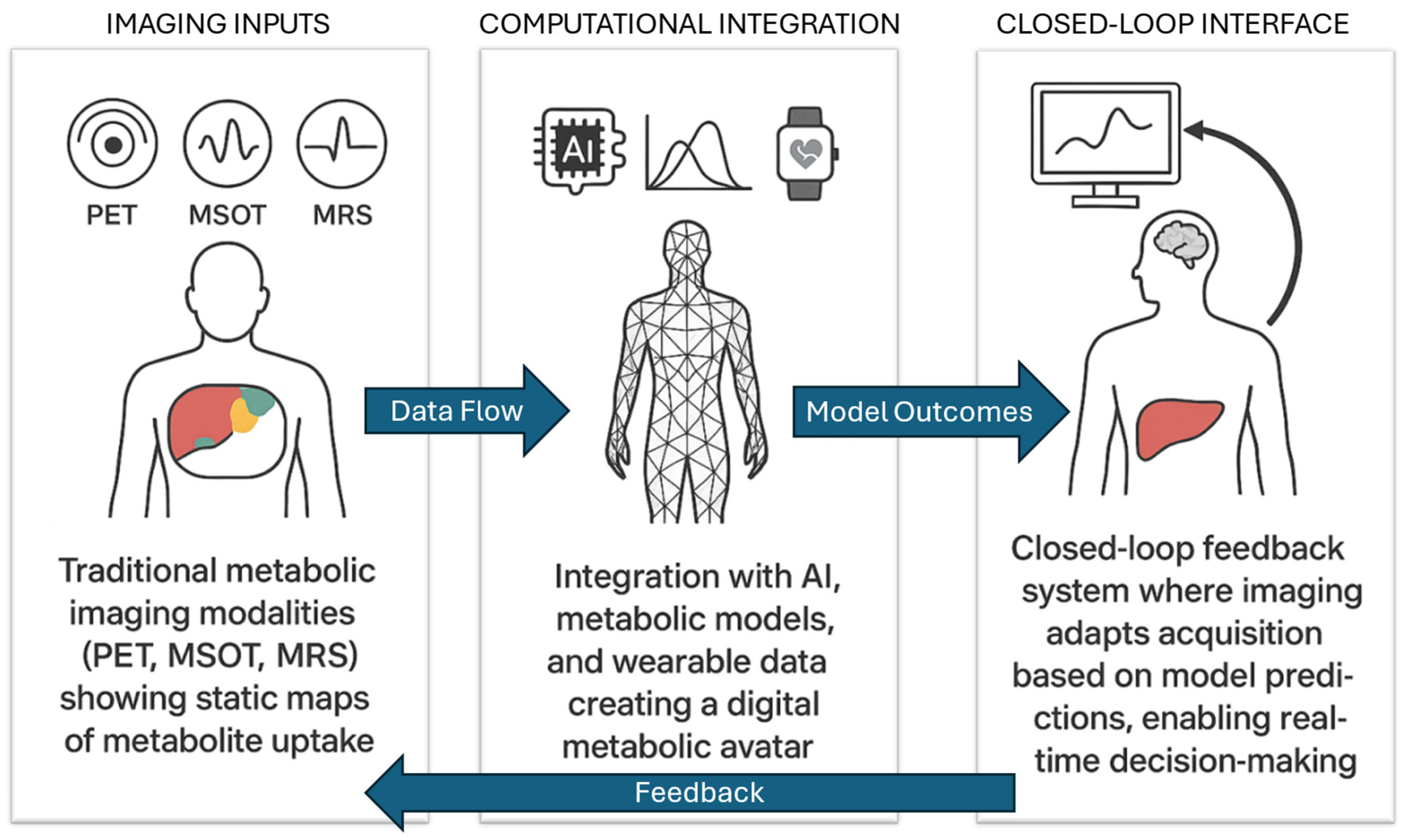
Figure 1. From static imaging to dynamic metabolic intelligence. Traditional imaging modalities (PET, MSOT, MRS) provide static maps of metabolite uptake (left). These data streams can be integrated with artificial intelligence, physics-based metabolic models, and wearable sensors to generate a digital metabolic avatar (center). Model outcomes then inform a closed-loop interface (right), where imaging parameters adapt in real time based on predictions and feedback, enabling personalized monitoring and decision-making. Directional arrows highlight the flow of information: ‘Data Flow’ from imaging to models, ‘Model Outcomes’ from computational integration to adaptive imaging, and ‘Feedback’ from model-guided interventions back to imaging acquisition.
Biophysica 05 00042 g001
Table 1. Advantages and limitations of major metabolic imaging modalities.
ModalityAdvantagesLimitations
PETHigh sensitivity; established clinical use; quantitative tracer uptakeRadiation exposure; high cost; relatively low spatial resolution; limited suitability for repeated longitudinal studies
MRSNon-invasive; direct metabolite quantification (e.g., lactate, creatine)Poor spatial localization; low sensitivity; long acquisition times
MSOTReal-time imaging; combines optical contrast with ultrasound resolutionLimited penetration depth (~2–3 cm); best suited for superficial tissues or preclinical models
Fluorescence imagingHigh specificity; subcellular resolution; sensitive metabolic readoutsRequires exogenous probes; limited deep tissue applicability; photobleaching and invasiveness
Hyperpolarized MRIDynamic tracking of metabolic fluxes (e.g., pyruvate, lactate)Short polarization lifetime; technically complex; expensive; limited availability
Table 2. Modular architecture of a dynamic metabolic intelligence platform.
ModuleComponentsFunction
Data SourcesImaging (PET, MSOT, MRS, hyperpolarized MRI, fluorescence imaging); wearables (HR, steps, glucose, sleep); clinical recordsProvide multimodal, real-time, and contextual inputs reflecting the metabolic state
Computational LayersPhysics-based models (ODEs, GEMs, constraint-based); machine learning (deep learning, physics-informed ML); hybrid avatarsSimulate, predict, and reconcile metabolic trajectories using complementary mechanistic and data-driven methods
InterfacesImaging–model feedback; model-informed acquisition (adaptive scanning, timing windows); clinical dashboardsEnable bidirectional flow: data feeding into models, and models guiding imaging parameters and interventions
Table 3. Foreseeable challenges and mitigation strategies for dynamic metabolic intelligence platforms.
CategoryChallengesMitigation Strategies
TechnologicalLack of standardization across imaging modalities and acquisition protocolsAdoption of open standards; development of harmonized acquisition protocols; federated multi-center initiatives
WorkflowDifficulty integrating real-time streaming data into clinical workflowsModular software design; use of interoperability frameworks (e.g., FHIR); embedding within clinical dashboards
RegulatoryComputational opacity of deep learning models; difficulty in regulatory approvalPrioritize physics-informed and explainable AI; ensure traceability and uncertainty quantification
Ethical and PrivacyData security and privacy risks when linking wearables and EHRs; patient consent managementPrivacy-preserving federated learning; strong encryption; GDPR/HIPAA compliance; transparent informed consent
Disclaimer/Publisher’s Note: The statements, opinions and data contained in all publications are solely those of the individual author(s) and contributor(s) and not of MDPI and/or the editor(s). MDPI and/or the editor(s) disclaim responsibility for any injury to people or property resulting from any ideas, methods, instructions or products referred to in the content.