Article

Identification and Expression Analysis of Na+/K+-ATPase and NKA-Interacting Protein in Ark Shells

1
National Engineering Research Center for Marine Aquaculture, Zhejiang Ocean University, Zhoushan 316022, China
2
Zhejiang Marine Ecology and Environment Monitoring Center, Zhoushan 316021, China
*
Author to whom correspondence should be addressed.
Biophysica 2025, 5(2), 22; https://doi.org/10.3390/biophysica5020022
Submission received: 29 April 2025 / Revised: 3 June 2025 / Accepted: 10 June 2025 / Published: 11 June 2025

Abstract

:
Ark shells are a group of bivalves that exhibit extraordinary adaptability to the dual environmental pressures of low oxygen and osmotic imbalance. These challenges are particularly pronounced in intertidal zones, where organisms are subjected to rapid and drastic changes in their surroundings. This research investigated the molecular mechanisms that underpin their survival and adaptive strategies, with particular focused on sodium–potassium ATPase (NKA), a pivotal enzyme responsible for maintaining cellular ion transmembrane gradients and ensuring cellular homeostasis under stress conditions. By utilizing genome assemblies and transcriptomics datasets from multiple ark shell species, we successfully identified two distinct NKA-α subunits and two NKA-β subunits, which are essential components of the NKA complex. Moreover, the discovery of a conserved NKA-interacting protein (NKAIN) highlights the complexity and evolutionary significance of the NKA-NKAIN system in ark shells. Phylogenetic analysis revealed a high degree of conservation in the NKA-α and NKA-β subunits across ark shells, suggesting strong selective pressures to preserve their functionality. However, the marked divergence observed between the two NKA-β subunits suggests that they may serve distinct roles in ion transport, potentially specialized for specific environmental conditions or stress responses. Comparative transcriptomic analysis further revealed the regulatory roles of NKA and NKAIN in the adaptive responses to hypoxia and osmotic stress, showing that these genes are dynamically modulated at the transcriptional level in response to environmental challenges. These findings provide a molecular foundation for understanding the osmotic adaptation mechanisms in ark shells and offer novel insights into their ability to thrive in mudflat habitats. This comprehensive exploration of the NKA-NKAIN system not only enhances our understanding of the resilience of ark shells but also provides valuable insights into the molecular and physiological strategies employed by bivalves in intertidal environments.

1. Introduction

Ark shells (family Arcidae Lamarck, 1809) represent one of the most ancient lineages of bivalve mollusks, with their evolutionary origins tracing back to the Lower Ordovician period, approximately 450 million years ago [1]. The Arcidae family encompasses approximately 260 species across 31 genera, underscoring their extensive evolutionary history and adaptability to diverse marine environments [2]. As quintessential filter feeders, ark shells play a pivotal role in maintaining marine ecosystem health by cleansing seawater, mitigating eutrophication, and contributing to the stability of marine habitats [3]. Ark shells, which are bivalve mollusks inhabiting mudflats, are particularly sensitive to fluctuations in water oxygen levels and dryness stress. Despite this sensitivity, they exhibit remarkable tolerance to low-oxygen conditions [3,4].
Like many marine invertebrates, ark shells possess an open circulatory system in which hemolymph and interstitial fluid are not distinctly separated. This anatomical feature renders their blood osmotic pressure highly sensitive to environmental changes, especially when they are exposed to air [5]. Ions, particularly sodium (Na⁺) and potassium (K⁺), are the primary contributors to osmotic pressure in both hemolymph and seawater. Transcellular ion transport has emerged as the principal mechanism enabling these organisms to respond rapidly to osmotic challenges [6]. This ion movement across cellular membranes is mediated by active transport processes, which are often facilitated by a variety of transporter proteins. Among these, sodium–potassium ATPase (Na⁺/K⁺-ATPase, or NKA) stands out as a key player. The NKA pump expends one ATP (Adenosine triphosphate) molecule to extrude three sodium ions (Na⁺) from the cell while importing two potassium ions (K⁺), thereby establishing a crucial ion concentration gradient essential for maintaining cellular homeostasis [7]. Remarkably, over half of the energy produced in biological systems is utilized by the NKA pump, highlighting its critical role in regulating osmotic pressure equilibrium [8]. However, ATP synthesis is highly dependent on oxygen availability, which implies that the operation of NKA is intricately tied to an organism’s ability to adapt and regulate its internal environment in response to variations in air exposure and osmotic stress [9]. This relationship underscores the importance of NKA in both normal physiological functions and stress responses.
The NKA complex is composed of α and β subunits, each with distinct roles in ion transport [10]. The α subunit serves as the central catalytic component of the pump, featuring dedicated binding sites for Na⁺ and K⁺ ions, as well as housing the ATP hydrolysis catalytic domain. In contrast, the β subunit functions primarily as a regulatory element, modulating the conformational dynamics of the α subunit, modulating the conformational dynamics of the α subunit [11]. Genomic studies have revealed that different organisms encode distinct types of α and β subunits, with notable differences in their tissue-specific expression patterns. These variations give rise to a multitude of unique α–β subunit combinations, which are likely tailored to meet the specific physiological demands of different tissues [12,13]. Furthermore, the functionality of the NKA β subunit is intricately regulated through interactions with NKA-interacting proteins (NKAINs) [14]. Research has shown that the transmembrane domains of NKAIN molecules can potentially form channel-like structures or interact with other membrane proteins, thereby playing essential roles in cellular signaling and functional regulation [14]. These findings suggest that NKAINs not only fine-tune the performance of the NKA pump but also participate in broader cellular signaling and functional regulation networks at the cell membrane level. Investigating the roles of these proteins in osmotic regulation and air exposure stress responses could provide valuable insights into the molecular basis of adaptation in ark shells and other marine organisms. Such knowledge could also inform strategies for enhancing their resilience in changing environmental conditions, making it a promising area for future research in both ecological and applied contexts.
To date, research focusing on the NKA in mollusks has been relatively limited. A study on the Pacific abalone revealed that under low-salinity stress conditions, both the expression levels and enzymatic activities of NKA subunits significantly decline [5]. Furthermore, it was demonstrated that the regulation of their expression is mediated by the cAMP signaling pathway. Building on these findings, investigations into oysters have utilized genomic assemblies and transcriptomics data from multiple species to identify one NKA-α subunit and two distinct NKA-β subunits [15]. These findings provide valuable insights into the structural composition and osmotic regulation mechanisms of the oyster NKA-NKAIN system, advancing our understanding of the comprehensive osmotic adaptation strategies employed by oysters under varying environmental conditions. However, the subunit composition profile of NKA in ark shells, as well as its specific role in osmotic and air exposure pressure regulation, remains largely unexplored. In the present study, we aimed to address this knowledge gap by conducting a thorough analysis of the NKA complex in ark shells. Leveraging high-quality genomic assembly data, we identified the fundamental composition of NKA in this species, providing a foundation for understanding its molecular architecture. Additionally, we performed a comprehensive transcriptomic analysis to investigate the presence of NKA variants and their expression patterns under different stress conditions. Our findings reveal distinct isoforms of NKA subunits in ark shells, suggesting a complex regulatory network that may contribute to their remarkable adaptability to environmental challenges.

2. Materials and Methods

2.1. Materials

To investigate the NKA complex and NKAIN in ark shells, genome assemblies from four distinct species across three genera were collected (Table 1). This comparative genomics approach not only captures evolutionary variations but also facilitates a comprehensive understanding of NKA’s roles in different species. Additionally, transcriptomic datasets for two ark shell species (T. granosa and A. broughtonii) were obtained for de novo assembly, complementing the identified NKA subunits and NKAIN from genome assemblies (Table 2). Additionally, transcriptomic datasets derived from various tissue and samples subjected to hypoxic conditions of T. granosa were collected to investigate the tissue-specific expression profiles and stress responses of the NKA subunits and its related protein NKAIN by calculating the fragments per kilobase million (FPKM) value of the reads (Table 2). This multi-faceted approach ensures a thorough investigation of the genetic, functional, and regulatory aspects of NKA and NKAIN in ark shells.

2.2. Methods

2.2.1. Identification of the NKA Subunits in Ark Shells

To date, the subunits of NKA in Pacific abalone have been among the rarest well-studied species in mollusks [5]. In this study, the published abalone NKA-α and -β subunit sequences (MG767304 and MG767305) were utilized as query sequences for conducting a de novo genome-wide identification of the NKA subunits in different ark shells. This was achieved using the local BLAST (v2.15.0) software with default parameters [16]. Currently, there is limited research on NKAIN in mollusks, with the mouse NKAIN sequence (ABN51166.1) representing the most original and comprehensive report on this homologous gene. Consequently, the mouse NKAIN sequence was employed to identify its corresponding orthologs in ark shells. To complement this approach, transcriptomic datasets were subjected to de novo assembly using the Trinity (v2.12) software, enabling the reconstruction of full-length transcripts from the short-read sequencing data [17]. Upon successful assembly, the de novo assembled transcripts were meticulously screened to retrieve the possible alternative splicing coding sequences of each NKA subunit and NKAIN in ark shells. This step was critical for identifying any differential isoforms that could be generated through the alternative splicing process, which may contribute to the functional diversity and adaptive capabilities of the NKA enzyme in ark shells, particularly in response to environmental changes.

2.2.2. Phylogenetic Analysis of the NKA Subunits in Ark Shells

The NKA-α and -β subunits, along with the NKAIN open reading frames (ORFs), were initially aligned using the L-INS-i algorithm in MAFFT (v7.520) software with the default parameters [18]. The alignment results for each orthologs were visually represented using the DNAMAN X (v10.3.516) software. To further analyze the evolutionary conservation among these orthologs, the identity matrices were computed based on the alignment results and graphically displayed in the form of heatmaps via the heatmap plugin in the TBtools toolkit (v2.080) [19]. A maximum likelihood (ML) phylogenetic tree was constructed using the “One Step Build a ML Tree” program in TBtools with ultrafast bootstrap mode. The best-fitting model was automatically detected, and a total of 5000 bootstrap replicates were computed. The generated phylogenetic tree was visualized and progressed with Figtree software (v1.4.5, https://github.com/rambaut/figtree/releases/tag/v1.4.5pre, accessed on 25 February 2025). Additionally, the Ka/Ks ratio was calculated using the KaKs Calculator plugin within TBtools to evaluate the evolutionary selection pressures acting on these orthologs. This analysis provided valuable insights into their functional conservation and adaptive evolution across different oyster species, shedding light on the potential roles of these subunits in oyster biology and environmental adaptation.

2.2.3. Gene Structure and Alternative Splicing Analysis

After the identification of the open reading frame (ORF) sequences of each NKA-related orthologs in ark shells, the splice junctions were meticulously determined using the GMAP (Version 17 December 2021) alignment tool [20]. The ORF sequences were used as queries and aligned against the reference genome with the default parameters. The splice junction information was then used to systematically visualize the exon–intron structures of each ortholog using the Gene Structure Display Server (GSDS) 2.0 web-based platform (http://gsds.gao-lab.org/, accessed on 5 March 2025) [21].

2.2.4. Synteny Analysis of the NKA Subunits in Ark Shells

Initially, the genomic distribution of the identified orthologs was systematically mapped and visually represented based on the available annotation information. To investigate the collinearity relationships among these orthologs across different ark shells, conserved syntenic blocks were analyzed using the JCVI comparative genomics pipeline (v1.1.15), which relies on encoding sequences and gene location information [22]. Furthermore, to examine the micro-synteny relationships of individual orthologs in greater detail, multiple collinear regions were analyzed and illustrated using the “Find Gene Block Evolutionary Path by Gene Pairs” plugin within the TBtools software, based on the collinearities derived from the JCVI analysis.

2.2.5. Protein Structure Analysis

Using the amino acid sequences of the identified NKA subunits, structural predictions were conducted using the trRosetta online server (http://yanglab.qd.sdu.edu.cn/trRosetta/, accessed on 10 March 2025), which employs deep learning algorithms to predict tertiary structures with high accuracy [23]. The predicted models were subsequently rendered and analyzed using PyMol software (v3.0.0), allowing for a detailed visualization of the three-dimensional architecture of these subunits [24].

2.2.6. Expression Analysis

To assess the expression levels of the identified NKA-related genes, transcriptomic sequencing reads were aligned to their respective genome databases using the Hisat2 aligner software (v2.2.1) [25]. The number of reads mapping to each gene region was quantified, and the matched reads were processed to calculate FPKM (fragments per kilobase of transcript per million mapped reads) values. This normalization procedure was executed using the StringTie software (v2.2.0), ensuring a standardized measure of gene expression that accounts for both gene length and sequencing depth [26]. Statistical significance of differentially expressed genes (DEGs) was analyzed using the DEseq methods based on a negative binomial distribution (p < 0.05).

3. Results

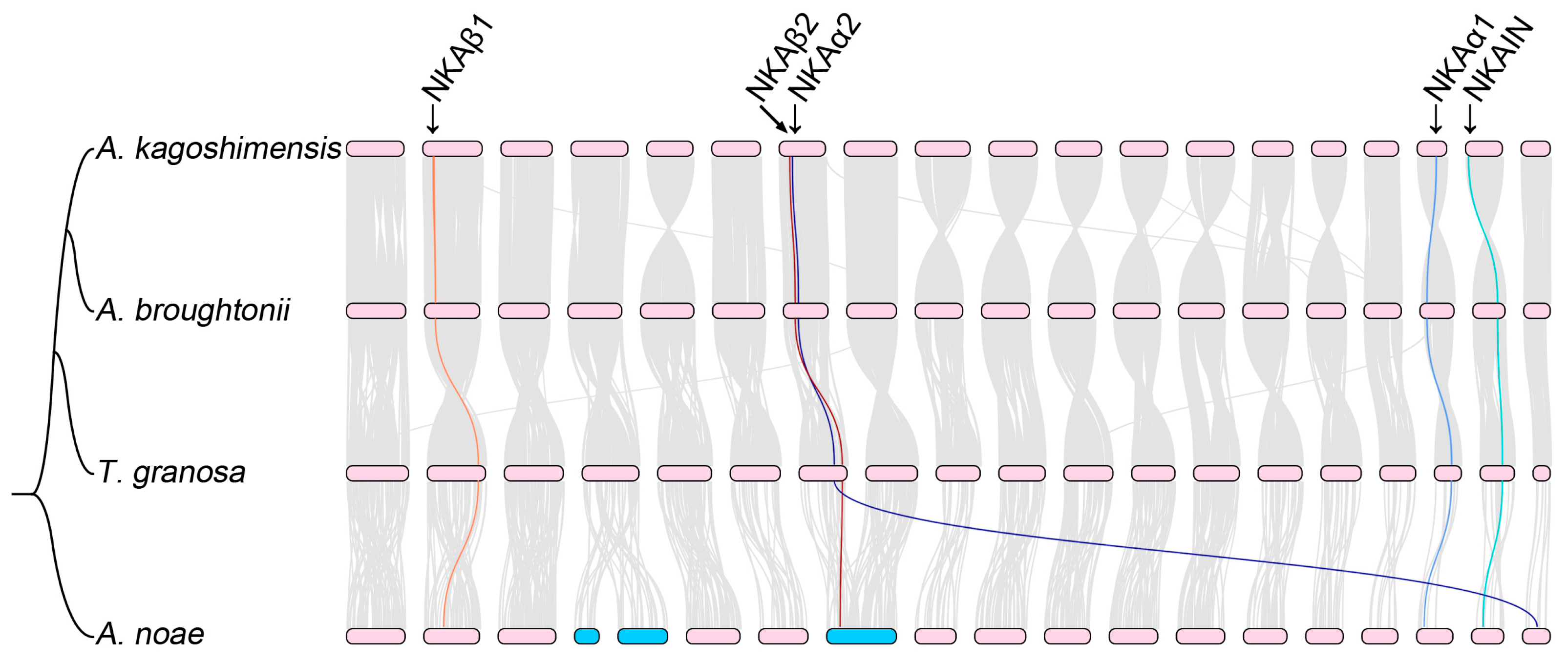
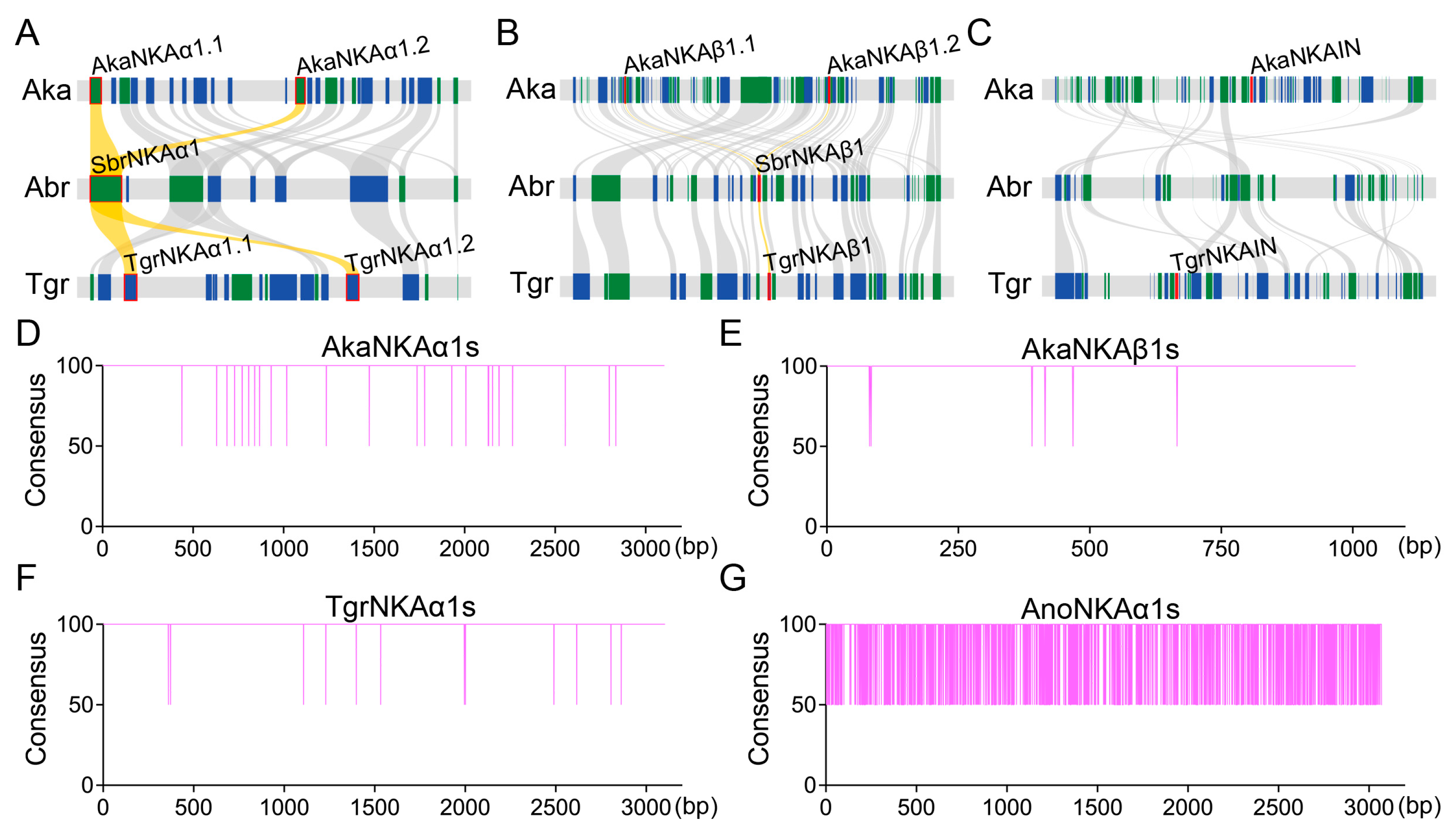
3.1. Conservation of Chromosomal Synteny in Ark Shells

We conducted a comparative analysis of four chromosome-level genome assemblies of ark shells and observed a broad-scale conservation in chromosomal synteny across their genomes. Notably, A. kagoshimensis, A. broughtonii, and T. granosa presented high levels of synteny, with minimal intrachromosomal rearrangements throughout their entire genomes (Figure 1). This pattern suggests a relatively stable genomic structure in these species, likely reflecting their close evolutionary relationships. In contrast, when A. noae was compared with the other three species, two distinct chromosome fusion/fission events were identified (Figure 1). These findings imply that A. noae has undergone a more complex evolutionary history, diverging from the last common ancestor of the other three species at an earlier stage within the ark shell lineage. Such ancient divergence events could have played a significant role in shaping the distinct genomic architecture observed in A. noae.

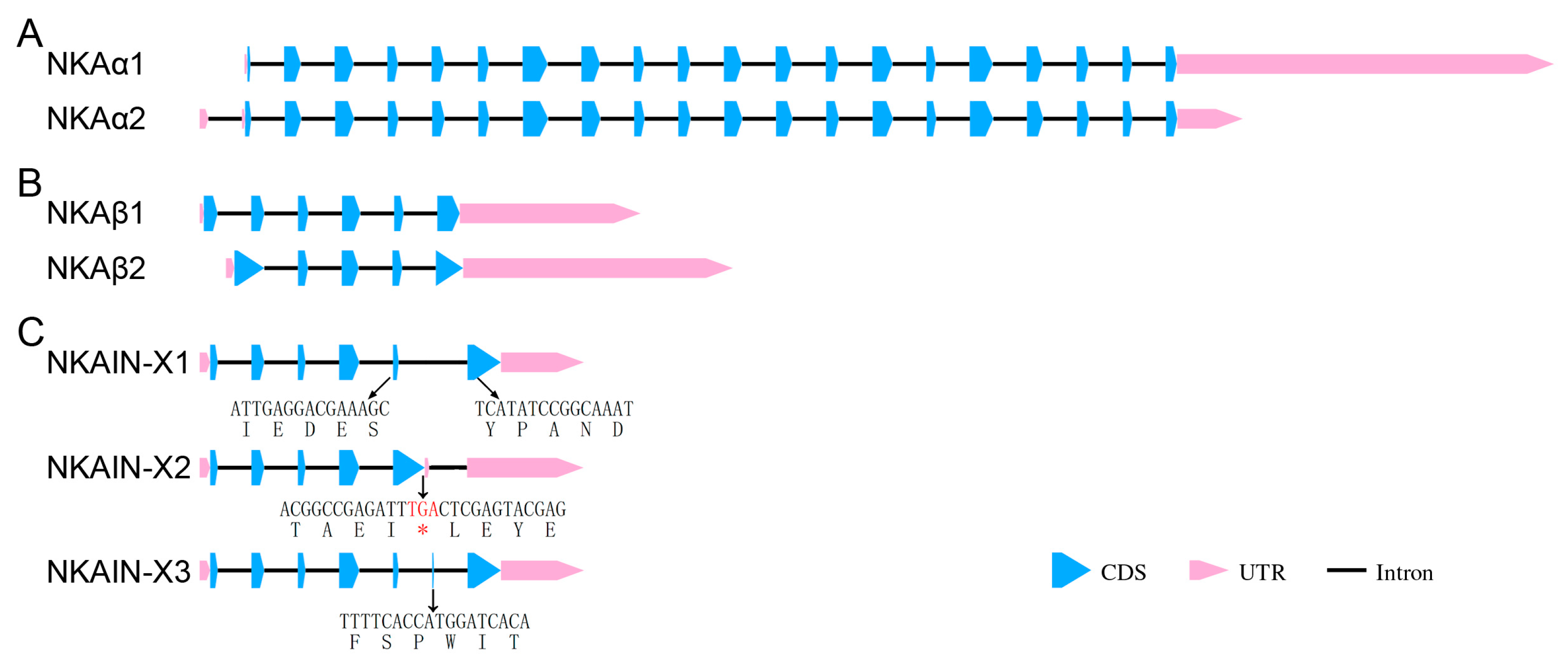
3.2. Identification of the NKA Subunits and NKAIN

To investigate the evolution and genomic organization of the NKA complex in ark shells, we performed genome-wide searches using selected genome assemblies. Our analysis revealed the presence of two NKA-α subunits (designated NKAα1 and NKAα2) and two distinct NKA-β subunits (named NKAβ1 and NKAβ2) in ark shells. Notably, the NKAα1 subunit exhibited independent duplication events within the genomes of A. noae, A. kagoshimensis, and T. granosa (Figure 2A–C). Additionally, the NKAβ1 subunit underwent a specific duplication event in A. kagoshimensis (Figure 2C). Comparative genomic analyses revealed that these duplications arose from independent large-scale intrachromosomal segmental duplication events (Figure 3A,B). Notably, the independently duplicated paralogs of NKAα1 and NKAβ1 displayed significant sequence identity conservation, suggesting that these duplications were preserved under strong evolutionary constraints (Figure 3D–F). However, in A. noae, the duplicated NKAα1 subunits displayed low sequence conservation, suggesting that they may have undergone subsequent functional differentiation (Figure 3G). In addition to the NKA subunits, we identified a single ortholog of NKAIN in most ark shells (Figure 2). Notably, the NKAIN ortholog was absent in A. broughtonii. Our comparative genomic analysis suggested that large-scale intrachromosomal rearrangements are the likely the cause of the absence of this ortholog (Figure 3C). These findings provide insights into the dynamic nature of genome evolution in ark shells and highlight potential differences in the functional roles of NKAIN across species.
To investigate potential alternative splicing (AS) events in the diversification and functional regulation of NKA subunits and NKAIN in ark shells, we performed de novo transcriptome assembly. Our analysis revealed that both the NKA-α and NKA-β subunits exhibited only a single splice form, indicating a lack of AS in these genes (Figure 4A,B). For the NKA-α subunits, the duplicated paralogs (NKAα1 and NKAα2) displayed a highly conserved exon–intron structure, which suggests strong purifying selection or functional constraints maintaining their structural integrity (Figure 4A). In contrast, the two NKA-β subunits (NKAβ1 and NKAβ2) demonstrated significant differences in their exon–intron compositions, which may be indicative of distinct evolutionary origins or functional divergence (Figure 4B). For NKAIN, three splice variants were identified through transcriptome assemblies (Figure 4C). All three variants were highly conserved in the first four exons at the 5′ end and the last exon at the 3′ end. Notably, the primary differences among the variants were concentrated in the last two exons (Figure 4C). Compared to the other variants, the NKAIN-X2 variant presented AS in the fifth exon, which extended the coding sequence. However, the presence of an in-frame stop codon within this exon resulted in a truncated protein product, which could impact its functionality (Figure 4C). In contrast, the NKAIN-X3 variant exhibited an 18 bp exon insertion, further altering the protein sequence and suggesting additional regulatory or functional complexity (Figure 4C). These findings underscore the importance of AS in shaping the diversity and functional regulation of NKAIN in ark shells. The lack of AS in the NKA-α and NKA-β subunits, combined with their conserved exon–intron structures, highlights the strong evolutionary and functional constraints acting on these genes.

3.3. Phylogenetic Analysis of the NKA Subunits and NKAIN

Multiple sequence alignments of the deduced protein sequences for the NKA subunits across diverse ark shells revealed a high degree of sequence conservation within orthologs of the NKA-α and NKA-β subunits (Figure 5). Interestingly, the orthologs identified in A. noae presented significantly reduced sequence conservation compared with those in other species, suggesting a unique evolutionary trajectory for this species (p < 0.05). Conversely, the orthologs shared between A. kagoshimensis and A. broughtonii presented a notably high level of conservation, with sequence identities exceeding 95%, which underscores a particularly close evolutionary relationship between these two species (p < 0.05). These findings highlight the complex evolutionary dynamics and varying patterns of sequence preservation among NKA subunits in these species.
The two NKA-α subunits present in ark shells share a similar gene structure, but their encoding sequences are significantly less conserved than their independent orthologs (Figure 5A). This divergence suggests that these subunits may have experienced distinct evolutionary pressures or selective forces. Analysis of the Ka/Ks ratio revealed that the NKAα1 subunit in ark shells has undergone strong purifying selection, suggesting that this ortholog has evolved under stringent functional constraints to preserve its original roles (Figure 5B). Furthermore, previous studies have demonstrated that oysters possess only one NKA-α subunit, which closely clusters with the NKAα1 subunit found in ark shells (Figure 5C). Collectively, these findings suggest that the two NKA-α subunits in ark shells have likely evolved independently, reflecting distinct evolutionary trajectories.
It should be noted that the two NKA-β subunits in ark shells exhibit considerable divergence from one another, which is consistent with the results of the gene structure analysis (Figure 4B). Similar phenomena were also identified in oysters, further supporting the notion of independent evolutionary origins for these two β subunit orthologs within bivalves. However, despite this divergence, the sequence of the NKAβ2 subunit was more conserved than that of the NKAβ1 subunits (65.64% vs. 50.97%, p < 0.05; Figure 5D). Calculations of the Ka/Ks ratio further provided additional evidence that both NKA-β subunits in ark shells have experienced strong purifying selection (Figure 5E). Phylogenetic analysis further revealed that the two NKA-β subunits formed distinct clusters with those in oysters (Figure 5F). The independent evolution of NKA-β subunits underscores the complexity of NKA subunit evolution and suggests that different subunits may have evolved to adapt to specific environmental pressures or functional requirements. These findings highlight the importance of these NKA subunits in maintaining physiological homeostasis and facilitating the survival of ark shells in their respective environments.

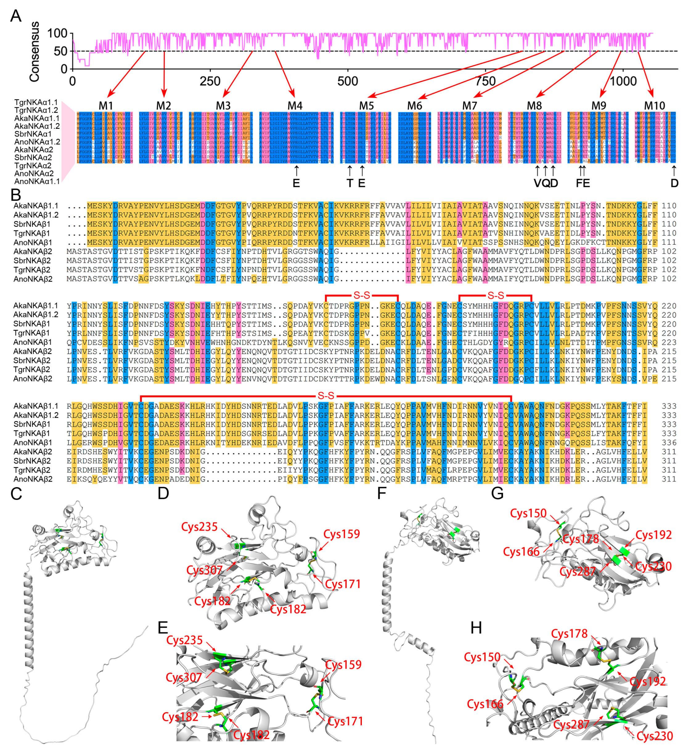
3.4. Structural Analysis of the NKA-α and NKA-β Subunits

Despite the overall low sequence identity observed between the two NKA-α subunits, their transmembrane regions displayed a significantly greater degree of sequence conservation (Figure 6A). Multiple sequence alignments further revealed that the cation-binding sites were remarkably conserved between the two NKA-α subunits, emphasizing their functional importance in ion transport and ATPase activity. These findings suggest that while other regions of the protein may have undergone more rapid evolution or relaxed selective pressures, the transmembrane regions and cation-binding sites have been preserved owing to their essential roles in protein function.
In the case of the NKA-β subunits, despite notable differences in gene structure and lower sequence similarity between the two subunits in ark shells, their protein structures exhibited significant conservation (Figure 6B–H). Both orthologs clearly share a common architectural feature characterized by a single transmembrane domain followed by an extracellular globular domain. This conserved structural motif suggests that both subunits likely play analogous functional roles in their interaction with the NKA complex, despite their evolutionary divergence. Of particular interest, both NKA-β subunits presented three cysteine bridges within their extracellular globular domain (Figure 6C–H). The consistent presence of these cysteine residues across the two subunits strongly highlights their potential importance in stabilizing the protein structure and facilitating interactions with other molecules. Cysteine bridges, also known as disulfide bonds, are essential for maintaining proper protein conformation and functionality. Therefore, the conservation of these cysteine residues in both NKA-β subunit orthologs strongly indicates their functional relevance to the overall activity of the NKA complex in ark shells. These findings emphasize the critical role of disulfide bond formation in ensuring the stability and functionality of the NKA complex, which may be integral to osmoregulation and air exposure adaptation in these organisms. The observed structural conservation despite sequence divergence highlights the importance of maintaining functional integrity while allowing for evolutionary innovation in other regions of the protein.

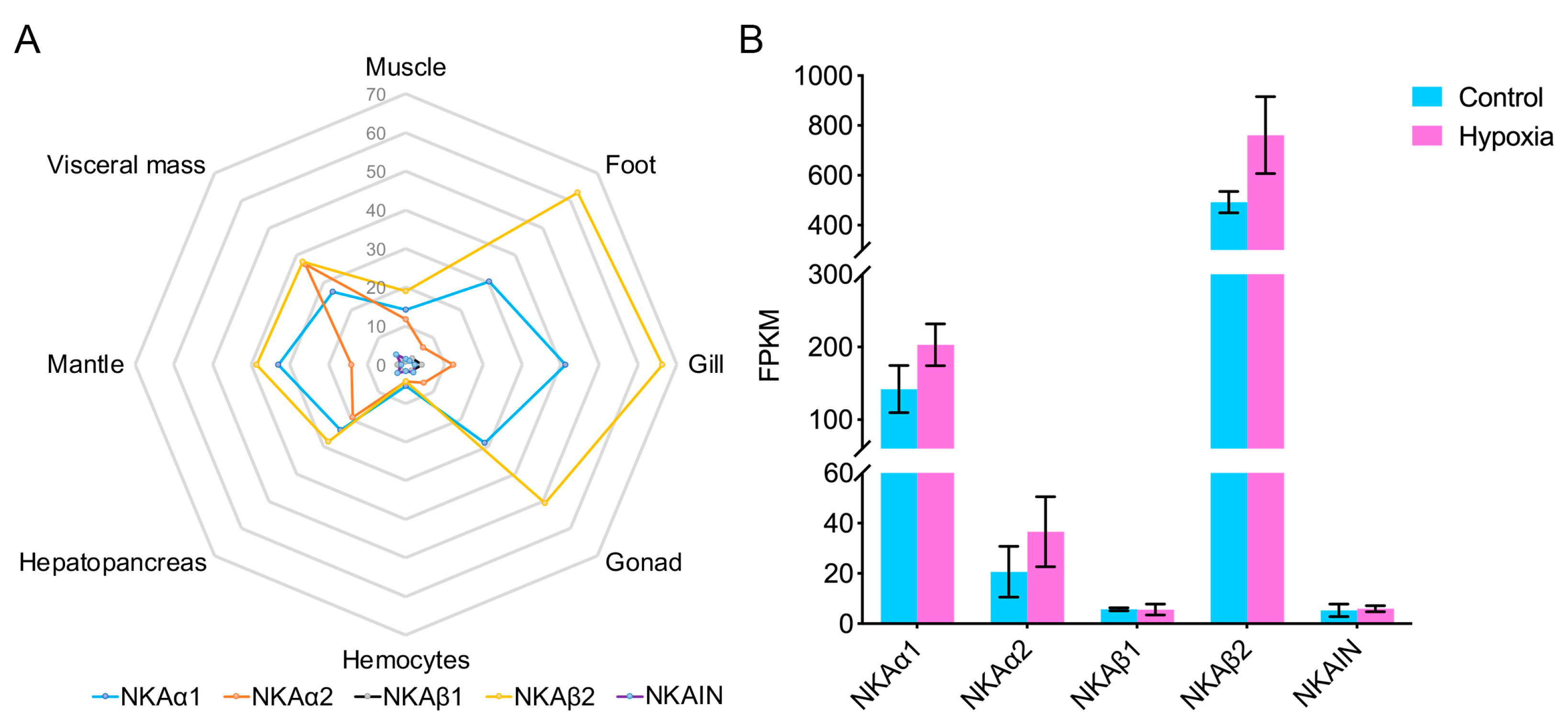
3.5. Expression of the NKA Subunits and NKAIN in Ark Shells

To elucidate the functional roles of the NKA subunits and NKAIN, we conducted transcriptomic analyses using T. granosa as a model organism. Our investigation demonstrated that both the NKA-α and NKA-β subunits in ark shells exhibit conserved expression profiles across various tissues, indicating their essential roles in maintaining cellular ion balance (Figure 7A). However, the expression levels of individual orthologs vary distinctly depending on the tissue type. Specifically, the NKAα1 subunit is generally expressed at higher levels than that of NKAα2, with the exception of the visceral mass. Interestingly, the expression level of NKAβ1 is consistently lower than that of NKAβ2 across different tissues (Figure 7A). These findings suggest that while NKAβ2 may serve as the primary structural subunit contributing to the NKA complex in ark shells, NKAβ1 could potentially play a more regulatory role. Turning to the NKAIN, we observed that it was widely expressed in different tissues, although its expression levels were relatively lower than those of the NKA subunits (Figure 7A). This pattern implies that NKAIN might have a specialized or modulatory function rather than being a core component of the NKA complex.
To further explore the adaptive responses of these genes, we analyzed their expression under hypoxic conditions. The results revealed distinct expression patterns among the NKA subunits under hypoxia. Specifically, both NKA-α subunits exhibited significantly increased expression levels when exposed to hypoxia conditions (p < 0.05, Figure 7B). These findings suggest that the α subunits play critical roles in the transcriptional regulation of NKA activity during oxygen deprivation. In contrast, the NKA-β subunits displayed differential responses. Among them, NKAβ2, which is proposed to function as the structural subunit of the NKA complex, showed notable upregulation in expression under hypoxic conditions. However, the other β subunit, which is potentially involved in regulatory roles, was maintained at a low expression level (Figure 7B). Parallel to the NKA subunits, the expression levels of NKAIN were also assessed under hypoxic conditions. Despite its proposed regulatory function in the NKA complex, no significant changes in expression were observed (Figure 7B). These findings suggest that the regulatory function of NKAIN may not be directly involved in the immediate transcriptional response to hypoxic stress, emphasizing the specificity of the roles of the NKA subunit in hypoxia adaptation mechanisms. Together, these results highlight the importance of the NKA complex in enabling ark shells to cope with environmental challenges such as low-oxygen conditions and suggest that NKAIN may contribute to more nuanced or tissue-specific regulatory processes.

4. Discussion

The NKA complex functions as a crucial ion channel responsible for actively transporting Na+ and K+ ions across cell membranes, thereby maintaining the balance of intracellular osmotic pressure [27]. It operates as a heterodimeric P-type 2C ATPase consisting of a catalytic α subunit and an auxiliary β subunit. The β subunit is essential for proper plasma membrane localization of the enzyme. In numerous animal species, multiple NKA isoforms exist, assembling into distinct isozymes that exhibit subtly different kinetic properties to fulfill the diverse ion transport demands of various cell types [13]. In a previous study, oysters, a group of bivalves that cluster as a sister group to ark shells, were found to possess only a single NKA-α subunit, significantly limiting their repertoire of NKA isozymes. However, in the present study, two NKA-α subunits and two NKA-β subunits were identified in ark shells. In this context, the repertoire of NKA isozymes in ark shells appears to be significantly diversified compared with that in oysters. These findings suggest that ark shells may have evolved a more sophisticated mechanism to regulate their ion homeostasis, potentially relying on other factors, such as posttranslational modifications or interacting partners such as NKAIN, to achieve the functional specificity and flexibility required for their physiological processes.
The NKA-β subunit, despite lacking direct ion-binding sites and catalytic activity, plays a crucial regulatory role in controlling the overall enzymatic function of the NKA complex [28]. Similar to those in oysters, we identified two distinct types of NKA-β subunits in ark shells. As expected, these two orthologs were highly conserved compared with those in oysters and exhibited marked differences in their gene structure and sequence similarity, suggesting significant functional divergence between them (Figure 5D–F). These data suggest that the duplication of the NKA-β subunits has anciently occurred early on bivalve evolution, providing the necessary diversity for ion transport mechanisms. This evidence supports the hypothesis that the duplication and subsequent expansion of the two NKA-β subunits predates speciation events that led to the modern diversity of bivalves. Moreover, this duplication may have allowed each NKA-β subunit to specialize in different functions, potentially through interactions with the α subunit or other proteins, or via posttranslational modifications [29]. The functional divergence between the two β subunits suggests that they contribute to the regulation of ion transport in different contexts or tissues, enhancing an organism’s ability to adapt to varying environmental conditions, such as salinity levels. The conservation of these two subunits throughout oyster and ark shell evolution points to their fundamental importance in the regulation and adaptation of the NKA complex in these marine bivalves. To fully understand the significance of these β subunits, further research into their specific functions, interactions, and the evolutionary pressures that lead to their conservation is needed. This deeper understanding could provide insights into the adaptation strategies of marine organisms and the mechanisms underlying ion regulation in ark shells.
When aquatic organisms leave the water environment and are directly exposed to the air, the oxygen concentration significantly decreases, causing severe hypoxic stress in the cells. Under hypoxic conditions, cellular energy metabolism is blocked, ATP production is reduced, and the function of ion pumps (such as NKA) is affected [30]. Despite extensive research on the role of NKA in air exposure regulation, much of this work has focused on teleost fish. Adaptation to lowering oxygen levels (hypoxia) requires coordinated downregulation of metabolic demand and supply to prevent mismatches in ATP utilization and production that might culminate in a bioenergetic collapse [31]. Hypoxia diminishes ATP utilization by downregulating protein translation and the activity of the NKA. Hypoxia diminishes ATP production in part by lowering the activity of the electron transport chain through activation of the transcription factor hypoxia-inducible factor-1 [4]. Previous research has suggested that a pattern of ionic gradients driven by NKA and channels allow, upon air exposure, the reversal of the direction of the brain/gut axis to the gut/brain axis upon recovery [30]. Moreover, air exposure decreases the degree of environmental osmic and typically stimulates an increase in NKA activity in the gills of teleost fish [32]. Transcript-level analyses revealed a more complex response: while high salinity decreases the expression of the NKA-α1a isoform, it increases the expression of the NKA-α1b isoform; interestingly, the remaining three NKA-α isoforms remain unchanged under salinity stress [32]. A comparison of these findings with those in ark shells revealed that both the NKA-α subunits and NKA-β subunits were upregulated under hypoxia conditions, which contrasts with the findings in teleost fish. These findings suggest that ark shells may employ distinct regulatory mechanisms to cope with salinity fluctuations, potentially involving other subunits or accessory proteins, such as the NKA complex subunits discussed earlier.
Indeed, research on the β subunit of NKA has been relatively rare compared with that on the α subunit because of its non-catalytic nature. In the present study, we found that the duplicated NKA-β subunits in ark shells were sensitive to hypoxia conditions. As previously mentioned, we categorized the two NKA-β subunits into a structural subunit and a regulatory subunit on the basis of their expression patterns. However, the structural subunit displayed a marked sensitivity to hypoxia adaptation compared with its regulatory counterpart. This differential response to hypoxia conditions shifts between the two NKA-β subunits in ark shells, opening new avenues for investigating the nuanced roles they play in hypoxia conditions and how these distinct subunits may contribute to the overall stability and efficiency of the NKA complex under varying environmental conditions. The finding that the structural subunit responds more acutely to hypoxia challenges could indicate its critical role in modulating the activity of the NKA complex [31]. To fully understand the significance of these β subunits, further research into their specific functions, interactions, and the evolutionary pressures that lead to their conservation is needed. This deeper understanding could provide insights into the adaptation strategies of marine organisms and the mechanisms underlying ion regulation in ark shells.

5. Conclusions

In conclusion, the present study has identified two NKA-α subunits and two NKA-β subunits in ark shells. Additionally, a conserved NKAIN with three distinct splice variants that interact with the NKA-β subunit has been discovered in ark shells. By elucidating the structural and functional aspects of the NKA system in ark shells, our work provides a valuable resource for advancing comparative studies on molluskan osmoregulation and for developing strategies to enhance the resilience of these species in the face of environmental stressors. Expression levels of the subunits and their response patterns to hypoxia conditions suggest that the two duplicated NKA-β subunits were classified as having distinct roles, with one acting as a structural subunit and the other as a regulatory subunit. Overall, these novel findings established a substantial foundation for deepening our understanding of the intrinsic distribution patterns and ecological adaptation mechanisms in ark shells, as well as significantly enhance our ability to investigate their responses to osmotic and air exposure stressors.

Author Contributions

Conceptualization, M.S. and Y.J.; software, X.L. and J.Z.; validation, W.L. and J.P.; data curation, M.S. and X.L.; original draft preparation, M.S. and Y.J.; funding acquisition, Y.J. All authors have read and agreed to the published version of the manuscript.

Funding

This research was funded by grants from the Natural Science Foundation of China (32301408), the China Postdoctoral Science Foundation (2023M741837), the Key Laboratory of Mariculture of Ministry of Education, Ocean University of China (KLM202203).

Data Availability Statement

All data analyzed during this study are included in this article.

Conflicts of Interest

The authors declare no conflicts of interest.

Abbreviations

The following abbreviations are used in this manuscript:
NKASodium–potassium ATPase, Na⁺/K⁺-ATPase
NKAINNKA-interacting protein
AkaA. kagoshimensis
AbrA. broughtonii
TgrT. granosa
AnoA. noae

References

  1. Sun, S.E.; Li, Q.; Kong, L.; Yu, H. Evolution of mitochondrial gene arrangements in Arcidae (Bivalvia: Arcida) and their phylogenetic implications. Mol. Phylogeneti. Evol. 2020, 150, 106879. [Google Scholar] [CrossRef] [PubMed]
  2. Oliver, P.G.; Holmes, A.M. The Arcoidea (Mollusca: Bivalvia): A review of the current phenetic-based systematics. Zool. J. Linn. Soc. 2006, 148, 237–251. [Google Scholar] [CrossRef]
  3. Park, H.J.; Lee, W.C.; Choy, E.J.; Kang, C.K. Reproductive cycle and gross biochemical composition of the ark shell Scapharca subcrenata (Lischke, 1869) reared on subtidal mudflats in a temperate bay of Korea. Aquaculture 2011, 322–323, 149–157. [Google Scholar] [CrossRef]
  4. Wheaton, W.W.; Chandel, N.S. Hypoxia. 2. Hypoxia regulates cellular metabolism. Am. J. Physiol.-Cell Physiol. 2011, 300, C385–C393. [Google Scholar] [CrossRef]
  5. Jia, Y.; Liu, X. Expression of Na+/K+-ATPase was affected by salinity change in Pacific abalone Haliotis discus hannai. Front. Physiol. 2018, 9, 1244. [Google Scholar] [CrossRef]
  6. Amende, L.M.; Pierce, S.K. Cellular volume regulation in salinity stressed molluscs: The response of Noetia ponderosa (Arcidae) red blood cells to osmotic variation. J. Comp. Physiol. 1980, 138, 283–289. [Google Scholar] [CrossRef]
  7. Skou, J.C. The identification of the Sodium–Potassium Pump (Nobel Lecture). Angew. Chem. Int. Ed. 1998, 37, 2320–2328. [Google Scholar] [CrossRef]
  8. Clausen, M.V.; Hilbers, F.; Poulsen, H. The structure and function of the Na, K-ATPase isoforms in health and disease. Front. Physiol. 2017, 8, 371. [Google Scholar] [CrossRef] [PubMed]
  9. Semenza, G.L. Life with oxygen. Science 2007, 318, 62–64. [Google Scholar] [CrossRef]
  10. Nyblom, M.; Poulsen, H.; Gourdon, P.; Reinhard, L.; Andersson, M.; Lindahl, E.; Fedosova, N.; Nissen, P. Crystal structure of Na+, K(+)-ATPase in the Na(+)-bound state. Science 2013, 342, 123–127. [Google Scholar] [CrossRef]
  11. Therien, A.G.; Blostein, R. Mechanisms of sodium pump regulation. Am. J. Physiol.-Cell Physiol. 2000, 279, C541–C566. [Google Scholar] [CrossRef] [PubMed]
  12. Malik, N.; Canfield, V.A.; Beckers, M.-C.; Gros, P.; Levenson, R. Identification of the mammalian Na, K-ATPase β3 subunit. J. Biol. Chem. 1996, 271, 22754–22758. [Google Scholar] [CrossRef] [PubMed]
  13. Guynn, S.R.; Scofield, M.A.; Petzel, D.H. Identification of mRNA and protein expression of the Na/K-ATPase α1-, α2- and α3-subunit isoforms in Antarctic and New Zealand nototheniid fishes. J. Exp. Mar. Biol. Ecol. 2002, 273, 15–32. [Google Scholar] [CrossRef]
  14. Gorokhova, S.; Bibert, S.; Geering, K.; Heintz, N. A novel family of transmembrane proteins interacting with beta subunits of the Na, K-ATPase. Hum. Mol. Genet. 2007, 16, 2394–2410. [Google Scholar] [CrossRef]
  15. Zhang, K.; Xue, J.; Li, W.; Gu, C.; Wang, P.; Jia, Y.; Liu, X. Identification and expression analysis of Na+/K+-ATPase and NKA-interacting protein in oyster. Invertebr. Surviv. J. 2024, 21, 99–108. [Google Scholar]
  16. Camacho, C.; Coulouris, G.; Avagyan, V.; Ma, N.; Papadopoulos, J.; Bealer, K.; Madden, T.L. BLAST+: Architecture and applications. BMC Bioinform. 2009, 10, 421. [Google Scholar] [CrossRef]
  17. Grabherr, M.G.; Haas, B.J.; Yassour, M.; Levin, J.Z.; Thompson, D.A.; Amit, I.; Adiconis, X.; Fan, L.; Raychowdhury, R.; Zeng, Q.; et al. Trinity: Reconstructing a full-length transcriptome without a genome from RNA-Seq data. Nat. Biotechnol. 2011, 29, 644–652. [Google Scholar] [CrossRef] [PubMed]
  18. Katoh, K.; Toh, H. Recent developments in the MAFFT multiple sequence alignment program. Brief. Bioinform. 2008, 9, 286–298. [Google Scholar] [CrossRef]
  19. Chen, C.; Wu, Y.; Li, J.; Wang, X.; Zeng, Z.; Xu, J.; Liu, Y.; Feng, J.; Chen, H.; He, Y.; et al. TBtools-II: A “one for all, all for one” bioinformatics platform for biological big-data mining. Mol. Plant 2023, 16, 1733–1742. [Google Scholar] [CrossRef]
  20. Wu, T.D.; Watanabe, C.K. GMAP: A genomic mapping and alignment program for mRNA and EST sequences. Bioinformatics 2005, 21, 1859–1875. [Google Scholar] [CrossRef]
  21. Hu, B.; Jin, J.; Guo, A.-Y.; Zhang, H.; Luo, J.; Gao, G. GSDS 2.0: An upgraded gene features visualization server. Bioinformatics 2014, 31, 1296–1297. [Google Scholar] [CrossRef] [PubMed]
  22. Wang, Y.; Tang, H.; Debarry, J.D.; Tan, X.; Li, J.; Wang, X.; Lee, T.H.; Jin, H.; Marler, B.; Guo, H.; et al. MCScanX: A toolkit for detection and evolutionary analysis of gene synteny and collinearity. Nucleic Acids Res. 2012, 40, e49. [Google Scholar] [CrossRef]
  23. Du, Z.; Su, H.; Wang, W.; Ye, L.; Wei, H.; Peng, Z.; Anishchenko, I.; Baker, D.; Yang, J. The trRosetta server for fast and accurate protein structure prediction. Nat. Protoc. 2021, 16, 5634–5651. [Google Scholar] [CrossRef] [PubMed]
  24. Delano, W.L. The PyMol molecular graphics system. Proteins Struct. Funct. Bioinform. 2002, 30, 442–454. [Google Scholar]
  25. Kim, D.; Paggi, J.M.; Park, C.; Bennett, C.; Salzberg, S.L. Graph-based genome alignment and genotyping with HISAT2 and HISAT-genotype. Nat. Biotechnol. 2019, 37, 907–915. [Google Scholar] [CrossRef]
  26. Kovaka, S.; Zimin, A.V.; Pertea, G.M.; Razaghi, R.; Pertea, M.; Salzberg, S.L. Transcriptome assembly from long-read RNA-seq alignments with StringTie2. Genome Biol. 2019, 20, 278. [Google Scholar] [CrossRef]
  27. Fambrough, D.M.; Lemas, M.V.; Hamrick, M.A.; Emerick, M.A.; Renaud, K.J.; Inman, E.M.; Hwang, B.; Takeyasu, K.U. Analysis of subunit assembly of the Na-K-ATPase. Am. J. Physiol. 1994, 266, C579. [Google Scholar] [CrossRef]
  28. Hasler, U.; Wang, X.; Crambert, G.; Béguin, P.; Jaisser, F.; Horisberger, J.D.; Geering, K. Role of β-subunit domains in the assembly, stable expression, intracellular routing, and functional properties of Na, K-ATPase. J. Biol. Chem. 1998, 273, 30826–30835. [Google Scholar] [CrossRef]
  29. Chakraborti, S.; Rahaman, S.M.; Alam, M.N.; Mandal, A.; Ghosh, B.; Dey, K.; Chakraborti, T. Na+/K+-ATPase: Aperspective in Regulation of Membrane Na+-K+ ATPase; Chakraborti, S., Dhalla, N.S., Eds.; Springer International Publishing: Cham, Switzerland, 2016; pp. 3–30. [Google Scholar]
  30. Pius, J.; Peter, M.C.S. Air exposure promotes subunit isoform expression of Na+/K+-ATPase and vH+-ATPase, and recovery reverses NKCC1 and NCC expressions in the brain/gut axis of zebrafish. Proc. Zool. Soc. 2025, 78, 16–28. [Google Scholar] [CrossRef]
  31. Bogdanova, A.; Petrushanko, I.; Boldyrev, A.; Gassmann, M. Oxygen- and redox-induced regulation of the Na/K ATPase. Curr. Enzym. Inhib. 2006, 2, 37–59. [Google Scholar] [CrossRef]
  32. Richards, J.G.; Semple, J.W.; Bystriansky, J.S.; Schulte, P.M. Na+/K+-ATPase α-isoform switching in gills of rainbow trout (Oncorhynchus mykiss) during salinity transfer. J. Exp. Biol. 2003, 206, 4475–4486. [Google Scholar] [CrossRef] [PubMed]
Figure 1. Comparative genomic analysis of the selected assemblies. The collinearity of gene orthologs is represented by gray lines. The collinearities of the NKA subunits are distinctly highlighted with differently colored lines. Chromosome fusion and fission events in A. noae are marked in light blue.
Biophysica 05 00022 g001
Figure 2. Chromosome distribution of the NKA subunits and NKAIN in different ark shells. The heatmap within the rounded rectangle illustrates the distribution of chromosomes, visualized based on gene density.
Biophysica 05 00022 g002
Figure 3. Independent duplication or absence of the NKA subunits and NKAIN across different ark shells. (A) Independent duplication of the NKAα1 subunit in A. kagoshimensis and T. granosa. (B) Independent duplication of the NKAβ1 subunit in A. kagoshimensis. (C) Absence of the NKAIN ortholog in A. broughtonii. (DG) Conservation of the independently duplicated NKAα1 and NKAβ1 subunits in A. kagoshimensis, T. granosa, and A. noae.
Biophysica 05 00022 g003
Figure 4. Gene structure and organization of the NKA subunits and NKAIN in ark shells. (A) Gene structure of two NKA-α subunits in ark shells. (B) Gene structure of two NKA-β subunits in ark shells. (C) Gene structure of different NKAIN variants in ark shells.
Biophysica 05 00022 g004
Figure 5. Phylogenetic analyses of the NKA subunits and NKAIN in ark shells. (A) Sequence identity of NKA-α subunits. (B) Selective pressures on NKA-α subunits. (C) Phylogenetic relationships of NKA-α subunits. (D) Sequence identity of NKA-β subunits. (E) Selective pressures on NKA-β subunits. (F) Phylogenetic relationships of NKA-β subunits. (G) Sequence identity of NKAIN orthologs. (H) Selective pressures on NKAIN orthologs. (I) Phylogenetic relationships of NKAIN orthologs.
Biophysica 05 00022 g005
Figure 6. Structural analyses of the NKA subunits in ark shells. (A) Transmembrane region and cation-binding sites in NKA-α subunits. (B) Multiple alignments of the two NKA-β subunits in ark shells. (C) Protein structure of the NKAβ1 subunit in ark shells. (D) Enlarged view of the cysteine bridges in the extracellular globular domain of the NKAβ1 subunit. (E) Top view of the cysteine bridges in the extracellular globular domain of the NKAβ1 subunit. (F) Protein structure of the NKAβ2 subunit in ark shells. (G) Enlarged view of the cysteine bridges in the extracellular globular domain of the NKAβ2 subunit. (H) Top view of the cysteine bridges in the extracellular globular domain of the NKAβ2 subunit.
Biophysica 05 00022 g006
Figure 7. Expression analyses of NKA subunits and NKAIN in ark shells. (A) Tissue-specific expression of NKA subunits and NKAIN in ark shells. (B) Expression level of NKA subunits and NKAIN in response to hypoxia in hamolymph.
Biophysica 05 00022 g007
Table 1. Selected genome assemblies for different mussels in this study.
FamilyGenusSpeciesGenomeLengthScaffoldsScaffold N50
ArcidaeAnadaraA. kagoshimensishttps://www.ncbi.nlm.nih.gov/datasets/genome/GCA_021292105.1, accessed on 12 January 20251,115,236,3083660,635,260
A. broughtoniihttp://gigadb.org/dataset/100607, accessed on 12 January 2025884,566,040102644,995,656
TegillarcaT. granosahttps://www.ncbi.nlm.nih.gov/datasets/genome/GCA_029721355.1, accessed on 12 January 2025915,485,25192345,661,362
ArcaA. noaehttps://www.ncbi.nlm.nih.gov/datasets/genome/GCA_964261245.2, accessed on 12 January 20251,495,912,61411884,746,044
Table 2. Selected transcriptomic data for different species in this study.
Run No.SpeciesTissueBytes (bp)Application
SRR491670T. granosaMix1,287,848,281Trinity assemble
SRR10855671A. broughtoniiFoot2,351,898,942
SRR10713949T. granosaGonad3,361,847,283Tissue expression analysis
SRR10713950T. granosaGill3,627,355,286
SRR10713951T. granosaGill2,993,728,725
SRR10713952T. granosaGill3,138,959,394
SRR10713953T. granosaFoot3,176,163,402
SRR10713958T. granosaVisceral Mass2,995,318,000
SRR10713959T. granosaMantle2,725,940,705
SRR10713960T. granosaMuscle3,133,933,436
SRR10713961T. granosaMantle3,908,781,169
SRR10713962T. granosaMantle2,903,531,436
SRR10713963T. granosaHepatopancreas2,498,636,342
SRR10713964T. granosaHepatopancreas3,370,949,865
SRR10713965T. granosaHepatopancreas2,777,483,605
SRR10713966T. granosaHemocytes3,300,438,409
SRR10713967T. granosaHemocytes2,648,750,164
SRR10713968T. granosaHemocytes2,658,545,770
SRR10713969T. granosaGonad2,985,742,068
SRR10713970T. granosaGonad2,535,690,855
SRR10713971T. granosaMuscle3,140,842,716
SRR10713972T. granosaMuscle2,923,365,727
SRR10713954T. granosaFoot3,273,166,707
SRR10713955T. granosaFoot3,746,359,568
SRR10713956T. granosaVisceral Mass3,753,476,460
SRR10713957T. granosaVisceral Mass3,068,244,064
SRR23345051T. granosaHaemolymph (Hypoxia)1,904,248,299Hypoxia treatment analysis
SRR23345052T. granosaHaemolymph (Hypoxia)1,929,010,199
SRR23345053T. granosaHaemolymph (Hypoxia)2,164,158,072
SRR23345054T. granosaHaemolymph (Control)2,003,282,116
SRR23345055T. granosaHaemolymph (Control)1,925,848,892
SRR23345056T. granosaHaemolymph (Control)1,869,022,616
Disclaimer/Publisher’s Note: The statements, opinions and data contained in all publications are solely those of the individual author(s) and contributor(s) and not of MDPI and/or the editor(s). MDPI and/or the editor(s) disclaim responsibility for any injury to people or property resulting from any ideas, methods, instructions or products referred to in the content.

