Article

Insights into Cysteine Protease Complexes with Grafted Chitosan–Poly(N-vinylpyrrolidone) Copolymers: Catalytic Activity and Storage Stability

by
Maria S. Lavlinskaya
1,*,
Andrey V. Sorokin
1,
Anastasia N. Dubovitskaya
1,
Anastasia I. Yutkina
1,
Maxim S. Kondratyev
1,2,
Marina G. Holyavka
1,3,
Yuriy F. Zuev
4 and
Valeriy G. Artyukhov
1
1
Biophysics and Biotechnology Department, Voronezh State University, 1 Universitetskaya Square, 394018 Voronezh, Russia
2
Laboratory of Structure and Dynamics of Biomolecular Systems, Institute of Cell Biophysics of the RAS, 3 Institutskaya Street, 142290 Pushchino, Russia
3
Physics Department, Sevastopol State University, 33 Studencheskaya Street, 299053 Sevastopol, Russia
4
Kazan Institute of Biochemistry and Biophysics, FRC Kazan Scientific Center of the RAS, 2/31 Lobachevsky Street, 420111 Kazan, Russia
*
Author to whom correspondence should be addressed.
Biophysica 2025, 5(2), 18; https://doi.org/10.3390/biophysica5020018
Submission received: 27 March 2025 / Revised: 17 April 2025 / Accepted: 7 May 2025 / Published: 8 May 2025
(This article belongs to the Special Issue Investigations into Protein Structure)

Abstract

:
The investigation of structure–function relationships in enzyme polysaccharide complexes provides a theoretical foundation for modulating enzyme properties and expanding their industrial applications. In this study, the interaction of cysteine proteases—bromelain, ficin, and papain—with a grafted chitosan–poly(N-vinylpyrrolidone) copolymers, Cs-g-PVP, was examined, and its effect on the catalytic and stability properties of the enzymes was assessed. Molecular docking and Fourier-transform infrared spectroscopy were used to analyze the topology of the resulting complexes and identify macromolecular fragments involved in binding. Based on the obtained results, it was hypothesized that complex formation would lead to a slight reduction in the catalytic activity of cysteine proteases. In vitro studies of the complexes confirmed this hypothesis, showing that the enzymes retained more than 63% of their proteolytic activity while their half-inactivation time during storage increased by up to ~12-fold. The investigated Cs-g-PVP copolymers demonstrated high efficiency as supports for the studied enzymes, capable of retaining up to 100% of the added enzymes.

1. Introduction

The significance of proteins cannot be overstated, as these fundamental building blocks of life play a crucial role in ensuring the functionality of living systems at all levels of biological organization. Consequently, the study of structure–function relationships in protein systems remains one of the most pressing topics in modern molecular biophysics. Upon exposure to different microenvironments, protein structures may undergo conformational changes, leading to alterations in their functional properties [1]. Understanding the patterns of these structural transitions in response to environmental factors enables the modulation of protein (including enzymes) properties, such as enhancing their stability against denaturation or improving catalytic activity [2]. In turn, these fundamental insights serve as the theoretical foundation for the development of novel pharmaceuticals and other practical applications of proteins [3].
A special class of proteins is occupied by enzymes, which are proteins with catalytic activity. These macromolecules are arguably the most frequently extracted from their natural environment and employed to address various industrial challenges. Given their high potential for practical applications, fundamental knowledge of the structure–function relationships of enzymes in artificially designed microenvironments is of particular value. To enhance the “technical” properties of enzymes—such as extending and shifting their activity optima or enabling repeated use—they are often subjected to immobilization [4,5]. An ideal enzyme support should maintain an optimal balance between enzymatic activity and stability while also being cost-effective and readily available for practical applications [6]. Currently, due to the lack of a well-established theoretical framework and principles for selecting enzyme supports, this process is performed empirically, which significantly slows down research and increases its cost.
A comprehensive understanding of the impact of the immobilizing microenvironment can be achieved through a combination of biophysical methods, incorporating both in vitro and in silico approaches. In silico predictions of binding sites and the nature of interactions driving enzyme-support complex formation enhance the efficiency of screening potential substrates and streamline the rational design process [7,8]. Simultaneously, in vitro evaluation of structural changes using spectroscopic techniques allows for validation of computational predictions and provides insight into the conformational state of the enzyme globule within the artificial microenvironment. Thus, the development of an optimal immobilization protocol is a multifaceted task, and the integration of biophysical approaches significantly accelerates and simplifies the process.
Among all classes of industrially applied enzymes, proteases are the undisputed leaders, accounting for more than 60% of the enzyme market [9]. It is no exaggeration to state that proteases, which are prone to autolysis, require the most optimization in terms of structural and functional properties to prolong their activity. Moreover, a distinct subclass of proteases—cysteine proteases—stands out due to the presence of a highly reactive thiol group in their active site, which must be protected from oxidation to ensure high enzymatic efficiency [10,11].
Plant-derived cysteine proteases, such as bromelain (EC 3.4.22.32), ficin (EC 3.4.22.3), and papain (EC 3.4.22.2), are widely used in biomedicine [12,13,14,15,16] and the food industry [17,18,19], further tightening the requirements for their supports. These supports must be non-toxic and, ideally, possess antibacterial properties to prevent microbial contamination of the final products.
Chitosan, an amino polysaccharide derived from the exoskeletons of insects and crustaceans, as well as from fungi, meets these criteria to a considerable extent. This polymer exhibits both antibacterial and antioxidant activity [20,21], and its solutions are characterized by high viscosity, making them suitable for the formation of elastic and porous films. However, chitosan has limited solubility in aqueous media, dissolving only at pH < 6.5 [22], which poses challenges for its application as an effective enzyme support in liquid formulations that function in neutral or alkaline environments. Additionally, this solubility limitation reduces the accessibility of interaction sites between the polysaccharide and proteins, leading to relatively low sorption capacity.
Previously, we demonstrated that graft copolymers of chitosan and poly(N-vinylpyrrolidone), Cs-g-PVP, with various compositions exhibit solubility in broader pH ranges, while also maintaining sufficient viscosity and antibacterial activity for potential applications in the food industry and biomedicine [23]. Thus, this material could serve as a promising support for bromelain, ficin, and papain.
In this study, we aim to assess the potential of the synthesized copolymers as supports for bromelain, ficin, and papain and to investigate the structural and functional characteristics of the resulting complexes with Cs-g-PVP copolymers using a set of biophysical methods.

2. Materials and Methods

2.1. Materials

The cysteine proteases—bromelain (B4882), papain (P4762), and ficin (F4165)—as well as azocasein and N-benzoyl-DL-arginine-p-nitroanilide (BAPNA), used as hydrolysis substrates, were purchased from Sigma-Aldrich, Saint Louis, MO, USA and used as received without further treatment. Grafted chitosan–poly(N-vinylpyrrolidone) copolymers (Cs-g-PVP) were previously synthesized [23]. Briefly, 0.5 g of chitosan and 50 mL of a 2% (w/w) acetic acid solution were placed in a Schlenk flask equipped with a magnetic stirrer and an argon purge line. N-vinylpyrrolidone (VP) in the molar ratios of Cs/VP of 1/5 and 1/10 was added to the flask after the chitosan was completely dissolved. Then, the obtained mixture was degassed by three freeze–pump–thaw cycles. The initiator mixture (potassium persulfate (PPS) and sodium metabisulfite in molar ratio 1/1, c(PPS) = 2 × 10³ M) was added against the argon flow. After polymerization was completed, the mixture was poured into acetone, and the precipitate was isolated by centrifugation. The crude copolymer was dried under vacuum and purified in a Soxhlet apparatus with ethanol to extract impurities. The final product was vacuum-dried to a constant weight. The main characteristics of the copolymers are presented in Table 1.
The Z-average intensity-distributed hydrodynamic diameters, Dh, of the copolymers were measured in 0.05% (w/v) aqueous solutions using a Nano Zetasizer ZS (Malvern Panalytical B.V., Almelo, The Netherlands). Backscattered light from a 4 mW He-Ne laser (632.8 nm) was collected at a scattering angle of 173°.

2.2. Molecular Docking

The structures of ficin (PDB ID: 4YYW, https://www.rcsb.org/structure/4YYW, accessed on 6 March 2025), papain (PDB ID: 9PAP, https://www.rcsb.org/structure/9PAP, accessed on 6 March 2025), and bromelain (PDB ID: 1W0Q, https://www.rcsb.org/structure/1W0Q, accessed on 6 March 2025) were prepared for docking following the standard AutoDock Vina protocol (https://sourceforge.net/projects/autodock-vina-1-1-2-64-bit/, accessed on 6 March 2025), as described by the software developers.
The initial 3D model of Cs-g-PVP was constructed using the molecular modeling software HyperChem (https://hyperchem.software.informer.com, accessed on 6 March 2025). Its geometry was then optimized through quantum-chemical calculations utilizing the PM3 method implemented in the MOPAC package (http://openmopac.net/, accessed on 6 March 2025).
For docking simulations, the ligand was granted full conformational flexibility, allowing the rotation of functional groups around all single bonds. The assignment of charges on the polysaccharide molecule and its protonation/deprotonation states was automatically managed using the MGLTools 1.5.6 package (https://ccsb.scripps.edu/mgltools/1-5-6, accessed on 6 March 2025).

2.3. Cysteine Protease Complexation with the Cs-g-PVP Copolymers

An amount of 1 gram of the Cs-g-PVP copolymer, dissolved in 50 mL of 0.05 M borate buffer with 0.1 M KCl (pH 9.0), was mixed with 20 mL of a 1 mg·mL1 enzyme solution prepared in the same buffer, maintaining an enzyme-to-support ratio of 1:50 (w/w). The enzyme solution also contained 8 × 10⁴ M cysteine, as the active sites of cysteine proteases are prone to oxidation by dissolved oxygen present in the buffer. Following incubation for 4 h (Figure S1) at 25 °C under continuous stirring, the resulting gel-like complex was centrifuged (5 min at 5000 rpm). The precipitate was then washed with the same buffer until no absorbance was detected at 280 nm in the wash solution, indicating the removal of unbound protein. The purified immobilized enzymes were lyophilized to constant weight.

2.4. Fourier-Transform Infrared Spectroscopy

Fourier-transform infrared spectroscopy (FTIR) with attenuated total reflectance (ATR) was used for the structural characterization of cysteine protease complexes. Spectra were recorded using a Bruker Vertex 70 instrument (Bruker Corporation, Billerica, MA, USA) with a Fourier transducer in the 400–4000 cm1 range. Each measurement consisted of 32 scans per cycle, with a total of 4 cycles. The samples were analyzed in powder form.

2.5. Protein Content Assay

The protein content in the enzyme complexes was determined using the modified Lowry method [24]. Prior to analysis, the enzyme complexes were treated with potassium/sodium tartrate (20 mg·mL1, or 0.7 M) prepared in 1 M NaOH at 50 °C for 10 min to remove the enzymes from the supports. The integrity of the enzymes following treatment was verified by recording their absorption spectra using a UV-2550PC spectrophotometer (Shimadzu Scientific Instruments Inc., Kyoto, Japan), confirming the absence of enzyme degradation.

2.6. Enzyme Activity Assay

The proteolytic activity of the complexed enzymes was assessed using the azocasein substrate [25]. Briefly, a weighed sample was dissolved in 200 μL of 0.05 M Tris-HCl buffer (pH 7.5), mixed with 800 μL of azocasein solution (0.5% w/v in the same buffer), and incubated for 30 min at 25 °C. Subsequently, 800 μL of 5% trichloroacetic acid (TCA) was added. After 10 min of incubation at 4 °C, the unhydrolyzed azocasein was removed by centrifugation (3 min at 13,000 rpm). The supernatant (1200 μL) was then mixed with 240 μL of 1 M NaOH, and the absorbance was measured at 410 nm. For the reference sample, TCA was added prior to enzyme addition. One unit of catalytic activity was defined as the amount of enzyme required to hydrolyze 1 μmol of substrate per minute under the experimental conditions.
To evaluate storage stability, the enzymes and their complexes were incubated in 0.05 M Tris-HCl buffer (pH 7.5) at 37 °C for 1, 3, 5, 7, 14, and 21 days. After each time point, proteolytic activity was measured. The activity at day 0 was defined as the enzyme/complex activity measured without prior incubation at 37 °C.
For amidase activity assays, 400 μL of BAPNA solution (1 mg·mL1) was added to 400 μL of enzyme solution (1 mg·mL1 in 0.05 M Tris-HCl buffer, pH 7.5) or to a dispersion of 50 mg of immobilized enzyme in 400 μL of the same buffer. The mixtures were incubated for 2 h at 60 °C for bromelain and papain and at 37 °C for ficin. The reaction was terminated by the addition of 800 μL of 1 M HCl. Absorbance was measured at 410 nm.

2.7. Statistical Assay

All the experimental studies were carried out with at least 8 repetitions. Statistical processing of the results was carried out using the Stadia 8.0 Professional software package (http://protein.bio.msu.ru/~akula/Podr2~1.htm, accessed on 6 March 2025). The statistical significance of differences between the control and experimental values was determined using Student’s t-test (at p < 0.05) since all indicators were characterized by a normal distribution.

3. Results and Discussions

3.1. In Silico Study of the Interaction Between Cysteine Proteases and the Cs-g-PVP Copolymers

One of the most important methods for detailing protein macromolecule interactions down to the level of specific amino acid residues in the sequence is flexible molecular docking. This approach enables the visualization of the protein–ligand complex, accurately reflecting the topology and spatial positioning of the ligand relative to the enzyme′s active site. Moreover, in silico calculations provide insights into the specific types of interactions formed by individual amino acid residues within the protein macromolecule [26,27].
Figure 1 presents the topology of the enzyme complexes with a Cs-g-PVP copolymer. The studied enzymes are closely related, sharing both similar amino acid sequences and spatial organization. These enzymes consist of a polypeptide chain folded into a globular structure, which distinctly features two domains: the L-domain, containing three α-helices (αL1, αL2, αL3), and the R-domain, which is characterized by β-sheet structures and a single α-helix in the case of bromelain or two α-helices in ficin and papain (αR1, αR2, and βR). At the junction of these domains, a cavity known as the catalytic pocket is formed, where the enzyme′s active site is located. This active site includes a cysteine residue, which is part of the αL1-helix, and a histidine residue associated with the βR-sheet structure. The catalytic process occurs via the nucleophilic attack of a deprotonated thiol group of cysteine on the carbonyl group of the substrate, with the imidazole ring of histidine facilitating SH-group dissociation [18].
The results of the in silico study indicate that, for all investigated enzymes, the Cs-g-PVP molecule is localized within the catalytic pocket. Based on these findings, it can be hypothesized that the activity of immobilized enzymes may decrease, as the presence of a bulky ligand could hinder the diffusion of both the substrate and hydrolysis products to and from the active site. A more detailed investigation of interaction processes (Table 2, Figure S2) also indicates that the amino acid residues of the enzyme active sites directly participate in interactions with the copolymer. In the case of papain and ficin, histidine forms hydrogen bonds, whereas cysteine in bromelain engages in ionic interactions. Notably, hydrogen bonds, which exhibit the highest energy among all interactions observed in the studied systems, are predominantly formed by the carbohydrate backbone of the copolymer.

3.2. Spectroscopic Investigation of the Interaction Between Cysteine Proteases and Cs-g-PVP Copolymers

Fourier-transform infrared spectroscopy (FTIR) is an effective and method for identifying functional groups or molecular fragments involved in interactions between different components. In some cases, it also allows for the assessment of structural changes that biomacromolecules undergo as a result of their interactions.
The FTIR study of enzyme–copolymer interactions further confirms the contribution of the carbohydrate backbone to complex formation. Figure 2 presents the FTIR spectra of dry powders of the grafted copolymer Cs-g-PVP-1 and its complexes with cysteine proteases obtained in 0.05 M borate buffer with 0.1 M KCl at pH 9.0. As shown in the data, the characteristic protein bands—primarily the amide I band (~1650 cm1)—are not observed in the spectrum due to (1) the relatively low enzyme content in the sample compared to the carrier content and (2) overlap with the absorption bands of amide group vibrations from pyrrolidone cycles and chitosan.
The FTIR spectrum of the copolymer contains the following characteristic absorption bands: bands in the 1028–1148 cm1 region, corresponding to vibrations of the glucopyranose rings of chitosan, including C-OH and glycosidic bond vibrations; bands in the 1377–1410 cm1 and 2880–2922 cm1 regions, associated with C-H bond vibrations; bands at 1537 cm1 and 1655 cm1, corresponding to amide II and amide I of the lactam cycles and N-acetyl chitosan residues, respectively; and a broad absorption band in the 3098–3400 cm1 region, attributed to the vibrations of associated hydroxyl and amino groups [23].
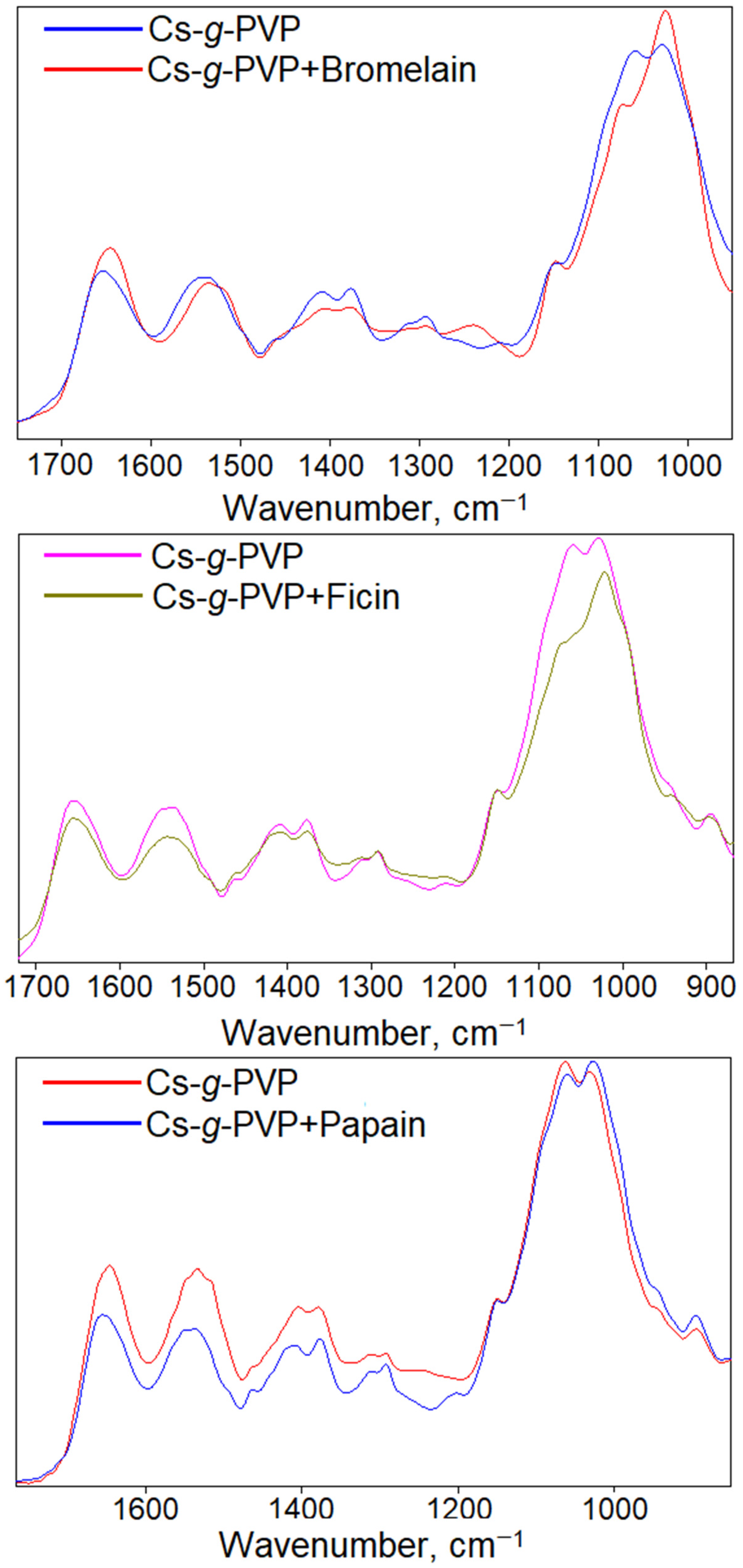
As previously noted, new absorption bands do not appear in the spectra of the enzyme–copolymer complexes. However, a detailed analysis of the spectra, normalized to the glycosidic bond absorption band (1148 cm1) in the 1000–1700 cm1 region (Figure 3), reveals that interactions with enzymes lead to several changes in the band associated with pyranose ring vibrations (1028–1148 cm1). This band is complex, consisting of one to ten modes, and changes in their number or shape indicate that the interaction involves the hydroxyl groups of chitosan attached to the carbon skeleton of the pyranose ring, disrupting the intrinsic self-organization of carbohydrate chains. In the case of papain, a shift in the intensity ratio of the modes at 1028 and 1059 cm1 is observed, whereas for ficin and bromelain, there is a sharp decrease in the intensity of the mode at 1059 cm1. Thus, FTIR data support the findings of molecular docking, further confirming that proteins predominantly bind to the hydroxyl groups of the carbohydrate backbone of the grafted copolymer.
The most informative approach for studying proteins and their interactions with various ligands using FTIR is the quantitative assessment of secondary structure, which is typically analyzed through the deconvolution of the amide I band. However, for the selected ligands—grafted copolymers of chitosan and poly(N-vinylpyrrolidone)—attempts to conduct such an analysis were unsuccessful due to the overlap between the amide I band of the protein and the absorption of the complex copolymer band, which includes contributions from the C=O groups of the lactam cycle and residual N-acetyl groups of chitosan.
Nevertheless, literature data indicate that non-covalent interactions do not cause significant alterations in the secondary structure of globular proteins [8,28]. Therefore, it can be inferred that the binding of cysteine proteases via hydrogen bonding and other weak physical interactions does not substantially affect the protein′s structure or its enzymatic activity.

3.3. In Vitro Studies of Cysteine Protease Complexes with the Cs-g-PVP Copolymers

Based on in silico and FTIR studies, a hypothesis can be formulated suggesting that the interaction with the Cs-g-PVP copolymers should lead to a slight decrease in catalytical activity of all researched enzymes. To validate this assumption, the proteolytic activity of the complexes was assessed in hydrolysis reactions using the macromolecular substrate azocasein. However, it is important to consider that the catalytic activity of the complexes may also depend directly on the enzyme content within them.
As the first stage of this study, the protein content in the complexes was evaluated (Table 3). The presented data indicate that the highest efficiency of enzyme interaction is observed for the copolymer characterized by the largest particle size in an aqueous medium and the highest proportion of grafted poly(N-vinylpyrrolidone) links (Table 1), highlighting their contribution to the interaction process.
For bromelain, the highest protein content in the complex was achieved: 20 and 18 mg·g−1 for the Cs-g-PVP-1 and Cs-g-PVP-2 copolymers, respectively. In contrast, ficin exhibited the lowest values: 13 and 9 mg·g−1 under the same conditions. Overall, this result correlates with docking calculations (Table 2). The affinity of bromelain for the copolymer was the highest (−8.1 kcal/mol), while that of papain was the lowest (−6.1 kcal/mol), with a similar trend observed for enzyme content in the complexes.
The slight deviation of ficin from this trend may be attributed to the fact that the in silico calculations did not account for the pH of the medium. Complex formation was carried out in 0.05 M borate buffer with 0.1 M KCl at pH 9.0, which corresponds to the isoelectric point of ficin, whereas for bromelain and papain, these values are lower—8.43 and 8.75, respectively [29]. Thus, when complex formation occurs at a pH equal to the isoelectric point of ficin, the contribution of electrostatic forces to the interaction between the enzyme and the polysaccharide decreases, which is reflected in the final protein content in the complex.
Table 3 also presents the results of the study on the proteolytic activity of cysteine protease complexes. The highest percentage of retained activity was observed for papain in complex with the Cs-g-PVP-2 copolymer, where no statistically significant decrease in enzyme activity was detected. Overall, the Cs-g-PVP-2 copolymer, which contains a lower amount of poly(N-vinylpyrrolidone), demonstrated a higher percentage of preserved activity. The tested proteases can be ranked in the following order based on their residual activity in the obtained complexes: papain > bromelain > ficin. These results are in good agreement with the in silico study. Bromelain, whose active-site amino acid residues form only weak ionic interactions, retains a high level of activity. In contrast, ficin, whose active site is more strongly bound by hydrogen bonding, exhibits a greater reduction in catalytic activity. The catalytic mechanism of papain differs somewhat from that of bromelain and ficin. It is known that the thiol group in the active site of papain can dissociate independently of histidine even before the catalytic process begins [30]. As a result, the binding of His159 via a hydrogen bond does not significantly affect the activity of this protease. Consequently, when complexed with the Cs-g-PVP-2 copolymer, papain retains nearly 100% of the activity.
It is well established that the complexation of enzymes with bulky ligands can reduce the catalytic activity of the resulting complexes due to diffusion hindrance, which impairs substrate and product transport to and from the enzyme’s active site [31,32]. In the present study, enzyme complexes containing branched Cs-g-PVP molecules located within the catalytic pockets of cysteine protease globules were obtained. Accordingly, it is of interest to investigate how substrate type influences the catalytic activity of these enzyme complexes. To this end, the activity of the complexes toward the small-molecular substrate BAPNA (amidase activity) was assessed (Table 3). The results indicate that the activity values of the enzyme complexes are statistically indistinguishable from those of the free enzymes. This suggests that diffusion limitations do not significantly affect the activity of cysteine protease complexes when acting on BAPNA. In contrast, during the hydrolysis of the macromolecular substrate azocasein, a decrease in catalytic activity is observed, indicating that diffusion limitations plays a more pronounced role under these conditions.
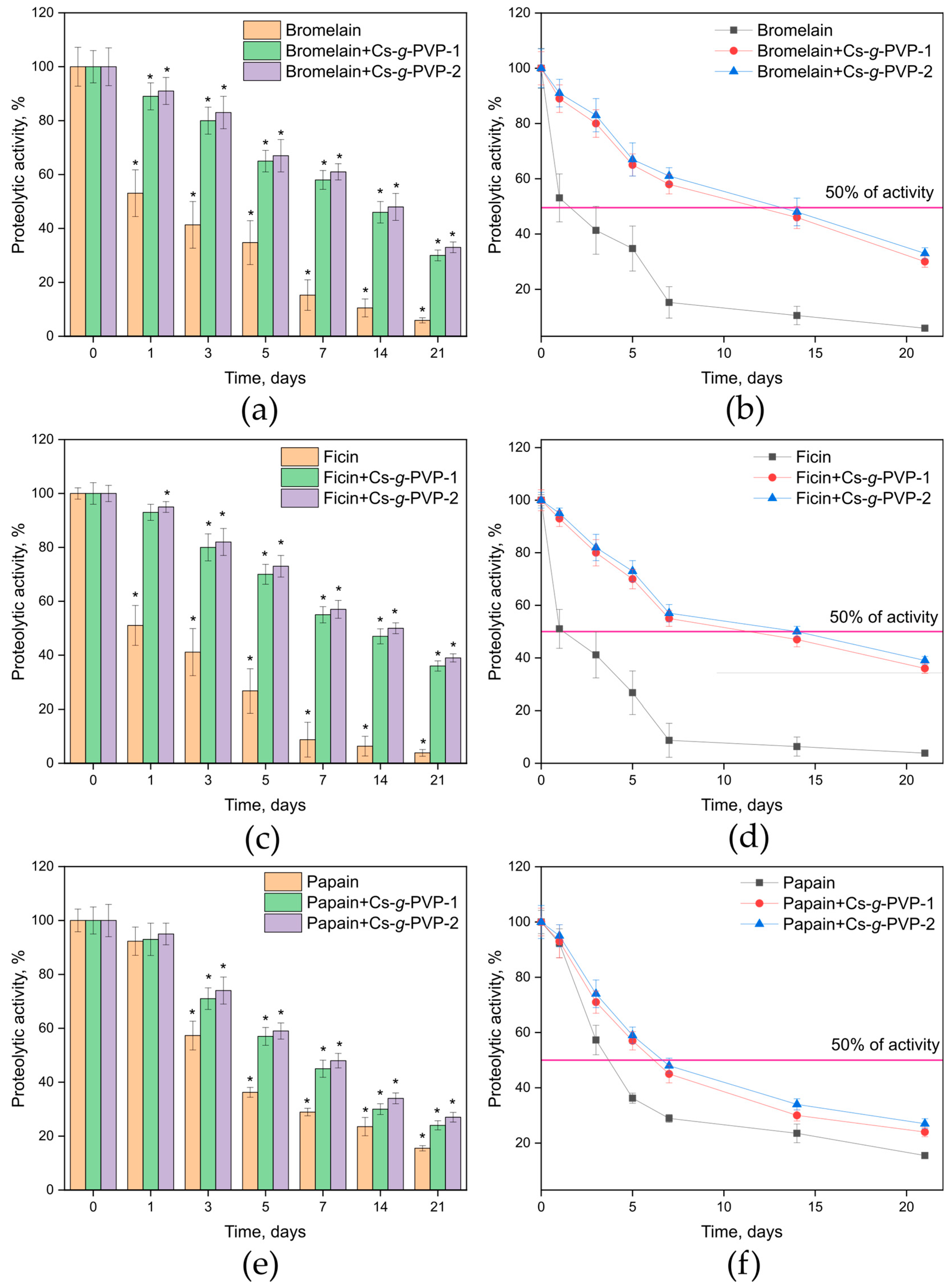
Enzyme interaction with a support often leads to prolonged activity, i.e., increased storage stability. The study of storage stability at 37 °C in 0.05 M Tris-HCl buffer with pH 7.5 revealed the following trends (Figure 4). Free bromelain and ficin exhibited a significant decrease in catalytic activity, with a reduction of 49–47% within the first 24 h of incubation, whereas their complexes retained up to 95% of their initial activity. This trend became more pronounced over time: by the third day of incubation, papain retained approximately 57% of its activity, while ficin and bromelain retained no more than 42%. In contrast, the enzyme–polymer complexes preserved up to 83% of their initial activity. The most significant differences in activity retention were observed at 14 and 21 days of incubation: free enzymes exhibited no more than 23 and 15% of their initial activity, respectively, whereas their immobilized forms retained up to 50 and 39%, respectively.
Based on the obtained experimental data, the half-life times (t1/2) of the free and complexed enzymes were calculated (Table 4, Figure 4). Interaction of the studied enzymes with Cs-g-PVP copolymers led to an increase in t1/2 by approximately 8.5-fold for bromelain, 2-fold for papain, and 12-fold for ficin. The greatest enhancement of this parameter was observed with the copolymer containing the lowest proportion of grafted poly(N-vinylpyrrolidone) links. The most pronounced stabilization effect was achieved for ficin, which had the lowest t1/2 value in its free form.

4. Conclusions

Thus, based on the conducted study, it can be concluded that the combination of molecular docking and FTIR methods allows for a reliable prediction of the potential use of Cs-g-PVP copolymers as supports for cysteine proteases. The proposed supports exhibit high capacity for bromelain, ficin, and papain (up to 100%), retain more than 63% of their proteolytic activity, and increase the half-life time by up to ~12 times. Due to these structural–-functional features, the resulting immobilized enzyme complexes are promising for practical applications, and the use of various biophysical methods rationalizes the process of their research and development.

Supplementary Materials

The following supporting information can be downloaded at: https://www.mdpi.com/article/10.3390/biophysica5020018/s1, Figure S1. Kinetis of enzyme binding with Cs-g-PVP-1 (a) and Cs-g-PVP-2 (b). Figure S2. Hydrogen bonds (indicated by dashed lines) and interactions between Cs-g-PVP and papain (a), ficin (b) and bromelain (c).

Author Contributions

Conceptualization, M.G.H., Y.F.Z. and V.G.A.; methodology, M.G.H., Y.F.Z. and V.G.A.; software, M.S.K.; investigation, A.V.S., A.N.D., A.I.Y. and M.S.K.; resources, M.G.H. and Y.F.Z.; writing—original draft preparation, M.S.L.; writing—review and editing, M.G.H., M.S.L., Y.F.Z. and V.G.A.; visualization, M.S.L.; supervision, M.G.H., Y.F.Z. and V.G.A.; project administration, M.G.H., Y.F.Z. and V.G.A.; funding acquisition, Y.F.Z. All authors have read and agreed to the published version of the manuscript.

Funding

This research was funded by Russian Science Foundation, grant number RSF-23-64-10020.

Data Availability Statement

The original contributions presented in the study are included in the article/Supplementary Materials, and further inquiries can be directed to the corresponding author.

Acknowledgments

FTIR data were obtained using the equipment at the Research Core Centre of Voronezh State University.

Conflicts of Interest

The authors declare no conflicts of interest.

References

  1. Prabantu, V.M.; Gadiyaram, V.; Vishveshwara, S.; Srinivasan, N. Understanding Structural Variability in Proteins Using Protein Structural Networks. Curr. Res. Struc. Biol. 2022, 4, 134–145. [Google Scholar] [CrossRef]
  2. Accardo, F.; Leni, G.; Tedeschi, T.; Prandi, B.; Sforza, S. Structural and Chemical Changes Induced by Temperature and pH Hinder the Digestibility of Whey Proteins. Food Chem. 2022, 387, 132884. [Google Scholar] [CrossRef]
  3. Śledź, P.; Caflisch, A. Protein Structure-Based Drug Design: From Docking to Molecular Dynamics. Curr. Opin. Struct. Biol. 2018, 48, 93–102. [Google Scholar] [CrossRef]
  4. Garcia-Galan, C.; Berenguer-Murcia, Á.; Fernandez-Lafuente, R.; Rodrigues, R.C. Potential of Different Enzyme Immobilization Strategies to Improve Enzyme Performance. Adv. Synth. Catal. 2011, 353, 2885–2904. [Google Scholar] [CrossRef]
  5. Rodrigues, R.C.; Berenguer-Murcia, Á.; Carballares, D.; Morellon-Sterling, R.; Fernandez-Lafuente, R. Stabilization of Enzymes via Immobilization: Multipoint Covalent Attachment and Other Stabilization Strategies. Biotechnol. Adv. 2021, 52, 107821. [Google Scholar] [CrossRef]
  6. Cipolatti, E.P.; Manoel, E.A.; Fernandez-Lafuente, R.; Freire, D.M.G. Support Engineering: Relation between Development of New Supports for Immobilization of Lipases and Their Applications. Biotechnol. Res. Innov. 2017, 1, 26–34. [Google Scholar] [CrossRef]
  7. Holyavka, M.G.; Goncharova, S.S.; Sorokin, A.V.; Lavlinskaya, M.S.; Redko, Y.A.; Faizullin, D.A.; Baidamshina, D.R.; Zuev, Y.F.; Kondratyev, M.S.; Kayumov, A.R.; et al. Novel Biocatalysts Based on Bromelain Immobilized on Functionalized Chitosans and Research on Their Structural Features. Polymers 2022, 14, 5110. [Google Scholar] [CrossRef] [PubMed]
  8. Sorokin, A.V.; Goncharova, S.S.; Lavlinskaya, M.S.; Holyavka, M.G.; Faizullin, D.A.; Zuev, Y.F.; Kondratyev, M.S.; Artyukhov, V.G. Complexation of Bromelain, Ficin, and Papain with the Graft Copolymer of Carboxymethyl Cellulose Sodium Salt and N-Vinylimidazole Enhances Enzyme Proteolytic Activity. Int. J. Mol. Sci. 2023, 24, 11246. [Google Scholar] [CrossRef]
  9. Aruna, V.; Chandrakala, V.; Angajala, G.; Nagarajan, E.R. Proteases: An Overview on Its Recent Industrial Developments and Current Scenario in the Revolution of Biocatalysis. Mater. Today Proceed. 2023, 92, 565–573. [Google Scholar] [CrossRef]
  10. Lalmanach, G.; Saidi, A.; Bigot, P.; Chazeirat, T.; Lecaille, F.; Wartenberg, M. Regulation of the Proteolytic Activity of Cysteine Cathepsins by Oxidants. Int. J. Mol. Sci. 2020, 21, 1944. [Google Scholar] [CrossRef]
  11. Holyavka, M.G.; Goncharova, S.S.; Redko, Y.A.; Lavlinskaya, M.S.; Sorokin, A.V.; Artyukhov, V.G. Novel Biocatalysts Based on Enzymes in Complexes with Nano- and Micromaterials. Biophys. Rev. 2023, 15, 1127–1158. [Google Scholar] [CrossRef] [PubMed]
  12. Kansakar, U.; Trimarco, V.; Manzi, M.V.; Cervi, E.; Mone, P.; Santulli, G. Exploring the Therapeutic Potential of Bromelain: Applications, Benefits, and Mechanisms. Nutrients 2024, 16, 2060. [Google Scholar] [CrossRef]
  13. Morellon-Sterling, R.; El-Siar, H.; Tavano, O.L.; Berenguer-Murcia, Á.; Fernández-Lafuente, R. Ficin: A Protease Extract with Relevance in Biotechnology and Biocatalysis. Int. J. Biol. Macromol. 2020, 162, 394–404. [Google Scholar] [CrossRef]
  14. Tacias-Pascacio, V.G.; Castañeda-Valbuena, D.; Morellon-Sterling, R.; Tavano, O.; Berenguer-Murcia, Á.; Vela-Gutiérrez, G.; Rather, I.A.; Fernandez-Lafuente, R. Bioactive Peptides from Fisheries Residues: A Review of Use of Papain in Proteolysis Reactions. Int. J. Biol. Macromol. 2021, 184, 415–428. [Google Scholar] [CrossRef]
  15. Baidamshina, D.R.; Koroleva, V.A.; Trizna, E.Y.; Pankova, S.M.; Agafonova, M.N.; Chirkova, M.N.; Vasileva, O.S.; Akhmetov, N.; Shubina, V.V.; Porfiryev, A.G.; et al. Anti-Biofilm and Wound-Healing Activity of Chitosan-Immobilized Ficin. Int. J. Biol. Macromol. 2020, 164, 4205–4217. [Google Scholar] [CrossRef]
  16. Hu, R.; Chen, G.; Li, Y. Production and Characterization of Antioxidative Hydrolysates and Peptides from Corn Gluten Meal Using Papain, Ficin, and Bromelain. Molecules 2020, 25, 4091. [Google Scholar] [CrossRef]
  17. Mohd Azmi, S.; Kumar, P.; Sharma, N.; Sazili, A.; Lee, S.-J.; Ismail-Fitry, M. Application of Plant Proteases in Meat Tenderization: Recent Trends and Future Prospects. Foods 2023, 12, 1336. [Google Scholar] [CrossRef]
  18. Fernández-Lucas, J.; Castañeda, D.; Hormigo, D. New Trends for a Classical Enzyme: Papain, a Biotechnological Success Story in the Food Industry. Trends Food Sci. Technol. 2017, 68, 91–101. [Google Scholar] [CrossRef]
  19. Aider, M. Potential Applications of Ficin in the Production of Traditional Cheeses and Protein Hydrolysates. JDS Commun. 2021, 2, 233–237. [Google Scholar] [CrossRef]
  20. Guarnieri, A.; Triunfo, M.; Scieuzo, C.; Ianniciello, D.; Tafi, E.; Hahn, T.; Zibek, S.; Salvia, R.; De Bonis, A.; Falabella, P. Antimicrobial Properties of Chitosan from Different Developmental Stages of the Bioconverter Insect Hermetia Illucens. Sci. Rep. 2022, 12, 8084. [Google Scholar] [CrossRef]
  21. Muthu, M.; Gopal, J.; Chun, S.; Devadoss, A.J.P.; Hasan, N.; Sivanesan, I. Crustacean Waste-Derived Chitosan: Antioxidant Properties and Future Perspective. Antioxidants 2021, 10, 228. [Google Scholar] [CrossRef]
  22. Qin, C.; Li, H.; Xiao, Q.; Liu, Y.; Zhu, J.; Du, Y. Water-Solubility of Chitosan and Its Antimicrobial Activity. Carbohyd. Polym. 2006, 63, 367–374. [Google Scholar] [CrossRef]
  23. Lavlinskaya, M.S.; Sorokin, A.V.; Mikhaylova, A.A.; Kuznetsov, E.I.; Baidamshina, D.R.; Saranov, I.A.; Grechkina, M.V.; Holyavka, M.G.; Zuev, Y.F.; Kayumov, A.R.; et al. The Low-Waste Grafting Copolymerization Modification of Chitosan Is a Promising Approach to Obtaining Materials for Food Applications. Polymers 2024, 16, 1596. [Google Scholar] [CrossRef]
  24. Lowry, O.H.; Rosebrough, N.J.; Farr, A.L.; Randall, R.J. Protein measurement with the folin phenol reagent. J. Biol. Chem. 1951, 193, 265–275. [Google Scholar] [CrossRef]
  25. Sorokin, A.V.; Goncharova, S.S.; Lavlinskaya, M.S.; Holyavka, M.G.; Faizullin, D.A.; Kondratyev, M.S.; Kannykin, S.V.; Zuev, Y.F.; Artyukhov, V.G. Carboxymethyl Cellulose-Based Polymers as Promising Matrices for Ficin Immobilization. Polymers 2023, 15, 649. [Google Scholar] [CrossRef]
  26. Tu, M.; Zheng, X.; Liu, P.; Wang, S.; Yan, Z.; Sun, Q.; Liu, X. Typical Organic Pollutant-Protein Interactions Studies through Spectroscopy, Molecular Docking and Crystallography: A Review. Sci. Total Environ. 2021, 763, 142959. [Google Scholar] [CrossRef]
  27. Vavra, O.; Damborsky, J.; Bednar, D. Fast Approximative Methods for Study of Ligand Transport and Rational Design of Improved Enzymes for Biotechnologies. Biotechnol. Adv. 2022, 60, 108009. [Google Scholar] [CrossRef]
  28. Secundo, F. Conformational Changes of Enzymes upon Immobilisation. Chem. Soc. Rev. 2013, 42, 6250. [Google Scholar] [CrossRef]
  29. Rawlings, N.D.; Salvesen, G. Handbook of Proteolytic Enzymes, 3rd ed.; Academic Press: Boston, MA, USA; London, UK, 2013; ISBN 9780123822192. [Google Scholar]
  30. Beveridge, A.J. A Theoretical Study of the Active Sites of Papain and S195C Rat Trypsin: Implications for the Low Reactivity of Mutant Serine Proteinases. Protein Sci. 1996, 5, 1355–1365. [Google Scholar] [CrossRef]
  31. Bahamondes, C.; Illanes, A.; Pouchucq, L. Effect of External Diffusional Restrictions in Immobilized Enzymes in Stirred Reactors. Biocatal. Biotransfor. 2025, 1–17. [Google Scholar] [CrossRef]
  32. Bolivar, J.M.; Woodley, J.M.; Fernandez-Lafuente, R. Is Enzyme Immobilization a Mature Discipline? Some Critical Considerations to Capitalize on the Benefits of Immobilization. Chem. Soc. Rev. 2022, 51, 6251–6290. [Google Scholar] [CrossRef] [PubMed]
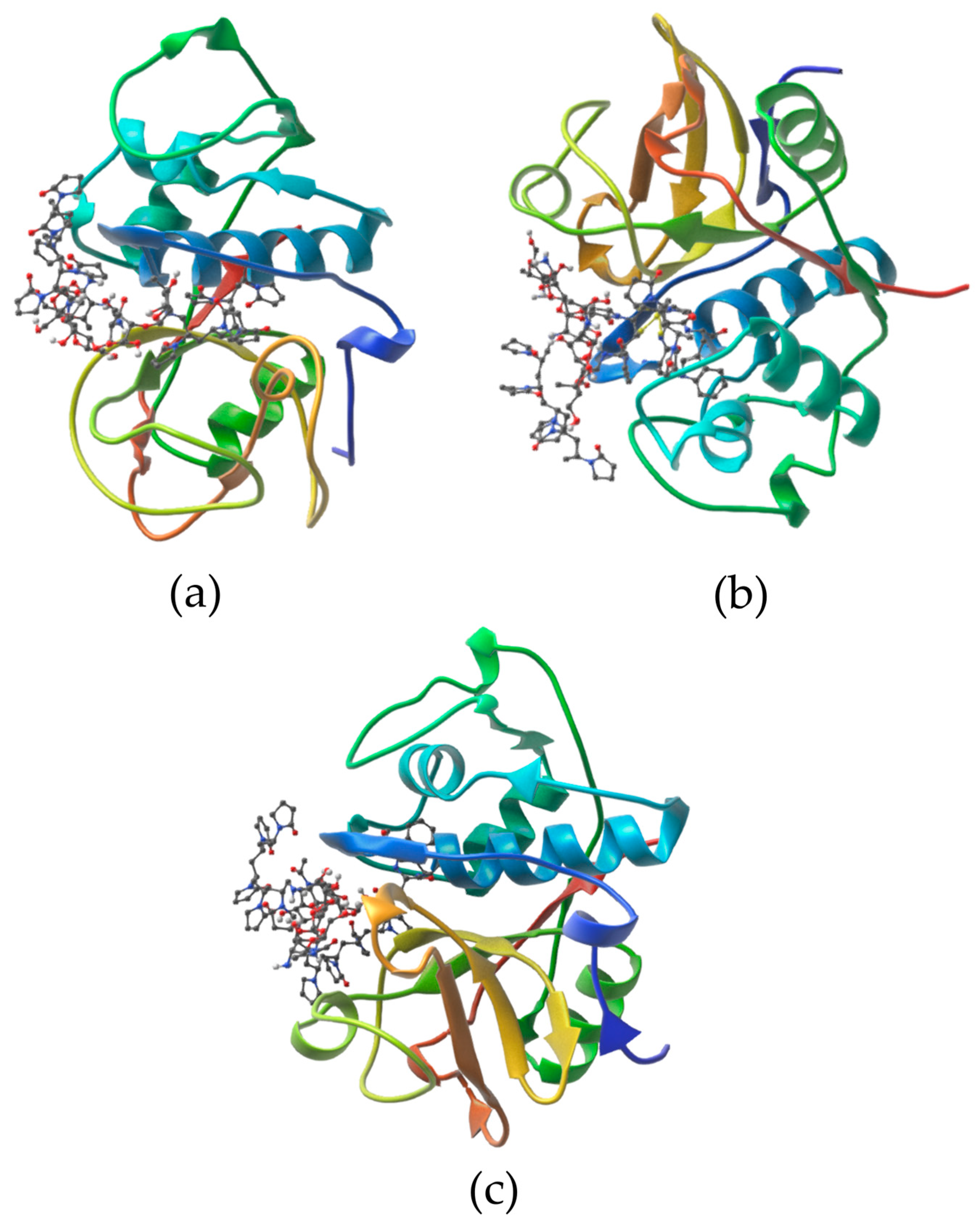
Figure 1. Topology of Cs-g-PVP complexes with bromelain (a), ficin (b), and papain (c). Different elements of the protein′s secondary structure are shown in different colors.
Biophysica 05 00018 g001
Figure 2. FTIR spectra of the Cs-g-PVP copolymer and its complexes with cysteine proteases.
Biophysica 05 00018 g002
Figure 3. Normalized FTIR spectra of the Cs-g-PVP copolymer and its complexes with cysteine proteases.
Biophysica 05 00018 g003
Figure 4. Left panel: residual catalytic activity of bromelain (a), ficin (c), and papain (e) and their complexes after incubation at 37 °C in 0.05 M Tris-HCl buffer with pH 7.5, expressed as a percentage of the initial activity. An asterisk (*) indicates values that are statistically different from the activity values of enzymes and their complexes before incubation. Right panel: graphical assessment of the half-life time of bromelain (b), ficin (d), and papain (f) and their complexes.
Biophysica 05 00018 g004
Table 1. Some parameters of the Cs-g-PVP copolymers.
CopolymerPVP Content (% wt.)PVP Molecular WeightZ-Average Dh (nm)PDI
Cs-g-PVP-15110,000189 ± 160.359 ± 0.02
Cs-g-PVP-2248000136 ± 110.405 ± 0.03
Table 2. Amino acid residues of the enzymes interacting with the Cs-g-PVP copolymer simulated by molecular docking *.
Affinity
(kcal/mol)
Amino Acid Residues Forming
H-bonds, Length, (Å)Other Physical Interactions
Bromelain
−8.1Asn19, 2.97; Asn21, 3.08; Gly66, 2.98 and 3.03; Ala136, 3.05; Gln141, 2.94Thr15, Ser16, Val17, Lys18, Asn19, Gln20, Asn21, Pro22, Gly24, Cys26 (αL1), Phe29 (αL1), Ala33 (αL1), Glu51 (αL2), Cys63, Lys64, Gly65, Gly66, Ala136, Phe140, Gln141, Leu156, Asn157, His158 (βR), Ala159 (βR), Thr161 (βR), Ile163 (βR), Ala178, Lys179, Trp180, Gly184, Trp185
Ficin
−7.3Gly20, 2.87; Cys22, 2.95 and 3.25; Gly23, 2.90; Glu145, 3.09 and 3.23; Trp184, 3.11; His162 (βR), 3.30; Cys65, 2.85; Ser66, 2.89 and 3.27Gln19, Gly20; Arg21, Cys22, Gly23, Cys25 (αL1), Tyr60, Leu63, Cys65, Ser66, Gly68, Trp69, Met70 (αL3), Lys94, Lys95, Glu145 (αR2), Leu160, Asp161, His162 (βR), Trp184, Asn187, Trp188
Papain
−6.1Gly20, 2.68 and 3.07; Cys22, 2.93 and 3.00; His159 (βR), 3.04; Trp177, 2.70Gln19, Gly20, Ser21, Cys22, Gly23, Cys63, Asn64, Gly65, Val133 (βR), Ala137, Gln142, Leu143 (αR2), Lys156, Asp158 (βR), His159 (βR), Ala160 (βR), Trp177, Gly180, Trp181
* Catalytically valuable amino acid residues are in bold; protein secondary structure elements are in brackets.
Table 3. Some characteristics of cysteine protease complexes with Cs-g-PVP copolymers.
Enzyme/ComplexEnzyme Content (mg g−1) *Enzyme Complexation Efficiency (%)Proteolytic Activity (U·mL−1) **Proteolytic Activity
Complexation Efficiency (%)
Amidase Activity (U·mL−1) **Amidase Activity
Complexation Efficiency (%)
Bromelain97.2 ± 7.21007.1 ± 0.2100
Bromelain + Cs-g-PVP-120.1 ± 2.110077.8 ± 4.5 a806.8 ± 0.295
Bromelain + Cs-g-PVP-218.2 ± 1.29084.5 ± 3.1 a876.9 ± 0.397
Ficin96.5 ± 2.31002.1 ± 0.2100
Ficin + Cs-g-PVP-113.3 ± 1.16560.7 ± 3.7 b631.9 ± 0.189
Ficin + Cs-g-PVP-29.1 ± 2.14572.9 ± 6.1 b762.0 ± 0.193
Papain95.4 ± 3.81004.4 ± 0.4100
Papain + Cs-g-PVP-117.2 ± 2.38576.2 ± 5.9 c804.1 ± 0.394
Papain + Cs-g-PVP-210.5 ± 2.15396.8 ± 4.21004.3 ± 0.197
* Enzyme content is expressed as milligrams of enzyme per gram of support. ** Activity values that are statistically different from those of the free enzyme are indicated by superscript letters.
Table 4. Half-life times of enzymes and their complexes with the Cs-g-PVP copolymers.
Enzyme/ComplexHalf-Life Time t1/2 (Days)
Bromelain1.53
Bromelain + Cs-g-PVP-111.68
Bromelain + Cs-g-PVP-212.95
Papain3.69
Papain + Cs-g-PVP-16.18
Papain + Cs-g-PVP-26.65
Ficin1.18
Ficin + Cs-g-PVP-111.38
Ficin + Cs-g-PVP-214.01
Disclaimer/Publisher’s Note: The statements, opinions and data contained in all publications are solely those of the individual author(s) and contributor(s) and not of MDPI and/or the editor(s). MDPI and/or the editor(s) disclaim responsibility for any injury to people or property resulting from any ideas, methods, instructions or products referred to in the content.