Review

Microplastic and Extracellular Vesicle Interactions: Recent Studies on Human Health and Environment Risks

1
Biochemistry and Molecular Biology Section, Department of Chemistry, Biology and Biotechnology, University of Perugia, Via del Giochetto, 06126 Perugia, Italy
2
Department of Civil and Environmental Engineering, University of Perugia, Via G. Duranti 93, 06125 Perugia, Italy
3
Centro di Eccellenza Materiali Innovativi Nanostrutturati (CEMIN), University of Perugia, Via del Giochetto, 06126 Perugia, Italy
*
Author to whom correspondence should be addressed.
These authors contributed equally to this work.
Biophysica 2024, 4(4), 724-746; https://doi.org/10.3390/biophysica4040047
Submission received: 20 November 2024 / Revised: 12 December 2024 / Accepted: 19 December 2024 / Published: 21 December 2024

Abstract

:
Microplastics (MPs) are widespread environmental pollutants that have drawn significant attention due to their possible health risks to humans and animals, as well as their extensive presence in ecosystems. Recent growing evidence highlights a remarkable relationship between MPs and extracellular vesicles (EVs), nanoscale particles involved in intercellular communication. The purpose of this review was to investigate how the relationships between MPs and EVs can affect cellular functions and how this interaction could impact environmental conditions leading to broader ecological risks. The interaction patterns and bioactivity of both MPs and EVs are strongly influenced by biophysical characteristics such as hydrophobicity, surface charge, and particle size, which have received particular attention from the scientific community. Recent studies indicate that MPs affect EV distribution and their capacity to function appropriately in biological systems. Additionally, MPs can modify the molecular cargo of EVs, which may result in alterations of cell signaling pathways. Understanding the interactions between MPs and EVs could provide important opportunities to comprehend their potential effects on human health and environmental systems, especially when it comes to cancer development, endocrine, metabolic, and inflammatory disorders, and ecological disruptions. This review emphasizes the necessity of multidisciplinary research to clarify the molecular and biophysical mechanisms regulating the interaction between MPs and EVs.

Graphical Abstract

1. Introduction

In recent decades, microplastics (MPs) have captured the attention of the scientific community as emerging contaminants. MPs are defined as plastic particles or fibers less than 5 mm in size and can be produced directly from the breakdown of larger plastic waste or from the direct manufacturing of microbeads, which are utilized in many different products (e.g., skin creams and cosmetic products such as facial scrubs) [1,2,3]. Significant ecological and health hazards have been related to their prevalence in the environment [4]. A lot of studies show that MPs can accumulate in food chains and be ingested by a range of animals, including humans [5,6,7]. It is estimated that an individual ingests between 39,000 and 52,000 MP particles annually from food and water sources, with inhalation having the ability to significantly increase this number [8,9]. To understand the impact of MPs on the environment, the annual production of plastic must be considered, which is around 400 million tons per year [10]. Furthermore, there are also numerous economic implications deriving from MP pollution, since sectors such as fish industries and tourism are strongly and negatively affected by the presence of these contaminants [11]. For example, it has been estimated that MP pollution causes an annual economic loss of over 13 billion dollars [12]. The health effects of MPs are now widely documented, with particularly serious consequences derived from direct and prolonged contact with significantly high concentrations of these pollutants. Chronic exposure to MPs is associated with an increased risk of developing serious pathologies, such as cancer, endocrine and inflammatory diseases, and metabolic disorders [13,14,15,16,17]. Although the extent of human exposure has not yet been precisely quantified, the mechanisms of exposure have been partially identified, and it is known with certainty that the presence of MPs in the human body increases the risk of contracting severe diseases [16,18]. Furthermore, because of their small size (reaching the nanometer scale) [19], they can pass through biological membranes and into the bloodstream, which raises concerns about their long-term impact on human health. A further alarm bell comes from a recent discovery that detected the presence of MPs also in the placenta and the fetus [20,21]; the consequences of this discovery could be more serious than previously assumed. The placenta, in fact, crucially regulates the interactions between the fetus and the mother’s body, as well as acting as a link to the external environment. It is therefore essential to address the problem of MP pollution, as it represents not only an environmental challenge but also a serious threat to human health [13,14,15,21,22]. In this regard, it would be important to develop advanced technologies for the ultrasensitive detection of MPs, such as single-molecule techniques (e.g., atomic force microscopy) and vibrational spectroscopy, which offer excellent capabilities to identify these contaminants even in complex biological matrices, including the placenta, thus contributing to a better understanding of their effects on human health [23,24]. However, it should be taken into account that biopolymeric nanoparticles (with a size of hundreds of nm), which can be referred to as nanoplastics (NPs), can also be introduced into the body for therapeutic purposes in biomedical applications [25,26,27]. For example, recent studies on polylactic acid (PLA) showed that a therapeutic enzyme can be effectively immobilized on biopolymeric compounds for the treatment of some metabolic diseases [26,28].
Given their small size and potential to interact with biological systems, MPs and especially NPs may influence cellular processes in ways similar to other nanoscale particles. In this context, extracellular vesicles (EVs) come into play. EVs are lipid-bilayer membranous vesicles secreted by cells that play a crucial role in intercellular communication. They are classified into three main types based on their biogenesis: exosomes (40–120 nm), microvesicles (50–1000 nm), and apoptotic bodies (500–2000 nm) [29,30,31,32]. EVs contribute significantly to cell cross-talk by carrying many metabolites such as proteins, lipids, mRNAs, and microRNAs (miRNA—small non-coding RNA which can interfere with gene expression) that influence recipient cell behavior affecting processes such as immunity, inflammation, and tissue repair [33,34,35]. Recent studies have emphasized the possible function of EVs in modulating the responses of cells to environmental stressors, such as MP-induced stresses [36,37,38]. This suggests that MPs may alter cellular communication pathways through EVs, potentially exacerbating health issues related to exposure.
Therefore, the study of the interaction between MPs and EVs is extremely fascinating. There is still limited scientific work in this area, but early evidence indicates that MPs can influence the biogenesis and release of EVs from cells. This interaction could facilitate the transfer of toxic or signaling molecules between cells, further complicating the biological impacts of MP exposure. Clarifying the mechanisms behind the health impacts linked to MP contamination requires an understanding of these interactions. The expanding body of information indicates that EVs and MPs are important components of environmental health and disease processes.

2. Microplastics: Environmental Prevalence and Biological Impact

2.1. Classification and Sources of Microplastics

Microplastics (MPs) are insoluble solid particles of plastic with a size ≤ 5 mm [3,39]. Based on their size, these residues are classified as large MPs (1 mm–5 mm), small MPs (1 µm–1 mm), submicron plastics (SMPs) (<1 μm) [40], and nanoplastics (NPs) (with a size ≤100 nm) [3,41,42]. These can also have variable shapes such as microbeads, fibers, and fragments, and based on their origin they are discriminated into primary and secondary MPs. Primary MPs are those intentionally produced and released into the environment such as those produced by the cosmetic industries, while secondary MPs are formed following the degradation of plastic residues in the environment due to biological and physical factors such as microorganisms, photodegradation, water corrosion, and mechanical stresses [3,43,44,45]. These same factors also act on the MPs themselves by modifying some of their peculiar properties such as morphology, color, and density which affect their chemical properties and therefore their interaction with the environment and living organisms [3,46]. Depending on the environmental matrix (soil, marine, freshwater, food, and atmosphere), different degradation mechanisms prevail. MP degradation in the environment occurs through physico-chemical processes such as UV-induced bond cleavage, mechanical comminution, and temperature or pH changes; additionally, microbial biodegradation driven by enzyme secretion can also occur. The latter represents a more sustainable route to degradation; however, it is limited by the complexity and diversity of environmental microbial communities. For example, in marine environments, UV radiation and salt-induced oxidation play key roles, while in soil, microbial activity and temperature fluctuations are more relevant, and in freshwater systems, biodegradation is often enhanced by microbial biofilms forming on plastic surfaces [47]. MPs have also been found in wastewater [48], water that has been contaminated by human activity, including domestic, industrial, and agricultural use. A large number of by-products are also produced through agricultural activities; these agricultural wastes could contain MPs [49,50]. It has been estimated that the EU releases between 0.7 and 1.8 × 10⁶ tons of secondary MPs and 42,000 tons of primary MPs into the environment annually [51]. The most prevalent MP shape categories, in both water and sediment, are fibers (48.5%), pieces (31%), particles (6.5%), films (5.5%), and foams (3.5%) [51,52]. Over time, MPs typically take on irregular or amorphous shapes and create even smaller nanoparticles as they further degrade or break into reduced sizes.
The most common MPs derive from plastic polymers such as polyethylene (PE), both at high-density (HDPE) and low-density (LDPE); polypropylene (PP); polyamide (PA); polystyrene (PS); polyethylene terephthalate (PET); and polyvinyl chloride (PVC) [53,54]. Additionally, MPs contain a lot of compounds linked to plastic, since manufacturing processes of plastics involve the addition of chemical additives including phthalates and polybrominated diphenyl ethers (PBDEs) that are mixed into polymers in order to modify their chemical, physical, and mechanical properties, with the aim to derive products with improved performance. Furthermore, plastics frequently contain two chemical groups of known health concern: bisphenols and per- or poly-fluoroalkyl substances (PFAS) [55,56]. In particular, it has been demonstrated that PFAS can be considered endocrine disruptors capable of influencing reproduction and cancer development [57].
The most common MPs identified in the environment include PS, PE, and PVC, with sizes ranging from less than 50 nm to 10 µm. These MPs have been detected in different environmental matrices, such as river water, seawater, agricultural soil, sand, and air, indicating a widespread distribution in various environmental compartments (Table 1).
Sampling, analysis, and identification of MPs are particularly complex due to their small size. The most commonly used technique to separate them is density separation, which uses ZnCl2, CaCl2, NaBr, or other salts as a separating agent after the removal of organic matter with H2O2 or Fenton reagent [64]. For the identification and characterization of MPs, FTIR or Raman and NIR spectroscopy are widely used, while methods such as pyrolysis-gas chromatography/mass spectrometry (Py-GC/MS) are preferred when quantitative analysis is requested [64]. However, there is still no technique of choice for the analysis of MPs [3]. MPs represent a growing threat as they are present in all environmental compartments, where they can generate pollution, mainly due to the life cycle of products and the progressive fragmentation of larger plastic wastes [43,65,66]. In addition, MPs can serve as important organic vectors for various environmental contaminants, such as heavy metals, because of their high adsorption capability and persistence in the environment [67,68].

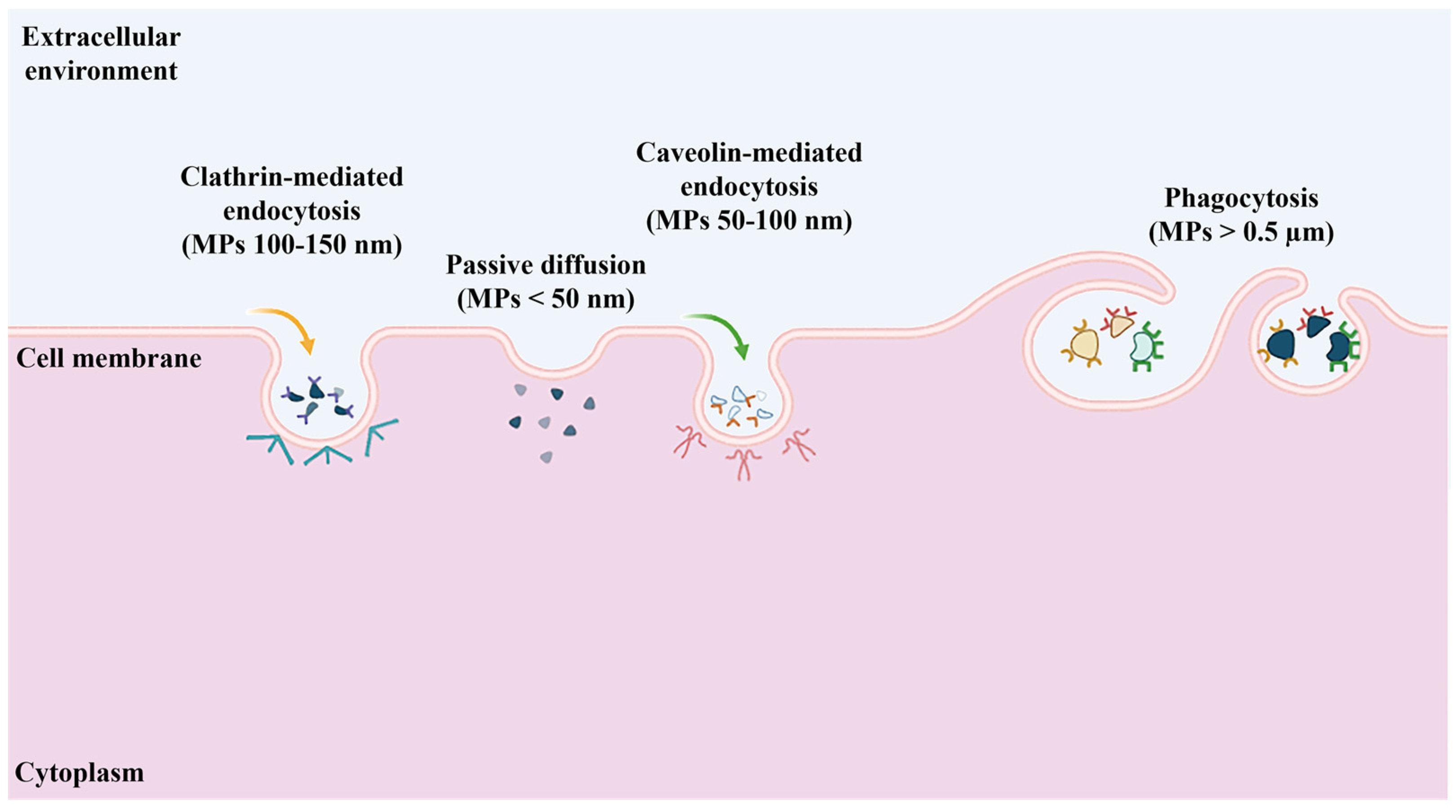
2.2. Biological Uptake of Microplastics

In recent years, attention to the risks that plastic waste poses to human health and the environment has increased significantly. However, accurately quantifying the real level of risk remains challenging. Although MPs are considered emerging contaminants, their long-term effect on human health remains unclear. This is because data on MP ingestion are relatively recent, and results are often not comparable. Furthermore, many studies are based on laboratory tests, with concentrations that do not reflect those actually present in the environment [69,70]. Despite these uncertainties, it is clear that the spread of plastic fragments in the biosphere represents a far-reaching problem, not only ecological but also economic and social. Because plastic debris is ubiquitous, virtually all living organisms, regardless of their trophic level, come into contact with it. Biological effects can occur through physical interactions with MPs, exposure to chemicals contained in the plastic itself (e.g., additives, heavy metals), adsorbed toxins (both synthetic and natural), or through organisms associated with their surfaces, such as pathogens [67,68,71]. Living organisms, especially humans, are exposed to MPs through three main routes: ingestion of food and water, inhalation of contaminated air, and skin contact through cosmetics and pharmaceuticals (Figure 1) [72,73,74]. The potential toxic effects of MPs and NPs on various organ systems, the mechanisms of cellular uptake, and the molecular pathways that determine their toxicity are not yet fully understood, mainly due to the limited amount of scientific evidence available. Once exposed, MPs and NPs can be absorbed by cells and accumulate within various tissues and organs [72,73]. This intracellular accumulation occurs when these pollutants interact with cell membrane components, such as receptors and lipids, leading to bioaccumulation [75]. In fact, traces of these particles have been detected in various body systems.
The main route of exposure for animals, including humans, is through the ingestion of contaminated food and water [72,76]. Many animals, both marine and terrestrial, such as fishes or birds, ingest large quantities of MPs and NPs, either by accident or by mistaking them for food. These particles accumulate in the tissues of organisms and, through the food chain, can reach the human body. Humans are indeed directly exposed to these particles through the consumption of contaminated food or through contact with packaging materials. It is estimated that unintentional ingestion of MPs from food containers can reach 203 particles per week per person. Currently, there are no global regulatory frameworks specifically regulating MP contamination in food. However, despite this regulatory gap, several directives govern the use of plastics in food-related applications, such as the European Union Directive on single-use plastics (Directive (EU) 2019/904) [77] and the Regulation (EC) No 1907/2006 [78] concerning the Registration, Evaluation, Authorisation, and Restriction of Chemicals (REACH). In addition, during the fifth session of the United Nations Environment Assembly (UNEA-5.2) [79], the proposed global treaty on plastic pollution was discussed. Nevertheless, many food products, including sugar, salt, bottled water, and most seafood, remain contaminated by MPs due to persistent environmental pollution [80,81,82]. A particularly worrying scenario is the fragmentation of MPs into NPs, which facilitates their absorption and potential accumulation in the body [83]. Despite efforts to monitor the quality of food and beverages, it is estimated that an individual in the United States ingests between 39,000 and 52,000 MP particles annually, while in Europe the estimated annual intake is around 11,000 particles [76,84]. The maximum predicted yearly consumption per person for tap water and shellfish is around 4000 and 11,000 MP particles, respectively [85,86]. When MPs are ingested, they travel through several digestive system sections. At first, the stomach’s acidic environment may cause MPs to degrade chemically, releasing plastic-related compounds more quickly, reducing their size, and changing their surface [87,88]. Then, the interaction between MPs and the gut microbiota may impact intestinal digestion or further speed up biotransformation and/or the release of additives and contaminants [89].
Inhalation is another important route of exposure to MPs and NPs for the human body. Numerous studies have detected the presence of MP fibers in the atmosphere, which are dispersed in the air from different sources, such as wear of synthetic clothing, construction materials, plastic waste, and landfills [90,91]. A lot of MPs and NPs have been found in human biological samples, including lungs and sputum [92,93]. According to a recent study, at the alveolar–capillary interface, MPs were observed forming aggregates with the pulmonary surfactant [94]. In addition to their intrinsic potential toxic effects, these particles act as vectors of other chemical and biological contaminants, due to their low polarity and high surface roughness [95]. Once inhaled and having entered the airways, MPs and their toxic loads can be easily absorbed by the alveolar epithelium, causing local inflammation [96]. Subsequently, they can be translocated into the circulatory system, causing systemic effects or stimulating the production of pro-inflammatory mediators, resulting in systemic inflammation [96,97].
Absorption of MPs and NPs can also occur through skin contact, with the use of topical products such as cosmetics, detergents, creams, and medical devices [74,90]. In fact, MPs are frequently included in cosmetic and skin care products for functions like emulsification, peeling, and viscosity correction [98]. MPs have been identified, for example, in detergents, masks, sunscreens, and toothpaste, in the form of small beads, which can be absorbed and cause skin lesions [99,100,101]. Although it is not yet entirely clear whether MPs and NPs are able to cross the skin barrier under normal conditions, it has been shown that they accumulate in hair follicles [72,102]. It has been hypothesized that NPs smaller than 6–7 nm can be absorbed via the trans-epidermal and trans-follicular lipid pathways, respectively, or through aqueous pores. In contrast, larger particles seem to prefer penetration via the trans-follicular pathway [102]. Moreover, dermal contact speeds up the cellular uptake of released MP-associated compounds or environmental pollutants, even if MPs themselves barely penetrate the skin barrier [103].

2.3. Impact of Microplastics on Cellular Health

To comprehend the impact of MPs on cellular health, it is essential to ascertain the extent of their interaction with cells, their uptake, and the specific cells and mechanisms involved. The size, chemical surface area, and charge of particles significantly influence MP interactions with the surrounding biological components, including phospholipids, proteins, and carbohydrates, thereby affecting cellular uptake. Various uptake mechanisms may come into play for MPs to approach and eventually enter cells. The majority of immune cells and epithelial cells possess the ability to absorb MPs through endocytosis. This process takes place in two distinct forms: phagocytosis for particles larger than 0.5 μm, predominantly in macrophages and also in monocytes, and pinocytosis represented by clathrin-mediated endocytosis and caveolin-mediated endocytosis taking place primarily in the epithelial cells for particles smaller than 0.5 μm (Figure 2) [15,104,105,106]. Phagocytosis, in particular, is active in specialized cells such as macrophages, dendritic cells, and neutrophils, which recognize MPs through specific phagocytic receptors, such as Fcγ and complement receptors. Opsonins, including immune and serum proteins, bind to the surface of MPs, facilitating receptor-mediated phagocytosis and particle internalization [107,108]. In addition to phagocytosis, caveolin-mediated endocytosis is another important mechanism of MP internalization, involving the formation of caveolae, small caveolin-rich invaginations of the plasma membrane [108,109]. Adipocytes, endothelial cells, and fibroblasts are among the cells that utilize this process to internalize small particles. It has been demonstrated that 50 nm PS microspheres can interact with caveolin and be transported into the cell to organelles such as the Golgi and endoplasmic reticulum. Similarly, nanoparticles of 50 and 100 nm have been observed in the proximity of the nucleus, where the distribution of the endoplasmic reticulum occurs [110,111]. These particles, once captured by lysosomes, are degraded, but in some cases, nanoparticles that exploit caveolin-mediated endocytosis can avoid fusion with lysosomes and move to other cellular regions. Caveolin-mediated endocytosis often leads to the formation of caveosomes, vesicles that follow a trafficking pathway distinct from traditional endocytic pathways. Caveosomes can avoid immediate fusion with lysosomes due to their unique trafficking pathway, which can bypass or delay lysosomal degradation. This endocytic pathway allows for a more controlled release of nanoparticles or the possibility of trafficking to other cellular compartments, such as the Golgi apparatus or endoplasmic reticulum, depending on the properties of the nanoparticles [112]. Another internalization way is clathrin-mediated endocytosis, which occurs in areas of the plasma membrane rich in this protein, organized in complexes that deform the membrane, leading to the formation of vesicles of about 100–150 nm. This process is able to absorb particles smaller than 200 nm, such as MPs, which can be internalized with the captured extracellular fluid [106,108,113].
In addition to these active mechanisms, MPs can also enter cells via passive permeation, a process that does not require receptor–ligand interaction. In some studies, 20 nm carboxylated MPs have been observed crossing the phospholipid bilayer of the membrane via passive diffusion [106]. Passive permeation, favored by van der Waals forces, spatial interactions, or electrostatic synergies, represents an important internalization route for very small particles (<50 nm).
The efficiency of cellular uptake of MPs depends not only on their size but also on their surface characteristics. It appears that MPs with different physicochemical characteristics are taken up and distributed within cells in different ways. While the exact mechanism of surface characteristic-dependent uptake is not fully understood, it is known that MPs with specific physicochemical modifications, such as negative charge and hydrophobicity, demonstrate a greater entrapment capacity compared to their unmodified counterparts [114,115].
Research has shown that exposure to MPs can have a detrimental impact on cellular health. Inflammatory responses can be triggered by MPs, leading to the production of various pro-inflammatory cytokines [116,117]. For example, immune cells have exhibited responses, such as increased pro-inflammatory cytokine production (particularly IL-1β, IL-8, and IL-6) in macrophages and peripheral blood mononuclear cells, which result in a decrease in macrophage phagocytic capacity [116,118]. Additionally, genotoxicity and the production of reactive oxygen species (ROS) have been observed, indicating that MPs have the potential to cause damage and provoke dysfunction in mitochondria [119,120,121]. It has been reported that MPs affect subcellular organelles, inducing pathological and morphological changes. Consequently, exposure to MPs may bring about inflammation and the development of chronic diseases, as this can continue to occur and harm the cells either directly or through the immune system [121,122]. A study on the in vitro toxicology of MPs proposed that even at low concentrations, MPs can cause the release of ROS, leading to cellular dysfunction and death [123]. These free radicals have the potential to cause slow but lasting damage to nucleic acids, lipids, and proteins. Concerning the activation of inflammatory pathways, it has been shown that exposure to MPs released from baby bottles, triggered an inflammatory response in human intestinal cells (Caco-2) through the production of ROS, which led to the activation of the NLRP3 inflammasome. This process subsequently triggered an inflammatory cascade, with an increase in the levels of pro-inflammatory cytokines such as IL-6 and TNFα, marking the onset of acute intestinal inflammation [124]. Consequently, these findings indicate that MPs can provoke a pro-inflammatory response significant enough to affect cellular integrity. Long-term exposure to MPs has been associated with chronic inflammation, which has been linked to various diseases, including diabetes and heart disease, thus suggesting that prolonged exposure to MPs could give rise to lasting physiological damage [125].

3. Extracellular Vesicles: Biological Roles and Environmental Relevance

3.1. Extracellular Vesicles: Definition and Classification

Extracellular vesicles (EVs) are lipid-bilayer nanoparticles naturally released from a cell into the surrounding extracellular space. Nowadays, it is known that the production of EVs is a phenomenon that crosses all three domains of life, playing a key role in intercellular communication and the transport of molecules between cells [126]. As well as a complex cargo of proteins and lipids, EVs contain a variety of nucleic acids, including mRNAs and non-coding RNAs. The presence of potentially bioactive molecules suggests that EVs are adapted to act as safe and effective carriers of molecular signals with the capacity to modulate cell behavior [29,127]. EVs are a huge and different category of membranous structures with diameters ranging from 40 to 2000 nm. The classification of these nanostructures can be based on their size, cellular origin, biological function, and/or biogenesis pathways. Based on their subcellular origin, vesicle sizes, and typical marker proteins, EVs are typically classified as exosomes, microvesicles (MVs), and apoptotic bodies. Exosomes are the smallest EVs (40–120 nm) released from cells upon fusion of endosomal multivesicular bodies with the plasma membrane. MVs are vesicles that are 50–1000 nm in diameter and are produced by the outward budding of the plasma membrane, whereas apoptotic bodies are vesicles that are 500–2000 nm in diameter and are discharged as blebs of cells going through apoptosis [31,32]. Different categories of EVs (Table 2) can be recognized by the presence of specific protein markers. Exosomes, for example, are characterized by proteins linked to the endosomal pathway, such as tetraspanins (CD9, CD63, and CD81), TSG101, and ALIX. Microvesicles, on the other hand, present proteins associated with the plasma membrane, including integrins, selectins, and the ligand CD40. Apoptotic bodies are distinguished by their selective histone content [31,128]. However, none of these markers are exclusive, as they can be shared between different types of vesicles. This lack of molecular distinctiveness makes it difficult to distinguish EV subtypes, such as exosomes and microvesicles. Consequently, the classification and nomenclature of these vesicles remain a complex and controversial topic. As mentioned above, EVs can enclose a large array of different biological molecules and macromolecules like proteins, lipids, biologically active amines, and nucleic acids [129]. They can also affect surrounding biofluids, tissues, and cells provided that some specific kind of molecular signature is present in the EVs. Moreover, EVs have also been found in the extracellular environment either in vivo or in vitro [130].

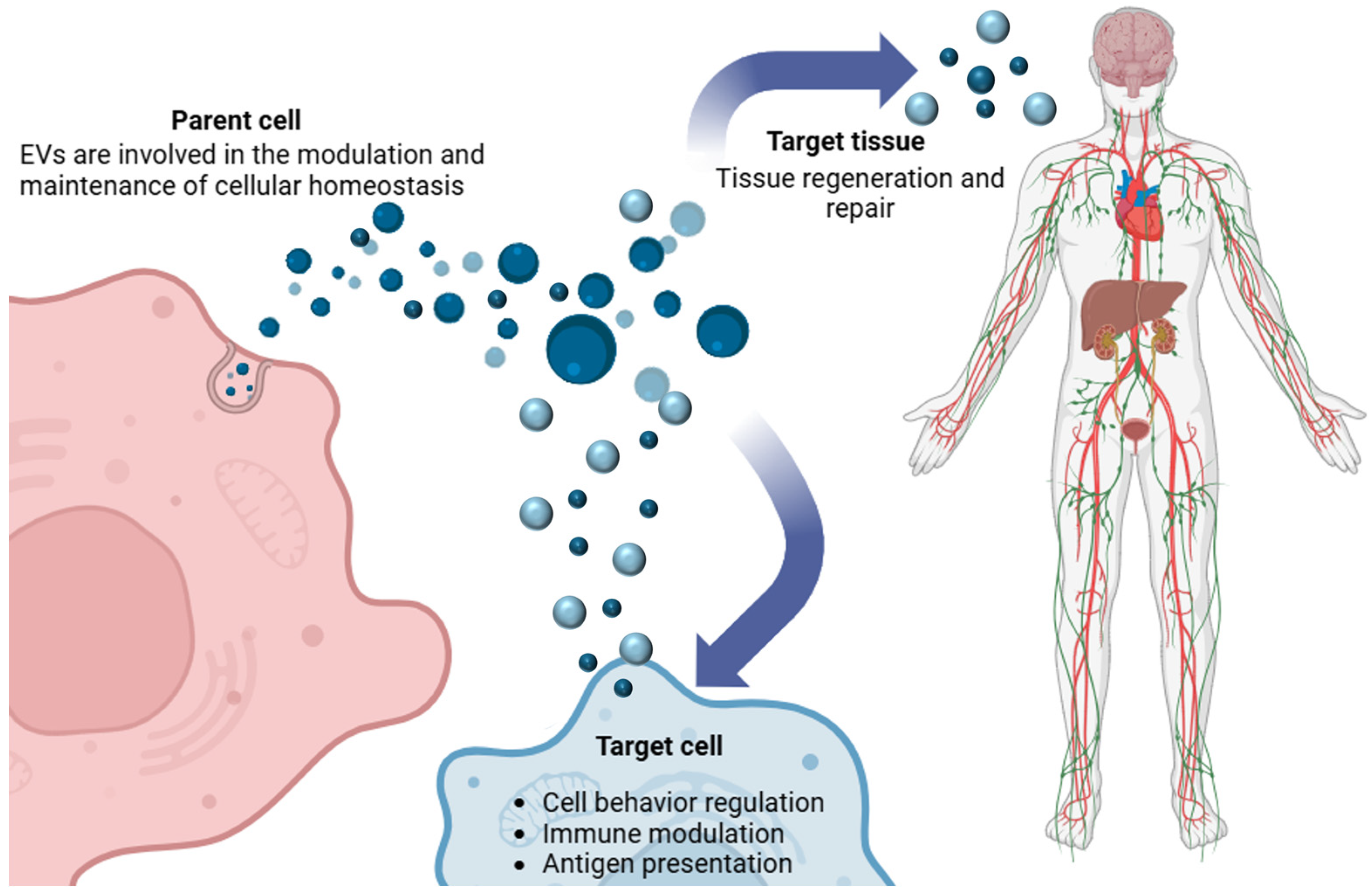
3.2. Biological Functions of Extracellular Vesicles

EVs play crucial and multifaceted roles in cellular function, both in healthy and pathological states. EVs can act as efficient shuttles of a large variety of molecules, both on their surface and their interior, between cells and/or tissues. They are important mediators in continuously adjustable and complex communication networks, working at both local and long distances in inter-organ and inter-organism communication [131].
The broadest and most complex function of EVs is that of mediating intercellular, horizontal communication, needed for the inter-organism interactions of multicellular eukaryotes. Indeed, EVs are principal actors in intercellular communication that subverts classical cellular limits through their capacity for long-distance delivery of information. EVs are equipped with molecular substrates, in particular, a complex profile of lipids, proteins, nucleic acids, and polysaccharides, which make them suitable for a wide-ranging function in modulating cell signaling processes, modulating the immune system, and many biological outcomes, including therapeutic effects [132,133,134]. In addition, multiple subpopulations of EVs, characterized by their molecular profile and biological effects, suggest a high degree of specificity in cellular and tissue usage.

Intercellular Communication

Nowadays EVs are well-characterized carriers of bioactive cargo that ranges from proteins and lipids to genetic material. In the parent cell where they are generated, EVs are involved in the modulation and maintenance of cellular homeostasis. In addition, as signal-bearing structures, EVs impart information both within the complex milieu of bodily fluids and between discrete cell populations (Figure 3) [135]. Indeed, EVs are indispensable for the healthy functioning of multiple physiological phenomena, including development, cell behavior regulation, immune responses, antigen presentation, and tissue regeneration and repair [136,137]. These incoming signals from other cells trigger a host of outcomes, including the unfolding of signaling pathways to induce cellular phenotypes, and may be different across cell types or stimuli. For example, EV cargoes can contribute to cellular “decisions” about migration, the uptake of glucose, the production of new proteins, and even participation in the coagulation cascade [138]. Examples of biological EV cargo include transcription factors, functional messenger RNAs, miRNAs, proteins, inflammatory mediators, and lipids that contribute to membrane structure and signaling [139,140]. Once released, EVs may fuse with other cell populations to initiate new signals or hit metabolic targets such as the mitochondria, a key site for strategic modulation of cellular metabolism. More recent research has revealed that much structural complexity exists within EVs. By analyzing EV membranes, proteins, and nucleic acids, researchers can identify the most probable types of cells that released the vesicles, the medical conditions those cells might be experiencing, and the prognosis for the associated diseases [141,142].

3.3. Extracellular Vesicles in Environmental Pollution

EVs and their cargoes can demonstrate the physiology of the organisms from which they are released and the diseases from which they originate. The presence of molecular signatures derived from the environment in circulating EVs could offer insights into the current state of the impact of environmental exposures on individual organisms [143]. Examining the presence of certain classes of small RNAs carried in EVs could be informative, more broadly for the status of ecosystems, with relevant roles in regulating the interaction between organisms and responses to local environmental stressors [144,145]. Specific vesicles can be released following various stress factors, including exposure to environmental hazards [143]. Studies on animals revealed that exposure to environmental pollutants such as heavy metals and oxidants can affect the quantity of EVs released, as well as the protein or nucleic cargo [145,146]. Moreover, EVs can mediate changes in cellular function in response to these environmental stresses. They might act to share information or to instigate responses in recipient cells. Furthermore, the presence in environmental samples of biomolecules, similar to those of EVs, suggests that EVs could also potentially work as biomarkers for environmental pollution in higher organisms, thereby complementing cellular stress markers [143]. Indeed, the presence of pollutants can influence the behavior of cells through several different molecular mechanisms. Since EVs can mediate communication between cells and involve complex cellular pathways, including immune system activation, proliferation, bone remodeling, oxidative stress responses, and protection from apoptosis or cytotoxic effects, they could possibly be used to study specific responses to environmental stressors.
Historically, EVs have been associated with human and animal pathology rather than with responses to environmental pollution. This is currently an increasingly developing field of study. The first paper focusing specifically on EVs attributed to responses to environmental stress appeared in 2017 [147]. Most investigations have then focused on the role of EVs as carriers of cellular proteins and/or RNAs and their changes with environmental stress. In such cases, one can expect a disruption of informational pathways, primarily between exposed and unexposed cell populations. In addition, recent in vitro studies suggest protection against the harmful effects of environmental xenobiotics. In a mouse model of pollution, in which mice were exposed to air ultrafine particulate matter, it was shown that pulmonary cells launched mesenchymal stem cell-derived (MSC) EVs to reduce lung inflammation through the activation of T-helper cells secreting anti-inflammatory cytokines [148]. In another set of experiments using the same mouse model, it was found that after exposure of MSCs to air pollutants, EVs isolated from these cells were enriched in miRNAs involved in cellular pathways that are activated after exposure to contaminants [148].
As our understanding of EVs continues to evolve, it is becoming increasingly evident that EVs play a crucial role in the dynamic interactions between organisms and their environments. Through their ability to carry and transport vital substances, EVs not only facilitate communication within and between cells but also serve as key messengers in maintaining the integrity and functioning of ecosystems. The exchange of materials and information facilitated by EVs allows for the rapid response and adaptation of organisms to environmental stressors, empowering them to better cope with and potentially mitigate the harmful effects of pollution.

4. Mechanisms of Microplastic–Extracellular Vesicle (MP–EV) Interaction

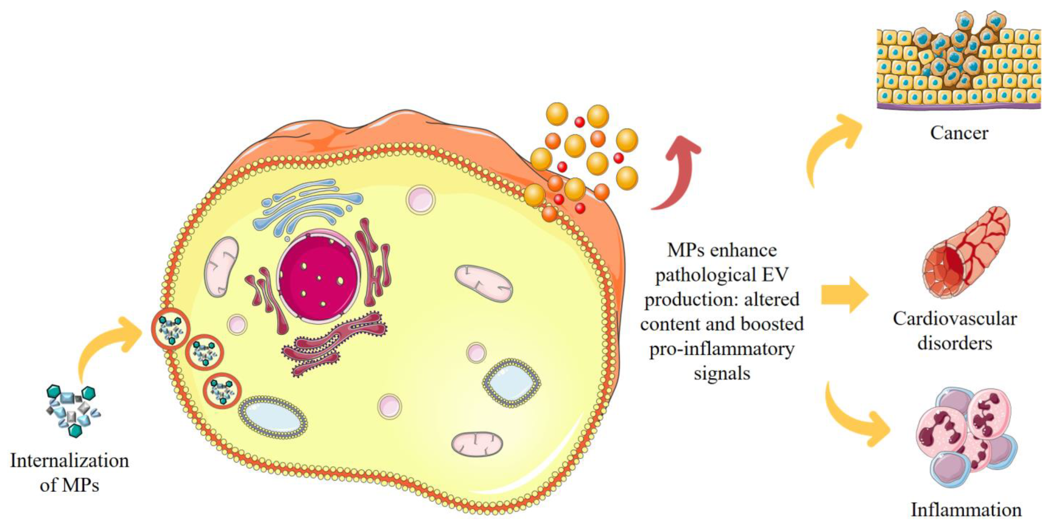
4.1. Impact of MP–EV Interaction on Cellular Function

In recent years, MPs have emerged as pervasive contaminants, finding their place also in clinical settings as a possible cause of cellular damage and promotion of pathologies [13,14,15,16,149]. These small particles, resulting from the degradation of plastic, have been detected in numerous human tissues, including the placenta, lungs, and the gastrointestinal tract, where they accumulate thus representing a tangible threat to health. One of the most fascinating aspects is to understand how MPs influence cell signaling. In this light, the interaction with cell signaling mechanisms mediated by EVs is certainly one of the most fascinating aspects to investigate. MPs can disrupt the release and composition of EVs in pathological settings, changing their content and boosting pro-inflammatory signals. This can boost pro-inflammatory signaling pathways, exacerbate oxidative stress, and contribute to pathological conditions such as cancer and cardiovascular disorders (Figure 4) [134,150,151]. This interaction suggests that MPs may act as bioactive pollutants, not only exerting direct cytotoxic effects but also indirectly amplifying pathological processes through their impact on EV-mediated intercellular communication. The interaction between MPs and EVs is a captivating area of research that highlights the complex interplay between environmental pollutants and biological systems. The literature on this topic is still incomplete and reveals critical insights into the roles and interactions of MPs with EVs. However, emerging studies provide critical insights into the roles and mechanisms through which MPs influence EV function, offering a new perspective to evaluate the broader implications of MP contamination on human health.
Numerous negative effects have been linked to the accumulation of MPs in human and animal cells; recent research showed that plastic exposure increases the number of EVs released by cells, but the mechanisms by which MPs accumulate and move between cells are unknown [108]. The impact of MPs and EVs on cellular systems has mainly been studied in the context of their potential synergistic effect on cellular toxicities [152]. EVs can act as vectors for MPs, facilitating their transport and increasing the activation of toxic cellular signals. The cargo of EVs and their biological effects on recipient cells can therefore be significantly influenced by the uptake of MPs.

4.2. MP–EV Interaction: Implications for Human Health

There is extensive literature documenting the role of EVs in cell-to-cell communication under normal conditions, but it is important to emphasize that EVs also mediate communication in response to stress conditions, releasing signals that reflect the state of cellular health. For example, in response to an acidifying environment, hypoxia, radiation, or cytotoxic drugs, cells release EVs enriched with effector molecules that modulate immune responses and cell growth or build the microenvironment that will be more suitable for their survival. [153,154]. In the last years, the nature of the MP–EV interaction was the topic of some studies that demonstrated distinct outcomes (Table 3). In human melanoma cells, the interaction with MPs was involved in the alteration of several signaling pathways, including those related to mitochondrial function, apoptosis, metabolic/anabolic processes, fatty acid synthesis, autophagy, and mitochondrial dynamics [155]. In a recent paper, after the injection of polystyrene MPs (PS-MPs) in zebrafish, in addition to the expression of genes involved in detoxification processes, lipid oxidation, and some ATPase complexes, kidney cells exhibited increased injury by enhancing oxidative stress, autophagy, apoptosis, and fibrosis. In particular, it was observed that exposure to aged PS-MPs caused oxidative damage, evidenced by a significant increase in ROS and DNA damage. This resulted in a reduction in mitochondrial membrane potential and the release of cytochrome c (cyt c) from mitochondria with subsequent activation of caspase-9/-3 signaling pathways that can induce cell death through mitochondrial apoptosis [121,156]. In a study conducted by Mierzejewski et al. [38], 27 miRNAs were detected within EVs isolated from the serum of pigs treated with low or high doses of PET-MPs. The results suggest that some of these miRNAs, whose expression was altered by PET, could contribute to the pathogenesis of lifestyle-related diseases. In particular, miRNAs differentially expressed in serum EVs of gilts exposed to different doses of PET-MPs have been associated with conditions such as obesity, insulin resistance, diabetes, and metabolic syndrome. Furthermore, an increase in the expression of ssc-miR-222, ssc-miR-146a-5p, and ssc-let-7d-3p was observed in EVs following administration of a low dose of PET-MPs. Numerous studies have shown for example that miR-222 is involved in the reduction of insulin sensitivity, through the negative regulation of ERα and GLUT4 receptors in adipose tissue, and that it may play a key role in the development of insulin resistance [157]. miRNAs involved in the pathogenesis of cardiovascular diseases have also been investigated and, among these, miR-136-3p within serum EVs was recorded as downregulated in both experimental groups. In fact, overexpression of miR-136-3p exerts beneficial effects on histopathological damage of the myocardium, helping to reduce oxidative stress and the inflammatory response in cases of coronary artery disease. Finally, an alteration in the expression of miRNAs related to carcinogenesis was also detected, such as the miRNA ssc-miR-31 which was upregulated in EVs collected from the group exposed to a low dose of PET-derived MPs. This miRNA was overexpressed in several types of cancer, including non-small cell lung cancer, colorectal cancer, pancreatic cancer, and cervical cancer. On the contrary, a decrease in ssc-miR-31 expression was observed in tumors such as breast, ovarian, prostate, hepatocellular, and gastric cancer [38,158]. In a study by Wang et al. [159], it was demonstrated that PS-MPs can be incorporated into and transported by EVs through a process characterized by cellular stress and the production of ROS. Using repeated doses of PS-MPs in mouse models, researchers observed that MPs accumulate in the kidneys and stimulate renal cells to release an increased number of EVs, which present with a variable size range in response to the concentration of PS-MPs. ROS generated by PS-MPs interacted with cellular proteins, thus altering their conformation and inducing pro-fibrotic signals that contribute to scar tissue formation in the kidneys. In particular, conditioned medium obtained from cells treated with PS-MPs was found to induce endoplasmic reticulum stress, with an increase in protein markers associated with this type of stress. This effect was observed in both renal tubular cells and fibroblasts, indicating that PS-MP accumulation may systemically impair cellular function. A further focus of the study was on autophagy and its regulation by EVs. Using a cellular model with a knockdown of Beclin 1, a regulator of autophagy, it was shown that the reduction of Beclin 1 significantly decreased EV production. The increase in the autophagy marker LC3 observed in cells exposed to PS-MPs suggests that MP accumulation stimulates autophagy as an adaptive mechanism. However, since some plastic particles are not degradable, autophagy itself may be ineffective, contributing to the release of EVs containing PS-MPs that can carry damage signals to other cells and organs. Recent studies have shown that PS-MPs and PS-nanoplastics (PS-NPs) can alter the intestinal barrier function in rats through the transport mediated by exosomes, small EVs that facilitate the transfer of molecular signals between cells. Prolonged exposure to PS-MPs and PS-NPs showed significant changes in the miRNA profile in serum and intestinal exosomes, in particular with the reduction of miR-126a-3p. This miRNA regulates the PI3K-Akt pathway, critical for the protection of cells from oxidative stress and apoptosis [160]. The reduction of miR-126a-3p in rats exposed to PS-MPs and PS-NPs is associated with the weakening of the barrier function and the increase in intestinal permeability, phenomena related to an increase in apoptosis and oxidative stress in epithelial cells. In another study conducted by Yan et al. [161], gastric adenocarcinoma (AGS) cells were exposed to a combined toxicity of PS-NPs and oleic acid, and the content of expressed miRNAs in exosomes derived from AGS cells was analyzed by miRNA sequencing. Both nanoparticles and oleic acid were observed to significantly affect the composition of miRNAs in exosomes, suggesting a mechanism of interaction between pollutants and cell-to-cell communication. Among the identified miRNAs, miR-1-3p and miR-1248 stood out as key indicators of toxic effects resulting from the tested exposures. These analyses demonstrated that exosomes can indirectly reflect the toxicity of donor cells, highlighting how exposure to MPs (or NPs) and other environmental pollutants induces significant changes in the released miRNA profiles. These results suggest that the analysis of exosomes, and in particular of the miRNAs contained therein, represents a new frontier for the assessment of biotoxicity associated with MP pollution. Although the data are mainly based on miRNA sequencing and cellular toxicity, they provide a theoretical basis for considering EVs as promising biomarkers to monitor the impact of MPs and other pollutants. It has been shown that, following alteration of the bowel tissue microenvironment due to exposure to MPs (and NPs), such as in the case of vascular or intestinal damage, platelets can be activated and release a variety of mediators. These include lipids, such as thromboxane (TX) A2, prostaglandin (PG) E2, and 12S-hydroxyeicosatetraenoic acid (12S-HETE); proteins including growth factors, proteases, and cytokines; genetic material; and miRNA-rich EVs. This release of mediators activates stromal cells, including fibroblasts, inflammatory cells, and endothelial cells, thus promoting chronic inflammation and contributing to the risk of intestinal tumorigenesis [150,162,163].

4.3. Evidence of MP Transport Mediated by EVs

In addition to altering EV-mediated signals, MPs have been shown to be transported into EVs. In the same study cited above and conducted by Mierzejewski et al. [38], it was demonstrated, via an approach based on the unique autofluorescent properties of PET and those of EV-associated proteins, that PET particles can be transported into EVs. PET emits in the wavelength range between 290 nm and 500 nm with a specific peak at approximately 380 nm. To distinguish the PET signal from other cellular components, EVs isolated from serum were analyzed with fluorescence spectroscopic and time-resolved measurements. In particular, the interaction between MPs and EV-associated proteins was examined (molecular interactions and motions monitored in the picosecond–nanosecond time range) by analyzing the fluorescence of the amino acid residues tyrosine (Tyr) and tryptophan (Trp), known to respond to conformational changes in proteins. High-PET samples showed significant changes in the Tyr signal compared to control samples, suggesting a conformational change due to the presence of PET. Time-resolved measurements confirmed that the fluorescence decay, modeled with a four-exponential function, was consistent with Trp emission, but also contained a decay time specific to PET, thus directly identifying the presence of PET in EVs [38]. It was also demonstrated that PS-MPs can be transferred between cells by EVs using a dual-staining and real-time imaging system. First, fluorescent PS-MPs were used to treat cells, and EVs were labeled with a second fluorophore to allow simultaneous detection of PS-MP and EV fluorescence. This dual-staining was confirmed by flow cytometry, which revealed the presence of PS-MPs within EVs extracted from treated cells. Then, EVs containing PS-MPs were introduced into new cells to monitor the internalization of fluorescent particles with real-time detection. Using a live-cell imaging system, it was possible to observe how the labeled EVs gradually approached and fused with cell membranes, transferring PS-MPs into host cells. The observation was further intensified by using 3D stack confocal microscopy, which allowed detailed visualization of the interaction and entry of PS-MPs into cells through EVs [164]. This procedure thus confirmed the role of EVs in the cell-to-cell transfer of PS-MPs, demonstrating that MPs can indeed use EVs as vehicles to migrate and accumulate in new cellular environments. In the study conducted by Wang et al., in 2024, the presence of PS-MPs in renal cell-derived EVs was confirmed by Raman analysis, highlighting an EV-mediated cell-to-cell transfer mechanism [159]. A recent study showed that PS-NPs are internalized by primary astroglial cells, accumulating in the cytoplasm and interacting with the endosomal–lysosomal system [165]. PS-NPs, once incorporated into cells, are transported through early endosomes and lysosomes, following an intracellular transport pathway common to other nanoparticles. This dynamic was confirmed by observing the co-localization of PS-NPs with endosome- and lysosome-specific markers. With increasing exposure time, PS-NPs form fluorescent aggregates inside endosomes, creating large vesicle-like structures that reflect a progressive stabilization of the nanoparticles in the internal membranes of endosomes, due to the interaction with the cellular microenvironment. In addition to the internalization process, a portion of PS-NPs was observed inside EVs released by astrocytes, suggesting a possible mechanism of EV-mediated lysosomal exocytosis. The presence of PS-NPs in EVs was evidenced by both transmission electron microscopy (TEM), which showed spherical structures containing fluorescent signals corresponding to PS-NPs, and confocal microscopy which revealed a green fluorescence intensity proportional to the PS-NP concentration and exposure time. These results suggest that EVs mediate the transport of PS-NPs between cells, facilitating the excretion of nanoparticles from the cytoplasm and potentially propagating toxic effects to the surrounding cells. This mechanism of extracellular release through EVs may represent a route of elimination of PS-NPs, reducing toxicity in donor cells; however, it could also promote the transfer of PS-NPs towards recipient cells, contributing to the spread of pro-inflammatory and cytotoxic effects in the surrounding microenvironment.

5. Ecological Implications: Microplastic and Extracellular Vesicle Interaction in Ecosystems

The interaction of MPs and EVs in natural ecosystems is an emerging and critical environmental concern. MPs, widespread in aquatic, terrestrial, and even atmospheric environments, have been shown to alter biological systems at all levels, influencing cellular function, organism health, and ecosystem stability [166]. In this context, EVs can act as inadvertent vectors of MPs within and between organisms, amplifying the reach and potential harm of plastic pollutants. This interaction adds a new dimension to the threat posed by MPs, suggesting far-reaching implications for biodiversity, ecosystem resilience, and, ultimately, global ecological balance. The ability of MPs to influence biological processes is widely recognized, and their presence has been documented in different environments, from the most remote ocean waters to agricultural soils and the atmosphere. The effects of MPs on ecosystems are complex and manifest through multiple pathways, by altering the microbial ecosystem of soil and water [167,168] or interfering with fundamental biological mechanisms of higher organisms, such as cellular signaling and hormonal regulation, following ingestion by numerous species [169].

5.1. MPs as Bioactive Pollutants in Ecosystems

MPs can modify biological and ecological systems through their chemical composition and surface properties, which attract and accumulate contaminants such as heavy metals and persistent organic pollutants. In aquatic ecosystems, MPs are ingested by filter feeders, zooplankton, and other keystone species, disrupting nutrient uptake, growth, and reproduction rates [170]. When these MPs are encapsulated or transported by EVs, they can penetrate deeper into cells and tissues, exacerbating toxic effects on key physiological processes. This persistence and bioactivity in organisms pose a major threat to biodiversity, especially in species with critical ecological roles for food networks and ecosystem functions, such as primary producers and key predators. The presence of MPs in the ecosystem processes may threaten the natural cycles that support life. MPs could disrupt habitat development, nutrient cycling, and predator–prey dynamics by affecting the health and function of keystone species. For instance, MPs may impair the primary productivity of species like phytoplankton or zooplankton in marine ecosystems, influencing carbon sequestration and oxygen production [171]. Over time, such disruptions could compromise ecosystem resilience, reduce biodiversity, and increase vulnerability to climate change and other environmental stressors. While these potential consequences are alarming, further studies are needed to quantify and fully understand the long-term impacts of chronic MP exposure on ecosystem stability [172,173].

5.2. Biological Amplification via EV-Mediated MP Transport

The role of EVs as natural molecular carriers allows MPs to reach previously inaccessible cellular environments, increasing their biological impact. This transport allows MPs to cross cellular barriers, affecting tissues and organs that would otherwise be protected. As EVs move across organisms, from cells to organs, and potentially from species to species within food networks, they increase the likelihood that MPs will disrupt intercellular signaling, immune responses, and metabolic functions. This increased cellular infiltration leads to a cascade of physiological changes that, if widespread across species, can affect entire ecosystems [174]. The amplification effect suggests that even low concentrations of MPs could have disproportionately large ecological impacts, especially in delicate ecosystems such as coral reefs, wetlands, and estuaries, where biodiversity is essential for resilience. The full ecological significance of these processes is still being explored due to the lack of long-term monitoring programs and the lack of comprehensive regulatory frameworks addressing MP pollution in vulnerable ecosystems. EVs containing MPs contribute to the bioaccumulation and biomagnification of plastics and associated contaminants within food networks [175]. MPs, once ingested by lower trophic level organisms, can be transferred upward to larger predators via EV-mediated pathways. This movement of MPs through trophic levels can alter the health of entire populations, compromising reproduction, growth, and survival rates in higher trophic-level organisms, including fish, birds, and marine mammals. This form of EV transfer is particularly insidious because vesicles not only facilitate the movement of MPs across biological barriers but can also protect particles from immediate degradation, thereby increasing their stability and persistence in host organisms. Furthermore, biomagnification amplifies the toxic load on predators, some of which are ecologically significant or endangered. The potential for MPs to reach humans through the consumption of contaminated food or water also poses serious health risks, underscoring the need for a more complete understanding of these ecological pathways and their toxicological ramifications.

6. Conclusions

The interaction between MPs and EVs represents an emerging field of research aimed at understanding the potential risks for human health and the environment associated with the extensive distribution of MPs. Recent studies suggest that EVs have the ability to carry MPs, facilitating their penetration into tissues and amplifying their harmful effects by altering critical cellular processes. These complex interaction mechanisms raise particular concerns since MPs transported by vesicles can bypass biological barriers and accumulate in sensitive organs leading to toxicological effects that are still unpredictable. From an environmental perspective, such interactions could negatively affect food chains and biodiversity, posing significant threats to ecological integrity. Further research should investigate how EVs interact with MPs of different compositions and sizes, in order to better characterize the associated risks and develop effective mitigation strategies. In addition, future directions should include social awareness to reduce the use and dispersion of MPs in the environment, promoting sustainable waste management, and adopting more restrictive policies to limit their impact on the environment and human health.

Author Contributions

Conceptualization, E.C., N.M. and A.C.; writing—original draft preparation, E.C., N.M., A.B. and G.C.; writing—review and editing, E.C., N.M. and A.C.; visualization, E.C., N.M., A.B. and G.C.; supervision, A.C., G.G. and C.E. All authors have read and agreed to the published version of the manuscript.

Funding

This work was funded by the European Union—NextGenerationEU under the Italian Ministry of University and Research (MUR) National Innovation Ecosystem grant ECS00000041-VITALITY-CUP J97G22000170005 (to Prof. Carla Emiliani).

Acknowledgments

We acknowledge Università degli Studi di Perugia and the Italian Ministry of University and Research (MUR) for their support within the VITALITY project.

Conflicts of Interest

The authors declare no conflicts of interest.

References

  1. Rodríguez-Seijo, A.; Pereira, R. Chapter 3—Morphological and Physical Characterization of Microplastics. In Comprehensive Analytical Chemistry; Rocha-Santos, T.A.P., Duarte, A.C., Eds.; Characterization and Analysis of Microplastics; Elsevier: Amsterdam, The Netherlands, 2017; Volume 75, pp. 49–66. [Google Scholar]
  2. Caputo, F.; Vogel, R.; Savage, J.; Vella, G.; Law, A.; Della Camera, G.; Hannon, G.; Peacock, B.; Mehn, D.; Ponti, J.; et al. Measuring Particle Size Distribution and Mass Concentration of Nanoplastics and Microplastics: Addressing Some Analytical Challenges in the Sub-Micron Size Range. J. Colloid Interface Sci. 2021, 588, 401–417. [Google Scholar] [CrossRef] [PubMed]
  3. Tirkey, A.; Upadhyay, L.S.B. Microplastics: An Overview on Separation, Identification and Characterization of Microplastics. Mar. Pollut. Bull. 2021, 170, 112604. [Google Scholar] [CrossRef]
  4. Gwenzi, W.; Simbanegavi, T.T.; Mahdi, H.I.; Azelee, N.I.W.; Muisa-Zikali, N.; Rangabhashiyam, S. Chapter 14—Occurrence and Ecological Health Risks of Microplastics. In Emerging Contaminants in the Terrestrial-Aquatic-Atmosphere Continuum; Gwenzi, W., Ed.; Elsevier: Amsterdam, The Netherlands, 2022; pp. 243–270. ISBN 978-0-323-90051-5. [Google Scholar]
  5. Mamun, A.A.; Prasetya, T.A.E.; Dewi, I.R.; Ahmad, M. Microplastics in Human Food Chains: Food Becoming a Threat to Health Safety. Sci. Total Environ. 2023, 858, 159834. [Google Scholar] [CrossRef] [PubMed]
  6. Saeedi, M. How Microplastics Interact with Food Chain: A Short Overview of Fate and Impacts. J. Food Sci. Technol. 2024, 61, 403–413. [Google Scholar] [CrossRef] [PubMed]
  7. Yee, M.S.-L.; Hii, L.-W.; Looi, C.K.; Lim, W.-M.; Wong, S.-F.; Kok, Y.-Y.; Tan, B.-K.; Wong, C.-Y.; Leong, C.-O. Impact of Microplastics and Nanoplastics on Human Health. Nanomaterials 2021, 11, 496. [Google Scholar] [CrossRef] [PubMed]
  8. Ziani, K.; Ioniță-Mîndrican, C.-B.; Mititelu, M.; Neacșu, S.M.; Negrei, C.; Moroșan, E.; Drăgănescu, D.; Preda, O.-T. Microplastics: A Real Global Threat for Environment and Food Safety: A State of the Art Review. Nutrients 2023, 15, 617. [Google Scholar] [CrossRef] [PubMed]
  9. Jung, Y.S.; Sampath, V.; Prunicki, M.; Aguilera, J.; Allen, H.; LaBeaud, D.; Veidis, E.; Barry, M.; Erny, B.; Patel, L.; et al. Characterization and Regulation of Microplastic Pollution for Protecting Planetary and Human Health. Environ. Pollut. 2022, 315, 120442. [Google Scholar] [CrossRef]
  10. Pilapitiya, P.N.T.; Ratnayake, A.S. The World of Plastic Waste: A Review. Clean. Mater. 2024, 11, 100220. [Google Scholar] [CrossRef]
  11. Ghosh, S.; Sinha, J.K.; Ghosh, S.; Vashisth, K.; Han, S.; Bhaskar, R. Microplastics as an Emerging Threat to the Global Environment and Human Health. Sustainability 2023, 15, 10821. [Google Scholar] [CrossRef]
  12. Xia, C.; Cai, L.; Lam, S.S.; Sonne, C. Microplastics Pollution: Economic Loss and Actions Needed. Eco Environ. Health 2023, 2, 41–42. [Google Scholar] [CrossRef] [PubMed]
  13. Goswami, S.; Adhikary, S.; Bhattacharya, S.; Agarwal, R.; Ganguly, A.; Nanda, S.; Rajak, P. The Alarming Link between Environmental Microplastics and Health Hazards with Special Emphasis on Cancer. Life Sci. 2024, 355, 122937. [Google Scholar] [CrossRef] [PubMed]
  14. Kumar, N.; Lamba, M.; Pachar, A.K.; Yadav, S.; Acharya, A. Microplastics—A Growing Concern as Carcinogens in Cancer Etiology: Emphasis on Biochemical and Molecular Mechanisms. Cell Biochem. Biophys. 2024, 82, 3109–3121. [Google Scholar] [CrossRef]
  15. Park, J.H.; Hong, S.; Kim, O.-H.; Kim, C.-H.; Kim, J.; Kim, J.-W.; Hong, S.; Lee, H.J. Polypropylene Microplastics Promote Metastatic Features in Human Breast Cancer. Sci. Rep. 2023, 13, 6252. [Google Scholar] [CrossRef] [PubMed]
  16. Kehinde, S.A.; Fatokun, T.P.; Olajide, A.T.; Praveena, S.M.; Sokan-Adeaga, A.A.; Adekunle, A.P.; Fouad, D.; Papadakis, M. Impact of Polyethylene Microplastics Exposure on Kallikrein-3 Levels, Steroidal-Thyroidal Hormones, and Antioxidant Status in Murine Model: Protective Potentials of Naringin. Sci. Rep. 2024, 14, 23664. [Google Scholar] [CrossRef]
  17. Zheng, P.C.; Li, R.; Lai, K.P.; Zhang, X.X. Biological Exposure to Microplastics and Nanoplastics and Plastic Additives: Impairment of Glycolipid Metabolism and Adverse Effects on Metabolic Diseases. Environ. Sci. Pollut. Res. 2024, 31, 60778–60791. [Google Scholar] [CrossRef]
  18. Wright, S.L.; Kelly, F.J. Plastic and Human Health: A Micro Issue? Environ. Sci. Technol. 2017, 51, 6634–6647. [Google Scholar] [CrossRef] [PubMed]
  19. Pelegrini, K.; Pereira, T.C.B.; Maraschin, T.G.; Teodoro, L.D.S.; Basso, N.R.D.S.; De Galland, G.L.B.; Ligabue, R.A.; Bogo, M.R. Micro- and Nanoplastic Toxicity: A Review on Size, Type, Source, and Test-Organism Implications. Sci. Total Environ. 2023, 878, 162954. [Google Scholar] [CrossRef] [PubMed]
  20. Luo, T.; Zhang, Y.; Wang, C.; Wang, X.; Zhou, J.; Shen, M.; Zhao, Y.; Fu, Z.; Jin, Y. Maternal Exposure to Different Sizes of Polystyrene Microplastics during Gestation Causes Metabolic Disorders in Their Offspring. Environ. Pollut. 2019, 255, 113122. [Google Scholar] [CrossRef] [PubMed]
  21. Ragusa, A.; Svelato, A.; Santacroce, C.; Catalano, P.; Notarstefano, V.; Carnevali, O.; Papa, F.; Rongioletti, M.C.A.; Baiocco, F.; Draghi, S.; et al. Plasticenta: First Evidence of Microplastics in Human Placenta. Environ. Int. 2021, 146, 106274. [Google Scholar] [CrossRef] [PubMed]
  22. Sun, H.; Chen, N.; Yang, X.; Xia, Y.; Wu, D. Effects Induced by Polyethylene Microplastics Oral Exposure on Colon Mucin Release, Inflammation, Gut Microflora Composition and Metabolism in Mice. Ecotoxicol. Environ. Saf. 2021, 220, 112340. [Google Scholar] [CrossRef]
  23. Singh, B.; Kumar, A. Advances in Microplastics Detection: A Comprehensive Review of Methodologies and Their Effectiveness. TrAC Trends Anal. Chem. 2024, 170, 117440. [Google Scholar] [CrossRef]
  24. Marcuello, C. Present and Future Opportunities in the Use of Atomic Force Microscopy to Address the Physico-Chemical Properties of Aquatic Ecosystems at the Nanoscale Level. Int. Aquat. Res. 2022, 14, 231–240. [Google Scholar] [CrossRef]
  25. Calzoni, E.; Cesaretti, A.; Montegiove, N.; Di Michele, A.; Emiliani, C. Enhanced Stability of Long-Living Immobilized Recombinant β-d-N-Acetyl-Hexosaminidase A on Polylactic Acid (PLA) Films for Potential Biomedical Applications. J. Funct. Biomater. 2021, 12, 32. [Google Scholar] [CrossRef] [PubMed]
  26. Calzoni, E.; Cesaretti, A.; Montegiove, N.; Di Michele, A.; Pellegrino, R.M.; Emiliani, C. HexA-Enzyme Coated Polymer Nanoparticles for the Development of a Drug-Delivery System in the Treatment of Sandhoff Lysosomal Storage Disease. J. Funct. Biomater. 2022, 13, 37. [Google Scholar] [CrossRef]
  27. Montegiove, N.; Calzoni, E.; Emiliani, C.; Cesaretti, A. Biopolymer Nanoparticles for Nose-to-Brain Drug Delivery: A New Promising Approach for the Treatment of Neurological Diseases. J. Funct. Biomater. 2022, 13, 125. [Google Scholar] [CrossRef]
  28. Zangi, A.R.; Amiri, A.; Borzouee, F.; Bagherifar, R.; Pazooki, P.; Hamishehkar, H.; Javadzadeh, Y. Immobilized Nanoparticles-Mediated Enzyme Therapy; Promising Way into Clinical Development. Discov. Nanoscale Res. Lett. 2023, 18, 55. [Google Scholar] [CrossRef]
  29. Kalluri, R.; LeBleu, V.S. The Biology, Function, and Biomedical Applications of Exosomes. Science 2020, 367, eaau6977. [Google Scholar] [CrossRef] [PubMed]
  30. Cerrotti, G.; Buratta, S.; Latella, R.; Calzoni, E.; Cusumano, G.; Bertoldi, A.; Porcellati, S.; Emiliani, C.; Urbanelli, L. Hitting the Target: Cell Signaling Pathways Modulation by Extracellular Vesicles. Extracell. Vesicles Circ. Nucleic Acids 2024, 5, 627–652. [Google Scholar] [CrossRef] [PubMed]
  31. Chen, X.; Yang, F. Classification and Nomenclature of Extracellular Vesicles. In Extracellular Vesicles: From Bench to Bedside; Wang, Q., Zheng, L., Eds.; Springer Nature: Singapore, 2024; pp. 3–7. ISBN 978-981-9983-65-0. [Google Scholar]
  32. EL Andaloussi, S.; Mäger, I.; Breakefield, X.O.; Wood, M.J.A. Extracellular Vesicles: Biology and Emerging Therapeutic Opportunities. Nat. Rev. Drug Discov. 2013, 12, 347–357. [Google Scholar] [CrossRef]
  33. Mir, B.; Goettsch, C. Extracellular Vesicles as Delivery Vehicles of Specific Cellular Cargo. Cells 2020, 9, 1601. [Google Scholar] [CrossRef]
  34. Dellar, E.R.; Hill, C.; Melling, G.E.; Carter, D.R.F.; Baena-Lopez, L.A. Unpacking Extracellular Vesicles: RNA Cargo Loading and Function. J. Extracell. Biol. 2022, 1, e40. [Google Scholar] [CrossRef]
  35. Calzoni, E.; Argentati, C.; Cesaretti, A.; Montegiove, N.; Tortorella, I.; Bazzucchi, M.; Morena, F.; Martino, S.; Emiliani, C. RNA Modifications in Neurodegenerations. In Epitranscriptomics; Jurga, S., Barciszewski, J., Eds.; Springer International Publishing: Cham, Switzerland, 2021; pp. 23–77. ISBN 978-3-030-71612-7. [Google Scholar]
  36. Trianni, A.; Brossa, A.; Catalano, F.; Saraceni, A.; Bongiovanni, S.; Merlo, G.R.; Cottone, E.; Bovolin, P. Unraveling the molecular mechanisms of microplastics internalization and their intracellular impact. Eur. J. Histochem. 2024, 68, 47–48. [Google Scholar] [CrossRef]
  37. Bovolin, P.; Saraceni, A.; Cottone, E.; Mognetti, B.; Gallo, M.P.; Merlo, G.; Dal Bello, F. Microplastics and associated contaminants: Potential impact on cells and developing organisms. Eur. J. Histochem. 2023, 67, 5. [Google Scholar] [CrossRef]
  38. Mierzejewski, K.; Kurzyńska, A.; Golubska, M.; Całka, J.; Gałęcka, I.; Szabelski, M.; Paukszto, Ł.; Andronowska, A.; Bogacka, I. New Insights into the Potential Effects of PET Microplastics on Organisms via Extracellular Vesicle-Mediated Communication. Sci. Total Environ. 2023, 904, 166967. [Google Scholar] [CrossRef]
  39. Xu, Z.; Sui, Q.; Li, A.; Sun, M.; Zhang, L.; Lyu, S.; Zhao, W. How to Detect Small Microplastics (20–100 Μm) in Freshwater, Municipal Wastewaters and Landfill Leachates? A Trial from Sampling to Identification. Sci. Total Environ. 2020, 733, 139218. [Google Scholar] [CrossRef] [PubMed]
  40. Bandekar, M.; Abdolahpur Monikh, F.; Kekäläinen, J.; Tahvanainen, T.; Kortet, R.; Zhang, P.; Guo, Z.; Akkanen, J.; Leskinen, J.T.T.; Gomez-Gonzalez, M.A.; et al. Submicron Plastic Adsorption by Peat, Accumulation in Sphagnum Mosses and Influence on Bacterial Communities in Peatland Ecosystems. Environ. Sci. Technol. 2022, 56, 15661–15671. [Google Scholar] [CrossRef] [PubMed]
  41. Nigina; Sajith, V.; Nair, B.G. Microplastics and Nanoplastics in Terrestrial Systems. In Toxic Effects of Micro and Nanoplastics; John Wiley & Sons Ltd.: Hoboken, NJ, USA, 2024; pp. 163–213. ISBN 978-1-394-23816-3. [Google Scholar]
  42. Fu, W.; Min, J.; Jiang, W.; Li, Y.; Zhang, W. Separation, Characterization and Identification of Microplastics and Nanoplastics in the Environment. Sci. Total Environ. 2020, 721, 137561. [Google Scholar] [CrossRef] [PubMed]
  43. Kwak, J.I.; Liu, H.; Wang, D.; Lee, Y.H.; Lee, J.-S.; An, Y.-J. Critical Review of Environmental Impacts of Microfibers in Different Environmental Matrices. Comp. Biochem. Physiol. Part C Toxicol. Pharmacol. 2022, 251, 109196. [Google Scholar] [CrossRef]
  44. Nur, A.; Setiyawan, A.S.; Oginawati, K.; Soewondo, P. Degradation of polyethylene terephthalate (pet) as secondary microplastics under three different environmental conditions. GEOMATE J. 2022, 22, 10–16. [Google Scholar] [CrossRef]
  45. Siddiqui, S.A.; Bahmid, N.A.; Salman, S.H.M.; Nawaz, A.; Walayat, N.; Shekhawat, G.K.; Gvozdenko, A.A.; Blinov, A.V.; Nagdalian, A.A. Chapter Eight—Migration of Microplastics from Plastic Packaging into Foods and Its Potential Threats on Human Health. In Advances in Food and Nutrition Research; Özogul, F., Ed.; Nano/micro-Plastics Toxicity on Food Quality and Food Safety; Academic Press: Cambridge, MA, USA, 2023; Volume 103, pp. 313–359. [Google Scholar]
  46. Lambert, S.; Wagner, M. Characterisation of Nanoplastics during the Degradation of Polystyrene. Chemosphere 2016, 145, 265–268. [Google Scholar] [CrossRef] [PubMed]
  47. Lin, Z.; Jin, T.; Zou, T.; Xu, L.; Xi, B.; Xu, D.; He, J.; Xiong, L.; Tang, C.; Peng, J.; et al. Current Progress on Plastic/Microplastic Degradation: Fact Influences and Mechanism. Environ. Pollut. 2022, 304, 119159. [Google Scholar] [CrossRef] [PubMed]
  48. Sun, J.; Dai, X.; Wang, Q.; van Loosdrecht, M.C.M.; Ni, B.-J. Microplastics in Wastewater Treatment Plants: Detection, Occurrence and Removal. Water Res. 2019, 152, 21–37. [Google Scholar] [CrossRef] [PubMed]
  49. Sa’adu, I.; Farsang, A. Plastic Contamination in Agricultural Soils: A Review. Environ. Sci. Eur. 2023, 35, 13. [Google Scholar] [CrossRef]
  50. Porterfield, K.K.; Hobson, S.A.; Neher, D.A.; Niles, M.T.; Roy, E.D. Microplastics in Composts, Digestates, and Food Wastes: A Review. J. Environ. Qual. 2023, 52, 225–240. [Google Scholar] [CrossRef] [PubMed]
  51. Alijagic, A.; Suljević, D.; Fočak, M.; Sulejmanović, J.; Šehović, E.; Särndahl, E.; Engwall, M. The Triple Exposure Nexus of Microplastic Particles, Plastic-Associated Chemicals, and Environmental Pollutants from a Human Health Perspective. Environ. Int. 2024, 188, 108736. [Google Scholar] [CrossRef] [PubMed]
  52. Preston, C.A.; McKenna Neuman, C.L.; Aherne, J. Effects of Shape and Size on Microplastic Atmospheric Settling Velocity. Environ. Sci. Technol. 2023, 57, 11937–11947. [Google Scholar] [CrossRef]
  53. Sorolla-Rosario, D.; Llorca-Porcel, J.; Pérez-Martínez, M.; Lozano-Castelló, D.; Bueno-López, A. Study of Microplastics with Semicrystalline and Amorphous Structure Identification by TGA and DSC. J. Environ. Chem. Eng. 2022, 10, 106886. [Google Scholar] [CrossRef]
  54. Mortula, M.M.; Atabay, S.; Fattah, K.P.; Madbuly, A. Leachability of Microplastic from Different Plastic Materials. J. Environ. Manag. 2021, 294, 112995. [Google Scholar] [CrossRef]
  55. Hahladakis, J.N.; Velis, C.A.; Weber, R.; Iacovidou, E.; Purnell, P. An Overview of Chemical Additives Present in Plastics: Migration, Release, Fate and Environmental Impact during Their Use, Disposal and Recycling. J. Hazard. Mater. 2018, 344, 179–199. [Google Scholar] [CrossRef]
  56. Seewoo, B.J.; Goodes, L.M.; Mofflin, L.; Mulders, Y.R.; Wong, E.V.; Toshniwal, P.; Brunner, M.; Alex, J.; Johnston, B.; Elagali, A.; et al. The Plastic Health Map: A Systematic Evidence Map of Human Health Studies on Plastic-Associated Chemicals. Environ. Int. 2023, 181, 108225. [Google Scholar] [CrossRef]
  57. Campanale, C.; Massarelli, C.; Savino, I.; Locaputo, V.; Uricchio, V.F. A Detailed Review Study on Potential Effects of Microplastics and Additives of Concern on Human Health. Int. J. Environ. Res. Public Health 2020, 17, 1212. [Google Scholar] [CrossRef] [PubMed]
  58. Zhou, X.-X.; Liu, R.; Hao, L.-T.; Liu, J.-F. Identification of Polystyrene Nanoplastics Using Surface Enhanced Raman Spectroscopy. Talanta 2021, 221, 121552. [Google Scholar] [CrossRef]
  59. Lv, L.; He, L.; Jiang, S.; Chen, J.; Zhou, C.; Qu, J.; Lu, Y.; Hong, P.; Sun, S.; Li, C. In Situ Surface-Enhanced Raman Spectroscopy for Detecting Microplastics and Nanoplastics in Aquatic Environments. Sci. Total Environ. 2020, 728, 138449. [Google Scholar] [CrossRef]
  60. Gillibert, R.; Balakrishnan, G.; Deshoules, Q.; Tardivel, M.; Magazzù, A.; Donato, M.G.; Maragò, O.M.; Lamy de La Chapelle, M.; Colas, F.; Lagarde, F.; et al. Raman Tweezers for Small Microplastics and Nanoplastics Identification in Seawater. Environ. Sci. Technol. 2019, 53, 9003–9013. [Google Scholar] [CrossRef] [PubMed]
  61. Wahl, A.; Le Juge, C.; Davranche, M.; El Hadri, H.; Grassl, B.; Reynaud, S.; Gigault, J. Nanoplastic Occurrence in a Soil Amended with Plastic Debris. Chemosphere 2021, 262, 127784. [Google Scholar] [CrossRef] [PubMed]
  62. Davranche, M.; Lory, C.; Juge, C.L.; Blancho, F.; Dia, A.; Grassl, B.; El Hadri, H.; Pascal, P.-Y.; Gigault, J. Nanoplastics on the Coast Exposed to the North Atlantic Gyre: Evidence and Traceability. NanoImpact 2020, 20, 100262. [Google Scholar] [CrossRef]
  63. Xu, G.; Cheng, H.; Jones, R.; Feng, Y.; Gong, K.; Li, K.; Fang, X.; Tahir, M.A.; Valev, V.K.; Zhang, L. Surface-Enhanced Raman Spectroscopy Facilitates the Detection of Microplastics <1 μm in the Environment. Environ. Sci. Technol. 2020, 54, 15594–15603. [Google Scholar] [CrossRef]
  64. Thomas, D.; Schütze, B.; Heinze, W.M.; Steinmetz, Z. Sample Preparation Techniques for the Analysis of Microplastics in Soil—A Review. Sustainability 2020, 12, 9074. [Google Scholar] [CrossRef]
  65. Barili, S.; Bernetti, A.; Sannino, C.; Montegiove, N.; Calzoni, E.; Cesaretti, A.; Pinchuk, I.; Pezzolla, D.; Turchetti, B.; Buzzini, P.; et al. Impact of PVC Microplastics on Soil Chemical and Microbiological Parameters. Environ. Res. 2023, 229, 115891. [Google Scholar] [CrossRef] [PubMed]
  66. Guerranti, C.; Martellini, T.; Perra, G.; Scopetani, C.; Cincinelli, A. Microplastics in Cosmetics: Environmental Issues and Needs for Global Bans. Environ. Toxicol. Pharmacol. 2019, 68, 75–79. [Google Scholar] [CrossRef] [PubMed]
  67. Abbasi, S.; Moore, F.; Keshavarzi, B.; Hopke, P.K.; Naidu, R.; Rahman, M.M.; Oleszczuk, P.; Karimi, J. PET-Microplastics as a Vector for Heavy Metals in a Simulated Plant Rhizosphere Zone. Sci. Total Environ. 2020, 744, 140984. [Google Scholar] [CrossRef]
  68. Liu, S.; Huang, J.; Zhang, W.; Shi, L.; Yi, K.; Yu, H.; Zhang, C.; Li, S.; Li, J. Microplastics as a Vehicle of Heavy Metals in Aquatic Environments: A Review of Adsorption Factors, Mechanisms, and Biological Effects. J. Environ. Manag. 2022, 302, 113995. [Google Scholar] [CrossRef]
  69. Weis, J.S.; Palmquist, K.H. Reality Check: Experimental Studies on Microplastics Lack Realism. Appl. Sci. 2021, 11, 8529. [Google Scholar] [CrossRef]
  70. Liu, K.; Li, Q.; Andrady, A.L.; Wang, X.; He, Y.; Li, D. Underestimated Activity-Based Microplastic Intake under Scenario-Specific Exposures. Environ. Sci. Ecotechnol. 2024, 18, 100316. [Google Scholar] [CrossRef]
  71. Hale, R.C.; Seeley, M.E.; La Guardia, M.J.; Mai, L.; Zeng, E.Y. A Global Perspective on Microplastics. J. Geophys. Res. Ocean. 2020, 125, e2018JC014719. [Google Scholar] [CrossRef]
  72. Khan, A.; Jia, Z. Recent Insights into Uptake, Toxicity, and Molecular Targets of Microplastics and Nanoplastics Relevant to Human Health Impacts. iScience 2023, 26, 106061. [Google Scholar] [CrossRef] [PubMed]
  73. Sangkham, S.; Faikhaw, O.; Munkong, N.; Sakunkoo, P.; Arunlertaree, C.; Chavali, M.; Mousazadeh, M.; Tiwari, A. A Review on Microplastics and Nanoplastics in the Environment: Their Occurrence, Exposure Routes, Toxic Studies, and Potential Effects on Human Health. Mar. Pollut. Bull. 2022, 181, 113832. [Google Scholar] [CrossRef] [PubMed]
  74. Kwak, J.I.; An, Y.-J. Post COVID-19 Pandemic: Biofragmentation and Soil Ecotoxicological Effects of Microplastics Derived from Face Masks. J. Hazard. Mater. 2021, 416, 126169. [Google Scholar] [CrossRef] [PubMed]
  75. Chojnacka, K.; Mikulewicz, M. Bioaccumulation. In Encyclopedia of Toxicology, 3rd ed.; Wexler, P., Ed.; Academic Press: Oxford, UK, 2014; pp. 456–460. ISBN 978-0-12-386455-0. [Google Scholar]
  76. Cox, K.D.; Covernton, G.A.; Davies, H.L.; Dower, J.F.; Juanes, F.; Dudas, S.E. Human Consumption of Microplastics. Environ. Sci. Technol. 2019, 53, 7068–7074. [Google Scholar] [CrossRef] [PubMed]
  77. European Union. Directive (EU) 2019/904 of the European Parliament and of the Council of 5 June 2019 on the Reduction of the Impact of Certain Plastic Products on the Environment (Text with EEA Relevance). Off. J. Eur. Union 2019, 155, 1–19. [Google Scholar]
  78. EUR-Lex—L:2006:396:TOC—EN—EUR-Lex. Available online: https://eur-lex.europa.eu/legal-content/EN/TXT/?uri=OJ%3AL%3A2006%3A396%3ATOC (accessed on 11 December 2024).
  79. The Resumed Session of UNEA-5 (UNEA-5.2). Available online: http://www.unep.org/environmentassembly/unea-5.2 (accessed on 11 December 2024).
  80. Oßmann, B.E. Microplastics in Drinking Water? Present State of Knowledge and Open Questions. Curr. Opin. Food Sci. 2021, 41, 44–51. [Google Scholar] [CrossRef]
  81. Thiele, C.J.; Hudson, M.D.; Russell, A.E.; Saluveer, M.; Sidaoui-Haddad, G. Microplastics in Fish and Fishmeal: An Emerging Environmental Challenge? Sci. Rep. 2021, 11, 2045. [Google Scholar] [CrossRef]
  82. Amran, N.H.; Zaid, S.S.M.; Mokhtar, M.H.; Manaf, L.A.; Othman, S. Exposure to Microplastics during Early Developmental Stage: Review of Current Evidence. Toxics 2022, 10, 597. [Google Scholar] [CrossRef]
  83. Dawson, A.L.; Kawaguchi, S.; King, C.K.; Townsend, K.A.; King, R.; Huston, W.M.; Bengtson Nash, S.M. Turning Microplastics into Nanoplastics through Digestive Fragmentation by Antarctic Krill. Nat. Commun. 2018, 9, 1001. [Google Scholar] [CrossRef] [PubMed]
  84. Expósito, N.; Rovira, J.; Sierra, J.; Gimenez, G.; Domingo, J.L.; Schuhmacher, M. Levels of Microplastics and Their Characteristics in Molluscs from North-West Mediterranean Sea: Human Intake. Mar. Pollut. Bull. 2022, 181, 113843. [Google Scholar] [CrossRef] [PubMed]
  85. Kosuth, M.; Mason, S.A.; Wattenberg, E.V. Anthropogenic Contamination of Tap Water, Beer, and Sea Salt. PLoS ONE 2018, 13, e0194970. [Google Scholar] [CrossRef] [PubMed]
  86. Van Cauwenberghe, L.; Janssen, C.R. Microplastics in Bivalves Cultured for Human Consumption. Environ. Pollut. 2014, 193, 65–70. [Google Scholar] [CrossRef]
  87. Wu, J.; Lu, J.; Wu, J. Effect of Gastric Fluid on Adsorption and Desorption of Endocrine Disrupting Chemicals on Microplastics. Front. Environ. Sci. Eng. 2021, 16, 104. [Google Scholar] [CrossRef]
  88. Wagstaff, A.; Petrie, B. Enhanced Desorption of Fluoxetine from Polyethylene Terephthalate Microplastics in Gastric Fluid and Sea Water. Environ. Chem. Lett. 2022, 20, 975–982. [Google Scholar] [CrossRef]
  89. Jiménez-Arroyo, C.; Tamargo, A.; Molinero, N.; Reinosa, J.J.; Alcolea-Rodriguez, V.; Portela, R.; Bañares, M.A.; Fernández, J.F.; Moreno-Arribas, M.V. Simulated Gastrointestinal Digestion of Polylactic Acid (PLA) Biodegradable Microplastics and Their Interaction with the Gut Microbiota. Sci. Total Environ. 2023, 902, 166003. [Google Scholar] [CrossRef]
  90. Dris, R.; Gasperi, J.; Mirande, C.; Mandin, C.; Guerrouache, M.; Langlois, V.; Tassin, B. A First Overview of Textile Fibers, Including Microplastics, in Indoor and Outdoor Environments. Environ. Pollut. 2017, 221, 453–458. [Google Scholar] [CrossRef] [PubMed]
  91. Prata, J.C. Airborne Microplastics: Consequences to Human Health? Environ. Pollut. 2018, 234, 115–126. [Google Scholar] [CrossRef]
  92. Amato-Lourenço, L.F.; Carvalho-Oliveira, R.; Júnior, G.R.; dos Santos Galvão, L.; Ando, R.A.; Mauad, T. Presence of Airborne Microplastics in Human Lung Tissue. J. Hazard. Mater. 2021, 416, 126124. [Google Scholar] [CrossRef] [PubMed]
  93. Huang, S.; Huang, X.; Bi, R.; Guo, Q.; Yu, X.; Zeng, Q.; Huang, Z.; Liu, T.; Wu, H.; Chen, Y.; et al. Detection and Analysis of Microplastics in Human Sputum. Environ. Sci. Technol. 2022, 56, 2476–2486. [Google Scholar] [CrossRef] [PubMed]
  94. Xu, X.; Goros, R.A.; Dong, Z.; Meng, X.; Li, G.; Chen, W.; Liu, S.; Ma, J.; Zuo, Y.Y. Microplastics and Nanoplastics Impair the Biophysical Function of Pulmonary Surfactant by Forming Heteroaggregates at the Alveolar–Capillary Interface. Environ. Sci. Technol. 2023, 57, 21050–21060. [Google Scholar] [CrossRef] [PubMed]
  95. Shen, M.; Zhang, Y.; Zhu, Y.; Song, B.; Zeng, G.; Hu, D.; Wen, X.; Ren, X. Recent Advances in Toxicological Research of Nanoplastics in the Environment: A Review. Environ. Pollut. 2019, 252, 511–521. [Google Scholar] [CrossRef]
  96. Lehner, R.; Weder, C.; Petri-Fink, A.; Rothen-Rutishauser, B. Emergence of Nanoplastic in the Environment and Possible Impact on Human Health. Environ. Sci. Technol. 2019, 53, 1748–1765. [Google Scholar] [CrossRef]
  97. Yacobi, N.R.; DeMaio, L.; Xie, J.; Hamm-Alvarez, S.F.; Borok, Z.; Kim, K.-J.; Crandall, E.D. Polystyrene Nanoparticle Trafficking across Alveolar Epithelium. Nanomed. Nanotechnol. Biol. Med. 2008, 4, 139–145. [Google Scholar] [CrossRef] [PubMed]
  98. Patil, P.M.; Mahamuni-Badiger, P.; Ingavale, R.R.; Patel, P.R.; Dhanavade, M.J. Usage of Microplastic Beads in Pharmaceuticals and Cosmetics Industry: A Review. In Microplastic Pollution; Shahnawaz, M., Adetunji, C.O., Dar, M.A., Zhu, D., Eds.; Springer Nature: Singapore, 2024; pp. 51–72. ISBN 978-981-9983-57-5. [Google Scholar]
  99. Napper, I.E.; Bakir, A.; Rowland, S.J.; Thompson, R.C. Characterisation, Quantity and Sorptive Properties of Microplastics Extracted from Cosmetics. Mar. Pollut. Bull. 2015, 99, 178–185. [Google Scholar] [CrossRef] [PubMed]
  100. Duis, K.; Coors, A. Microplastics in the Aquatic and Terrestrial Environment: Sources (with a Specific Focus on Personal Care Products), Fate and Effects. Environ. Sci. Eur. 2016, 28, 2. [Google Scholar] [CrossRef]
  101. Sharma, S.; Chatterjee, S. Microplastic Pollution, a Threat to Marine Ecosystem and Human Health: A Short Review. Environ. Sci. Pollut. Res. 2017, 24, 21530–21547. [Google Scholar] [CrossRef] [PubMed]
  102. Baroli, B. Penetration of Nanoparticles and Nanomaterials in the Skin: Fiction or Reality? J. Pharm. Sci. 2010, 99, 21–50. [Google Scholar] [CrossRef]
  103. Damaj, S.; Trad, F.; Goevert, D.; Wilkesmann, J. Bridging the Gaps between Microplastics and Human Health. Microplastics 2024, 3, 46–66. [Google Scholar] [CrossRef]
  104. Wolff, C.M.; Singer, D.; Schmidt, A.; Bekeschus, S. Immune and Inflammatory Responses of Human Macrophages, Dendritic Cells, and T-Cells in Presence of Micro- and Nanoplastic of Different Types and Sizes. J. Hazard. Mater. 2023, 459, 132194. [Google Scholar] [CrossRef]
  105. Yang, W.; Jannatun, N.; Zeng, Y.; Liu, T.; Zhang, G.; Chen, C.; Li, Y. Impacts of Microplastics on Immunity. Front. Toxicol. 2022, 4, 956885. [Google Scholar] [CrossRef] [PubMed]
  106. Hua, X.; Wang, D. Cellular Uptake, Transport, and Organelle Response After Exposure to Microplastics and Nanoplastics: Current Knowledge and Perspectives for Environmental and Health Risks. Rev. Environ. Contam. Toxicol. 2022, 260, 12. [Google Scholar] [CrossRef]
  107. Swanson, J.A. Shaping Cups into Phagosomes and Macropinosomes. Nat. Rev. Mol. Cell Biol. 2008, 9, 639–649. [Google Scholar] [CrossRef] [PubMed]
  108. Huang, D.; Chen, H.; Shen, M.; Tao, J.; Chen, S.; Yin, L.; Zhou, W.; Wang, X.; Xiao, R.; Li, R. Recent Advances on the Transport of Microplastics/Nanoplastics in Abiotic and Biotic Compartments. J. Hazard. Mater. 2022, 438, 129515. [Google Scholar] [CrossRef] [PubMed]
  109. Behzadi, S.; Serpooshan, V.; Tao, W.; Hamaly, M.A.; Alkawareek, M.Y.; Dreaden, E.C.; Brown, D.; Alkilany, A.M.; Farokhzad, O.C.; Mahmoudi, M. Cellular Uptake of Nanoparticles: Journey inside the Cell. Chem. Soc. Rev. 2017, 46, 4218–4244. [Google Scholar] [CrossRef] [PubMed]
  110. Liu, L.; Xu, K.; Zhang, B.; Ye, Y.; Zhang, Q.; Jiang, W. Cellular Internalization and Release of Polystyrene Microplastics and Nanoplastics. Sci. Total Environ. 2021, 779, 146523. [Google Scholar] [CrossRef]
  111. Sendra, M.; Saco, A.; Yeste, M.P.; Romero, A.; Novoa, B.; Figueras, A. Nanoplastics: From Tissue Accumulation to Cell Translocation into Mytilus Galloprovincialis Hemocytes. Resilience of Immune Cells Exposed to Nanoplastics and Nanoplastics plus Vibrio Splendidus Combination. J. Hazard. Mater. 2020, 388, 121788. [Google Scholar] [CrossRef]
  112. Kiss, A.L.; Botos, E. Endocytosis via Caveolae: Alternative Pathway with Distinct Cellular Compartments to Avoid Lysosomal Degradation? J. Cell. Mol. Med. 2009, 13, 1228–1237. [Google Scholar] [CrossRef] [PubMed]
  113. Kaksonen, M.; Roux, A. Mechanisms of Clathrin-Mediated Endocytosis. Nat. Rev. Mol. Cell Biol. 2018, 19, 313–326. [Google Scholar] [CrossRef]
  114. Jiang, H.; Li, Q.; Sun, J.; Mao, Y.; Liu, X.; Que, S.; Yu, W.; Kan, Y. The Adsorption and Desorption Behavior of Bisphenol A on Five Microplastics Under Simulated Gastrointestinal Conditions. Water Air Soil Pollut. 2023, 234, 106. [Google Scholar] [CrossRef]
  115. Liu, X.; Gharasoo, M.; Shi, Y.; Sigmund, G.; Hüffer, T.; Duan, L.; Wang, Y.; Ji, R.; Hofmann, T.; Chen, W. Key Physicochemical Properties Dictating Gastrointestinal Bioaccessibility of Microplastics-Associated Organic Xenobiotics: Insights from a Deep Learning Approach. Environ. Sci. Technol. 2020, 54, 12051–12062. [Google Scholar] [CrossRef]
  116. Mattioda, V.; Benedetti, V.; Tessarolo, C.; Oberto, F.; Favole, A.; Gallo, M.; Martelli, W.; Crescio, M.I.; Berio, E.; Masoero, L.; et al. Pro-Inflammatory and Cytotoxic Effects of Polystyrene Microplastics on Human and Murine Intestinal Cell Lines. Biomolecules 2023, 13, 140. [Google Scholar] [CrossRef]
  117. Pulvirenti, E.; Ferrante, M.; Barbera, N.; Favara, C.; Aquilia, E.; Palella, M.; Cristaldi, A.; Conti, G.O.; Fiore, M. Effects of Nano and Microplastics on the Inflammatory Process: In Vitro and In Vivo Studies Systematic Review. Front. Biosci. 2022, 27, 287. [Google Scholar] [CrossRef] [PubMed]
  118. Kwon, W.; Kim, D.; Kim, H.-Y.; Jeong, S.W.; Lee, S.-G.; Kim, H.-C.; Lee, Y.-J.; Kwon, M.K.; Hwang, J.-S.; Han, J.E.; et al. Microglial Phagocytosis of Polystyrene Microplastics Results in Immune Alteration and Apoptosis in Vitro and in Vivo. Sci. Total Environ. 2022, 807, 150817. [Google Scholar] [CrossRef] [PubMed]
  119. Das, A. The Emerging Role of Microplastics in Systemic Toxicity: Involvement of Reactive Oxygen Species (ROS). Sci. Total Environ. 2023, 895, 165076. [Google Scholar] [CrossRef]
  120. Kadac-Czapska, K.; Ośko, J.; Knez, E.; Grembecka, M. Microplastics and Oxidative Stress—Current Problems and Prospects. Antioxidants 2024, 13, 579. [Google Scholar] [CrossRef]
  121. Ding, P.; Xiang, C.; Yao, Q.; Li, X.; Zhang, J.; Yin, R.; Zhang, L.; Li, A.J.; Hu, G. Aged Polystyrene Microplastics Exposure Affects Apoptosis via Inducing Mitochondrial Dysfunction and Oxidative Stress in Early Life of Zebrafish. J. Environ. Manag. 2024, 367, 121995. [Google Scholar] [CrossRef]
  122. Cheng, Y.; Yang, S.; Yin, L.; Pu, Y.; Liang, G. Recent Consequences of Micro-Nanaoplastics (MNPLs) in Subcellular/Molecular Environmental Pollution Toxicity on Human and Animals. Ecotoxicol. Environ. Saf. 2023, 249, 114385. [Google Scholar] [CrossRef]
  123. Ma, S.; Xiao, Y.; Zhang, X.; Xu, Y.; Zhu, K.; Zhang, K.; Li, X.; Zhou, H.; Chen, G.; Guo, X. Dietary Exposure to Polystyrene Microplastics Exacerbates Liver Damage in Fulminant Hepatic Failure via ROS Production and Neutrophil Extracellular Trap Formation. Sci. Total Environ. 2024, 907, 167403. [Google Scholar] [CrossRef] [PubMed]
  124. Xu, Z.; Shen, J.; Lin, L.; Chen, J.; Wang, L.; Deng, X.; Wu, X.; Lin, Z.; Zhang, Y.; Yu, R.; et al. Exposure to Irregular Microplastic Shed from Baby Bottles Activates the ROS/NLRP3/Caspase-1 Signaling Pathway, Causing Intestinal Inflammation. Environ. Int. 2023, 181, 108296. [Google Scholar] [CrossRef]
  125. Deng, Y.; Chen, H.; Huang, Y.; Zhang, Y.; Ren, H.; Fang, M.; Wang, Q.; Chen, W.; Hale, R.C.; Galloway, T.S.; et al. Long-Term Exposure to Environmentally Relevant Doses of Large Polystyrene Microplastics Disturbs Lipid Homeostasis via Bowel Function Interference. Environ. Sci. Technol. 2022, 56, 15805–15817. [Google Scholar] [CrossRef]
  126. Raposo, G.; Stahl, P.D. Extracellular Vesicles: A New Communication Paradigm? Nat. Rev. Mol. Cell Biol. 2019, 20, 509–510. [Google Scholar] [CrossRef] [PubMed]
  127. Yáñez-Mó, M.; Siljander, P.R.-M.; Andreu, Z.; Bedina Zavec, A.; Borràs, F.E.; Buzas, E.I.; Buzas, K.; Casal, E.; Cappello, F.; Carvalho, J.; et al. Biological Properties of Extracellular Vesicles and Their Physiological Functions. J. Extracell. Vesicles 2015, 4, 27066. [Google Scholar] [CrossRef] [PubMed]
  128. Shao, H.; Im, H.; Castro, C.M.; Breakefield, X.; Weissleder, R.; Lee, H. New Technologies for Analysis of Extracellular Vesicles. Chem. Rev. 2018, 118, 1917–1950. [Google Scholar] [CrossRef]
  129. Urbanelli, L.; Magini, A.; Buratta, S.; Brozzi, A.; Sagini, K.; Polchi, A.; Tancini, B.; Emiliani, C. Signaling Pathways in Exosomes Biogenesis, Secretion and Fate. Genes 2013, 4, 152–170. [Google Scholar] [CrossRef]
  130. Latella, R.; Calzoni, E.; Urbanelli, L.; Cerrotti, G.; Porcellati, S.; Emiliani, C.; Buratta, S.; Tancini, B. Isolation of Extracellular Vesicles from Agri-Food Wastes: A Novel Perspective in the Valorization of Agri-Food Wastes and By-Products. Foods 2024, 13, 1492. [Google Scholar] [CrossRef] [PubMed]
  131. Marzan, A.L.; Nedeva, C.; Mathivanan, S. Extracellular Vesicles in Metabolism and Metabolic Diseases. In New Frontiers: Extracellular Vesicles; Mathivanan, S., Fonseka, P., Nedeva, C., Atukorala, I., Eds.; Springer International Publishing: Cham, Switzerland, 2021; pp. 393–410. ISBN 978-3-030-67171-6. [Google Scholar]
  132. Takahashi, Y.; Takakura, Y. Extracellular Vesicle-Based Therapeutics: Extracellular Vesicles as Therapeutic Targets and Agents. Pharmacol. Ther. 2023, 242, 108352. [Google Scholar] [CrossRef] [PubMed]
  133. Izquierdo-Altarejos, P.; Moreno-Manzano, V.; Felipo, V. Pathological and Therapeutic Effects of Extracellular Vesicles in Neurological and Neurodegenerative Diseases. Neural Regen. Res. 2024, 19, 55–61. [Google Scholar] [CrossRef] [PubMed]
  134. Mahmoudi, F.; Hanachi, P.; Montaseri, A. Extracellular Vesicles of Immune Cells; Immunomodulatory Impacts and Therapeutic Potentials. Clin. Immunol. 2023, 248, 109237. [Google Scholar] [CrossRef]
  135. Veziroglu, E.M.; Mias, G.I. Characterizing Extracellular Vesicles and Their Diverse RNA Contents. Front. Genet. 2020, 11, 700. [Google Scholar] [CrossRef]
  136. Silva, A.M.; Teixeira, J.H.; Almeida, M.I.; Gonçalves, R.M.; Barbosa, M.A.; Santos, S.G. Extracellular Vesicles: Immunomodulatory Messengers in the Context of Tissue Repair/Regeneration. Eur. J. Pharm. Sci. 2017, 98, 86–95. [Google Scholar] [CrossRef] [PubMed]
  137. Bahram Sangani, N.; Gomes, A.R.; Curfs, L.M.G.; Reutelingsperger, C.P. The Role of Extracellular Vesicles during CNS Development. Prog. Neurobiol. 2021, 205, 102124. [Google Scholar] [CrossRef] [PubMed]
  138. Cao, Z.; Jiang, X.; He, Y.; Zheng, X. Metabolic Landscape in Venous Thrombosis: Insights into Molecular Biology and Therapeutic Implications. Ann. Med. 2024, 56, 2401112. [Google Scholar] [CrossRef] [PubMed]
  139. Wang, W.; Han, Y.; Jo, H.A.; Lee, J.; Song, Y.S. Non-Coding RNAs Shuttled via Exosomes Reshape the Hypoxic Tumor Microenvironment. J. Hematol. Oncol. 2020, 13, 67. [Google Scholar] [CrossRef]
  140. Kalinec, G.M.; Gao, L.; Cohn, W.; Whitelegge, J.P.; Faull, K.F.; Kalinec, F. Extracellular Vesicles From Auditory Cells as Nanocarriers for Anti-Inflammatory Drugs and Pro-Resolving Mediators. Front. Cell. Neurosci. 2019, 13, 530. [Google Scholar] [CrossRef]
  141. de Freitas, R.C.C.; Hirata, R.D.C.; Hirata, M.H.; Aikawa, E. Circulating Extracellular Vesicles As Biomarkers and Drug Delivery Vehicles in Cardiovascular Diseases. Biomolecules 2021, 11, 388. [Google Scholar] [CrossRef]
  142. Carnino, J.M.; Hao Kwok, Z.; Jin, Y. Extracellular Vesicles: A Novel Opportunity for Precision Medicine in Respiratory Diseases. Front. Med. 2021, 8, 661679. [Google Scholar] [CrossRef] [PubMed]
  143. Eckhardt, C.M.; Baccarelli, A.A.; Wu, H. Environmental Exposures and Extracellular Vesicles: Indicators of Systemic Effects and Human Disease. Curr. Environ. Health Rep. 2022, 9, 465–476. [Google Scholar] [CrossRef] [PubMed]
  144. D’Amico, G.; Santonocito, R.; Vitale, A.M.; Scalia, F.; Marino Gammazza, A.; Campanella, C.; Bucchieri, F.; Cappello, F.; Caruso Bavisotto, C. Air Pollution: Role of Extracellular Vesicles-Derived Non-Coding RNAs in Environmental Stress Response. Cells 2023, 12, 1498. [Google Scholar] [CrossRef] [PubMed]
  145. Qin, Y.; Long, L.; Huang, Q. Extracellular Vesicles in Toxicological Studies: Key Roles in Communication between Environmental Stress and Adverse Outcomes. J. Appl. Toxicol. 2020, 40, 1166–1182. [Google Scholar] [CrossRef]
  146. Liu, S.; Costa, M.; Ortiz, A. Chapter 12—Effects of Metals on Extracellular Vesicle Signaling. In Handbook on the Toxicology of Metals, 5th ed.; Nordberg, G.F., Costa, M., Eds.; Academic Press: Cambridge, MA, USA, 2022; pp. 279–298. ISBN 978-0-12-823292-7. [Google Scholar]
  147. Neven, K.Y.; Nawrot, T.S.; Bollati, V. Extracellular Vesicles: How the External and Internal Environment Can Shape Cell-To-Cell Communication. Curr. Environ. Health Rep. 2017, 4, 30–37. [Google Scholar] [CrossRef] [PubMed]
  148. Gao, Y.; Huang, X.; Lin, H.; Zhao, M.; Liu, W.; Li, W.; Han, L.; Ma, Q.; Dong, C.; Li, Y.; et al. Adipose Mesenchymal Stem Cell-Derived Antioxidative Extracellular Vesicles Exhibit Anti-Oxidative Stress and Immunomodulatory Effects under PM2.5 Exposure. Toxicology 2021, 447, 152627. [Google Scholar] [CrossRef]
  149. Sun, J.; Yang, S.; Zhou, G.-J.; Zhang, K.; Lu, Y.; Jin, Q.; Lam, P.K.S.; Leung, K.M.Y.; He, Y. Release of Microplastics from Discarded Surgical Masks and Their Adverse Impacts on the Marine Copepod Tigriopus japonicus. Environ. Sci. Technol. Lett. 2021, 8, 1065–1070. [Google Scholar] [CrossRef]
  150. Bruno, A.; Dovizio, M.; Milillo, C.; Aruffo, E.; Pesce, M.; Gatta, M.; Chiacchiaretta, P.; Di Carlo, P.; Ballerini, P. Orally Ingested Micro- and Nano-Plastics: A Hidden Driver of Inflammatory Bowel Disease and Colorectal Cancer. Cancers 2024, 16, 3079. [Google Scholar] [CrossRef]
  151. Cheng, Y.; Yang, Y.; Bai, L.; Cui, J. Microplastics: An Often-Overlooked Issue in the Transition from Chronic Inflammation to Cancer. J. Transl. Med. 2024, 22, 959. [Google Scholar] [CrossRef]
  152. Donisi, I.; Colloca, A.; Anastasio, C.; Balestrieri, M.L.; D’Onofrio, N. Micro(Nano)Plastics: An Emerging Burden for Human Health. Int. J. Biol. Sci. 2024, 20, 5779–5792. [Google Scholar] [CrossRef] [PubMed]
  153. van Niel, G.; Carter, D.R.F.; Clayton, A.; Lambert, D.W.; Raposo, G.; Vader, P. Challenges and Directions in Studying Cell–Cell Communication by Extracellular Vesicles. Nat. Rev. Mol. Cell Biol. 2022, 23, 369–382. [Google Scholar] [CrossRef]
  154. Mantile, F.; Franco, P.; Stoppelli, M.P.; Liguori, G.L. Chapter Two—Biological Role and Clinical Relevance of Extracellular Vesicles as Key Mediators of Cell Communication in Cancer. In Advances in Biomembranes and Lipid Self-Assembly; Bongiovanni, A., Pocsfalvi, G., Manno, M., Kralj-Iglič, V., Eds.; Advances in Biomembranes and Lipid Self-Assembly; Academic Press: Cambridge, MA, USA, 2021; Volume 33, pp. 37–117. [Google Scholar]
  155. Wang, Y.; Xu, X.; Jiang, G. Microplastics Exposure Promotes the Proliferation of Skin Cancer Cells but Inhibits the Growth of Normal Skin Cells by Regulating the Inflammatory Process. Ecotoxicol. Environ. Saf. 2023, 267, 115636. [Google Scholar] [CrossRef] [PubMed]
  156. Zou, H.; Chen, Y.; Qu, H.; Sun, J.; Wang, T.; Ma, Y.; Yuan, Y.; Bian, J.; Liu, Z. Microplastics Exacerbate Cadmium-Induced Kidney Injury by Enhancing Oxidative Stress, Autophagy, Apoptosis, and Fibrosis. Int. J. Mol. Sci. 2022, 23, 14411. [Google Scholar] [CrossRef]
  157. Deiuliis, J.A. MicroRNAs as Regulators of Metabolic Disease: Pathophysiologic Significance and Emerging Role as Biomarkers and Therapeutics. Int. J. Obes. 2016, 40, 88–101. [Google Scholar] [CrossRef]
  158. Yu, T.; Ma, P.; Wu, D.; Shu, Y.; Gao, W. Functions and Mechanisms of microRNA-31 in Human Cancers. Biomed. Pharmacother. 2018, 108, 1162–1169. [Google Scholar] [CrossRef] [PubMed]
  159. Wang, Y.-L.; Huang, C.C.-Y.; Zheng, C.-M.; Liu, W.-C.; Lee, Y.-H.; Chiu, H.-W. Polystyrene Microplastic-Induced Extracellular Vesicles Cause Kidney-Related Effects in the Crosstalk between Tubular Cells and Fibroblasts. Ecotoxicol. Environ. Saf. 2024, 273, 116098. [Google Scholar] [CrossRef] [PubMed]
  160. Huang, Z.; Weng, Y.; Shen, Q.; Zhao, Y.; Luo, T.; Xiao, Y.; Yang, G.; Jin, Y. Nano- and Micro-Polystyrene Plastics Interfered the Gut Barrier Function Mediated by Exosomal miRNAs in Rats. Environ. Pollut. 2023, 335, 122275. [Google Scholar] [CrossRef] [PubMed]
  161. Yan, L.; Lin, P.; Wu, Z.; Lu, Z.; Ma, L.; Dong, X.; He, L.; Dai, Z.; Zhou, C.; Hong, P.; et al. Exosomal miRNA Analysis Provides New Insights into Exposure to Nanoplastics and Okadaic Acid. Sci. Total Environ. 2023, 905, 167010. [Google Scholar] [CrossRef] [PubMed]
  162. Contursi, A.; Sacco, A.; Grande, R.; Dovizio, M.; Patrignani, P. Platelets as Crucial Partners for Tumor Metastasis: From Mechanistic Aspects to Pharmacological Targeting. Cell. Mol. Life Sci. 2017, 74, 3491–3507. [Google Scholar] [CrossRef]
  163. Ballerini, P.; Contursi, A.; Bruno, A.; Mucci, M.; Tacconelli, S.; Patrignani, P. Inflammation and Cancer: From the Development of Personalized Indicators to Novel Therapeutic Strategies. Front. Pharmacol. 2022, 13, 838079. [Google Scholar] [CrossRef]
  164. Kim, N.; Park, J.H.; Lee, I.; Jung, G.S.; Lee, J.H.; Lee, M.J.; Im, W.; Cho, S.; Choi, Y.S. Investigation of Cell-to-Cell Transfer of Polystyrene Microplastics through Extracellular Vesicle-Mediated Communication. Biochem. Biophys. Res. Commun. 2024, 734, 150719. [Google Scholar] [CrossRef] [PubMed]
  165. Adamiak, K.; Sidoryk-Węgrzynowicz, M.; Dąbrowska-Bouta, B.; Sulkowski, G.; Strużyńska, L. Plastic Nanoparticles Interfere with Extracellular Vesicle Pathway in Primary Astrocytes. Ecotoxicol. Environ. Saf. 2024, 286, 117180. [Google Scholar] [CrossRef] [PubMed]
  166. Wang, J.; Peng, C.; Li, H.; Zhang, P.; Liu, X. The Impact of Microplastic-Microbe Interactions on Animal Health and Biogeochemical Cycles: A Mini-Review. Sci. Total Environ. 2021, 773, 145697. [Google Scholar] [CrossRef] [PubMed]
  167. Yi, M.; Zhou, S.; Zhang, L.; Ding, S. The Effects of Three Different Microplastics on Enzyme Activities and Microbial Communities in Soil. Water Environ. Res. 2021, 93, 24–32. [Google Scholar] [CrossRef]
  168. Yang, Y.; Liu, W.; Zhang, Z.; Grossart, H.-P.; Gadd, G.M. Microplastics Provide New Microbial Niches in Aquatic Environments. Appl. Microbiol. Biotechnol. 2020, 104, 6501–6511. [Google Scholar] [CrossRef]
  169. Zhu, L.; Xie, C.; Chen, L.; Dai, X.; Zhou, Y.; Pan, H.; Tian, K. Transport of Microplastics in the Body and Interaction with Biological Barriers, and Controlling of Microplastics Pollution. Ecotoxicol. Environ. Saf. 2023, 255, 114818. [Google Scholar] [CrossRef] [PubMed]
  170. Athey, S.N.; Albotra, S.D.; Gordon, C.A.; Monteleone, B.; Seaton, P.; Andrady, A.L.; Taylor, A.R.; Brander, S.M. Trophic Transfer of Microplastics in an Estuarine Food Chain and the Effects of a Sorbed Legacy Pollutant. Limnol. Oceanogr. Lett. 2020, 5, 154–162. [Google Scholar] [CrossRef]
  171. Botterell, Z.L.R.; Beaumont, N.; Cole, M.; Hopkins, F.E.; Steinke, M.; Thompson, R.C.; Lindeque, P.K. Bioavailability of Microplastics to Marine Zooplankton: Effect of Shape and Infochemicals. Environ. Sci. Technol. 2020, 54, 12024–12033. [Google Scholar] [CrossRef]
  172. Malafeev, K.V.; Apicella, A.; Incarnato, L.; Scarfato, P. Understanding the Impact of Biodegradable Microplastics on Living Organisms Entering the Food Chain: A Review. Polymers 2023, 15, 3680. [Google Scholar] [CrossRef]
  173. Galloway, T.S.; Cole, M.; Lewis, C. Interactions of Microplastic Debris throughout the Marine Ecosystem. Nat. Ecol. Evol. 2017, 1, 0116. [Google Scholar] [CrossRef] [PubMed]
  174. Morrison, M.; Trevisan, R.; Ranasinghe, P.; Merrill, G.B.; Santos, J.; Hong, A.; Edward, W.C.; Jayasundara, N.; Somarelli, J.A. A Growing Crisis for One Health: Impacts of Plastic Pollution across Layers of Biological Function. Front. Mar. Sci. 2022, 9, 980705. [Google Scholar] [CrossRef]
  175. Miller, M.E.; Hamann, M.; Kroon, F.J. Bioaccumulation and Biomagnification of Microplastics in Marine Organisms: A Review and Meta-Analysis of Current Data. PLoS ONE 2020, 15, e0240792. [Google Scholar] [CrossRef] [PubMed]
Figure 1. Representation of main microplastic (MP) uptake routes in the human body: oral exposure, respiratory exposure, and dermal exposure.
Biophysica 04 00047 g001
Figure 2. Cellular uptake of MPs from the extracellular environment.
Biophysica 04 00047 g002
Figure 3. Representation of the biological functions of extracellular vesicles (EVs) in the parent cell and the target cell or tissue.
Biophysica 04 00047 g003
Figure 4. Impact of MPs on pathological EV production and their biological effects.
Biophysica 04 00047 g004
Table 1. Identification and characterization of the most widespread microplastics (MPs) in the environment.
MP TypeMP SizeEnvironmental SourceReferences
PS˂50 nmRiver water[58]
PS100 nm, 500 nm, and 10 μmSeawater[59]
PE2 μmSeawater[60]
PS, PE, PVC20–150 nmAgricultural soil[61]
PVC10–500 nmSand[62]
PS200–400 nmSand[62]
PS˂450 nmAir[63]
PS, polystyrene; PE, polyethylene; and PVC, polyvinyl chloride.
Table 2. Extracellular vesicle (EV) size classification and associated protein markers.
EV Size RangeEV Major CategoryEV Protein Markers
500–2000 nmApoptotic bodiesHistone
50–1000 nmMicrovesiclesCD40
40–120 nmExosomesCD9, CD63, CD81, TSG101, and ALIX
Table 3. Interaction of MPs and EVs: molecular impacts on cellular functions.
Type of MPEV Biological SourceMP SizeMolecular Alterations Induced by the MP–EV ComplexReferences
PSHuman melanoma cellsNanoscale (<100 nm)Alters signaling pathways (mitochondrial function, apoptosis, and autophagy).[155]
Fluorescent PSZebrafish kidney cells50–500 nmGene expression changes (oxidative stress response and immune modulation).[121]
PETPig serum (gilts exposed to PET MPs)Microscale
(<1 µm)
Alters miRNA expression linked to obesity, insulin resistance, and metabolic syndrome.[38]
PSMouse kidney cellsNanoscaleInduces ROS, cellular stress, and renal fibrosis markers.[159]
PSRat intestinal cellsNanoscale (<100 nm)Alters intestinal barrier function and increases apoptosis and oxidative stress.[160]
PS and oleic acidGastric adenocarcinoma
(AGS) cells
NanoscaleModifies miRNA composition in exosomes indicating potential toxicity.[161]
MPs and NPsPlateletsNano/micronRelease of lipids (TXA2, PGE2, 12S-HETE), proteins (growth factors, proteases, cytokines), and miRNA-rich EVs. Promotion of chronic inflammation, and increased risk of intestinal tumorigenesis.[150,162,163]
PS, polystyrene; PET, polyethylene terephthalate; MPs, microplastics, and NPs, nanoplastics, represent tiny pieces of synthetic polymers commonly found in the environment—i.e., high-density polyethylene (HDPE), low-density polyethylene (LDPE), polypropylene (PP), PS, polyvinyl chloride (PVC), and PET.
Disclaimer/Publisher’s Note: The statements, opinions and data contained in all publications are solely those of the individual author(s) and contributor(s) and not of MDPI and/or the editor(s). MDPI and/or the editor(s) disclaim responsibility for any injury to people or property resulting from any ideas, methods, instructions or products referred to in the content.

