Article

Modulatory Impact of Tefluthrin, Telmisartan, and KB-R7943 on Voltage-Gated Na+ Currents

1
Department of Dentistry, Ditmanson Medical Foundation Chia-Yi Christian Hospital, Chiayi City 60002, Taiwan
2
Department of Stomatology, Ditmanson Medical Foundation Chia-Yi Christian Hospital, Chiayi City 60002, Taiwan
3
Department of Medical Research, Ditmanson Medical Foundation Chia-Yi Christian Hospital, Chiayi City 60002, Taiwan
4
Department of Research and Education, An Nan Hospital, China Medical University, No. 66, Section 2, Changhe Road, An Nan District, Tainan 70965, Taiwan
5
School of Medicine, College of Medicine, National Sun Yat-sen University, Kaohsiung 80424, Taiwan
6
Department of Physiology, College of Medicine, National Cheng Kung University, Tainan 70101, Taiwan
*
Author to whom correspondence should be addressed.
These authors contributed equally to this work.
Biophysica 2024, 4(4), 488-506; https://doi.org/10.3390/biophysica4040032
Submission received: 30 August 2024 / Revised: 1 October 2024 / Accepted: 8 October 2024 / Published: 12 October 2024

Abstract

:
Tefluthrin (Tef) is categorized as a type-I pyrethroid insecticide, telmisartan (Tel) functions as an angiotensin II receptor blocker, and KB-R7943 has been identified as an inhibitor of the Na+-Ca2+ exchange process. However, the influence of these compounds on the amplitude and gating properties of voltage-gated Na+ current (INa) in neurons associated with pain signaling remains unclear. In cultured dorsal root ganglion (DRG) neurons, whole-cell current recordings revealed that Tef or Tel increased the peak amplitude of INa, concomitant with an elevation in the time constant of INa inactivation, particularly in the slow component. Conversely, exposure to KB-R7943 resulted in a depression in INa, coupled with a decrease in the slow component of the inactivation time constant of INa. Theoretical simulations and bifurcation analyses were performed on a modeled interneuron in the spinal dorsal horn. The occurrence of INa inactivation accentuated the subthreshold oscillations (SO) in the membrane potential. With an increase in applied current, SO became more pronounced, accompanied by the emergence of high-frequency spiking (HS) with a frequency of approximately 150 Hz. Moreover, an elevation in INa conductance further intensified both SO and HF. Consequently, through experimental and in silico studies, this work reflects that Tef, Tel, or KB-R7943 significantly impacts the magnitude and gating properties of INa in neurons associated with pain signaling. The alterations in INa magnitude and gating in these neurons suggest a close relationship with pain transmission.

1. Introduction

It is established that nine isoforms, specifically NaV1.1–1.9 (or SCN1A-SCN5A and SCN8A-SCN11A), of voltage-gated Na+ (NaV) channels are distributed in mammalian excitable tissues, encompassing the central or peripheral nervous system, as well as the endocrine or neuroendocrine system [1,2,3,4]. These NaV channel proteins in eukaryotes have a single subunit, each containing four six-transmembrane pseudomains [3].
Upon rapid depolarization, NaV channels, which encode macroscopic voltage-gated Na+ currents (INa), undergo swift transitions from the resting (closed) state to the open state. Subsequently, there is a rapid shift to the inactivated state of the channel [1,2]. The inactivation of INa can also accumulate before being triggered during repetitive brief depolarizing pulses [4,5,6,7]. Once evoked, the increased magnitude of INa can depolarize the cell membrane through a positive feedback cycle, thereby eliciting the upstroke of the action potentials (APs) and governing the amplitude, frequency, and/or pattern of AP firing in different excitable cells such as sensory neurons [1,2,4].
Tefluthrin (Tef) is a synthetic type-I pyrethroid insecticide that is characterized by the presence of a cyano group at the α-position of the alcohol moiety. It is recognized as an activator of INa and a slowing in current inactivation [8,9]. Permethrin or λ-cyhalothrin, another pyrethroid, has been previously noticed to influence pain signaling [10,11]. Telmisartan (Tel) is a medication belonging to the class of angiotensin II receptor blockers. Despite its primary therapeutic effects on cardiovascular function, there is some evidence to reflect that Tel has additional effects on pain signaling [12]. Whether the presence of Tef or Tel alters the amplitude and gating kinetics of INa in sensory or dorsal root ganglion (DRG) neurons is unclear. Alternatively, KB-R7943, an inhibitor of Na+-Ca2+ exchange process [13,14], has been reported to cause an antinociceptive effect in the neuropathic pain model [15,16]. However, notably, how KB-R7943 can cause any perturbations on INa linked to nociceptive signaling is unknown.
Although Tef is classified as a pyrethroid insecticide, Tel acts as a blocker of angiotensin II receptors, and KB-R7943 inhibits Na+-Ca2+ exchange process, it remains unclear how these three compounds regulate neuronal firing associated with pain signaling.
Therefore, the primary aim of this study was to examine the impact of three compounds—Tef, Tel, and KB-R7943—on cultured DRG neurons. Specifically, the investigation focused on determing whether and how these compounds affect the magnitude and gating kinetics of INa. Additionally, a computational model of an interneuron, originally derived from the spinal dorsal horn [17], was utilized to investigate the influence of variations in INa inactivation and conductance, both individually and in combination, on dynamic changes in membrane potential. This investigation also employed the generation of bifurcation diagrams [18,19,20].

2. Materials and Methods

2.1. Chemicals, Reagents, and Solutions Used in This Study

KB-R7943 (2-[2-[4-(4-nitrobenzyloxy)phenyl]ethyl]isothiourea, 2-[4-[(4-nitrophenyl)methoxy]phenyl]ethyl ester carbamimidothioic acid, C16H17N3O3S·CH3SO3H) was acquired from Cayman (Excel Biomedical, Tainan, Taiwan). Ranolazine (Ranexa®, CV Therapeutics, Palo Alto, CA, USA) and Tel (Micardis®, Boehringer Ingelheim, Germany) were supplied by Tocris (Bristol, UK), while Tef, tetraethylammonium chloride (TEA), and tetrodotoxin (TTX) were by Sigma-Aldrich (St. Louis, MO, USA). The stock solutions of Tef, Tel, and KB-R7943 were prepared at a concentration of 10 mM and stored at −20 °C until use.
The extracellular solution, specifically HEPES-buffered normal Tyrode’s solution, had the following compositions (mM): NaCl 136.5, KCl 5.4, CaCl2 1.8, MgCl2 0.53, glucose 5.5, and HEPES 5.5 (pH 7.4). In order to assess macroscopic K+ currents or membrane potential, we utilized a recording electrode filled with a solution containing the following (in mM): K-aspartate 130, KCl 20, MgCl2 1, Na2ATP 3, Na2GTP 0.1, EGTA 0.1, and HEPES 5 m (pH 7.2). For recording INa, K+ ions within the intracellular solution were substituted with equimolar Cs+ ions, and the pH was then adjusted to 7.2 with CsOH.

2.2. Cell Preparation

The rat DRG neurons (neonatal) were a high-quality sensory neuron suspension acquired from Lonza Walkersville, Inc. (R-DRG-505; Walkersville, MD, USA) available at https://bioscience.lonza.com/lonza_bs/TW/en/document/27607 (accessed on 15 July 2024). Following the recommended conditions, the cells were cultured using the PNBMTM BulletKitTM (Lonza, Basel, Switzerland), which includes a 200 mL bottle of primary neuron basal medium (PNBMTM) and PNGMTM SingleQuotsTM. The dorsal horn of the spinal cord was isolated from Sprague–Dawley rat embryos at embryonic day 15–17. The cells were dissociated using mechanical trituration and enzyme digestion, followed by suspension in minimal essential medium. The cells were incubated at 37 °C in monolayer cultures within a humidified environment containing 5% CO2/95% air. Experiments were conducted five or six days after subculturing the cells, at a confluence of 60–80%. Experimental protocols were consistent with ethical principles for animal research approved by the Institutional Animal Care and Use Committee of National Cheng Kung University.

2.3. Electrophysiological Measurements

Before the experiments, we dispersed DRG neurons using a 1% trypsin/EDTA solution. A small volume of cell suspension was transferred to a specially crafted chamber affixed on the stage of an inverted microscope. Cells were bathed at room temperature (20–25 °C) in the standard Tyrode’s solution. Prior to each measurement, the cells were allowed to settle at the chamber’s bottom. The patch pipettes were fashioned from Kimax®-51 borosilicate glass tube (#DWK34500-99; Kimble®, VWR Scientific, West Chester, PA, USA) and polished to attain a resistance ranging between 2 and 4 MΩ. Recordings of INa or other ionic currents were conducted in the whole-cell mode using a modified patch-clamp technique, employing an RK-400 amplifier (Bio-Logic, Claix, France) [21].
To nullify liquid junction potentials resulting from disparities between the composition of the pipette solution and that of the bath, adjustments were made before giga-Ω formation, and subsequent corrections were applied to the whole-cell data. The capacitive transients induced during INa elicitation were counteracted by applying a hyperpolarizing pulse of equal magnitude. Cell-membrane capacitance of 28–49 pF (37.3 ± 5.6 pF; n = 29) was compensated. Figure 1 illustrates a histogram showing the relationship between cell number and cell capacitance.

2.4. Data Recordings

The data were stored online on a laptop computer at 5 kHz or higher. The computer was equipped with Digidata® 1440A device (Molecular Devices, Forest City, CA, USA), facilitating analog-to-digital and digital-to-analog conversions controlled by pCLAMP® 10.6 (Molecular Devices). Offline analyses of the acquired signals were performed using various analytical tools, including OriginPro® 2021 (OriginLab, Northampton, MA, USA), and custom-made macros developed in Excel® 2022 (Redmond, WA, USA).

2.5. Curve-Fitting Procedures for INa Inactivation Time Course

The decay of the inactivation time course of INa is elucidated through a family of exponential functions. This family can be defined as
y t = i = 1 n   A i × e x p t τ i
Here, y(t) symbolizes the inactivation trajectory of INa over time, and the index i commonly takes on two values corresponding to the fast and slow components in the observed inactivation of INa. The nonlinear curve-fitting was performed to extract the parameters for the fast (τinact(F)) and slow component (τinact(S)) of the inactivation time constant (τinact).

2.6. Statistical Analyses

The experimental results are expressed as the means ± SEM with sample sizes (n) indicating the cell number from which the data were taken. The paired or unpaired Student’s t-test and one- or two-way analysis of variance (ANOVA) followed by post hoc Fisher’s least-significant difference test were made for the evaluation of differences among means. Statistical analyses were performed using IBM SPSS Statistics 24.0 (Armonk, New York, NY, USA). The significance was determined at a p value of <0.05.

2.7. Computer Simulations

To explore the influence of changes in the amplitude and/or inactivation characteristics of INa on the AP firing related to pain signaling, we utilized a theoretical model of AP firing derived previously [17]. This model is based on the biophysical properties of parvalbumin-expressing interneurons (PVINs) situated within the dorsal horn of the spinal cord. Voltage-gated Ca2+ current with concurrent changes in intracellular Ca2+ concentrations ([Ca2+]i) were also included in the model [17,22]. The detailed descriptions of the modeled neuron were provided previously [17]. The mathematical model is given by the following:
C m · d V d t = I N a I K I A I K D R I l e a k + I a p p   x ˙ = x x τ x ,   x = m N a , h N a , n K , n A , I A , n K D R , h K D R  
To simulate changes in the rate of INa inactivation, we mathematically constructed a modified Hodgkin–Huxley (HH) type model. The detailed framework utilized in this work was previously described [17], and the biophysical characteristics of INa were noted to closely resemble those from earlier studies [23,24]. For INa, the modified HH scheme was used according to the following equation:
I N a = g N a × m 3 × h × ( V V N a )
The rate constants for INa activation used in the simulation are described by the following equations [17,25]:
m = 1 1 + e x p V + 17.5 11.4
The inactivation variable h in the paper of Ma et al. (2023) satisfies the dynamic equation
d h d t = α h · 1 h β h · h ,
where
α h = 0.0025 e x p V 23 10
β h = 0.094 · ( V + 31 ) 1 e x p V + 31 5.5
Moreover, an arbitrarily incorporated adjustable βh-inactivation parameter of INa, represented as , was integrated into the simulated neuron to mimic how variations in the inactivation time course of INa and current conductance (gNa) can influence the patterns of subthreshold oscillation (SO) or AP firing. This, in turn, leads to modifications in the bifurcation diagram through AUTO implementation [18,19,20,26].
In particular, the inactivation parameter of INa used for mimicking the increase or decrease in current inactivation is reformulated by
d h d t = α h · 1 h · β h · h
where h is inactivation gating variable, αh and βh are the rate constants for inactivation gating variable, and the parameter represents the magnitude of NaV-channel inactivation embedded in the βh inactivation component. As the value of is decreased, the time course of NaV channel inactivation elicited by membrane depolarization becomes slowed.
For detailed information on other onic currents involved in simulated changes in membrane potential, such as voltage-gated Ca2+ current, KV1.3 K+ current, KV3.1 K+ current, and small-conductance Ca2+-activated K+ current., please refer to the previous work by Ma et al. [17].

2.8. Numerical Simulation Technique Employed in This Study

The XPPAUT simulation package, accessible at https://sites.pitt.edu/~phase/bard/bardware/xpp/xpp.html (accessed on 27 December 2023), was utilized to conduct analyses related to linear stability and bifurcation. The term “bifurcation” refers to points in parameter space where the system undergoes a qualitative change in its dynamics, leading to the emergence of new solutions or the alteration of existing ones [18,26]. XPPAUT 8.0, a software tool for simulating and analyzing dynamical systems, provides a platform for conducting bifurcation analyses. The AUTO function in XPPAUT refers to a utility within the software that is used for bifurcation analysis [18,19,20,26].

3. Results

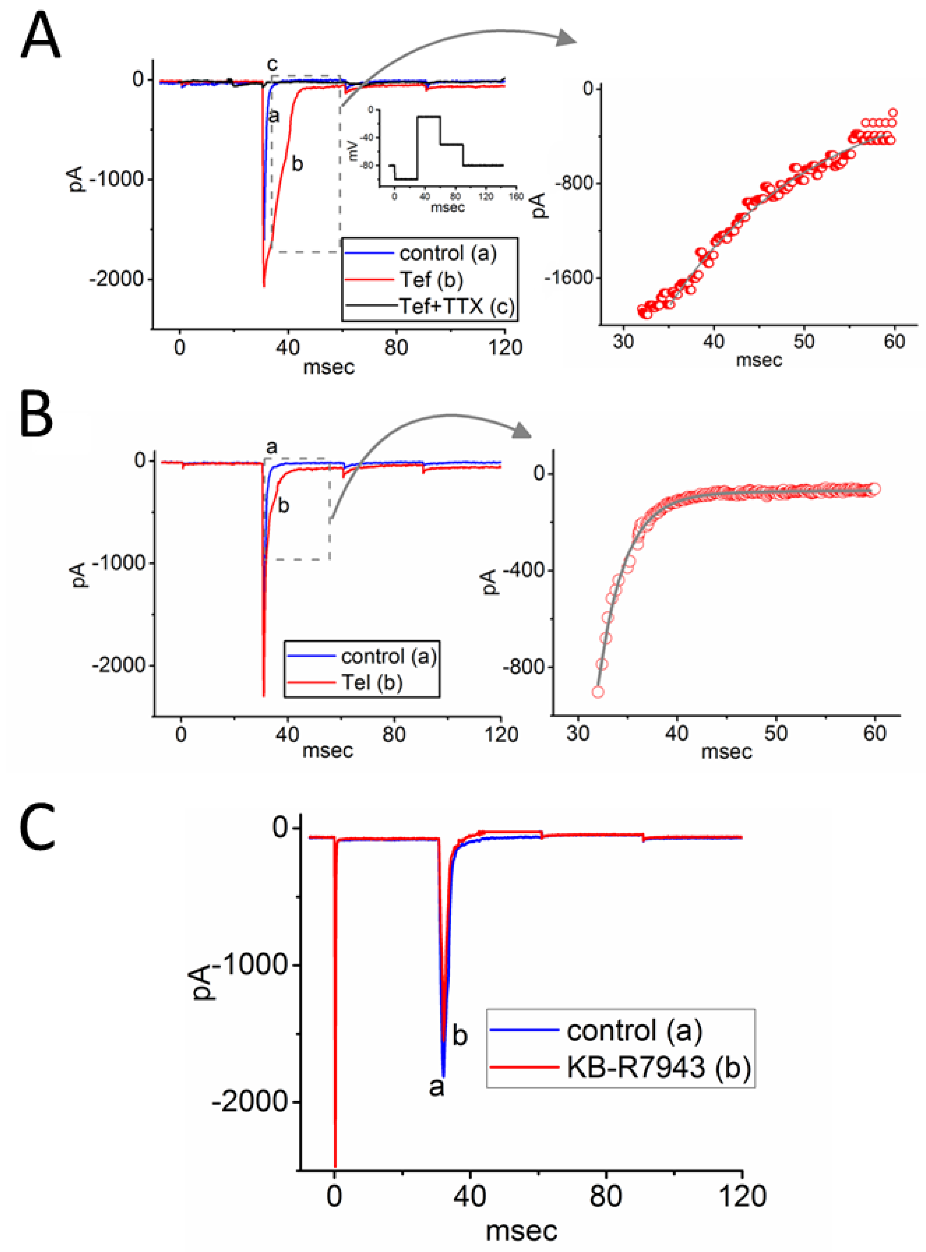
3.1. Effects of Either Tef, Tel, or KB-R7943 on INa Measured from Rat DRG Neurons

The initial series of experiments aimed to explore the possible impact of Tef, Tel, or KB-R7943 on INa elicited in response to short step depolarizations in these cells. The cells were bathed in Ca2+-free Tyrode’s solution supplemented with 0.5 mM CdCl2 and 10 mM TEA. The whole-cell voltage-clamp experiments were conducted using a pipette solution containing Cs+. As shown in Figure 2, the tested cell was depolarized from −100 to −10 mV for 30 ms, and the INa, which was sensitive to block by TTX (1 μM), could be readily elicited in these neurons. As shown in Figure 2A, further addition of TTX (1 μM) completely blocks the INa amplitude observed during continued exposure to 10 μM Tef.
Exposure to Tef or Tel exhibited an enhancement in the peak amplitude of INa, accompanied by a progressive slowing of the inactivation time course of the current (Figure 2A,B). For example, during a step depolarization from −100 to −10 mV, the addition of Tef (10 μM) or Tel (10 μM) increased the peak INa amplitude from 1.3 ± 0.3 to 2.4 ± 0.8 nA (n = 8, paired t-test, df = 7, p = 0.019) or from 1.1 ± 0.3 to 2.2 ± 0.9 nA (n = 8, paired t-test, df = 7, p = 0.032), respectively. After washout of Tef or Tel, INa amplitude returned to the control level. Additionally, during cell exposure to 10 μM Tef or 10 μM Tel, the slow component of the inactivation time constant of INainact(S)) was prolonged, shifting from 10.6 ± 1.8 to 23.6 ± 2.1 ms (n = 8, paired t-test, df = 7, p = 0.021) and from 12.2 ± 2.0 to 22.4 ± 2.1 ms (n = 8, paired t-test, df = 7, p = 0.024), respectively. However, the value in the fast component of the inactivation time constant (τinact(F)) obtained in the presence of Tef or Tel was not changed (1.3 ± 0.3 ms [control], 1.4 ± 0.4 ms [in the presence of Tef], and 1.4 ± 0.3 ms [in the presence of Tel]; n = 8, one-way ANOVA, dfbetween = 2. dfwithin = 21, p = 0.069). Furthermore, when cells were exposed to 10 μM KB-R7943, the peak INa amplitude decreased, accompanied by a concurrent reduction in the τinact(S) value of the current (Figure 2C). The presence of 10 μM KB-R7943 significantly decreased the peak INa amplitude from 1.76 ± 0.05 to 1.51 ± 0.04 nA (n = 8, paired t-test, df = 7, p = 0.028). Concurrently, the value of τinact(S) decreased from 6.1 ± 0.2 to 2.1 ± 0.1 ms (n = 8, paired t-test, df = 7, p = 0.030). However, the τinact(F) value obtained with or without the KB-R7943 presence remained unaltered. We isolated neurons from the dorsal horn of the spinal cord in rats and found that Tef enhanced the INa amplitude. When cells were exposed to Tef (10 μM), the peak amplitude of INa increased from 0.45 ± 0.02 to 0.67 ± 0.03 nA (n = 6, paired t-test, df = 5, p = 0.039). Additionally, the inactivation time constant (τinact(S)) increased from 4.5 ± 0.1 to 8.9 ± 0.2 ms (n = 6, paired t-test, df = 5, p = 0.021).

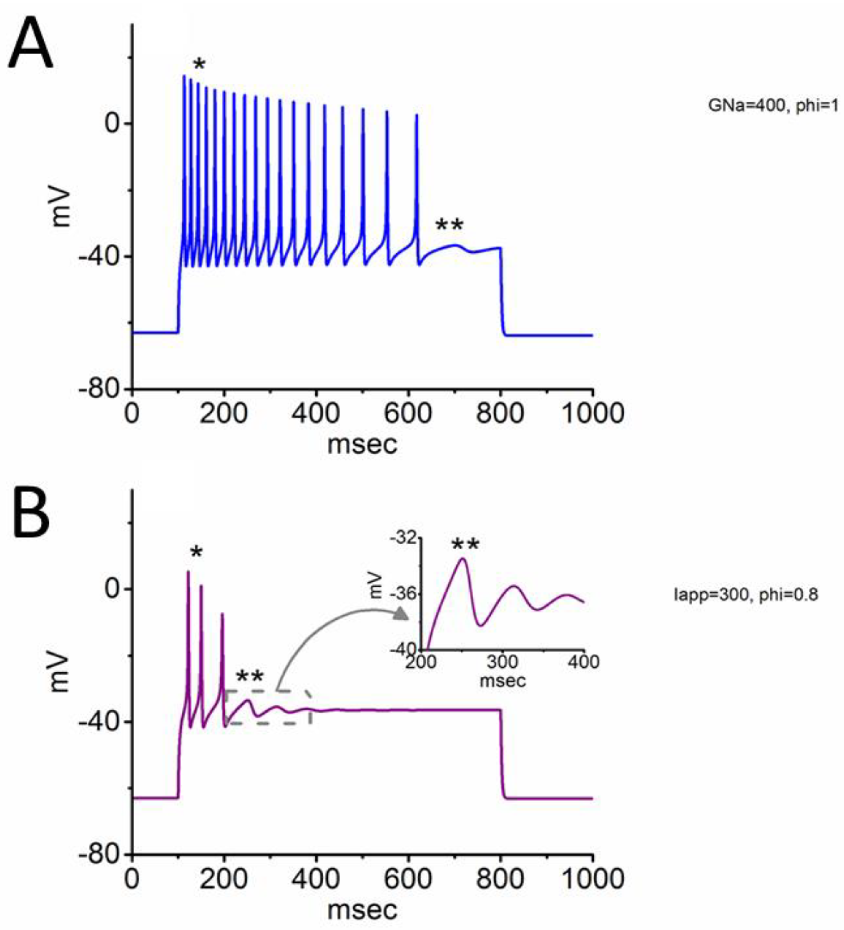
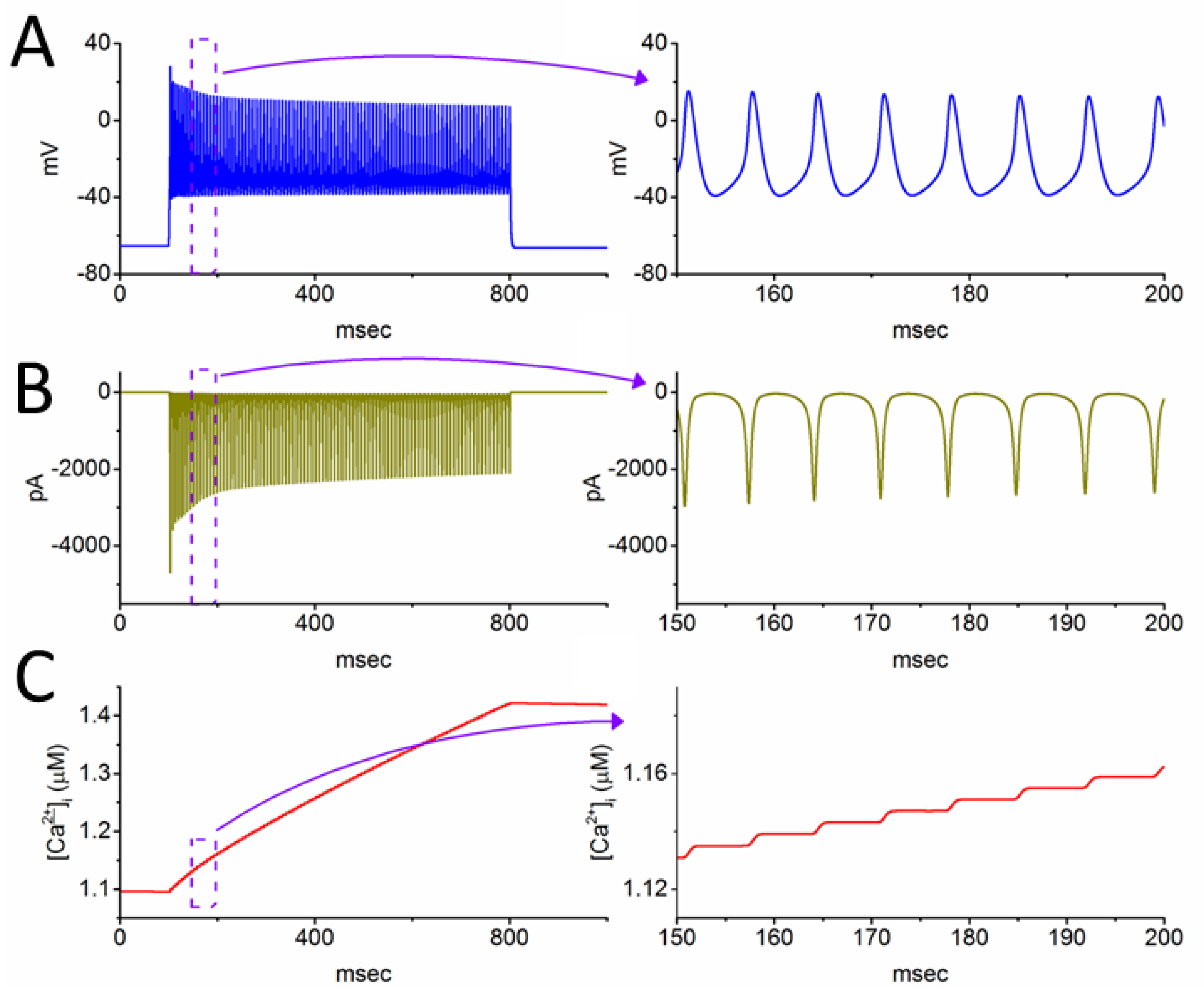
3.2. Simulated INa Traces Created from a Modified HH Model

In our endeavor to replicate the impact of Tef, Tel, or KB-R7943 on INa in cultured DRG neurons, we undertook a detailed exploration into the modulation of both INa amplitude and gating. This work aimed to elucidate the influence on the electrical properties of neurons related to nociceptive signaling. The INa model employed in these simulations was developed based on a modified HH model [17,20,27,28]. In this series of simulations, the values of gNa and VNa were set to be 120 nS and +58 mV, respectively. Other parameters are outlined in Table 1 and in a recent paper [17].
Under the specified conditions, when the value, particularly within the βh inactivation component of h inactivation variable, was increased in a stepwise fashion, the inactivation time course of the simulated INa progressively became enhanced. This was accompanied by a reduction in the late component of INa during rapid step depolarization. It is established that a stepwise increase of the value (from 0.4 to 1.2) embedded in h inactivation variable is capable of increasing the inactivation rate of the current with a reduction in the sustained component of INa (Figure 3), although the amplitude in the transient component of the current was slightly changed. Therefore, the simulated INa trajectories within this modeled neuron aligned closely well with the experimentally observed results, showing that the presence of Tef, Tel, or KB-R7943 altered the inactivation time course of this current in cultured DRG neurons (Figure 2).

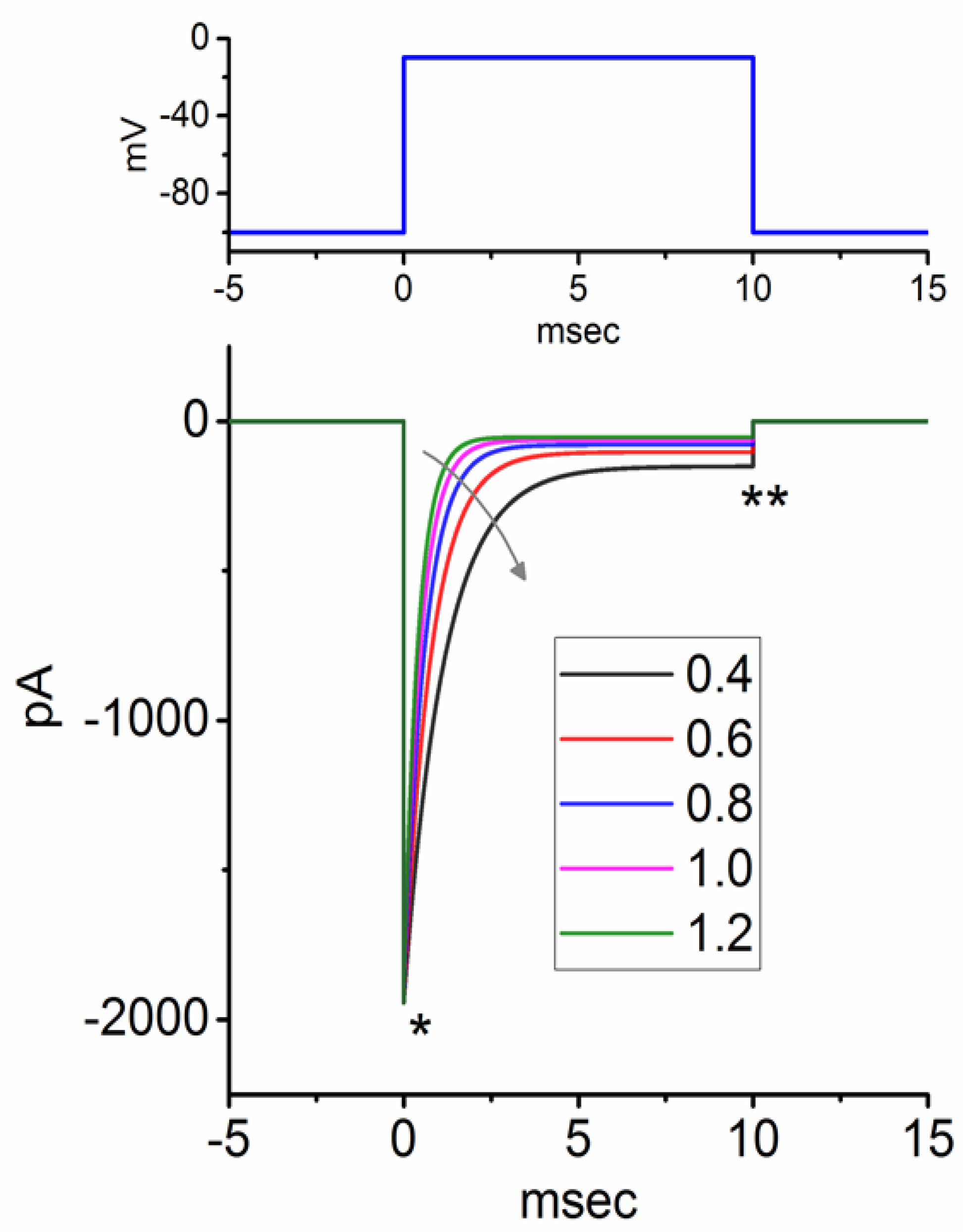
3.3. Changes of Membrane Potential Caused by Different Value of

The variations in membrane potential resulting from different values were further investigated. In Figure 4A, was set at 1.0, with gNa and the applied current (Iapp) set at 400 nS and 300 pA, respectively, and the resulting changes in membrane potential were plotted over time. Under these simulations, a transient oscillatory spike followed by subthreshold oscillations (SOs) was observed. Notably, the amplitude and frequency of APs exhibited progressive decreases, indicative of spike-frequency adaptation [29,30]. The emergence of SOs, as previously observed in interior olive neurons [13], was evident.
However, when the gNa value was decreased to 300 nS while maintaining the same values for Iapp and φ, both the amplitude and frequency of APs in response to current injection were reduced. Following the cessation of APs, the occurrence of SO was also observed (Figure 4B). Findings from these results highlight that changes in gNa alone can alter the electrical behavior of the modeled neurons.

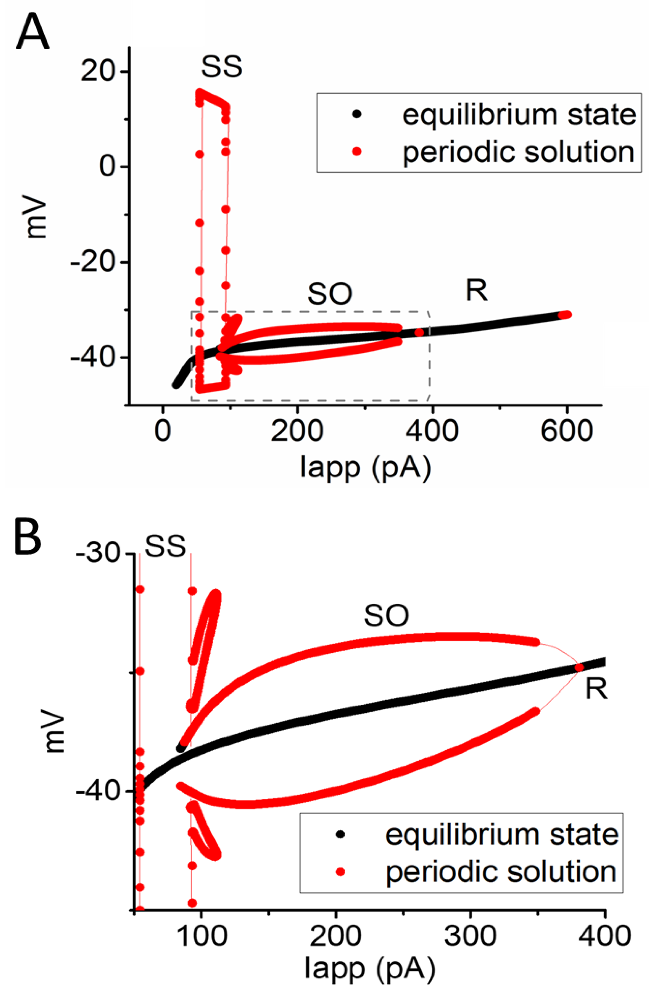
3.4. Relationship of Changes in Steady-State Membrane Potential vs. the Iapp Value

In this study, a bifurcation analysis was employed to explore alterations in the steady-state membrane potential in response to variations in the Iapp. A bifurcation diagraph is a graphical representation of the long-term behavior of a dynamical system as a parameter is changed [13,18,20]. Through these simulations, Iapp was considered a variable, while other parameters were maintained at default values (Table 1). With a constant value of set to 1, Figure 5 depicts the bifurcation diagram illustrating the correlation between Iapp and the change in membrane potential. Two critical values of Iapp, representing the subcritical and supercritical bifurcation points, were identified. Specifically, the Iapp values for these points at = 1 were noted to be 88 and 177 pA. Within the range of Iapp values between these two Hopf bifurcation points, a stable limit cycle emerged―a closed trajectory in the phase plane, indicating the SO occurrence in the modeled neuron, as demonstrated previously [13,18]. The range of membrane potential changes were found to be between −39.3 and −36.2 mV. The model therefore exhibits stability when Iapp is below the subcritical point or above the supercritical point. Conversely, self-sustained periodic oscillations, or SOs, occur when Iapp falls within the range between the subcritical and supercritical points.
However, a notable alteration in the bifurcation diagraph occurred when the value in the βh inactivation process of h inactivation variable decreased to 0.8 with a fixed gNa value of 300 nS, mimicking a slowing in the inactivation time course of INa (Figure 6A,B). Under these conditions, two types of periodical solutions were observed. The subcritical and supercritical points, determining the occurrence of preexisting SO, increased to 95 and 367 pA, respectively. Additionally, another phenomenon, namely, somatic spiking (SS) or somatic AP firing, was noted during the existence of Iapp ranging between 52 and 98 pA. Within the domain of SS, the voltage of membrane potential was found to range between −40 and +15 mV. It is also noted from Figure 6B that as the SO domain overlapped with SS, the cell behavior was denoted to be SS/SO bistable in the system, when the Iapp amplitude ranged between 79 and 118 pA.

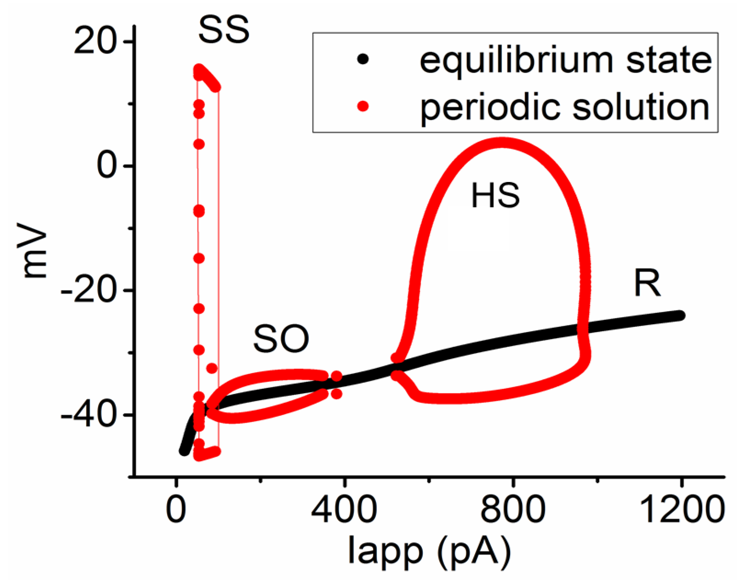
3.5. Further Modifications on Changes in Membrane Potential with the Increasing Iapp Value with a Fixed Value of 0.8

We made further adjustments to investigate how an additional large increase in Iapp amplitude, with the fixed and gNa values of 0.8 and 300 nS, influences changes in membrane potential, INa, and intracellular Ca2+ levels ([Ca2+]i) in the modeled neuron. As depicted in Figure 7A–C, setting the Iapp value of 700 pA resulted in high-frequency spiking (HS) of APs at a rate of 150 Hz. The membrane potential during this HS spiking exhibited periodical fluctuations with the range of −39 to +18 mV. Moreover, when HS occurred, the height of the AP gradually decreased over time, accompanied by a reduction of INa amplitude. This phenomenon corresponds to previous experimental reports, showing an accumulation of INa inactivation when excitable cells are subjected to high-frequency depolarizing stimuli [5,6,7,31]. Alternatively, the [Ca2+]i level rose during AP firing. Over the 700 ms duration of Iapp, the [Ca2+]i level concurrently increased from 1.11 to 1.42 μM.

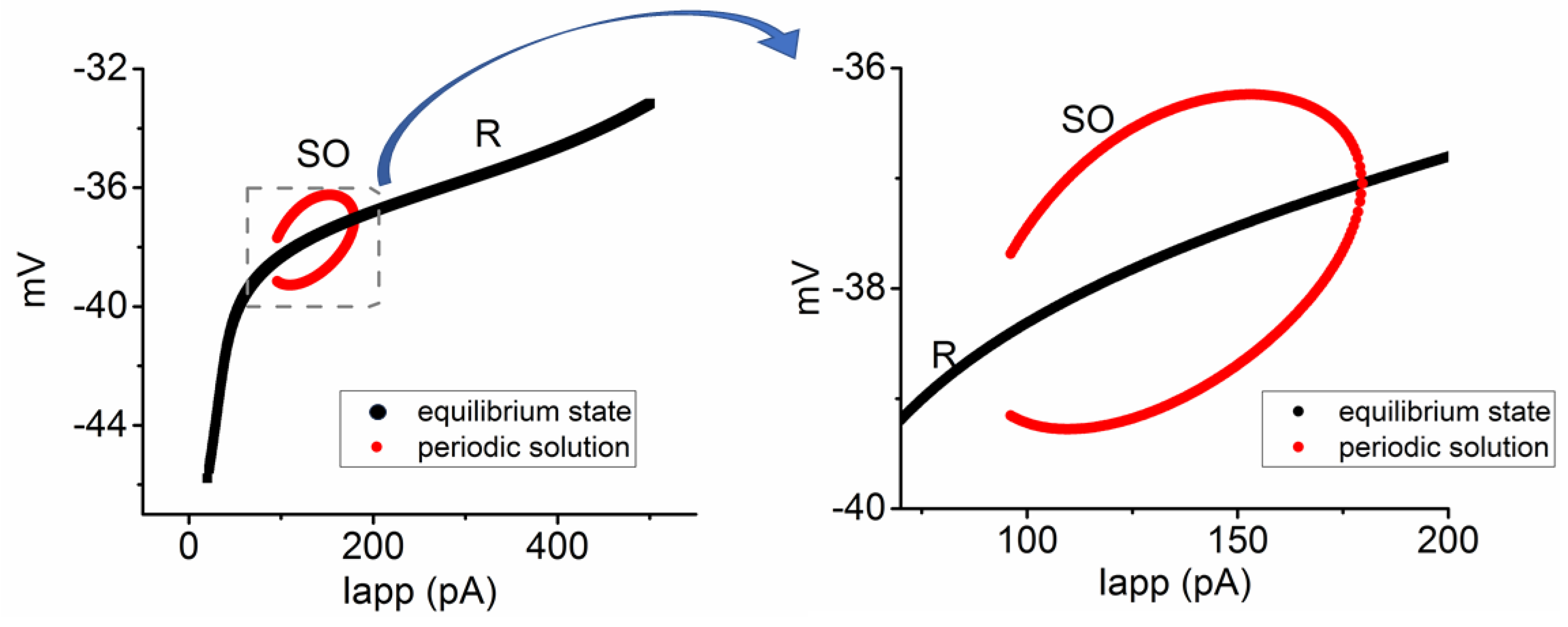
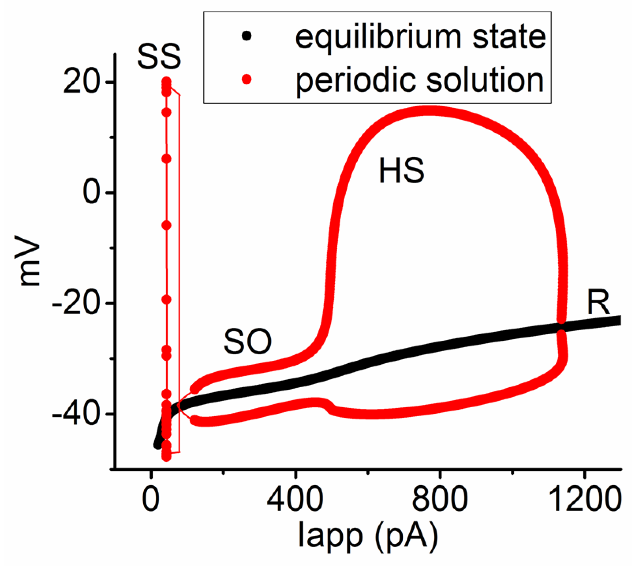
3.6. Bifurcation Analysis on the Relationship between Iapp and Membrane Potential at the Range between 0 and 1200 pA

As demonstrated in Figure 8, the bifurcation diagram, which illustrates the relationship between Iapp magnitude and membrane potential, was further examined. Three distinct periodic solutions, highlighted in red, are evident within the system. In these simulations, with gNa set at 300 nS and at 0.8, a gradual increase in Iapp amplitude within the 560 to 910 pA range leads to another self-sustained periodic solution of the system. That is, there appeared to be another closed trajectory in this phase plane. This result indicates the emergence of HS, with a rate reaching 150 Hz (Figure 8). Notably, when both the inactivation time course of INa slowed down and the amplitude of Iapp were concurrently increased within the range of 560 to 910 nA, the modeled neuron became more predisposed to generate HS, namely, high-frequency firing of APs. Changes in membrane potential were noted to range between −35 and +4 mV during the HS occurrence in the bifurcation diagram. It is thus noted that as the gNa value increased to 400 nS, the range of limit cycles generated by both SO and HS was observed to increase. Moreover, the pattern of SO was noticeably amalgamated with that of HS, as observed in Figure 9, in comparison to the bifurcation diagram in Figure 8. Therefore, simulated alterations in both the INa amplitude and the inactivation time course of the current, as observed experimentally in the presence of Tef or Tel, makes the membrane potential susceptible to SO occurrence and even the HS emergence. Additionally, as found during cell exposure to KB-R7943, both the decrease in INa amplitude and the inactivation time course of this current are anticipated to diminish the emergence of SO or HS.

4. Discussion

The results of this study demonstrate that in the presence of Tef, Tel, or KB-R7943, the amplitude and gating of INa in cultured DRG neurons (R-DRG-505) was affected. Tef or Tel increased the amplitude of INa while slowing down the inactivation time course of this current. The impact of Tel on INa is related to its effect on NaV channels and is not associated with its antagonistic action against angiotensin II receptors [12,20]. Furthermore, in addition to suppression of the Na+-Ca2+ exchanging process [1,32], KB-R7943 also inhibited the magnitude of INa while simultaneously accelerating the inactivation rate of INa in cultured DRG neurons.
This study shows that the presence of INa can be detected in cultured DRG neurons. However, these INa currents are blocked by TTX. In these cells, we did not observe clear TTX-resistant (TTX-R) INa [33,34]. The NaV1.6, NaV1.7, NaV1.8, and NaV1.9 isoforms were noticed to be expressed in DRG neurons. It is thus possible that the NaV1.8 expression in these cells (R-DRG-505 cells) could be relatively low [35]. However, we did observe that the current was enhanced by Tef or Tel and inhibited by KB-R7943, as reported previously [20,36,37,38,39]. Additionally, the inactivation time course of these currents was also altered by these drugs (Figure 2). Our in silico study proposed that the inactivation time course of INa (indicated by the variable value), which is activated at depolarized potentials, contributes to the presence, amplitude, and frequency of SO and HS [4,13,18,34].
In the modeled interneuron of this study, there were no parameters for the Na+-Ca2+ exchanging process. However, with the generation of AP firing, the level of [Ca2+]i also increased, as demonstrated in Figure 6. Therefore, at least in excitable cells such as neuronal cells, changes in the magnitude of INa or the inactivation time course of the current might affect the level of [Ca2+]i without the need for the participation of the Na+-Ca2+ exchanging process. Indeed, KB-R7943, an inhibitor of the Na+-Ca2+ exchanging process [14,15], was reported to inhibit the INa responsible for these effects [21].
Previous reports have indicated that ranolazine, a blocker of late INa, has an inhibitory effect on the INa in DRG neurons, concurrently demonstrating relief in painful sensations [23,40,41]. This study confirms this phenomenon and additionally reveals that KB-R7943, an inhibitor of the Na+-Ca2+ exchanging process, can inhibit INa magnitude while accelerating the inactivation time course of the INa, as described recently [21]. Therefore, KB-R7943 may alleviate a certain type of pain signaling [25]. Furthermore, further investigations are needed to explore how Tef or Tel influences the biophysical properties of INa in DRG neurons or sensory neurons associated with pain signaling, consequently modulating the nociceptive transmission [10,11,42,43].
The results of this study demonstrate that when the INa in DRG neurons increased and its inactivation time course simultaneously slowed down, as mimicked by the presence of Tef or Tel, the cell membrane of these neurons became prone to generating the occurrence of SO. Moreover, excessive stimulation with strong electrical currents easily induced the HS emergence. Conversely, when INa decreased and the inactivation time course accelerated (as mimicked by cell exposure to KB-R7943), these cells were less likely to exhibit the pattern of SOs and even resulted in the HS development [13,17,18,28]. Therefore, alterations in the biophysical properties of INa impact the cell excitability of DRG neurons in vivo, subject to biophysical, pharmacological, or toxicological modulation of INa.
The modeled neuron used in this study includes two types of different delayed-rectifier K+ currents, namely, KV1.3 and KV3.1 currents [17]. Previous reports have indicated that changes in the magnitude and inactivation process of these K+ currents significantly impact the HS emergence [44,45,46,47,48]. KCNQ channels have been also reported to enable reliable presynaptic spiking occurring at high frequency [49]. Therefore, further investigations are needed to understand how the modulation of these K+ currents may have effects on the HS emergence in the presence of Tef or Tel.
In this study, we employed a theoretical model of AP firing, which was adapted from a previous study by Ma et al. [17]. This model is based on the biophysical properties of PVINs located in the dorsal horn of the spinal cord. Although there are some differences between this model neuron and the DRG neuron, both of these types of neurons contain abundant NaV channels. We thus believe that it is valuable to investigate how changes in the amplitude and/or inactivation characteristics of INa affect the patterns of AP firing related to pain signaling. However, the effects of these compounds on the AP firing in DRG neurons occurring in vivo still require further analysis and research.
Although our study found that TTX-R INa was not detected in cultured DRG neurons (R-DRG-505), it is established that nociceptive DRG neurons (≤30 μm in a diameter) express TTX-R NaV channels, such as NaV1.8 and NaV1.9, and that previous studies have emphasized the role of these channels in nociceptive transmission [35,50,51]. However, the modified PVINs in lamina III of the spinal cord are very heterogeneous and mainly express NaV1.1, NaV1.2, and NaV1.6, and they do not express NaV1.7 abundant in nociceptive DRG neurons [52,53,54]. Nevertheless, the effects of three compounds on TTX-R NaV channels should be tested, possibly using native DRG neurons or other types of spinal neurons linked to pain transmission.
Plasma Tel concentration after intravenous administration of a single dose of 40 mg Tel was noted to reach about 2.32 μM [55]. As for Tel, it is a pyrethroid insecticide. Its concentration in the blood of organisms varies greatly, reaching up to several micromolar levels. After intravenous administration, KB-R7943 reached a concentration of about 0.12 μM [56], which is lower than the concentration used in this study. However, recent reports have found that KB-R7943 can cross the blood–brain barrier, with concentrations in brain tissue reaching 5 to 10 times higher [16].
The effects of these compounds on the dorsal root ganglia may extend beyond the necessity of the blood–brain barrier. However, in nociceptive transmission, the dorsal root ganglia is one of the first primary neurons; the nerve transmission also needs to reach the dorsal horn of the spinal cord, then pass and conduct through long fibers upward to the medulla oblongata, further up to the thalamus, and then to the cerebral cortex. In these regions, the blood–brain barrier is often present. Therefore, if these compounds, such as TelTef or KB-7943, can easily pass through the blood–brain barrier across those areas, it is believed that they could have a regulatory effect on these pain-linked transmission pathways. Furthermore, neurons of the dorsal horn of the spinal cord, including parbalbumin-expressing interneurons, were reported to exhibit greater heterogeneity than DRG neurons [54]. Nevertheless, the effects of Tel, Tef, or KB-R7943 may have implications for biological activity in the nervous system [8,9]. Among these effects, the regulation of ion channels in the cell membrane might be one of the important mechanisms.
Of note, the model used in the current study is not perfect. Indeed, there are many different formulations for Na+ currents. The simulated firing of neuronal action potentials derived from different regions linked to pain signaling can also be present in various possible forms, as detailed on the ModelDB website (http://modeldb.sicence/, accessed on 14 July 2024).
To ensure our experimental results are more substantial and effective in presentations, and to observe potential action potential firing patterns, we further explored this through bifurcation analysis. We indeed spent a significant amount of time searching for developments of various types of simulation models. Thus, we found the model developed by Ma et al. [17] in PVINs to be more suitable for our needs. However, we still believe it is not perfect, and in the future, we will continue to develop more suitable computational models so that the experimentally observed and theoretical results can be more consistent.
Tef has previously been reported to inhibit voltage-gated Ca2+ current and mildly suppress delayed-rectifier K+ currents [57]. Additionally, telmisartan has been shown to suppress erg-mediated K+ currents [37]. However, there are no reports indicating that KB-R7943 affects K+ currents. This paper focuses solely on the changes these compounds induce in the amplitude and gating of Na+ currents. Further analysis is required to understand the effects of these agents on other ion currents, such as K+ and Ca2+ currents, is necessary. Therefore, additional research is necessary to provide further clarification in this area.
Limitation of study. In our cultured DRG neurons, namely, R-DRG-505 cells, we employed current-clamp voltage recordings to observe changes in membrane potential. However, we were unable to detect HS. Even in the presence of Tef or Tel, HS did not manifest. However, through our investigations using modeled interneurons in the spinal dorsal horn [17], we found that enhancing the INa and slowing down the inactivation time course can simulate the exposure to Tef or Tel. The simulations suggest that these modeled neurons can generate SO and HS. Furthermore, when subjected to strong applied currents, HS with a rate of about 150 Hz was found to emerge. The theoretical analyses of these scenarios are presented in the bifurcation diagrams provided herein.
Additionally, to induce differentiation-like events in cell culture and observe variable AP firing patterns, the medium might be replaced with one containing 1 μM nerve growth factor. The experimental and theoretical observations in this study may serve as a foundation for future research aimed in these directions. Furthermore, the concentrations used in this research project are very close to the potential clinical, pharmaceutical, or toxicological application ranges of these compounds. Therefore, the research findings will hold practical significance for the appropriate administration or use of these compounds.

Author Contributions

Conceptualization, H.-Y.H., C.-L.W. and S.-N.W.; Methodology, Y.-B.H. and S.-N.W.; Validation, Y.-B.H.; Formal analysis, Y.-B.H. and S.-N.W.; Investigation, H.-Y.H.; Data curation, C.-L.W.; Writing—original draft, C.-L.W.; Writing—review and editing, S.-N.W.; Supervision, S.-N.W.; Project administration, C.-L.W.; Funding acquisition, H.-Y.H. All authors have read and agreed to the published version of the manuscript.

Funding

This work was partly supported by grants from the Ministry of Science and Technology (NSTC- 113-2923-B-906-001), Taiwan, from An Nan Hospital (ANHR-112-42, ANHR-112-43, and ANHR-112-44), and Ditmanson Medical Foundation Chia-Yi Christian Hospital Research Program. The funders were not involved in the study, design, data collection, analyses, or interpretation.

Data Availability Statement

The data will be made available upon request.

Acknowledgments

The authors of this study express gratitude to the late Ching-Hsing Luo of the Department of Electrical Engineering at National Cheng Kung University, Tainan, Taiwan, for his assistance in the earlier work on bifurcation analyses.

Conflicts of Interest

The authors declare that the research was conducted in the absence of any commercial or financial relationships that could be construed as potential conflicts of interest.

Abbreviations

AP, action potential; [Ca2+]i, intracellular Ca2+ concentration; df, degree of freedom; HH, Hodgkin-Huxley; HS, high-frequency spiking; DRG neuron, dorsal root ganglion neuron; Iapp, applied current; INa, voltage-gated Na+ current; NaV channel, voltage-gated Na+ channel; SK channel, small-conductance Ca2+-activated K+ channel; PVINs, parvalbumin-expressing interneurons; SO, subthreshold oscillation; SS, somatic spiking; τinact(F), fast component of inactivation time constant; τinact(S), slow component of inactivation time constant; Tef, tefluthrin; Tel, telmisartan; TTX, tetrodotoxin; TTX-R, tetrodotoxin-resistant. (The other abbreviations related to parameters used in the simulations are detailed in Table 1).

References

  1. Catterall, W.A. Voltage-gated sodium channels at 60: Structure, function and pathophysiology. J. Physiol. 2012, 590, 2577–2589. [Google Scholar] [CrossRef] [PubMed]
  2. De Lera Ruiz, M.; Kraus, R.L. Voltage-Gated Sodium Channels: Structure, Function, Pharmacology, and Clinical Indications. J. Med. Chem. 2015, 58, 7093–7118. [Google Scholar] [CrossRef] [PubMed]
  3. Shen, H.; Zhou, Q.; Pan, X.; Li, Z.; Wu, J.; Yan, N. Structure of a eukaryotic voltage-gated sodium channel at near-atomic resolution. Science 2017, 355, eaal4326. [Google Scholar] [CrossRef]
  4. Bennett, D.L.; Clark, A.J.; Huang, J.; Waxman, S.G.; Dib-Hajj, S.D. The Role of Voltage-Gated Sodium Channels in Pain Signaling. Physiol. Rev. 2019, 99, 1079–1151. [Google Scholar] [CrossRef] [PubMed]
  5. Taddese, A.; Bean, B.P. Subthreshold Sodium Current from Rapidly Inactivating Sodium Channels Drives Spontaneous Firing of Tuberomammillary Neurons. Neuron 2002, 33, 587–600. [Google Scholar] [CrossRef]
  6. Carter, B.C.; Bean, B.P. Incomplete Inactivation and Rapid Recovery of Voltage-Dependent Sodium Channels During High-Frequency Firing in Cerebellar Purkinje Neurons. J. Neurophysiol. 2011, 105, 860–871. [Google Scholar] [CrossRef]
  7. Huang, C.-W.; Hung, T.-Y.; Wu, S.-N. The inhibitory actions by lacosamide, a functionalized amino acid, on voltage-gated Na+ currents. Neuroscience 2015, 287, 125–136. [Google Scholar] [CrossRef]
  8. So, E.C.; Wu, S.-N.; Lo, Y.-C.; Su, K. Differential regulation of tefluthrin and telmisartan on the gating charges of INa activation and inactivation as well as on resurgent and persistent INa in a pituitary cell line (GH3). Toxicol. Lett. 2018, 285, 104–112. [Google Scholar] [CrossRef]
  9. Lin, M.-H.; Lin, J.-F.; Yu, M.-C.; Wu, S.-N.; Wu, C.-L.; Cho, H.-Y. Characterization in Potent Modulation on Voltage-Gated Na+ Current Exerted by Deltamethrin, a Pyrethroid Insecticide. Int. J. Mol. Sci. 2022, 23, 14733. [Google Scholar] [CrossRef]
  10. Jiang, N.; Nutter, T.J.; Cooper, B.Y. Molecular and cellular influences of permethrin on mammalian nociceptors at physiological temperatures. NeuroToxicology 2013, 37, 207–219. [Google Scholar] [CrossRef]
  11. Silwal, P.M.; Adhikari, R.; Yadav, B.M.; Sah, S.K.M.; Bhatt, A.; Basnet, S.M. Lambda-cyhalothrin ingestion: An infrequent yet concerning presentation of pyrethroid poisoning. Ann. Med. Surg. 2023, 85, 5250–5254. [Google Scholar] [CrossRef] [PubMed]
  12. Hegazy, N.; Rezq, S.; Fahmy, A. Renin-angiotensin system blockade modulates both the peripheral and central components of neuropathic pain in rats: Role of calcitonin gene–related peptide, substance P and nitric oxide. Basic Clin. Pharmacol. Toxicol. 2020, 127, 451–460. [Google Scholar] [CrossRef] [PubMed]
  13. Schweighofer, N.; Doya, K.; Kawato, M.; Davoine, F.; Curti, S.; Hakimian, S.; Norris, S.A.; Greger, B.; Keating, J.G.; Anderson, C.H.; et al. Electrophysiological properties of inferior olive neurons: A compartmental model. J. Neurophysiol. 1999, 82, 804–817. [Google Scholar] [CrossRef] [PubMed]
  14. Amran, S.; Homma, N.; Hashimoto, K. Pharmacology of KB-R7943: A Na+–Ca2+ exchange inhibitor. Cardiovasc. Drug Rev. 2003, 21, 255–276. [Google Scholar] [CrossRef]
  15. Huang, Y.; Wen, L.-L.; Xie, J.-D.; Ouyang, H.-D.; Chen, D.-T.; Zeng, W.-A. Antinociceptive effectiveness of the inhibition of NCX reverse-mode action in rodent neuropathic pain model. Mol. Pain 2019, 15, 1744806919864511. [Google Scholar] [CrossRef]
  16. Andreeva-Gateva, P.; Hristov, M.; Strokova-Stoilova, M.; Ivanova, N.; Sabit, Z.; Surcheva, S.; Beliakov, M.; Karakashev, G.; Sukhov, I.; Belinskaya, D.; et al. Therapeutic potential of orally applied KB-R7943 in streptozotocin-induced neuropathy in rats. Heliyon 2024, 10, e27367. [Google Scholar] [CrossRef] [PubMed]
  17. Ma, X.; Miraucourt, L.S.; Qiu, H.; Sharif-Naeini, R.; Khadra, A. Modulation of SK Channels via Calcium Buffering Tunes Intrinsic Excitability of Parvalbumin Interneurons in Neuropathic Pain: A Computational and Experimental Investigation. J. Neurosci. 2023, 43, 5608–5622. [Google Scholar] [CrossRef]
  18. White, J.; Budde, T.; Kay, A. A bifurcation analysis of neuronal subthreshold oscillations. Biophys. J. 1995, 69, 1203–1217. [Google Scholar] [CrossRef]
  19. Enns-Ruttan, J.S.; Miura, R.M. Spontaneous Secondary Spiking in Excitable Cells. J. Theor. Biol. 2000, 205, 181–199. [Google Scholar] [CrossRef]
  20. Chang, T.-T.; Yang, C.-J.; Lee, Y.-C.; Wu, S.-N. Stimulatory Action of Telmisartan, an Antagonist of Angiotensin II Receptor, on Voltage-Gated Na^+ Current: Experimental and Theoretical Studies. Chin. J. Physiol. 2018, 61, 1–13. [Google Scholar] [CrossRef]
  21. Wu, S.-N.; Yu, M.-C. Inhibition of Voltage-Gated Na+ Currents Exerted by KB-R7943 (2-[2-[4-(4-nitrobenzyloxy)phenyl]ethyl]isothiourea), an Inhibitor of Na+-Ca2+ Exchanging Process. Int. J. Mol. Sci. 2023, 24, 1805. [Google Scholar] [CrossRef] [PubMed]
  22. Brown, A.M. A modeling study predicts the presence of voltage gated Ca2+ channels on myelinated central axons. Comput. Methods Programs Biomed. 2002, 71, 25–31. [Google Scholar] [CrossRef] [PubMed]
  23. Wu, S.-N.; So, E.C.; Liao, Y.-K.; Huang, Y.-M. Reversal by ranolazine of doxorubicin-induced prolongation in the inactivation of late sodium current in rat dorsal root ganglion neurons. Pain Med. 2015, 16, 1032–1034. [Google Scholar] [CrossRef]
  24. Ghovanloo, M.-R.; Tyagi, S.; Zhao, P.; Effraim, P.R.; Dib-Hajj, S.D.; Waxman, S.G. Sodium currents in naïve mouse dorsal root ganglion neurons: No major differences between sexes. Channels 2023, 18, 2289256. [Google Scholar] [CrossRef]
  25. Traub, R.D.; Miles, R. Multiple Modes of Neuronal Population Activity Emerge after Modifying Specific Synapses in a Model of the CA3 Region of the Hippocampus. Ann. N. Y. Acad. Sci. 1991, 627, 277–290. [Google Scholar] [CrossRef]
  26. Winslow, R.L. Bifurcation analysis of nonlinear retinal horizontal cell models. I. Properties of isolated cells. J. Neurophysiol. 1989, 62, 738–749. [Google Scholar] [CrossRef] [PubMed]
  27. Wu, S.-N. Simulations of the cardiac action potential based on the Hodgkin-Huxley kinetics with the use of Microsoft Excel spreadsheets. Chin. J. Physiol. 2004, 47, 15–22. [Google Scholar]
  28. Chen, B.-S. Effects of Transient Receptor Potential-Like Current on the Firing Pattern of Action Potentials in the Hodgkin-Huxley Neuron during Exposure to Sinusoidal External Voltage. Chin. J. Physiol. 2010, 53, 423–429. [Google Scholar] [CrossRef] [PubMed]
  29. Chiesa, N.; Rosati, B.; Arcangeli, A.; Olivotto, M.; Wanke, E. A Novel Role for HERG K+ Channels: Spike-Frequency Adaptation. J. Physiol. 1997, 501, 313–318. [Google Scholar] [CrossRef]
  30. Wu, S.; Yeh, C.; Huang, H.; So, E.C.; Lo, Y. Electrophysiological characterization of sodium-activated potassium channels in NG108-15 and NSC-34 motor neuron-like cells. Acta Physiol. 2012, 206, 120–134. [Google Scholar] [CrossRef]
  31. A Navarro, M.; Salari, A.; Lin, J.L.; Cowan, L.M.; Penington, N.J.; Milescu, M.; Milescu, L.S. Sodium channels implement a molecular leaky integrator that detects action potentials and regulates neuronal firing. eLife 2020, 9, e54940. [Google Scholar] [CrossRef] [PubMed]
  32. Schröder, U.H.; Breder, J.; Sabelhaus, C.F.; Reymann, K.G. The novel Na+/Ca2+ exchange inhibitor KB-R7943 protects CA1 neurons in rat hippocampal slices against hypoxic/hypoglycemic injury. Neuropharmacology 1999, 38, 319–321. [Google Scholar] [CrossRef] [PubMed]
  33. Roy, M.; Narahashi, T. Differential properties of tetrodotoxin-sensitive and tetrodotoxin- resistant sodium channels in rat dorsal root ganglion neurons. J. Neurosci. 1992, 12, 2104–2111. [Google Scholar] [CrossRef] [PubMed]
  34. Lee, H.M.; Kim, H.I.; Shin, Y.K.; Lee, C.S.; Park, M.; Song, J.-H. Diclofenac inhibition of sodium currents in rat dorsal root ganglion neurons. Brain Res. 2003, 992, 120–127. [Google Scholar] [CrossRef] [PubMed]
  35. Szulczyk, B.; Pasierski, M.; Gawlak, M. Prefrontal cortex pyramidal neurons express functional Nav1.8 tetrodotoxin-resistant sodium currents. Clin. Exp. Pharmacol. Physiol. 2021, 49, 350–359. [Google Scholar] [CrossRef]
  36. Kim, H.K.; Youm, J.B.; Lee, S.R.; Lim, S.E.; Ko, T.H.; Long, L.T.; Nilius, B.; Won, D.N.; Noh, J.-H.; Ko, K.S.; et al. The angiotensin receptor blocker and PPAR-γ agonist, telmisartan, delays inactivation of voltage-gated sodium channel in rat heart: Novel mechanism of drug action. Pflügers Arch. Eur. J. Physiol. 2012, 464, 631–643. [Google Scholar] [CrossRef]
  37. Chang, W.; Wu, S. Activation of voltage-gated sodium current and inhibition of erg-mediated potassium current caused by telmisartan, an antagonist of angiotensin II type-1 receptor, in HL-1 atrial cardiomyocytes. Clin. Exp. Pharmacol. Physiol. 2018, 45, 797–807. [Google Scholar] [CrossRef]
  38. Lai, M.-C.; Wu, S.-N.; Huang, C.-W. Telmisartan, an Antagonist of Angiotensin II Receptors, Accentuates Voltage-Gated Na+ Currents and Hippocampal Neuronal Excitability. Front. Neurosci. 2020, 14, 902. [Google Scholar] [CrossRef]
  39. Lu, T.-L.; Wu, S.-N. Investigating the Impact of Selective Modulators on the Renin–Angiotensin–Aldosterone System: Unraveling Their Off-Target Perturbations of Transmembrane Ionic Currents. Int. J. Mol. Sci. 2023, 24, 14007. [Google Scholar] [CrossRef]
  40. Gould, H.J.; Soignier, R.D.; Cho, S.R.; Hernandez, C.; Diamond, I.; Taylor, B.K.; Paul, D. Ranolazine Attenuates Mechanical Allodynia Associated with Demyelination Injury. Pain Med. 2014, 15, 1771–1780. [Google Scholar] [CrossRef]
  41. Gould, H.J.; Diamond, I. Ranolazine: A potential treatment for refractory neuropathic pain. J. Neurol. Sci. 2016, 369, 310–311. [Google Scholar] [CrossRef] [PubMed]
  42. Castellanos, A.; Andres, A.; Bernal, L.; Callejo, G.; Comes, N.; Gual, A.; Giblin, J.P.; Roza, C.; Gasull, X. Pyrethroids inhibit K2P channels and activate sensory neurons: Basis of insecticide-induced paraesthesias. Pain 2017, 159, 92–105. [Google Scholar] [CrossRef] [PubMed]
  43. Karádi, D.; Galambos, A.R.; Lakatos, P.P.; Apenberg, J.; Abbood, S.K.; Balogh, M.; Kiraly, K.; Riba, P.; Essmat, N.; Szucs, E.; et al. Telmisartan Is a Promising Agent for Managing Neuropathic Pain and Delaying Opioid Analgesic Tolerance in Rats. Int. J. Mol. Sci. 2023, 24, 7970. [Google Scholar] [CrossRef] [PubMed]
  44. Klemic, K.G.; Kirsch, G.E.; Jones, S.W. U-Type Inactivation of Kv3.1 and Shaker Potassium Channels. Biophys. J. 2001, 81, 814–826. [Google Scholar] [CrossRef] [PubMed]
  45. Lin, M.-W.; Wang, Y.-J.; Liu, S.-I.; Lin, A.-A.; Lo, Y.-C.; Wu, S.-N. Characterization of aconitine-induced block of delayed rectifier K+ current in differentiated NG108-15 neuronal cells. Neuropharmacology 2008, 54, 912–923. [Google Scholar] [CrossRef]
  46. Wang, Y.-J.; Lin, M.-W.; Lin, A.-A.; Peng, H.; Wu, S.-N. Evidence for state-dependent block of DPI 201-106, a synthetic inhibitor of Na+ channel inactivation, on delayed-rectifier K+ current in pituitary tumor (GH3) cells. Acta Physiol. Pol. 2008, 59, 409–423. [Google Scholar]
  47. Wu, S.-N.; Chen, B.-S.; Lin, M.-W.; Liu, Y.-C. Contribution of slowly inactivating potassium current to delayed firing of action potentials in NG108-15 neuronal cells: Experimental and theoretical studies. J. Theor. Biol. 2008, 252, 711–721. [Google Scholar] [CrossRef]
  48. Hsiao, H.-T.; Wang, J.C.-F.; Wu, S.-N. Inhibitory Effectiveness in Delayed-Rectifier Potassium Current Caused by Vortioxetine, Known to Be a Novel Antidepressant. Biomedicines 2022, 10, 1318. [Google Scholar] [CrossRef]
  49. Zhang, Y.; Li, D.; Darwish, Y.; Fu, X.; Trussell, L.O.; Huang, H. KCNQ Channels Enable Reliable Presynaptic Spiking and Synaptic Transmission at High Frequency. J. Neurosci. 2022, 42, 3305–3315. [Google Scholar] [CrossRef]
  50. Woolf, C.J.; Ma, Q. Nociceptors—Noxious Stimulus Detectors. Neuron 2007, 55, 353–364. [Google Scholar] [CrossRef]
  51. Nascimento de Lima, A.P.; Zhang, H.; Chen, L.; Effraim, P.R.; Gomis-Perez, C.; Cheng, X.; Huang, J.; Waxman, S.G.; Dib-Hajj, S.D. Nav1.8 in small dorsal root ganglion neurons contributes to vincristine-induced mechanical allodynia. Brain 2024, 147, 3157–3170. [Google Scholar] [CrossRef] [PubMed]
  52. Fukuoka, T.; Kobayashi, K.; Noguchi, K. Laminae-specific distribution of alpha-subunits of voltage-gated sodium channels in the adult rat spinal cord. Neuroscience 2010, 169, 994–1006. [Google Scholar] [CrossRef]
  53. Petitjean, H.; Pawlowski, S.A.; Fraine, S.L.; Sharif, B.; Hamad, D.; Fatima, T.; Berg, J.; Brown, C.M.; Jan, L.-Y.; Ribeiro-Da-Silva, A.; et al. Dorsal Horn Parvalbumin Neurons Are Gate-Keepers of Touch-Evoked Pain after Nerve Injury. Cell Rep. 2015, 13, 1246–1257. [Google Scholar] [CrossRef] [PubMed]
  54. Gradwell, M.A.; Boyle, K.A.; Browne, T.J.; Bell, A.M.; Leonardo, J.; Reyes, F.S.P.; Dickie, A.C.; Smith, K.M.; Callister, R.J.; Dayas, C.V.; et al. Diversity of inhibitory and excitatory parvalbumin interneuron circuits in the dorsal horn. Pain 2021, 163, e432–e452. [Google Scholar] [CrossRef] [PubMed]
  55. Stangier, J.; Schmid, J.; Türck, D.; Switek, H.; Verhagen, A.; Peeters, P.A.; van Marle, S.P.; Tamminga, W.J.; Sollie, F.A.; Jonkman, J.H. Absorption, metabolism, and excretion of intravenously and orally administered [14C]telmisartan in healthy volunteers. J. Clin. Pharmacol. 2000, 40, 1312–1322. [Google Scholar] [CrossRef] [PubMed]
  56. Miyata, A.; Zipes, D.P.; Hall, S.; Rubart, M. KB-R7943 Prevents Acute, Atrial Fibrillation–Induced Shortening of Atrial Refractoriness in Anesthetized Dogs. Circulation 2002, 106, 1410–1419. [Google Scholar] [CrossRef]
  57. Wu, S.-N.; Wu, Y.-H.; Chen, B.-S.; Lo, Y.-C.; Liu, Y.-C. Underlying mechanism of actions of tefluthrin, a pyrethroid insecticide, on voltage-gated ion currents and on action currents in pituitary tumor (GH3) cells and GnRH-secreting (GT1-7) neurons. Toxicology 2009, 258, 70–77. [Google Scholar] [CrossRef]
Figure 1. Histogram depicting the relationship between cell number and cell capacitance. Each red bar represents the number of cells used in this study. A smooth line overlaid on the histogram illustrates the Gaussian distribution used for curve fitting.
Biophysica 04 00032 g001
Figure 2. Effects of tefluthrin (Tef), telmisartan (Tel), and KB-R7943 on voltage-gated Na+ current (INa) in rat dorsal root ganglion (DRG) neurons using whole-cell current recordings. These experiments were conducted in Ca2+-free Tyrode’s solution containing CdCl2 (0.5 mM) and tetraethylammonium chloride (TEA, 10 mM). Panel (AC) displays INa traces under the conditions: in the absence (a, blue color) and in the presence (b, red color) of Tef (10 μM), Tel (10 μM), or KB-R7943 (10 μM), respectively. Inset in the left side of panel (A) indicates the voltage-clamp protocol applied. The right graph of panels (A,B) shows an expanded view of the dashed box from the left side, with the red data points fitted to a two-exponential function (gray curve). Please note that in panel (A), the current trace labeled c (black color) indicates further addition of tetrodotoxin (TTX, 1 μM), in the continued presence of Tef (10 μM).
Biophysica 04 00032 g002
Figure 3. Simulated INa traces in response to short depolarization from −100 to −10 mV for 10 ms. A detailed profile is described in the Section 2 and in a previous paper (Ma et al., 2023) [17]. INa traces were created when the value in the inactivation process (βh) of h variable decreased from 1.2 to 0.4 with 0.2 decrements (as indicated by curved arrow). The term “βh” in the HH kinetics refers to the rate at which the NaV channel undergoes inactivation in the h gating variable. * and ** represent the transient and late components of INa activated by an abrupt depolarizing pulse. The upper part illustrates the voltage-clamp protocol applied.
Biophysica 04 00032 g003
Figure 4. Two distinct spiking patterns simulated from the modeled neuron. When a current injection (700 ms in duration) was applied to this modeled neuron, two different spike patterns emerged, namely, somatic spike (SS) and subthreshold oscillation (SO). In panel (A), gNa = 400 nS, Iapp = 300 pA, = 1.0. In panel (B), gNa = 300 nS, Iapp = 300 pA, = 0.8. The symbols * and ** represent the occurrence of SS and SO, respectively. The inset in panel (B) is a magnified view from the dashed box in (B), highlighting the SO emergence.
Biophysica 04 00032 g004
Figure 5. Bifurcation diagram illustrating the relationship between Iapp and alterations in membrane potential in the modeled neuron. These simulations were conducted using default parameters detailed in Table 1, with a value of 1.0. Two types of solutions in the system were identified. That is, one is the resting, steady-state, or equilibrium state (R, black color), while the other is the unstable and periodic solution indicative of SO (red color). The enlarged graph on the (right) corresponds to the highlighted region within the dashed box on the (left).
Biophysica 04 00032 g005
Figure 6. Bifurcation diagram concerning the relationship between Iapp and membrane potential in the modeled neuron with a constant value of 0.8. Beside the equilibrium or resting state (R, in black color), two distinct types of periodic solutions (indicated in red color) were identified, namely, SS occurring within the Iapp range of 52 to 97 pA and SO at the Iapp range between 85 and 381 pA. Panel (B) is an enlarged view of the dashed box in panel (A).
Biophysica 04 00032 g006
Figure 7. Effect of elevated Iapp at 700 pA on spiking, INa, and intracellular Ca2+ concentration ([Ca2+]i) in the simulated neuron. Panels (AC), respectively, depict membrane potential, INa amplitude, and [Ca2+]i induced by a 700 ms Iapp with 700 pA. Each corresponding panel on the right provides an enlarged view of the graph contained within the dashed box on the left.
Biophysica 04 00032 g007
Figure 8. Bifurcation diagram showing the relationship between the applied current (Iapp) and membrane potential in the modeled neuron. The parameters were set at gNa = 300 nS and = 0.8, with the Iapp range spanning from 20 to 1300 pA. Three unstable bifurcations (indicated in red color) occur, namely, SS with low frequency, SO, and a high-frequency spiking pattern (HS). This caption suggests that in the presence of compounds like Tef or Tel, the modeled neuron exhibited HS occurrence, especially at higher intensities of applied current ranging between 500 and 950 pA. Red and black colors in this and the following figures denote the periodic solutions (SS, SO, and HS) and the equilibrium or resting state (R), respectively.
Biophysica 04 00032 g008
Figure 9. Phase diagram illustrating the impact of enhanced gNa coupled with a reduced φon the relationship of Iapp vs. membrane potential. In this diagram, we set = 0.8, and increased gNa to 320 nS. With the increased Iapp, the coalescence of SO and HS became evident. No existence in the stable steady-state for Iapp ranging between 20 and 1100 pA was noted.
Biophysica 04 00032 g009
Table 1. Default parametric values used for the modeling of interneurons in the dorsal horn of the spinal cord. For more detailed parameters of this model, please refer to Ma et al. [17].
Symbol or ParameterDescription *Value
CmMembrane capacitance (pF)30
gNaMaximum Na+ current conductance (nS)300
gCaMaximum Ca2+ current conductance (nS)8
gK1Maximum KV1 current conductance (nS)15
gK3Maximum KV3 current conductance (nS)180
gSKMaximum SK K+ current conductance (nS)10
gleakMaximum leak current conductance (nS)8
VNaNa+ reversal potential (mV)58
VKK+ reversal potential (mV)−80
VCaCa2+ reversal potential (mV)68
VLeakReversal potential for leak current (mV)−50
γCa2+ recovery rate (ms−1)0.01
kSKCa2+ sensitivity of SK channel (μM)0.8
ACell surface area (μm2)3000
Adjustable βh-inactivation parameter of h gating variable (dimensionless)1.0
* The unit of each parameter is indicated in parentheses.
Disclaimer/Publisher’s Note: The statements, opinions and data contained in all publications are solely those of the individual author(s) and contributor(s) and not of MDPI and/or the editor(s). MDPI and/or the editor(s) disclaim responsibility for any injury to people or property resulting from any ideas, methods, instructions or products referred to in the content.

