Article

Bay Laurel of Northern Morocco: A Comprehensive Analysis of Its Phytochemical Profile, Mineralogical Composition, and Antioxidant Potential

1
Laboratory of Physical Chemistry of Materials, Natural Substances and Environment (LAMSE), Chemistry Department, Faculty of Sciences and Techniques of Tangier, Abdelmalek Essaâdi University, P.O. Box 416, Tangier 90000, Morocco
2
Research Team: Materials, Environment and Sustainable Development (MEDD), Faculty of Sciences and Techniques of Tangier, Abdelmalek Essaâdi University, P.O. Box 416, Tangier 93000, Morocco
*
Author to whom correspondence should be addressed.
Biophysica 2024, 4(2), 238-255; https://doi.org/10.3390/biophysica4020017
Submission received: 20 April 2024 / Revised: 6 May 2024 / Accepted: 11 May 2024 / Published: 15 May 2024
(This article belongs to the Special Issue Biomedical Optics 2.0)

Abstract

:
Laurus nobilis, sometimes referred to as laurel, has been used for medicinal and culinary purposes for a very long time. The main subjects of this study are the phytochemical composition, mineralogical profile, and potential antioxidant properties of Laurus nobilis in Tangier, Northern Morocco. For phytochemical analysis of methanolic extracts, high-performance liquid chromatography (HPLC-UV-MS) was used, and Fourier transformation infrared spectroscopy (FT-IR) was used to identify each individual component. Minerals were studied by inductively coupled plasma atomic emission spectroscopy (ICP-AES) and wavelength dispersive X-ray fluorescence (WD-XRF). Total tannin, flavonoid, and phenolic amounts were quantified using aqueous and methanolic extracts. The antioxidant properties were assessed using 2,2-diphenyl-1-picrylhydrazyl (DPPH), 2,2′-azino-bis (3ethylbenzothiazoline-6-sulfonic acid) (ABTS), ferric reducing antioxidant power (FRAP), and oxygen radical absorbance capacity (ORAC) assays. Research has revealed a complex array of phytochemicals, including tannins, flavonoids, and phenolic acids. Mineral analysis has revealed the existence of vital components that are beneficial to health. Comparing the methanolic extract to the water extract, it demonstrated higher levels of phenols, flavonoids, and tannins as well as stronger antioxidant activity, indicating greater health benefits. This comprehensive study highlights the importance of Laurus nobilis from Northern Morocco as a reliable botanic resource with potential pharmaceutical, nutritional, and cosmetic uses.

1. Introduction

Native to the Mediterranean region, Laurus nobilis, also known as bay laurel, is a fragrant evergreen tree or big shrub [1,2]. Due to its many health-promoting qualities, it has been widely utilized in traditional medicine in addition to culinary uses, where it lends a unique flavor [3,4]. Its usage for treating conditions ranging from infections and inflammation to dyspepsia and arthritis has been described in historical writings and ethnobotanical investigations. Its diverse mix of bioactive chemicals is thought to be responsible for this broad variety of uses [5].
The pharmacological assessment of traditional medicinal plants is one aspect of the growing interest in natural and holistic approaches to health in the scientific community today [6]. Laurus nobilis is an important topic for these kinds of investigations because of its wide range of possible therapeutic benefits [2,7]. Although numerous regional studies have acknowledged the therapeutic benefits of Laurus nobilis, little is known about the unique qualities and possible advantages of the bay laurel from Tangier, Northern Morocco, especially with regard to its complete mineral composition and particular antioxidant mechanisms [8]. This gap offers a chance for comprehensive, comparative study rather than original investigation, which might uncover different phytochemical and mineralogical profiles as a result of local soil compositions and climates. Such variations may have significant effects on our comprehension of this Laurus nobilis variation from a scientific and practical standpoint [9].
Providing a thorough phytochemical profile of Laurus nobilis from Tangier, Northern Morocco, is the main goal of this study. With the use of advanced analytical methods, including Fourier-transform infrared (FT-IR) spectroscopy and high-performance liquid chromatography (HPLC-UV-MS), we hope to detect and measure the important phytochemicals, flavonoids, and phenolic components. This study uses wavelength dispersive X-ray fluorescence (WD-XRF) and inductively coupled plasma atomic emission spectroscopy (ICP-AES) to evaluate the mineralogical composition of the plant in addition to chemical analysis.
Furthermore, because oxidative stress has been linked to a number of chronic diseases, it is important to comprehend Laurus nobilis’s antioxidant properties. As a result, a variety of tests, including DPPH, ABTS, FRAP, and ORAC, will be used to assess the antioxidant potential of both methanolic and aqueous extracts. These discoveries will further our knowledge of the plant’s possible health advantages and open the door for its application in cosmetic, pharmaceutical, and nutraceutical fields.
This study promotes the responsible and efficient use of natural resources like Laurus nobilis and adds to the global effort to support traditional medicine with scientific research. We intend to support the plant’s practical use and maybe expand its uses in contemporary medicine by clarifying the phytochemical and mineral compositions of this plant as well as its antioxidant activity.

2. Results and Discussion

2.1. Mineralogical Analysis

ICP-AES and WD-XRF were used in the mineralogical investigation of Laurus nobilis to determine the plant’s elemental composition and oxide content, as indicated in Table 1 and Table 2.
Since ICP-AES is well renowned for its high sensitivity and precision in analyzing a wide range of elements, it is a great option for determining the elemental composition of plant specimens like Laurus nobilis.
Conversely, WD-XRF spectroscopy offers a non-destructive method for figuring out an element’s composition, which is highly useful for examining valuable or delicate materials.
The findings from the ICP-AES and WD-XRF investigations of Laurus nobilis, or bay laurel, offer important new information about the mineral and oxide components of the plant. The plant’s potential health advantages, nutritional value, and pharmaceutical uses may be affected by these findings.
Laurus nobilis has a substantial number of important minerals, especially calcium (Ca) and magnesium (Mg), measured at 10,012.75 mg/kg and 1635 mg/kg, respectively, which were detected in the greatest amounts, according to the elemental analysis conducted using ICP-AES. These minerals are essential to human health because they support enzyme activity, cardiovascular health, and bone health [10,11]. Potassium (K), another necessary nutrient found in significant concentrations, is critical for preserving cardiovascular health and cellular function [12,13]. Despite being found in lesser amounts than calcium and magnesium, iron (Fe) and manganese (Mn) are essential for the transfer of oxygen and the operation of enzymes, respectively [14,15]. Additionally, trace levels of copper (Cu) and aluminum (Al) were found. Copper is crucial for blood health, whereas aluminum is a prevalent, if non-essential, element in plants [16].
For instance, studies from diverse geographical locations, such as Ukraine [17], have reported varying mineral compositions of Laurus nobilis leaves. Notably, a study from Ukraine documented the presence of essential minerals, including calcium, phosphorus, potassium, iron, copper, magnesium, manganese, and zinc, in Laurus nobilis leaves, with particularly high concentrations of calcium and magnesium. Interestingly, calcium and magnesium were found in high concentrations, with calcium at 377 mg/100 g and magnesium at 550 mg/100 g. On the other hand, a study from Southern Algeria [18] revealed a high concentration of potassium (6665.92 mg/kg), copper (20.37 mg/kg), magnesium (1605.97 mg/kg), and manganese (10.95 mg/kg). Incorporating insights from such studies underscores the importance of considering geographical variations in elemental composition.
The oxide composition of Laurus nobilis was revealed by WD-XRF analysis, with calcium oxide (CaO) and potassium oxide (K2O) being the two most common oxides, with a concentration of 1.36% and 0.763%, respectively. This supports the plant’s prospective nutritional value by indicating a significant contribution from these elements to the plant’s total mineral makeup [2,19]. Smaller concentrations of sulfur trioxide (SO3) and silicon dioxide (SiO2) were also found; these compounds may have a role in the structural or metabolic traits of the plant [20]. Furthermore, the smaller quantities of aluminum oxide (Al2O3) and phosphorus pentoxide (P2O5) suggest that these substances may have a role in the growth and health of plants [21]. Other oxides, such as iron oxide (Fe2O3) and magnesium oxide (MgO), are correlated with vital nutrients that are good for human health [22].
Laurus nobilis has a significant mineral content that highlights its potential as a nutritional supplement and in pharmaceutical uses [2,19]. This mineral richness includes important elements, including calcium, magnesium, and potassium. The plant’s antioxidant effects may be attributed to trace elements and their associated oxides, as investigated in a number of tests, increasing the plant’s worth in the pharmaceutical and cosmetic sectors [22]. The plant’s importance in health-related applications is highlighted by the presence of these essential nutrients and advantageous chemicals in bay leaves, which not only makes them attractive candidates for nutritional supplements but also highlights their potential in therapeutic goods [21,23].

2.2. FT-IR Analysis

The FT-IR spectrum of Laurus nobilis extract was examined to determine the functional groups of its active components. Figure 1 and Table 3 present the findings. The analysis revealed a broad range of functional groups spanning from 400 to 4000 cm−1.
A prominent peak at 3278.80 cm−1, characterized by strong and sharp features, signifies the O-H stretching vibration, indicating the presence of alcohols or phenols—a characteristic feature of phytochemical compounds found in Laurus nobilis [24]. Additionally, strong peaks at 2928.34 cm−1 and 1030.33 cm−1 were observed, corresponding to C-H stretching and bending vibrations, respectively. These vibrations are typical of aliphatic hydrocarbons and alkanes, underscoring the organic compound content within the extract [25]. Medium-intensity peaks at 1688.28 cm−1 and 1267.57 cm−1 were identified as C=O stretching vibrations and C-N stretching vibrations, suggesting the potential presence of ketones or aldehydes and amines, respectively [26]. Further, strong peaks at 1598.39 cm−1, along with weaker peaks at 1516.59 cm−1 and 857.85 cm−1, indicate C=C stretching and C-H bending vibrations associated with aromatic rings and compounds [27]. These observations strongly suggest the existence of aromatic phytochemicals in the extract. Moreover, medium-intensity peaks at 560.61 cm−1, 514.41 cm−1, 458.82 cm−1, 446.30 cm−1, 429.91 cm−1, and 417.58 cm−1 point towards the presence of alkyl chlorides and alkyl bromides, corresponding to C-Cl and C-Br stretching vibrations, respectively [28]. Collectively, these findings provide valuable insights into the complex molecular composition of the methanolic extract of Laurus nobilis, showcasing a diverse phytochemical profile with potential medicinal properties.

2.3. HPLC-MS-UV Analysis

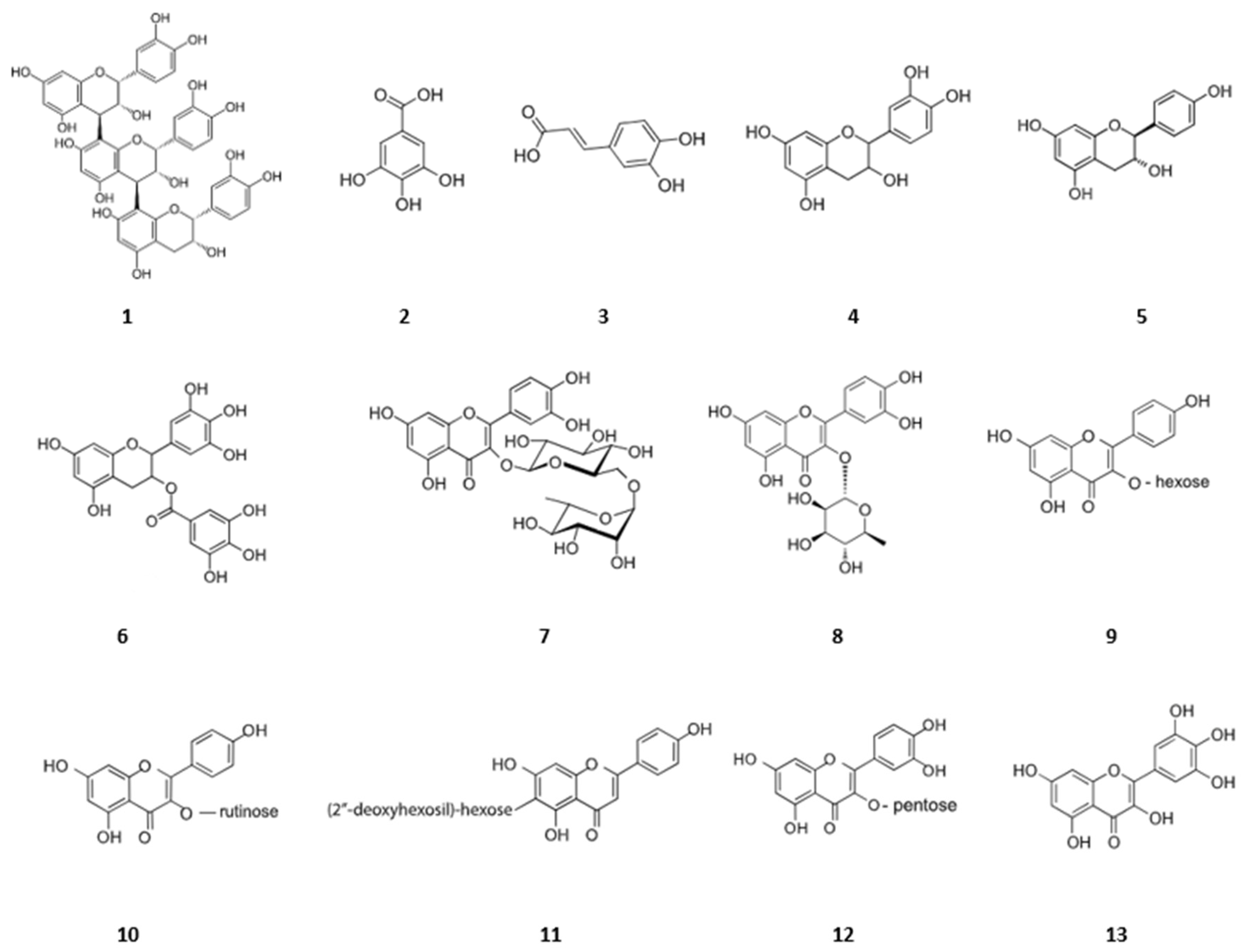
Figure 2 depicts the HPLC-UV-MS profiling of the methanolic extract of L. nobilis. Table 4 shows thirteen phenolic compounds, including flavonoids (flavonols, flavon-3-ols, and proanthocyanidins) and phenolic acids, with their Rt, precursor ion, m/z, fragment ions, molecular formula, and molecular weight, while Figure 3 represents structures of representative compounds.
These compounds were proposed based on a characteristic fragmentation pattern using ESI-MS experiments (key aglycone fragments of 285, 243 for kaempferol, 151 for quercetin, 139 for catechin) [29,30].
  • Phenolic acids
Compounds 2 and 3 were detected as gallic acid and caffeic acid based on previous data [31,32]. The MS/MS spectra of these compounds revealed fragment ions at m/z 125 and 135 owing to CO2 loss. This finding underscores the presence of basic phenolic acids in the Laurus nobilis extract.
2.
Flavonoids
The metabolite 9 (m/z 447) and the ion [M-H] collided to produce an aglycone radical (m/z 284) that is linked to the flavonol kaempferol. The molecular anion at m/z 593 in Compound 10’s ESI spectra was determined to be kaempferol-3-O-rutinoside. This kaempferol glycoside was cleaved at m/z 285 [M-H], yielding the anion aglycone [33,34,35].
Compound 7’s molecular anions at m/z 609 in its complete MS spectra were determined to be rutin; the loss of quercetin produced an MS ion at m/z 301 [36,37,38]. Compound 8 was defined as quercetin-3-rhamnoside ([M-H] ion at 447 m/z, MS2 ion at 301 m/z, formed by rhamnose moiety loss). The tentative identification of compound 11 was found to be quercetin-3-glucoside (also known as quercetin-3-O-hexose) ([M-H] ion at 463 m/z, MS2 ion at 301 m/z, formed by loss of hexose moiety). Compound 12’s mass spectrum supported the existence of quercetin-3-O-pentoside. Indeed, at m/z 434, it displayed the deprotonated molecular ion [M-H]. A radical aglycone with a mass of 301 was produced by the collision of the ion [M-H], pointing to the flavonol quercetin. The glyconic moiety’s pentose composition was suggested by the neutral loss of 132 m/z [39].
The [M-H] ion of compound 5 was 289, which corresponds to catechin, and its retention time was 21.77 min. The fragment at 245 m/z suggests that the hydroxyl group on C3 as well as the [CH2-COOH] group from C3 and C4 in ring C were lost. Compound 4 had a retention time of 20.34 min and a molecular ion [M-H] at 289, and it also had a fragment ion at 245 m/z as catechin [40,41,42]. The detection of catechin derivates emphasizes the presence of flavon-3-ols in the bay laurel methanolic extract.
The sixth compound (6), detected at 24.71 min, corresponds to epicatechin gallate, a flavan-3-ol gallate. With a molecular formula of C22H18O10 and a molecular weight of 442.4 g/mol, this compound indicates the presence of gallate-containing flavonoids in the bay laurel extract.
Compound 13 was identified by the detection of the [M-H] ion, which had an m/z value of 317.03 and characteristic MS/MS fragments at m/z 179 and 151, respectively. These fragments correlated with retrocyclization on the A-C ring and the subsequent loss of CO, indicating that compound 13 was myricetin [43]. This one contributed to the flavonol content of the Laurus nobilis methanolic extract.
3.
Proanthocyanidins
The extracted ion chromatogram’s molecular ion at m/z 577 pointed to a procyanidin trimer (compound 1) made up of catechin/epicatechin units connected by either a C4–C8 or C4–C6 interflavonoid bond. The procyanidin trimer’s [M-H] ion MS/MS spectrum displayed ions at 289 and 245 that serve as confirmation of catechin/epicatechin units [42,44]. This compound represents a member of the proanthocyanidin class, contributing to the complex phenolic profile of the bay laurel extract.

2.4. Qualitative Phytochemical Analysis

To identify the presence of several classes of secondary metabolites in both methanolic and aqueous plant extracts, Laurus nobilis underwent phytochemical screening and qualitative estimation. The results (Table 5) show that the methanol phytochemical profile had a similar trend as the aqueous extract. It was observed that the flavonoids, saponins, and tannins were intensively present in both plant extracts, whereas terpenoids were present only in the methanolic extract. However, anthocyanins were absent in both the methanolic and the aqueous extract. After qualitatively confirming the presence of essential phytochemicals, we performed a quantitative assessment. Methanol as an extraction solvent for bioactive compounds of plant extracts offers various benefits. Studies have shown that methanol extracts of plants like Haberlea rhodopensis, Acroptilon repens, Amaranthus retroflexus, and Andrographis paniculata exhibit significant anti-tumor, antimicrobial, antioxidant, and anti-proliferative activity [45,46,47]. Methanol extraction has been found to yield higher concentrations of phenolic and flavonoid contents compared to other solvents, indicating its efficiency in extracting phytochemical compounds [48]. Additionally, methanol extracts of plants like Citrullus colocynthis, Solanum nigrum, Solanum surattense, Calotropis procera, Agave americana, and Anagallis arvensis have shown potential as antioxidant, antibacterial, antifungal, and antidiabetic agents [49]. Overall, methanol extraction proves to be a versatile and effective solvent for obtaining bioactive compounds with various beneficial properties from plant materials.
The phytochemical substances found are recognized to have therapeutic value. Alkaloids, for instance, have been described as potent poisons, and many of their biologically active derivatives from medicinal plants have anti-inflammatory properties [50] and antimicrobial [51], antispasmodic, cytotoxicity, and pharmacological effects [52]. On the other hand, flavonoids have substantial anticancer action and are powerful water-soluble antioxidants and free radical scavengers that prevent oxidative cell damage [53]. Red blood cells can be precipitated and coagulated by saponin [54]. Research has shown that tannins have antibacterial [55], anticancer, and antiviral properties [56]. They function by precipitating microbial protein, which renders dietary protein inaccessible to them [57].

2.5. Quantitative Phytochemical Analysis

Significant antioxidant activity is seen in plants with high concentrations of secondary metabolites as phenolics, flavonoids, and tannins. Phytochemical analysis of laurel showed differences in phenolic compound content between aqueous and methanolic extracts. The data in Table 6 show that the highest concentration of those secondary metabolites was measured in the methanol extract, which contained 46.223 mg GAE/g dw, 42.386 mg QE/g dw, and 300.506 mg TAE/g dw, followed by the aqueous extract, which had a lower content of polyphenols, flavonoids, and tannins (33.766 mg GAE/g dw, 37.059 mg QE/g dw, and 98.43 mg TAE/g dw, respectively). This could be the case because the polarity of various extracting solvents elutes specific components in diverse ways. In a different study, laurel leaf extract contained 25.70 GAE/g dw of polyphenols and 12.11 mg CE/g dw of flavonoids [18]. The phenolic and flavonoid content was lower than what we found. The discrepancies in these data might be attributed to the extraction method, the species, or the stage of plant growth.

2.6. Antioxidant Activity

Due to the presence of several bioactive substances such phenolic compounds, flavonoids, and tannins, medicinal plants are recognized for their exceptional antioxidant qualities [58]. The body needs these antioxidants to protect it from oxidative stress and free radical damage. Medicinal herbs aid in preventing cellular and tissue damage, which is linked to the development of several chronic illnesses, including cancer, cardiovascular problems, and neurological diseases, by scavenging free radicals and stabilizing reactive oxygen species [59]. These organic antioxidants play a crucial role in conventional medical procedures and continue to receive a lot of attention in contemporary medicine due to their ability to improve health, strengthen the body’s defenses, and promote general wellbeing. Due to these factors, four complementary in vitro assays based on various mechanisms, including free radical scavenging assays (DPPH and ABTS), ferric reducing antioxidant power assay (FRAP), and oxygen radical absorbance capacity (ORAC), were used to assess the antioxidant properties of aqueous and methanolic extracts made from laurel leaves.
Both methanol and aqueous extracts demonstrated highly significant, exceptional antioxidant activity in all radical scavenging experiments, at p < 0.05. As indicated in Table 7, the IC50 values were used to express the scavenging effects of the radical scavenging assays DPPH and ABTS. Free radical DPPH is a synthetic, somewhat stable nitrogen radical, and this test technique is based on electron transfer, in which an antioxidant chemical decreases the oxidant by donating an electron, leading to a change in color and subsequent change in absorbance [60,61]. The ABTS radical scavenging technique relies on the decrease in the preformed radical cation ABTS+ by the addition of an antioxidant. The amount of ABTS+ chromophore decolorization measured spectrophotometrically at 734 nm provides a measure of the sample’s antioxidant activity [62]. By quantifying the decrease in the cation radical as the percentage of inhibition, the degree of ABTS+ inhibition was displayed as a function of concentration [63]. The methanolic extract exhibited a strong DPPH and ABTS scavenging effect of about 0.079 and 0.148 mg/mL compared to the aqueous extract with an IC50 of 0.169 and 0.221 mg/mL, respectively. The analyzed extracts from L. nobilis exhibited IC50 values that were obtained are greater than those found in published research, with an IC50 = 0.17 mg/mL (DPPH) [64] and 1.901 mg/mL (ABTS) [65].
The approach of ferric reducing antioxidant potential (FRAP) relies on a reduction in a colorless ferric complex (Fe3+-tripyridyltriazine) to a blue ferrous complex (Fe2+-tripyridyltriazine) at low pH caused by the action of antioxidants that donate electrons. The decrease in absorbance at 593 nm is used to gauge the reduction [66]. Table 7 shows that the methanolic extract (73.262 mg TE/g dw) had the highest reducing power compared to the aqueous extract (46.291 mg TE/g dw). Lu et al. [67] reported a significantly higher FRAP value of 504.25 μmol TE/g, which is higher than that in our study.
Based on an extract’s ability to shield the fluorescent probe fluorescein from peroxyl radicals, the oxygen radical absorbance capacity (ORAC) assay produces longer fluorescence decay curves that can be integrated [68]. The highest value was recorded in the methanolic extract (77.006 mg TE/g dw) in comparison to the aqueous extract (40.754 mg TE/g dw). ORAC test results showing the antioxidant potential of Laurus nobilis L. leaves vary significantly in the literature. As an example, Kratchanova et al. [69] identified an ORAC value lower than our study’s for a water extract, which was 170 μmol TE/g.
Besides that, the phenolic, flavonoid, and tannin contents were correlated with the outcomes of the DPPH, ABTS, FRAP, and ORAC experiments. The high phenolic content of the methanolic extract of Laurus n. leaf extract is what gives it its antioxidant capabilities. This study suggests that the polyphenol content of an extract may influence its ability to scavenge free radicals.

3. Materials and Methods

3.1. Plant Material

The collection of the leaves of Laurus nobilis was carried out in a forest called Rmilat (Tangier, Northern Morocco). They were taken to the laboratory of Physical Chemistry of Materials, Natural Substances and Environment, Faculty of Sciences and Technologies (Tangier, Morocco). The leaves were stripped from the plant and air-dried in the lab under shade. After complete shade drying, the dried leaves of the plants were crushed into a fine powder and stored in a small, airtight plastic bag to avoid contact with moisture.

3.2. Extract Preparation

The pulverized plant leaves were extracted with methanol 80% and dH2O (4.5 g of plant material was added to 45 mL of methanol 80% or dH2O), and shaken continuously (250 rpm) in the dark for 48 h at room temperature. The extracts were filtered with Whatman filter paper and then put in incubator at 37 °C so the solvents could evaporate. The dried extracts were weighed to determine the percent recovery of solvent [70].

3.3. ICP-AES Analysis

We carefully weighed 0.1 g of sample (leaf powder), to which we added 3 mL of hydrochloric acid and 1 mL of nitric acid. The mixture was allowed to stand for 24 h, followed by heating for 2 h at 95 °C. Subsequently, the sample was diluted to 25 mL using ultrapure water. Finally, the solution was filtered through a 45 µm sieve before being analyzed by an ICP ULTIMA EXPERT apparatus, HORIBA, Palaiseau, France.

3.4. WD-XRF Analysis

Quantitative analysis of the Laurus nobilis plant sample was carried out using a WDXRF spectrometer, which is an advanced instrument that enabled precise elemental analysis of various sample forms, including solids, fused beads, pressed or unpressed powders, and liquid samples. The instrument operated with a rhodium anode and had a maximum power of 4 kW and a maximum current of 160 mA. The X-ray tube in the WDXRF spectrometer recorded the characteristic radiation from major, minor, and trace elements through ten distinct scans to identify all elements present in the plant samples. This instrument (Axios 2005, PANalytical, Malvern, UK) is widely used for elemental analysis and is considered one of the most important tools in the field of analytical chemistry.

3.5. FT-IR Analysis

Little milligrams of methanolic freeze-dried extract were dispersed and encapsulated in potassium bromide (KBr) to form a thin translucent sample disc for FT-IR analysis. The disc was then placed in a sample cup of a diffuse reflectance accessory. FT-IR experiments were performed using a PerkinElmer 2000 infrared spectrometer (PerkinElmer, Inc. Waltham, MA, USA) to identify the characteristic functional groups in the sample. The spectra were recorded within a range of 4000–400 cm−1, with a resolution of 4 cm−1 [71].

3.6. HPLC-MS-UV Analysis

Samples were analyzed by HPLC-UV-MS using a Q-Exactive plus mass spectrometer (Thermo Scientific, Waltham, MA, USA) fitted with nanoflow reversed-phase HPLC (Ultimate3000 RSLC, Dionex, Sunnyvale, CA, USA). HPLC-MS-UV analyses of phenolic compounds were performed on a reverse-phase BDS Hypersil C18 (4.6 × 150 nm, 5 μm) using a gradient program with two solvents: water (solvent A) and acetonitrile (solvent B)—both with the addition of 0.1% formic acid to enhance the ionization of metabolites. The following composition of gradient was applied: 0 min 2% of B in A, 15 min 25% of B in A, 30 min 45% of B in A, 40 min 95% of B in A, 43 min 2% of B in A. The injection volume of all samples at a concentration of 10 mg/mL was 20 μL, the flow rate was 0.2 mL/min, the analysis run was 50 min, and the post run was 10 min. Mass spectrometry was operated in the m/z range of 200–2000 using an optimized negative electrospray ionization (ESI) spectrum. The identification of eluted components was detected spectrophotometrically at 280 nm [72].

3.7. Phytochemical Screening

The entirety of the qualitative procedures allowing for the identification of various chemical groups present in a plant organ is collectively referred to as phytochemical screening. Physical–chemical reactions allow us to recognize the presence of chemical compounds.
Although there are many phytochemical groups, the primary ones are the total polyphenols, which include flavonoids, anthocyanins, tannins, coumarins, alkaloids, saponins, and terpenoids/steroids. The qualitative results are expressed as (+) for the presence and (−) for the absence of phytochemicals.
The following phytochemical tests were performed in triplicate and relied on protocols described by several groups.

3.7.1. Flavonoids

In a test tube containing 10 mL of the extract, a solution of NH4OH was added; after 3 h, a light yellow color appeared in the upper part of the tube, which indicated the presence of flavonoids [73].

3.7.2. Tannins

A total of 0.5 g of leaf extracts was mixed with 10 mL of distilled water before being filtered. Then, 5% ferric chloride was added in a few drops. The occurrence of black or blue–green precipitation was seen as a sign that tannins were present [74].

3.7.3. Alkaloids

A total of 5 g of dried and roasted plant materials were macerated in 50 mL of HCl at 1%, filtered, and tested by Mayer’s reagent. The presence of aldehydes was indicated by a white precipitate [75].

3.7.4. Anthocyanins

To 5 mL of the infusion we add a few drops of HCl and then a few drops of NH4OH. The color change indicated the presence of anthocyanins [76].

3.7.5. Saponins

In a test tube, 0.5 g of each plant extract was mixed with 10 mL of distilled water individually. When heated in a water bath for five minutes, foaming took place and continued, indicating the presence of saponins [77].

3.7.6. Coumarins

A total of 5 mL of the etheric extract was evaporated, and the residue was taken up in 2 mL of hot water. Then, we added 0.5 mL of NH4OH to the tube at 25%. The presence of coumarins was indicated by fluorescence observed under UV at 366 nm [78].

3.7.7. Terpenoids/Steroids

Maceration of 4 g of the plant material was carried out in 20 mL of petroleum ether. After filtration, the organic phase was evaporated, and 0.5 mL of acetic acid was poured on the residue obtained. Then, 1 mL of sulfuric acid was concentrated. The formation of violet or maroon rings at the contact area of the two liquids revealed the presence of terpenes [79].

3.8. Determination of Total Phenolic Content

The total phenolic content was determined by the Folin–Ciocalteu method, following Ennoury et al. [80]. To 100 µL of the sample, 400 µL of Folin Ciocalteu reagent and 1 mL of saturated Na2CO3 (7%) were added, and the final volume was increased to 1.6 mL with distilled water. The tubes were left to stand in the dark for 30 min, after which the absorbance was read at 725 nm against a blank. The total phenolic content of the plant extracts was expressed as milligrams of gallic acid equivalent per gram of dry weight (mg GAE/g dw) through the calibration curve with gallic acid.

3.9. Determination of Total Flavonoid Content

The flavonoid content was assessed following the method of Ennoury et al., with some modifications [80]. Briefly, 40 µL of each sample were mixed with 10 µL of acetate potassium (1 M) and 10 µL of aluminum chloride (10%). Thereafter, 100 µL of 50% methanol were added and the total volume was increased to 400 µL with distilled water. The absorbance of the mixture was determined at 415 nm. Quercetin was used as standard. The flavonoid content was expressed as milligram of quercetin equivalent (QE) per gram of extract.

3.10. Determination of Total Tannin Content

The tannins were determined by the Folin–Ciocalteu method [74]. About 0.1 mL of the sample extract was added to a volumetric flask (10 mL) containing 7.5 mL of distilled water, 0.5 mL of Folin–Ciocalteu phenol reagent, and 1 mL of 35% sodium carbonate solution and was diluted to 10 mL with distilled water. The mixture was shaken well and kept at room temperature for 30 min. Absorbance for test and standard solutions were measured against a blank at 700 nm. The estimation of the tannin content was carried out in triplicate. The tannin content was expressed in terms of mg of tannic acid equivalent/g of dried sample.

3.11. DPPH Radical Scavenging Assay

The radical scavenging capacity was determined using the stable free radical DPPH (2,2-diphenyl-1-picrylhydrazyl) and following the method described by BenMrid et al. with some modifications [70]. Adequate dilutions of sample were realized to obtain a final volume of 50 µL. Extract solutions (50 µL) were mixed with 150 µL of a freshly prepared DPPH solution. The mixture was shaken vigorously and left to stand in the dark and at room temperature for 30 min. The reduction in the DPPH radical was measured at 517 nm. The DPPH scavenging activity was determined by calculating the percentage of DPPH discoloration using the following equation:
%Scavenging effect = [(ADPPH − AS)/ADPPH] × 100
where AS corresponds to the values of the sample and ADPPH refers to the absorbance of the DPPH solution. The extract concentration providing 50% inhibition (IC50) was determined from the graph of the scavenging effect percentage against the extract concentration in the solution.

3.12. ABTS Radical Scavenging Assay

The radical scavenging activity against the radical ABTS+ was evaluated following the method of BenMrid et al. [70]. ABTS+ was generated by the oxidation of ABTS with potassium persulfate. Prior to assay, the ABTS+ stock solution was diluted with methanol until it reached an absorbance of 0.700 ± 0.020 at 734 nm. Then, 185 µL of the diluted ABTS+ solution were mixed with 15 µL of the sample, and the absorbance was measured at 734 nm after 10 min. The radical scavenging activity was calculated using the following formula:
%Scavenging effect = [(A ABTS − AS)/AABTS] × 100
where AS corresponds to the values of the sample and AABTS refers to the absorbance of the ABTS solution. The extract concentration providing 50% inhibition (IC50) was calculated from the graph of the scavenging effect percentage against the extract concentration in the solution.

3.13. FRAP Assay

The ferric-reducing antioxidant power (FRAP) assay was estimated following the method reported by Olatunji et al. [81].
The FRAP reagent was prepared with acetate buffer 30 nM, TPTZ 10 nM, and FeCl3 in a proportion of 10:1:1. The diluted extract was put on the microplate with the FRAP reagent, which was incubated before at 30 °C.
The absorbance was read at 593 nm, and the Trolox was used as a calibration curve. The results are presented with the Trolox equivalent.

3.14. ORAC Assay

Antioxidant activity of the extracts were determined by the ORAC assay, as reported by Escribano et al. [82]
The sample was placed on a microplate with 120 µL of fluorescein 80 nM (diluted with PBS 13.3 mM). The microplate reader was prepared before use and fixed at 37 °C. The initial fluorescence was read and then the AAPH solution was read. The plate continued to be read quickly every 2 min for 120 min. The Trolox was used as the calibration curve.

3.15. Statictical Analysis

All the experiments were conducted in triplicate, and the data are presented as mean ± SD (standard deviation). SPSS (version 23; SPSS Inc., Chicago, IL, USA) was used to process the results. For the antioxidant activity assays, a one-way ANOVA test followed by Tukey’s test (p < 0.05) was used to analyze the differences among IC50 of the CEE and its various fractions.

4. Conclusions

In conclusion, our investigation into Laurus nobilis specimens collected from the northern region of Morocco has revealed compelling findings regarding the extraction methods and the subsequent quantification of secondary metabolites and antioxidant properties. This study is the first to evaluate the nutritional and therapeutic potential of Laurus nobilis leaves from this particular location using a wide range of analytical techniques, including infrared and several chromatographic methods. It is shown by tests utilizing WD-XRF and ICP-AES methods that vital minerals such calcium, magnesium, and potassium were present. These minerals are critical to human health. The noteworthy quantities of these constituents, in conjunction with the identification of advantageous trace elements such as iron, manganese, and copper, indicate that Laurus nobilis may represent a good dietary supplement for augmenting food intake and bolstering diverse physiological processes.
This plant is rich in flavonoids, especially those belonging to flavonols (myricetin, kaempferol, and quercetin), flavan-3-ols (catechin, epicatechin, and epicatechin gallate) and proanthocyanidins. The comparative analysis between the methanolic and aqueous extracts of laurel leaves yielded noteworthy disparities. The methanolic extract of laurel leaves displayed significantly higher values in terms of secondary metabolites, including total phenolic, flavonoid, and tannin content, compared to the aqueous extract. This discrepancy underscores the superiority of methanol as a solvent for extracting these bioactive compounds from Laurus nobilis leaves. These secondary metabolites are well known for their potential health benefits, including antioxidant, anti-inflammatory, and antimicrobial properties. Furthermore, the assessment of antioxidant properties through various assays, namely, DPPH, ABTS, FRAP, and ORAC, demonstrates that the methanolic extract outperformed the aqueous extract. This finding indicates that the methanolic extract of laurel leaves collected from the Northern Morocco region possessed stronger free radical scavenging abilities and a greater capacity to mitigate oxidative stress.
The observed disparities between the two extraction methods emphasize the importance of selecting an appropriate solvent for the extraction of bioactive compounds from Laurus nobilis. The methanolic extract in particular stands out as a more effective choice for harnessing the full potential of laurel leaves in terms of secondary metabolites and antioxidant properties. In conclusion, this study contributes valuable insights into the utilization of Laurus nobilis from the Northern Morocco region as a source of bioactive compounds with immense potential for improving human health and well-being.

Author Contributions

Conceptualization, A.M.; methodology, A.M. and B.A.; formal analysis and investigation, M.K.; resources, F.E.M.; writing—original draft preparation, A.M. and A.S.; writing—review and editing, A.M., F.E.M. and M.K.; supervision, F.E.M. and M.K. All authors have read and agreed to the published version of the manuscript.

Funding

This research received no external funding.

Data Availability Statement

All data are included in the article.

Conflicts of Interest

The authors declare no conflict of interest.

References

  1. Bektaş, S.; Özdal, M.; Gürkök, S. Determination of Antibacterial and Antibiofilm Activities for Laurel (Laurus nobilis L.) Essential Oil Against the Fish Pathogen Pseudomonas Species. Menba Kastamonu Üniversitesi Su Ürün. Fakültesi Derg. 2023, 9, 25–33. [Google Scholar] [CrossRef]
  2. Khodja, Y.K.; Bachir-Bey, M.; Belmouhoub, M.; Ladjouzi, R.; Dahmoune, F.; Khettal, B. The botanical study, phytochemical composition, and biological activities of Laurus nobilis L. leaves: A review. Int. J. Second. Metab. 2023, 10, 269–296. [Google Scholar] [CrossRef]
  3. Ross, I.A. Medicinal Plants of the World 3; Humana Press: Totowa, NJ, USA, 2005. [Google Scholar]
  4. Santoyo, S.; Lloría, R.; Jaime, L.; Ibañez, E.; Señoráns, F.J.; Reglero, G. Supercritical fluid extraction of antioxidant and antimicrobial compounds from Laurus nobilis L. Chemical and functional characterization. Eur. Food Res. Technol. 2006, 222, 565–571. [Google Scholar] [CrossRef]
  5. El Faqer, O.; Rais, S.; Elkoraichi, I.; El Amrani, A.; Dakir, M.; Zaid, Y.; Mtairag, E.M. Phytochemical characterization and immunomodulatory effects of aqueous and ethanolic extracts and essential oil of Moroccan Laurus nobilis L. (Lauraceae) on human neutrophils. J. Herbmed Pharmacol. 2022, 12, 92–99. [Google Scholar] [CrossRef]
  6. Yao, R.; Heinrich, M.; Zhang, B.; Wei, X.; Qi, Y.; Gao, W. Single botanical drugs in the Ayurvedic Pharmacopoeia of India—A quantitative ethnobotanical analysis. Front. Pharmacol. 2023, 14, 1136446. [Google Scholar] [CrossRef] [PubMed]
  7. Ben Ayed, A.; Aissa, E.; Haouala, F. Effect of salinity on germination capacity of laurel (Laurus nobilis L.). GSC Adv. Res. Rev. 2023, 15, 304–309. [Google Scholar] [CrossRef]
  8. Dobroslavić, E.; Garofulić, I.E.; Ilich, J.Z. Potential of Laurel (Laurus nobilis L.) Leaf Polyphenols for Modulation of Body Composition. Appl. Sci. 2023, 13, 2275. [Google Scholar] [CrossRef]
  9. Bahtiti, N.H.; Orabi, F.M.A.; Kailani, M.H.; Abdel-Rahman, I.; Ismail, L.S. A Comparative LC/MS Analysis of Jordanian Bay Leaf, Branches, and Roots (Laurus nobilis L.). J. Southwest Jiaotong Univ. 2022, 57, 6. Available online: http://jsju.org/index.php/journal/article/view/1424 (accessed on 15 April 2024). [CrossRef]
  10. Escobedo-Monge, M.F.; Barrado, E.; Parodi-Román, J.; Escobedo-Monge, M.A.; Torres-Hinojal, M.C.; Marugán-Miguelsanz, J.M. Magnesium Status and Ca/Mg Ratios in a Series of Children and Adolescents with Chronic Diseases. Nutrients 2022, 14, 2941. [Google Scholar] [CrossRef]
  11. Hibler, E.A.; Zhu, X.; Shrubsole, M.J.; Hou, L.; Dai, Q. Physical activity, dietary calcium to magnesium intake and mortality in the National Health and Examination Survey 1999–2006 cohort. Int. J. Cancer 2020, 146, 2979–2986. [Google Scholar] [CrossRef]
  12. Appel, L.J. Potassium. In Encyclopedia of Human Nutrition, 4th ed.; Caballero, B., Ed.; Academic Press: Oxford, UK, 2023; pp. 362–367. [Google Scholar] [CrossRef]
  13. Aljafary, M.A.; Alshwyeh, H.; Alahmadi, N.; Shehzad, A.; Tombuloglu, H.; Gaymalov, Z.; Homieda, A.; Al-Suhaimi, E. Physiological and Cellular Functions of Vitamin K on Cardiovascular Function. In Vitamin K—Recent Topics on the Biology and Chemistry; IntechOpen: London, UK, 2021. [Google Scholar] [CrossRef]
  14. Manalang, M.F.; Ocampo, G.; Alonto, A.-S.; Rimocal, G. Geo-Spatial Mapping of Iron (Fe) and Manganese (Mn) Present in Groundwater of Clark, Philippines. ASEAN Eng. J. 2023, 13, 101–107. [Google Scholar] [CrossRef]
  15. Xu, C.; Yang, H.; Huang, C.; Lan, M.; Zou, Z.; Zhang, F.; Zhang, L. Interaction Mechanism of Fe, Mg and Mn in Karst Soil-Mango System. Land 2023, 12, 256. [Google Scholar] [CrossRef]
  16. Bojórquez-Quintal, E.; Escalante-Magaña, C.; Echevarría-Machado, I.; Martínez-Estévez, M. Aluminum, a Friend or Foe of Higher Plants in Acid Soils. Front. Plant Sci. 2017, 8, 1767. [Google Scholar] [CrossRef] [PubMed]
  17. Khvorost, O.; Posohova, I.; Fedchenkova, Y.; Skrebtsova, K. Component composition of essential oil shoots and leaves of Laurus nobilis L. Ukrainian origian. Sci. Pharm. Sci. 2021, 4, 50–58. [Google Scholar] [CrossRef]
  18. Guenane, H.; Gherib, A.; Carbonell-Barrachina, Á.; Cano-Lamadrid, M.; Krika, F.; Berrabah, M.; Maatallah, M.; Bakchiche, B. Minerals analysis, antioxidant and chemical composition of extracts of Laurus nobilis from southern Algeria. J. Mater. Environ. Sci. 2016, 7, 4253–4261. [Google Scholar]
  19. Awada, F.; Hamade, K.; Kassir, M.; Hammoud, Z.; Mesnard, F.; Rammal, H.; Fliniaux, O. Laurus nobilis Leaves and Fruits: A Review of Metabolite Composition and Interest in Human Health. Appl. Sci. 2023, 13, 4606. [Google Scholar] [CrossRef]
  20. de cássia Basilio, L. Óleo Laurus nobilis L. (laurel) Essential Induces Activation of Reactive Oxygen Species and Cell Signaling Pathways during Larvicidal Action; Federal University of Viçosa: Viçosa, Brazil, 2022. [Google Scholar] [CrossRef]
  21. Jaradat, N.; Abualhasan, M.; Hawash, M.; Qadi, M.; Al-Maharik, N.; Abdallah, S.; Mousa, A.; Zarour, A.; Arar, M.; Sobuh, S.; et al. Chromatography analysis, in light of vitro antioxidant, antidiabetic, antiobesity, anti-inflammatory, antimicrobial, anticancer, and three-dimensional cancer spheroids’ formation blocking activities of Laurus nobilis aromatic oil from Palestine. Chem. Biol. Technol. Agric. 2023, 10, 25. [Google Scholar] [CrossRef]
  22. Obiandu, C.; Laz-Okenwa, J.O.A.; Owhorji, B.I.; Opubo, A.T.; Samuel, H.O.A. Evaluation of the Effects of Extracts of Laurus nobilis on some Biochemical Parameters of Wistar Rats. Sch. Int. J. Anat. Physiol. 2023, 6, 37–41. [Google Scholar] [CrossRef]
  23. Saleh, B.H.; Yahya, H.N.; Ibrahim, R.N. Study antibacterial activity of laurus nobilis leaves water extract on some isolates of pathogenic bacteria. Iraqi J. Agric. Sci. 2023, 54, 18–24. [Google Scholar] [CrossRef]
  24. Durocher, G.; Sandorfy, C. Anharmonicity and hydrogen bonding: Part II. Examples of moderately strong OH⋯Y bonds. J. Mol. Spectrosc. 1965, 15, 22–28. [Google Scholar] [CrossRef]
  25. Rizwana, H.; Al Kubaisi, N.; Al-Meghailaith, N.N.; Moubayed, N.M.; Albasher, G. Evaluation of Chemical Composition, Antibacterial, Antifungal, and Cytotoxic Activity of Laurus nobilis L Grown in Saudi Arabia. J. Pure Appl. Microbiol. 2019, 13, 2073–2085. [Google Scholar] [CrossRef]
  26. Deghles, A.; Hamed, O.; Azar, M.; Abu Lail, B.; Azzaoui, K.; Abu Obied, A.; Jodeh, S. Cellulose with Bidentate Chelating Functionality: An Adsorbent for Metal Ions from Wastewater. BioResources 2019, 14, 6247–6266. [Google Scholar] [CrossRef]
  27. Trivedi, M.K.; Tallapragada, R.M.; Branton, A.; Trivedi, D.; Nayak, G.; Km, R.; Jana, S. Biofield Treatment: A Potential Strategy for Modification of Physical and Thermal Properties of Indole. J. Environ. Anal. Chem. 2015, 6, 4. [Google Scholar] [CrossRef]
  28. De Lorenzi, A.; Giorgianni, S.; Bini, R. High-resolution FTIR spectroscopy of the C—Cl stretching mode of vinyl chloride. Int. J. Interface Chem. Phys. 2009, 96, 101–108. [Google Scholar] [CrossRef]
  29. Fabre, N.; Rustan, I.; de Hoffmann, E.; Quetin-Leclercq, J. Determination of flavone, flavonol, and flavanone aglycones by negative ion liquid chromatography electrospray ion trap mass spectrometry. J. Am. Soc. Mass Spectrom. 2001, 12, 707–715. [Google Scholar] [CrossRef]
  30. Rand, D.; Elizabeth, E. Identification of Digallated and Methylated Catechins Using UPLC/MS/MS and Development of a Rapid Analysis Method for Theanine in Tea (Camellia sinensis (L.) O. Kuntze) Utilizing Evaporative Light Scattering Detection. 2009. Available online: https://repository.up.ac.za/handle/2263/28932 (accessed on 2 January 2024).
  31. Santagati, N.A.; Salerno, L.; Attaguile, G.; Savoca, F.; Ronsisvalle, G. Simultaneous determination of catechins, rutin, and gallic acid in Cistus species extracts by HPLC with diode array detection. J. Chromatogr. Sci. 2008, 46, 150–156. [Google Scholar] [CrossRef] [PubMed]
  32. Barrajón-Catalán, E.; Fernández-Arroyo, S.; Roldán, C.; Guillén, E.; Saura, D.; Segura-Carretero, A.; Micol, V. A systematic study of the polyphenolic composition of aqueous extracts deriving from several Cistus genus species: Evolutionary relationship. Phytochem. Anal. PCA 2011, 22, 303–312. [Google Scholar] [CrossRef] [PubMed]
  33. Engels, C.; Gräter, D.; Esquivel, P.; Jiménez, V.M.; Gänzle, M.G.; Schieber, A. Characterization of phenolic compounds in jocote (Spondias purpurea L.) peels by ultra high-performance liquid chromatography/electrospray ionization mass spectrometry. Food Res. Int. 2012, 46, 557–562. [Google Scholar] [CrossRef]
  34. Perestrelo, R.; Lu, Y.; Santos, S.A.; Silvestre, A.J.; Neto, C.P.; Câmara, J.S.; Rocha, S.M. Phenolic profile of Sercial and Tinta Negra Vitis vinifera L. grape skins by HPLC–DAD–ESI-MSn: Novel phenolic compounds in Vitis vinifera L. grape. Food Chem. 2012, 135, 94–104. [Google Scholar] [CrossRef]
  35. Gattuso, G.; Barreca, D.; Gargiulli, C.; Leuzzi, U.; Caristi, C. Flavonoid Composition of Citrus Juices. Mol. J. Synth. Chem. Nat. Prod. Chem. 2007, 12, 1641–1673. [Google Scholar] [CrossRef]
  36. Simirgiotis, M.J.; Schmeda-Hirschmann, G.; Bórquez, J.; Kennelly, E.J. The Passiflora tripartita (Banana Passion) Fruit: A Source of Bioactive Flavonoid C-Glycosides Isolated by HSCCC and Characterized by HPLC–DAD–ESI/MS/MS. Molecules 2013, 18, 1672–1692. [Google Scholar] [CrossRef]
  37. Alonso-Salces, R.M.; Ndjoko, K.; Queiroz, E.F.; Ioset, J.R.; Hostettmann, K.; Berrueta, L.A.; Gallo, B.; Vicente, F. On-line characterisation of apple polyphenols by liquid chromatography coupled with mass spectrometry and ultraviolet absorbance detection. J. Chromatogr. A 2004, 1046, 89–100. [Google Scholar] [CrossRef]
  38. Silva, B.A.; Ferreres, F.; Malva, J.O.; Dias, A.C.P. Phytochemical and antioxidant characterization of Hypericum perforatum alcoholic extracts. Food Chem. 2005, 90, 157–167. [Google Scholar] [CrossRef]
  39. Ferreres, F.; Grosso, C.; Gil-Izquierdo, A.; Valentão, P.; Andrade, P.B. Phenolic compounds from Jacaranda caroba (Vell.) A. DC.: Approaches to neurodegenerative disorders. Food Chem. Toxicol. 2013, 57, 91–98. [Google Scholar] [CrossRef]
  40. Bravo, M.N.; Silva, S.; Coelho, A.V.; Boas, L.V.; Bronze, M.R. Analysis of phenolic compounds in Muscatel wines produced in Portugal. Anal. Chim. Acta 2006, 563, 84–92. [Google Scholar] [CrossRef]
  41. Borbalán, A.M.A.; Zorro, L.; Guillén, D.A.; Barroso, C.G. Study of the polyphenol content of red and white grape varieties by liquid chromatography–mass spectrometry and its relationship to antioxidant power. J. Chromatogr. A 2003, 1012, 31–38. [Google Scholar] [CrossRef]
  42. Benavides, A.; Montoro, P.; Bassarello, C.; Piacente, S.; Pizza, C. Catechin derivatives in Jatropha macrantha stems: Characterisation and LC/ESI/MS/MS quali–quantitative analysis. J. Pharm. Biomed. Anal. 2006, 40, 639–647. [Google Scholar] [CrossRef] [PubMed]
  43. Chernonosov, A.A.; Karpova, E.A.; Lyakh, E.M. Identification of phenolic compounds in Myricaria bracteata leaves by high-performance liquid chromatography with a diode array detector and liquid chromatography with tandem mass spectrometry. Rev. Bras. Farm. 2017, 27, 576–579. [Google Scholar] [CrossRef]
  44. Zywicki, B.; Reemtsma, T.; Jekel, M. Analysis of commercial vegetable tanning agents by reversed-phase liquid chromatography–electrospray ionization–tandem mass spectrometry and its application to wastewater. J. Chromatogr. A 2002, 970, 191–200. [Google Scholar] [CrossRef]
  45. Zasheva, D.; Mladenov, P.; Rusanov, K.; Simova, S.; Zapryanova, S.; Simova-Stoilova, L.; Moyankova, D.; Djilianov, D. Fractions of Methanol Extracts from the Resurrection Plant Haberlea rhodopensis Have Anti-Breast Cancer Effects in Model Cell Systems. Separations 2023, 10, 388. [Google Scholar] [CrossRef]
  46. Babaei-Ghaghelestany, A.; Alebrahim, M.T.; Farzaneh, S.; Mehrabi, M. The anticancer and antibacterial properties of aqueous and methanol extracts of weeds. J. Agric. Food Res. 2022, 10, 100433. [Google Scholar] [CrossRef]
  47. Anoor, P.K.; Yadav, A.N.; Rajkumar, K.; Kande, R.; Tripura, C.; Naik, K.S.; Burgula, S. Methanol extraction revealed anticancer compounds Quinic Acid, 2(5H)-Furanone and Phytol in Andrographis paniculata. Mol. Clin. Oncol. 2022, 17, 151. [Google Scholar] [CrossRef] [PubMed]
  48. Baeshen, N.A.; Almulaiky, Y.Q.; Afifi, M.; Al-Farga, A.; Ali, H.A.; Baeshen, N.N.; Abomughaid, M.M.; Abdelazim, A.M.; Baeshen, M.N. GC-MS Analysis of Bioactive Compounds Extracted from Plant Rhazya stricta Using Various Solvents. Plants 2023, 12, 960. [Google Scholar] [CrossRef] [PubMed]
  49. Aldughaylibi, F.S.; Raza, M.A.; Naeem, S.; Rafi, H.; Alam, M.W.; Souayeh, B.; Farhan, M.; Aamir, M.; Zaidi, N.; Mir, T.A. Extraction of Bioactive Compounds for Antioxidant, Antimicrobial, and Antidiabetic Applications. Molecules 2022, 27, 5935. [Google Scholar] [CrossRef] [PubMed]
  50. Souto, A.L.; Tavares, J.F.; Da Silva, M.S.; Diniz, M.d.F.F.M.; De Athayde-Filho, P.F.; Filho, J.M.B. Anti-Inflammatory Activity of Alkaloids: An Update from 2000 to 2010. Molecules 2011, 16, 8515–8534. [Google Scholar] [CrossRef] [PubMed]
  51. Amel, B.; Abdlouahab, Y.; Abdlhakim, B. Assessment of the antibacterial activity of crude alkaloids extracted from seeds and roots of the plant Peganum harmala L. J. Nat. Prod. Plant Resources. 2012, 2, 568–573. [Google Scholar]
  52. Ameyaw, Y.; Duker-Eshun, G. The alkaloid contents of the ethno-plant organs of three antimalarial medicinal plant species in the eastern region of ghana. Int. J. Chem. Sci. 2009, 7, 48–58. [Google Scholar]
  53. Benavente-García, O.; Castillo, J.; Marin, F.R.; Ortuño, A.; Del Río, J.A. Uses and Properties of Citrus Flavonoids. J. Agric. Food Chem. 1997, 45, 4505–4515. [Google Scholar] [CrossRef]
  54. Sodipo, O.A.; Akinniyi, J.A.; Ogunbameru, J.V. Studies on certain characteristics of extracts of bark of Pausinystalia johimbe and Pausinystalia macroceras (K Schum) Pierre ex Beille. Glob. J. Pure Appl. Sci. 2000, 6, 83–88. [Google Scholar] [CrossRef]
  55. Akiyama, H.; Fujii, K.; Yamasaki, O.; Oono, T.; Iwatsuki, K. Antibacterial action of several tannins against Staphylococcus aureus. J. Antimicrob. Chemother. 2001, 48, 487–491. [Google Scholar] [CrossRef]
  56. Kumari, D.M.; Jain, S. Tannin: An Antinutrient with Positive Effect to Manage Diabetes. Res. J. Recent Sci. 2012, 2277, 2502. [Google Scholar]
  57. Kren, V.; Martinkova, L. Glycosides in Medicine: “The Role of Glycosidic Residue in Biological Activity”. Curr. Med. Chem. 2001, 8, 1303–1328. [Google Scholar] [CrossRef]
  58. Tungmunnithum, D.; Thongboonyou, A.; Pholboon, A.; Yangsabai, A. Flavonoids and Other Phenolic Compounds from Medicinal Plants for Pharmaceutical and Medical Aspects: An Overview. Medicines 2018, 5, 93. [Google Scholar] [CrossRef] [PubMed]
  59. Cheng, D.; Li, W.; Wang, L.; Lin, T.; Poiani, G.; Wassef, A.; Hudlikar, R.; Ondar, P.; Brunetti, L.; Kong, A.-N. Pharmacokinetics, Pharmacodynamics, and PKPD Modeling of Curcumin in Regulating Antioxidant and Epigenetic Gene Expression in Healthy Human Volunteers. Mol. Pharm. 2019, 16, 1881–1889. [Google Scholar] [CrossRef]
  60. Huang, D.; Ou, B.; Prior, R.L. The Chemistry behind Antioxidant Capacity Assays. J. Agric. Food Chem. 2005, 53, 1841–1856. [Google Scholar] [CrossRef]
  61. Letelier, M.E.; Molina-Berríos, A.; Cortés-Troncoso, J.; Jara-Sandoval, J.; Holst, M.; Palma, K.; Montoya, M.; Miranda, D.; González-Lira, V. DPPH and oxygen free radicals as pro-oxidant of biomolecules. Toxicol. Vitr. 2008, 22, 279–286. [Google Scholar] [CrossRef]
  62. Re, R.; Pellegrini, N.; Proteggente, A.; Pannala, A.; Yang, M.; Rice-Evans, C. Antioxidant activity applying an improved ABTS radical cation decolorization assay. Free Radic. Biol. Med. 1999, 26, 1231–1237. [Google Scholar] [CrossRef]
  63. Chen, Y.; Xie, M.-Y.; Nie, S.-P.; Li, C.; Wang, Y.-X. Purification, composition analysis and antioxidant activity of a polysaccharide from the fruiting bodies of Ganoderma atrum. Food Chem. 2008, 107, 231–241. [Google Scholar] [CrossRef]
  64. Ereifej, K.I.; Feng, H.; Rababah, T.M.; Tashtoush, S.H.; Al-U’datt, M.H.; Gammoh, S.; Al-Rabadi, G.J. Effect of Extractant and Temperature on Phenolic Compounds and Antioxidant Activity of Selected Spices. Food Nutr. Sci. 2016, 07, 362–370. [Google Scholar] [CrossRef]
  65. Ünver, A.; Arslan, D.; Özcan, M.M.; Akbulut, M. Phenolic Content and Antioxidant Activity of Some Spices. World Appl. Sci. J. 2009, 6, 373–377. [Google Scholar]
  66. Gülçin, İ. Antioxidant and antiradical activities of l-carnitine. Life Sci. 2006, 78, 803–811. [Google Scholar] [CrossRef] [PubMed]
  67. Lu, M.; Yuan, B.; Zeng, M.; Chen, J. Antioxidant capacity and major phenolic compounds of spices commonly consumed in China. Food Res. Int. 2011, 44, 530–536. [Google Scholar] [CrossRef]
  68. Ou, B.; Hampsch-Woodill, M.; Prior, R.L. Development and Validation of an Improved Oxygen Radical Absorbance Capacity Assay Using Fluorescein as the Fluorescent Probe. J. Agric. Food Chem. 2001, 49, 4619–4626. [Google Scholar] [CrossRef] [PubMed]
  69. Kratchanova, M.; Denev, P.; Ciz, M.; Lojek, A.; Mihailov, A. Evaluation of antioxidant activity of medicinal plants containing polyphenol compounds. Comparison of two extraction systems. Acta Biochim. Pol. 2010, 57, 229–234. [Google Scholar] [CrossRef] [PubMed]
  70. Ben Mrid, R.; Bouchmaa, N.; Bouargalne, Y.; Ramdan, B.; Karrouchi, K.; Kabach, I.; El Karbane, M.; Idir, A.; Zyad, A.; Nhiri, M. Phytochemical Characterization, Antioxidant and In Vitro Cytotoxic Activity Evaluation of Juniperus oxycedrus Subsp. oxycedrus Needles and Berries. Molecules 2019, 24, 502. [Google Scholar] [CrossRef] [PubMed]
  71. Ghazali, A.; Azhar, N.H. Inscribing the Compositional Changes of Heterogeneous Bio-system through FTIR Spectroscopy—Demonstration of Guideline to Sound Interpretation. J. Adv. Res. Appl. Sci. Eng. Technol. 2023, 29, 276–290. [Google Scholar] [CrossRef]
  72. Gaweł-Bęben, K.; Kukula-Koch, W.; Hoian, U.; Czop, M.; Strzępek-Gomółka, M.; Antosiewicz, B. Characterization of Cistus × incanus L. and Cistus ladanifer L. Extracts as Potential Multifunctional Antioxidant Ingredients for Skin Protecting Cosmetics. Antioxidants 2020, 9, 202. [Google Scholar] [CrossRef] [PubMed]
  73. Effect of Crataegus Azarolus on Blood Glucose, Lipid Profile and Antioxidant Status in Streptozotocin Diabetic Rats Fed Zinc Deficient Diet. Available online: https://www.researchsquare.com (accessed on 31 July 2023).
  74. Mythili, K.; Reddy, C.U.; Chamundeeswari, D.; Manna, P.K. Determination of Total Phenol, Alkaloid, Flavonoid and Tannin in Different Extracts of Calanthe Triplicata. Res. Rev. J. Pharmacogn. Phytochem. 2014, 2, 40–44. [Google Scholar]
  75. Kancherla, N.; Dhakshinamoothi, A.; Chitra, K.; Komaram, R.B. Preliminary Analysis of Phytoconstituents and Evaluation of Anthelminthic Property of Cayratia auriculata (In Vitro). Mædica 2019, 14, 350–356. [Google Scholar]
  76. Mouissi, S.; Bouchelaghem, S.; Djabali, N. Phytochemical study of two medicinal plants (Rosmarinus officinalis and Anthémis nobili) from the Haddada region (El Tarf-Algeria). Ukr. J. Ecol. 2022, 12, 7–14. [Google Scholar]
  77. Angelina, M.; Mardhiyah, A.; Dewi, R.T.; Fajriah, S.; Muthiah, N.; Ekapratiwi, Y.; Dewijanti, I.D.; Sukirno, S.; Jamilah, J.; Hartati, S. Physicochemical and phytochemical standardization, and antibacterial evaluation of Cassia alata leaves from different locations in Indonesia. Pharmacia 2021, 68, 947–956. [Google Scholar] [CrossRef]
  78. Boufellous, M.; Lrhorfi, L.A.; Berrani, A.; Haoud, H.E.; Zaher, A.; Bouhaddioui, B.; Bengueddour, R. Phytochemical screening of a medicinal plant: Lavandula stoechas (Lamiaceae). J. Pharmacogn. Phytochem. 2017, 6, 56–62. [Google Scholar]
  79. Meharie, B.G.; Tunta, T.A. Evaluation of Diuretic Activity and Phytochemical Contents of Aqueous Extract of the Shoot Apex of Podocarpus falcactus. J. Exp. Pharmacol. 2020, 12, 629–641. [Google Scholar] [CrossRef] [PubMed]
  80. Ennoury, A.; BenMrid, R.; Nhhala, N.; Roussi, Z.; Latique, S.; Zouaoui, Z.; Nhiri, M. River’s Ulva intestinalis L. extract protects common bean plants (Phaseolus vulgaris L.) against salt stress. S. Afr. J. Bot. 2022, 150, 334–341. [Google Scholar] [CrossRef]
  81. Olatunji, O.J.; Zuo, J.; Olatunde, O.O. Securidaca inappendiculata stem extract confers robust antioxidant and antidiabetic effects against high fructose/streptozotocin induced type 2 diabetes in rats. Exploration of bioactive compounds using UHPLC-ESI-QTOF-MS. Arch. Physiol. Biochem. 2021, 129, 1187–1199. [Google Scholar] [CrossRef]
  82. Escribano, J.; Cabanes, J.; Jiménez-Atiénzar, M.; Ibañez-Tremolada, M.; Gómez-Pando, L.R.; García-Carmona, F.; Gandía-Herrero, F. Characterization of betalains, saponins and antioxidant power in differently colored quinoa (Chenopodium quinoa) varieties. Food Chem. 2017, 234, 285–294. [Google Scholar] [CrossRef]
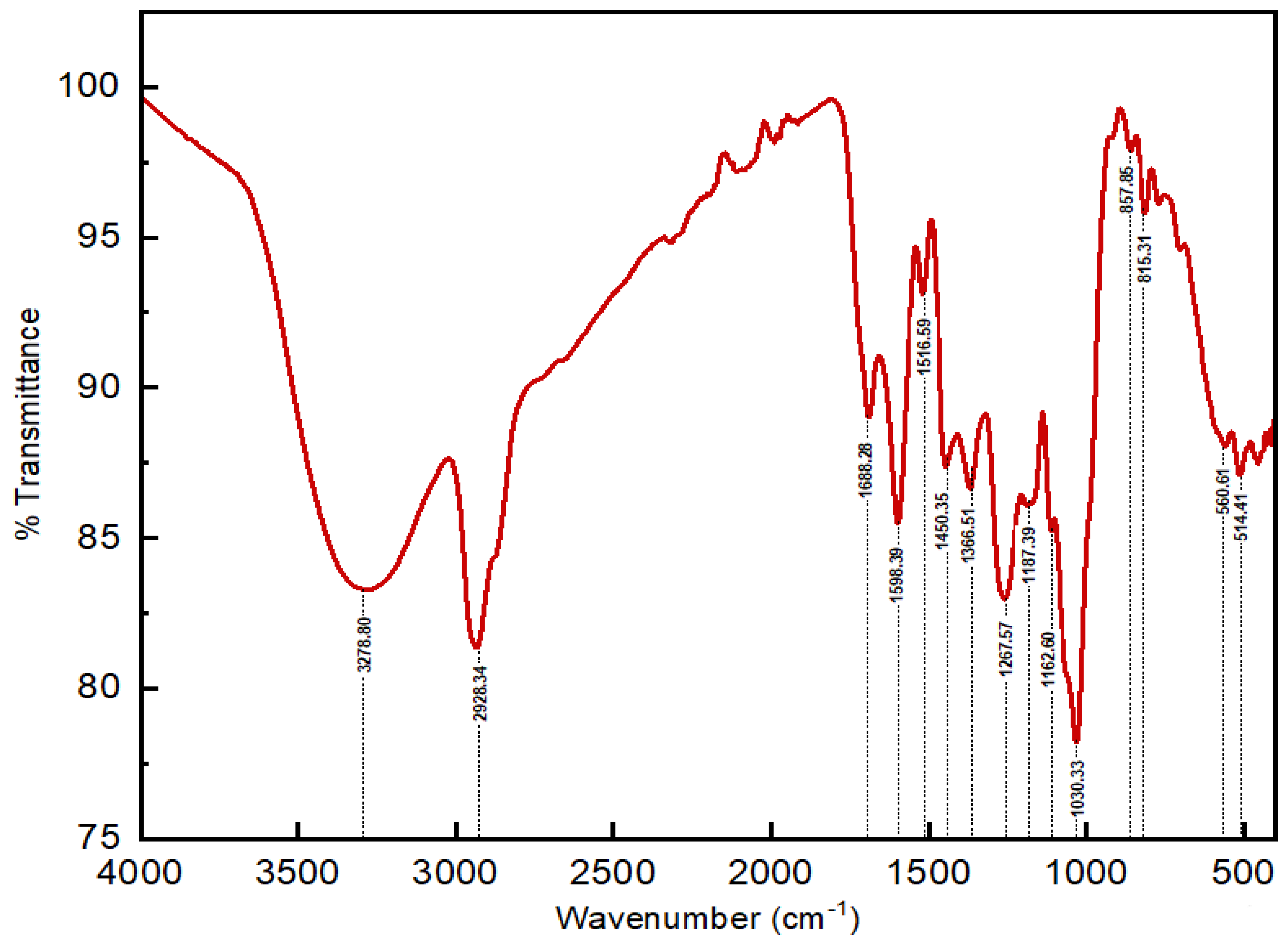
Figure 1. IR spectra of Laurus nobilis methanolic extract.
Biophysica 04 00017 g001
Figure 2. HPLC-UV-MS chromatogram profile of Laurus nobilis methanolic extract.
Biophysica 04 00017 g002
Figure 3. Chemical structures of 13 components identified from Laurus nobilis methanolic extract.
Biophysica 04 00017 g003
Table 1. Elemental composition of Laurus nobilis obtained by ICP-AES.
ElementCrZnNiMnFeMgCaCuAlK
Value (mg/kg)---72.25124.275163510,012.753.475144.454645.5
Table 2. Oxide content of Laurus nobilis obtained by WD-XRF.
ElementCaOK2OSiO2SO3P2O5Al2O3MgO
Concentration %1.3620.7630.2550.2550.1620.0840.074
ElementFe2O3MnONa2OZnOSrOCuONiO
Concentration %0.0610.0220.0190.0070.0060.0030.003
Table 3. FT-IR spectrum analysis of Laurus nobilis methanolic extract.
Peak No.Wavenumber (cm−1)Vibration TypeFunctional Group AssignmentIntensity
13278.80O-H stretching vibrationAlcohols or phenolsStrong sharp
22928.34C-H stretching vibrationAliphatic hydrocarbonsStrong
31688.28C=O stretching vibrationKetones or aldehydesMedium
41598.39C=C stretchingAromatic ringsStrong
51516.59C=C stretchingAromatic compoundsWeak
61450.35C-H (CH2) bendingAlkanesMedium
71366.51C-H bendingMethyl groupsMedium
81267.57C-N stretchingAminesMedium
91187.39C-N stretchingNitro compounds or aminesWeak
101162.60C-N stretchingAminesWeak
111116.62C-O stretching vibrationEthersWeak
121030.33C-H bendingAlkanesStrong
13857.85C-H bendingAromatic compoundsWeak
14815.31C-H bendingSubstituted aromatic compoundsWeak
15560.61C-Cl stretchingAlkyl chloridesMedium
16514.41C-Cl stretchingAlkyl chloridesMedium
17458.82C-Br stretchingAlkyl bromidesMedium
18446.30C-Cl stretchingAlkyl chloridesMedium
19429.91C-Cl stretchingAlkyl chloridesMedium
20417.58C-Br stretchingAlkyl bromidesMedium
Table 4. Phenolic compounds identified in laurel methanol extract by HPLC-MS-UV data.
Peak NumberRt (min)[M-H]/[M+H]+ (m/z)MS2 Ions (m/z)Molecular FormulaProposed CompoundMolecular Weight (g/mol)
116.64865.2577, 289C45H38O18Procyanidin trimer866.772
217.93169.015125C7H6O5Gallic acid170.120
319.82179.039135C9H8O4Caffeic acid180.159
420.34290.07152, 139, 123C15H14O6Epicatechin290.27
521.77289.072272, 152, 139, 123C15H14O6Catechin290.271
624.71443.09289, 139C22H18O10Epicatechin gallate442.4
727.79609.145301C27H30O16Rutin610.521
833.40447.09301, 271, 255, 151C21H20O11Quercetin-3-rhamnoside448.38
933.57447.093284C21H20O11Kaempferol-3-O-hexoside448.4
1036.33593.15285, 145C27H30O15Kaempferol-3-O-rutinoside594.526
1137.23463268, 179, 151C21H19O12Quercetin-3-glucoside463.4
1237.55434.085301, 151C20H18O11Quercetin-3-O-pentoside434.30
1338.92317.03179, 151C15H10O8Myricetin318.24
Table 5. Phytochemical constituents of Laurus nobilis extracts.
FlavonoidsTanninsAlkaloidsAnthocyaninsSaponinsCoumarinsTerpenoids/Steroids
Aqueous Extract+++++
Methanolic Extract+++++
+ = Present; − = absent.
Table 6. Quantitative analysis of total phenolic, flavonoid, and tannin content of methanolic and aqueous extracts of Laurus nobilis.
Extraction SolventTotal Phenolic Content
(mg GAE/g dw)
Flavonoid Content
(mg QE/g dw)
Tannin Content
(mg TAE/g dw)
Aqueous Extract33.766 ± 1.701 a37.059 ± 1.905 a98.439 ± 2.581 a
Methanolic Extract46.223 ± 0.637 b42.386 ± 0.514 b300.506 ± 7.747 b
GAE: gallic acid equivalents; QE: quercetin equivalents; TAE: Tannic acid equivalents; dw: dry weight; different letters in the same column indicate significant differences (p < 0.05) within conditions.
Table 7. The content of antioxidant activity in methanolic and aqueous extracts of Laurus nobilis.
Extraction SolventDPPH Scavenging IC50 (mg/mL)ABTS Scavenging IC50 (mg/mL)FRAP (mg TE/g dw)ORAC (mg TE/g dw)
Aqueous Extract0.169 ± 0.005 a0.221 ± 0.026 a46.291 ± 0.299 a40.754 ± 0.109 a
Methanolic Extract0.079 ± 0.002 b0.148 ± 0.006 b73.262 ± 0.535 b77.006 ± 2.682 b
DPPH: 2,2-diphenyl-1-picrylhydrazyl; ABTS: 2,2′-azino-bis (3ethylbenzothiazoline-6-sulfonic acid); ORAC: oxygen radical absorbance capacity; FRAP: ferric reducing antioxidant power. Different letters indicate significant differences (p < 0.05) within conditions.
Disclaimer/Publisher’s Note: The statements, opinions and data contained in all publications are solely those of the individual author(s) and contributor(s) and not of MDPI and/or the editor(s). MDPI and/or the editor(s) disclaim responsibility for any injury to people or property resulting from any ideas, methods, instructions or products referred to in the content.