Review

Optical Tweezers to Force Information out of Biological and Synthetic Systems One Molecule at a Time

by
Rebeca Bocanegra
,
María Ortiz-Rodríguez
,
Ismael Plaza Garcia-Abadillo
,
Carlos R-Pulido
and
Borja Ibarra
*
IMDEA Nanociencia, Campus Cantoblanco, Faraday 9, 28049 Madrid, Spain
*
Author to whom correspondence should be addressed.
Biophysica 2022, 2(4), 564-580; https://doi.org/10.3390/biophysica2040047
Submission received: 6 October 2022 / Revised: 15 November 2022 / Accepted: 23 November 2022 / Published: 9 December 2022
(This article belongs to the Special Issue State-of-the-Art Biophysics in Spain)

Abstract

:
Over the last few decades, in vitro single-molecule manipulation techniques have enabled the use of force and displacement as controlled variables in biochemistry. Measuring the effect of mechanical force on the real-time kinetics of a biological process gives us access to the rates, equilibrium constants and free-energy landscapes of the mechanical steps of the reaction; this information is not accessible by ensemble assays. Optical tweezers are the current method of choice in single-molecule manipulation due to their versatility, high force and spatial and temporal resolutions. The aim of this review is to describe the contributions of our lab in the single-molecule manipulation field. We present here several optical tweezers assays refined in our laboratory to probe the dynamics and mechano-chemical properties of biological molecular motors and synthetic molecular devices at the single-molecule level.

1. Introduction

Molecular processes are thermally activated and subjected to random fluctuations from their interaction with the thermal bath. As a result, inherent, intricate dynamic changes over time govern the operation of biological and synthetic systems at the molecular level. Access to these “molecular dynamics” using classical ensemble or bulk methods is virtually impossible. Even if all molecules were synchronized initially, they would lose coherence gradually, yielding an unphysical average of their behavior. Therefore, bulk methods provide valuable information about the average trajectory of the population and/or its kinetics but cannot inform on its real-time dynamics.
Two decades ago, the advent of in vitro single-molecule methods and their implementation to study the operation of biological systems allowed overcoming these limitations [1]. These methods offer the possibility to monitor the time evolution of single molecules as they undergo structural and biochemical changes and/or interact with other molecular partners, without having to synchronize a population of molecules. In this way, “molecular trajectories” become evident. These trajectories reveal real-time kinetics (pauses, backtrackings and rate fluctuations) and intermediate states, which allow drawing a much clearer picture of the dynamics of the reaction. In addition, single-molecule manipulation methods allow exerting mechanical forces on individual molecules and/or directly measuring forces generated in the course of their mechano-chemical reactions. Note that many proteins operate inside the cell as molecular motors or mechano-enzymes that convert chemical (ATP/GTP) and thermal energy into mechanical work [2]. Examples include proteins involved in nucleic acid metabolism (polymerases, helicases, ribosomes, etc.), ATP synthesis (F0F1ATP synthase), intracellular transport (kinesin and dynein), membrane remodeling (dynamins, endosomal sorting complex required for transport, etc.), cell motility and many others. The description of the operation of these biological motors must include mechanical forces, torques and displacements, which are produced by their reactions.
The possibility offered by single-molecule manipulation methods to measure the effect of mechanical force on the real-time kinetics of the operation of a biological system informs on the rates, equilibrium constants and free-energy landscapes of the mechanical steps of the reaction. In this way, these methods allow one to relate the chemical and mechanical coordinates of the reaction (mechano-chemistry).
Because of their versatility, force and spatial and temporal resolutions, optical tweezers have become the method of choice in single-molecule manipulation studies. Optical tweezers rely on forces imparted to matter by the scattering, emission and absorption of light [3,4,5]. The Nobel Prize in Physics 2018 was awarded to the inventor of optical tweezers, Arthur Ashkin, and recognized the far-reaching applications of this technique to study biological systems. In 1986, Ashkin et al. [6] showed that a tightly focused laser beam can be used to hold and maintain (or trap) microscopic plastic beads in all three dimensions. Near the focus, the optical trap behaves as a linear “Hookean” spring, generating forces on the bead proportional to its displacement from the center of the trap (Figure 1A). Ten years after Ashkin’s discoveries, biophysicists showed the possibility of using optically trapped beads as handles to manipulate biological systems and as probes to interrogate the displacement and forces resulting from their activities.
In a typical optical tweezers experiment, the system of interest is first biochemically linked to a plastic bead, which allows the handling of the system inside a sample chamber (Figure 1C). Monitoring the activity and exerting calibrated forces on the system requires attaching it at some other end. In the optical tweezers setups built in our laboratory, this second attachment point is provided by a second bead held on top of a micropipette by suction (Figure 1C and Figure 2A). The tether may be the substrate to a protein or a motor (i.e., nucleic acid molecules or membrane nanotubes, Figure 3B, Figure 4 and Figure 5) [9,10], a molecule undergoing conformational changes under mechanical tension (i.e., a structured RNA molecule or protein [11,12]), or a molecular motor (either natural or artificial) linked to the beads via double-stranded DNA (dsDNA) handles (Figure 3A and Figure 6) [13,14]. In each case, activities are monitored in real time for the motions of the bead in the optical trap due to changes in the end-to-end extension of the tether as a consequence of proteins acting on it or the (un)folding of structured or dynamic domains within the tether (Δx, Figure 3, Figure 4, Figure 5 and Figure 6). The detection of the bead position in the optical trap is carried out by back-focal plane interferometry, [15,16]. Under standard conditions, the typical spatial, force and temporal resolutions of optical tweezers are 5–10 nm, 0.1–1 picoNewton (pN) and 500–1000 Hz, respectively [5]. This resolution is ideal for studying biological systems, which operate within this range of forces and displacements.
Finally, we note that, in the last decades, the efficient operation of biological molecular motors has inspired research in the field of Supramolecular Chemistry to design and build an impressive array of synthetic molecules that work as switches, ratchets and propellers at the nanoscale [17]. These synthetically fabricated molecular devices have many potential applications in fields from biomedicine to nanorobotics [18]. Here, we review optical tweezers assays refined in our laboratory to study the activities of proteins and/or biological molecular motors that work on DNA or play a role in membrane remodeling. In addition, we describe recent optical tweezers assays developed by us to probe, for the first time, the dynamics and mechano-chemical properties of synthetic molecular devices at the single-molecule level under biocompatible aqueous conditions.

2. Optical Tweezers to Interrogate the Elastic Properties of DNA

DNA is a stiff and highly charged biological polymer. The mechanical properties of DNA determine the structure and function of proteins involved in its metabolism. Early in vitro single-molecule manipulation experiments set the basis for determining the characteristic mechanical parameters of ss- and dsDNA [19,20,21,22,23]. These pioneering works enabled the study of the activity of proteins involved in DNA metabolism with force-spectroscopy techniques (see below).
In a typical optical tweezers assay, the elastic responses of ss- and dsDNA are studied by attaching the nucleic acid between two beads and stretching its ends in opposite directions at a controlled rate (Figure 2A). The results of these experiments are force–extension curves (FEC), from which the mechanical parameters of each polymer can be extracted upon fitting the curves to polymer physics models (Figure 2B) [24]. The FEC of a dsDNA molecule can be accurately described by the extensible worm-like chain (eWLC) model (Figure 2B, red solid line) [25,26]. This model describes the elasticity of a molecule at equilibrium in a thermal bath in terms of its persistence length, P [27,28,29] and stretch modulus (S) [30,31]. The characteristic values for P and S of a regular dsDNA molecule are ~45 nm and ~1250 pN, respectively [19,32,33]. An exception to this rule is A:T-rich DNA sequences (A-tracts) [34,35,36,37,38]. A recent work led by Prof. Fernando Moreno-Herrero (CNB-CSIC), in which we participated, used optical and magnetic tweezers together with atomic force microscopy and theoretical modeling to show that A:T-rich sequences exhibit unique mechanical properties with smaller P (~20 nm) and higher S (~2400 nm) [39]. These particular mechanical properties may confer specific functions to these sequences in vivo, such as nucleosome organization and transcription regulation [40].
Figure 2. Optical tweezers to study the elastic properties of DNA. (A) A DNA molecule is stretched (Δx) between two polystyrene microspheres (beads) held in a micropipette and a force-measuring optical trap. Biotin and digoxigenin moieties at the distal ends of the DNA molecule serve to anchor the DNA to beads functionalized with streptavidin or anti-digoxigenin. (B) Force−extension curves (FECs) of dsDNA (orange) and ssDNA (blue). The eWLC model (red line) effectively describes the dsDNA elasticity (F < 50pN), whereas the FJC model (green line) fits the ssDNA curve at high forces (F > 15pN). The formation of a secondary structure in ssDNA condenses the molecule at low forces, diverting its extension from that predicted by the FJC model. Under these conditions, the helix-coil model described in [41] (blue line) effectively described the elastic properties of ssDNA. (C) Helix-coil model for secondary−structure formation. Each monomer can be either in the free state (blue) or in the compact state (orange). At low forces (<15 pN), the majority of the monomers are organized in compact domains. As force increases, compact domains unfold, gradually forming larger free domains. At higher forces (>15 pN), a fully stretched unfolded state is reached, where all the monomers are in the free domain (adapted from [41], Creative Commons license).
Biophysica 02 00047 g002
ssDNA, instead, behaves as a semiflexible polymer best described by the Freely Jointed Chain (FJC) model (Figure 2B green solid line) [19,24]. However, this description only accounts for the elasticity of the polymer at high forces (f>15pN). At lower forces, the FECs of ssDNA deviate from the ideal elastic behavior, showing consistent compaction in the form of a force shoulder, especially under moderate- to high-salt conditions [20,42]. This compaction indicates that ssDNA folds via secondary-structure formation (Figure 2B blue dots). We recently participated in a study led by Prof. Felix Ritort (Universitat de Barcelona), where they developed a mathematical model that predicts the formation of secondary structures in individual ssDNA molecules and explains the elastic properties of this polymer at all forces (Figure 2B,C, blue solid line). A major finding of this work was that the folding of ssDNA, at the most basic level of monomer interactions, is a cooperative process. In addition, the model also predicts the folding free energy and the average size of compacted and free domains in the absence of force. Importantly, these predictions are in good agreement with secondary-structure predictions by mfold [41]. Ongoing studies in our lab are testing the ability of this model to predict the folding of individual ssRNA molecules under mechanical tension.
In summary, pulling on single molecules of DNA with optical tweezers has resulted in the precise measurement of the mechanical properties of this fundamental biological polymer, provided a rigorous test of theories of polymer elasticity and, as shown in the following sections, established the conceptual and experimental basis for the design and analysis of mechanical assays of enzymes that act on DNA [9,28,43].

3. Optical Tweezers Assays to Study the Operation of Replicative DNA Polymerases
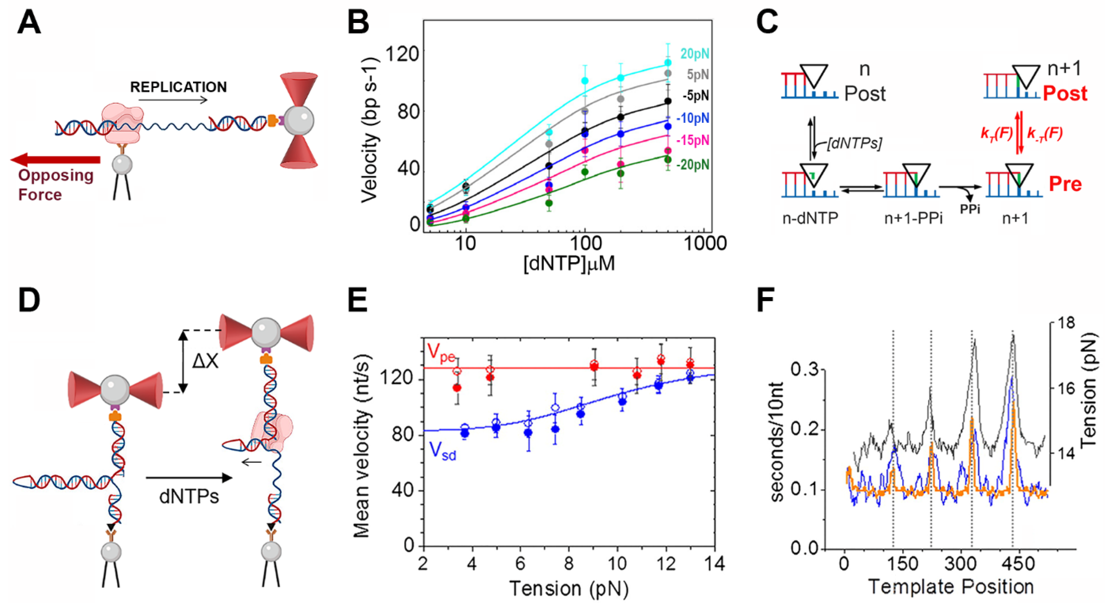
Replicative DNA polymerases (DNApol) are the molecular motors that catalyze template-directed DNA replication by the stepwise addition of the corresponding dNTPs onto the 3′ end of the nascent DNA strand (primer) [44]. Single-molecule manipulation techniques, such as optical tweezers and magnetic tweezers, have provided unique insight into the real-time replication kinetics of prokaryotic and eukaryotic replicative DNA polymerases [13,45,46,47,48,49,50,51,52].
In our lab, we used optical tweezers to study several aspects of the motor activities of the bacteriophage Phi29 DNApol. One of the first questions we aimed to address was how Phi29 DNApol couples chemical catalysis with mechanical motion. As in other replicative DNApols, the dNTP incorporation cycle of Phi29 DNApol involves large conformational changes and chemical steps that couple nucleotide binding and incorporation with the mechanical translocation of the enzyme along the DNA [53]. In order to investigate the coupling between the chemical and mechanical steps during replication, we used optical tweezers to measure the combined effects of mechanical force and dNTP/PPi concentration on the real-time kinetics of replication. A mechanical load directly applied on DNApol affects the mechanical steps, whereas dNTP/PPi concentrations modulate the chemical steps of the reaction [2]. In these experiments, we attached a Phi29 DNApol–DNA complex containing a biotin tag on the N-terminus of the enzyme to a streptavidin bead held on top of a micropipette, whereas the downstream end of the template DNA was attached to the second bead trapped in the optical trap (Figure 3A). The dependency of the real-time kinetics of a single DNA polymerase on force and nucleotide concentration (Figure 3B) suggested a loose-coupling mechanism between chemical catalysis and mechanical translocation (Figure 3C). In other words, translocation occurs by thermal diffusion of the polymerase along the DNA. The conformational changes resulting from the binding and hydrolysis of the incoming dNTP do not push or pull the enzyme to the next position (power-stroke mechanism). Instead, the binding of the next dNTP biases the diffusion of the enzyme toward the post-translocation step (Brownian ratchet model) [13].
Figure 3. Optical tweezers assays to study translocation and DNA unwinding activities of DNA polymerases. (A) Experimental configuration to apply mechanical load on a DNA polymerase. A single Phi29 DNApol (pink) labeled with biotin is attached to a streptavidin−covered bead held on a micropipette and tethered via the upstream end of the DNA template to the bead in the optical trap. At constant opposing force, the length of the DNA tether shortens as replication proceeds. (B) Average replication rates (without pauses) as a function of dNTP concentration measured at varying loads. Solid lines represent nonlinear least-square fits to the Michaelis−Menten equation. (C) The dependency of replication kinetics on force and dNTP concentration is compatible with a Brownian ratchet translocation mechanism. According to this model, translocation is driven by Brownian fluctuations, and binding of dNTP biases diffusion toward the post−translocation step. A, B and C adapted from [13] (Creative Commons license). (D) Experimental setup to study the strand displacement activity of Phi29 DNApol. A single DNA hairpin is tethered between the bead in the optical trap and the bead on top of the micropipette. At a constant tension, the strand displacement activity of the polymerase (pink) increases the distance between the beads (Δx). (E) The average strand displacement rates of Phi29 DNApol with and without pauses (full and empty blue circles, respectively) are similar to those measured during primer extension conditions (red circles) (F) However, analysis of the instantaneous velocities revealed that during strand displacement, DNApol spends longer times (blue) at positions with high GC content (dotted lines). The force (blue line in (E)) and sequence (orange in (F)) dependencies of the strand displacement rate were well explained by a model that considers that Phi29 DNA polymerase destabilizes the two nearest base pairs of the fork with ~2 kBT/bp.
Biophysica 02 00047 g003
We also used Phi29 DNApol as a model system to study how replicative DNA polymerases couple DNA replication and unwinding activities during strand-displacement DNA synthesis. Phi29 DNApol is a good model system to study this reaction because it presents processive DNA replication and efficient DNA unwinding activities [54,55]. To study the coordination between these two activities, we studied the real-time kinetics of the polymerase on a single DNA hairpin held at a constant force between a bead on the optical trap and a bead on the micropipette (Figure 3D). In these experiments, the activity of DNApol increased the distance between the two beads as the protein replicated and opened the hairpin (Figure 3D). We measured the dependence of the real-time DNApol kinetics on the applied force (Figure 3E) and DNA sequence (Figure 3F) to conclude that during each nucleotide incorporation step, Phi29 DNApol destabilizes the two nearest base pairs of the fork with an interaction energy of ~2 kBT per base pair [47]. The value of the interaction energy is similar to the average height of the energy barrier for DNA melting, providing a quantitative explanation of the strong strand displacement activity of this polymerase. This unique ability is conferred by an amino acid insertion named TPR2, which, together with other structural domains, forms a narrow tunnel around the template strand. The tunnel is required to tightly hold the template strand and generate mechanical stress at the fork junction, which, in turn, forces the separation of the two dsDNA strands of the fork [47,56].

4. Optical Tweezers Assays to Study the Activity of the Human Mitochondrial Replisome
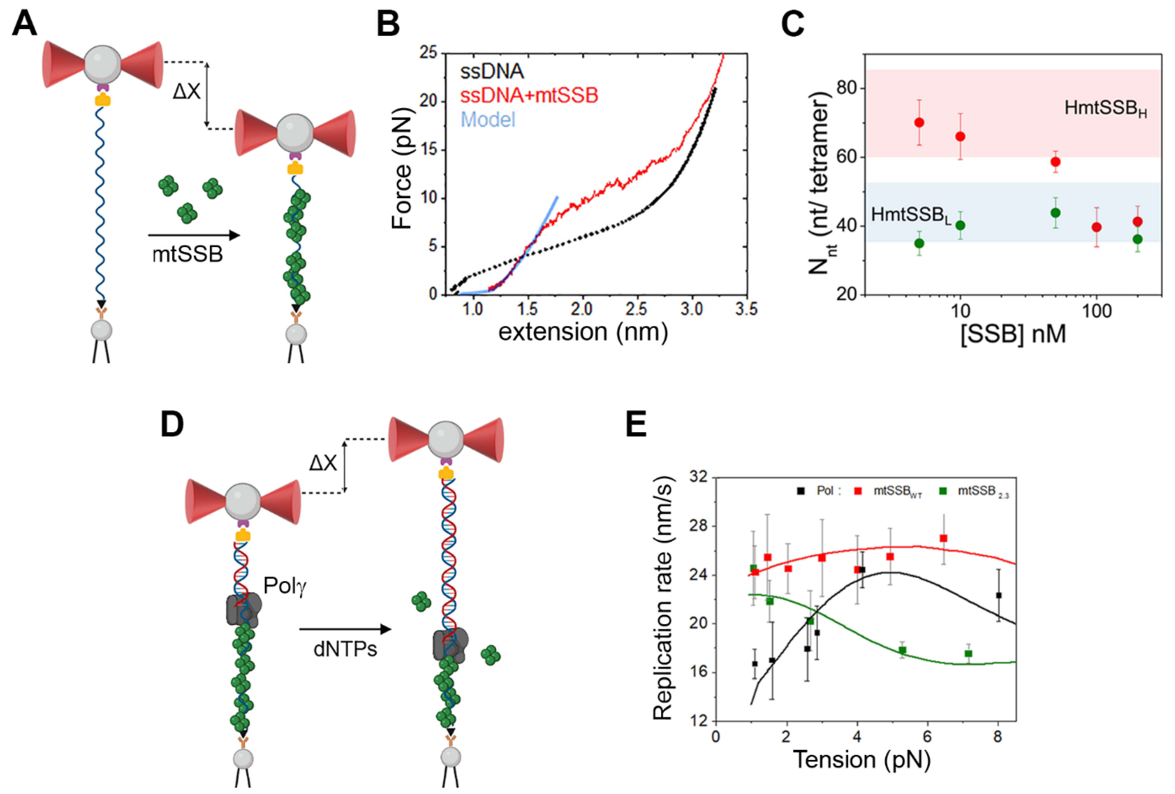
The previous section illustrates how single-molecule manipulation studies with optical tweezers can reveal insights about the operation of DNApols that are inaccessible to other techniques. However, DNApols work in coordination with a plethora of other molecular motors and specialized proteins to unravel, synthesize, edit and move unidirectionally along mega-base-pair-long genomes. Over the last 60 years, biochemical, structural and genetic studies have identified the components of these replication machineries (or replisomes) in different organisms and have defined their functions and structures. In the last few years, we have developed several optical tweezers assays to study the coordinated activity of the proteins that form the human mitochondrial DNA replisome. Mitochondria present their own DNA (mtDNA) [57], and failures in mitochondrial DNA replication (and/or maintenance) are directly linked to mitochondrial diseases [58,59,60]. The replication of mtDNA is carried out by a set of three proteins that form the mitochondrial replisome: DNA polymerase γ (Polγ), Twinkle helicase and mitochondrial single-stranded binding protein (mtSSB). We use optical tweezers to study the activity of each component in isolation and in combination with the other two factors to gain a general understanding of the function of every component on its own and the relationships between them [52,61].
The replication of circular mitochondrial DNA (~16 kb) occurs via a strand displacement mechanism with alternative light-strand origins, in contrast to the strand-coupled mechanism of nuclear DNA. During replication, the synthesis of the leading and lagging strands is not synchronized, and long lagging strands of ssDNA intermediates accumulate [62]. These intermediates are rapidly covered and organized by mtSSB to ensure the correct synthesis of the lagging strand by Polγ [63]. Initially, we used our optical tweezers to study the modes and kinetics of mtSSB binding to individual ssDNA molecules (Figure 4A). Fits of the mtSSB-ssDNA FECs (Figure 4B) to a theoretical model that explains the mechanics of ligand binding to biopolymers [64] showed that mtSSB binds ssDNA in two major binding modes: a low site-size binding mode (mtSSBL) that wraps ~35 nt of ssDNA and is dominant at low protein concentrations, and a high site-size binding mode (mtSSBH) that binds ~65 nt and predominates at high protein concentrations (Figure 4C, red dots). Similar footprints as a function of salt concentration have been described for the related E. coli SSB [65]. Interestingly, when mtSSB binding was coupled to strand-displacement DNA synthesis by a DNApol, only one of the two binding modes was observed under all experimental conditions (Figure 4C, green dots). Our results revealed a key role for the gradual generation of ssDNA during DNA replication in modulating the binding mode of a multimeric SSB protein and, consequently, in generating the appropriate nucleoprotein structure for the DNA synthesis reactions required for genome maintenance [61].
Figure 4. Optical tweezers assays to study the operation of proteins involved in human mtDNA replication. (A) Experimental setup to measure mtSSB binding to ssDNA. ssDNA is tethered between the bead in the optical trap and the bead on top of the micropipette. At constant tension, wrapping of ssDNA by mtSSB decreases the distance between the beads (Δx). (B) Force–extension curves of ssDNA (black) and ssDNA-mtSSB (red) polymers. Blue line shows the fit of the ssDNA-mtSSB curve (F < 8 pN) to a theoretical model described in [64]. The model predicts the number of nucleotides bound per mtSSB tetramer. Tension above 8 pN promotes the detachment of mtSSB from ssDNA, diverting the experimental curve from the model. (C) On long ssDNA molecules (such as in (A), the number of nucleotides bound per mtSSB depends on mtSSB concentration (red dots). However, this is not the case when mtSSB binds to ssDNA that is gradually generated during DNA replication (as in Figure 3D). Under these conditions, a low binding mode (mtSSBL) is favored at all mtSSB concentrations (green dots). Colored areas represent the approximate average site sizes in the two possible binding modes of the mtSSB. Reproduced with permission from [61]. (D) Experimental setup to measure the primer extension activity of Polγ DNApol in the presence of mtSSB. A single DNA molecule containing an ssDNA gap was tethered between the two beads. mtSSB (green tetramers) binds to the template, and the DNA polymerase (gray) loads at the 3′ end of the primer–template. In the presence of dNTPs, the polymerase starts DNA synthesis, releasing SSBs as it advances along the template. This activity is followed by the gradual increase in the distance between the beads (Δx). (E) Average primer extension rate of Polγ DNApol as a function of template tension in the absence (black) or presence of mtSSBwt (red) or mtSSB2,3 mutant (green). The presence of mtSSBwt promotes the maximum replication rate of Polγ DNApol at all tensions by elimination of the secondary structure of the template. Elimination of specific residues of mtSSB (loop2,3, mtSSB2,3 mutant) disrupted the functional interactions between DNApol and mtSSB. Under these conditions, the tight binding of mtSSB to ssDNA template hindered the activity of DNApol. Adapted with permission [52].
Biophysica 02 00047 g004
As mentioned above, the replication of mtDNA generates large stretches of ssDNA-mtSSB intermediates. The replication of these intermediates requires that Polγ removes the tightly bound SSBs from the template ssDNA [66,67]. To address the mechanism of mtSBB release from the template by Polγ, we compared the real-time replication kinetics of Polγ on free and mtSSB-covered ssDNA using optical tweezers (Figure 4D). The particular force dependencies of the instantaneous replication rate of Polγ on the different SSB-ssDNA substrates indicated that: (i) mtSSB promotes the activity of Polγ by disrupting secondary structures of the template, and (ii) strong specific polymerase–SSB repulsive interactions (up to ~12 kBT) are required for the polymerase to dislodge SSB from the template without compromising its instantaneous replication rate. We showed that these interactions may spring from the electrostatic repulsion between the negatively charged residues of loop 2.3 of mtSSB and the negative residue clusters on the surface of Polγ [52,68].
In our attempt to reconstitute the mitochondrial replisome in singulo and determine the coordinated activities of its constituents, we are currently studying the strand-displacement DNA synthesis of Polγ, the DNA unwinding activity of Twinkle helicase and the modulation of their activities by mtSSB. For these experiments, we are using experimental designs similar to that shown in Figure 3D.

5. Optical Tweezers Studies of Membrane Remodeling Reactions
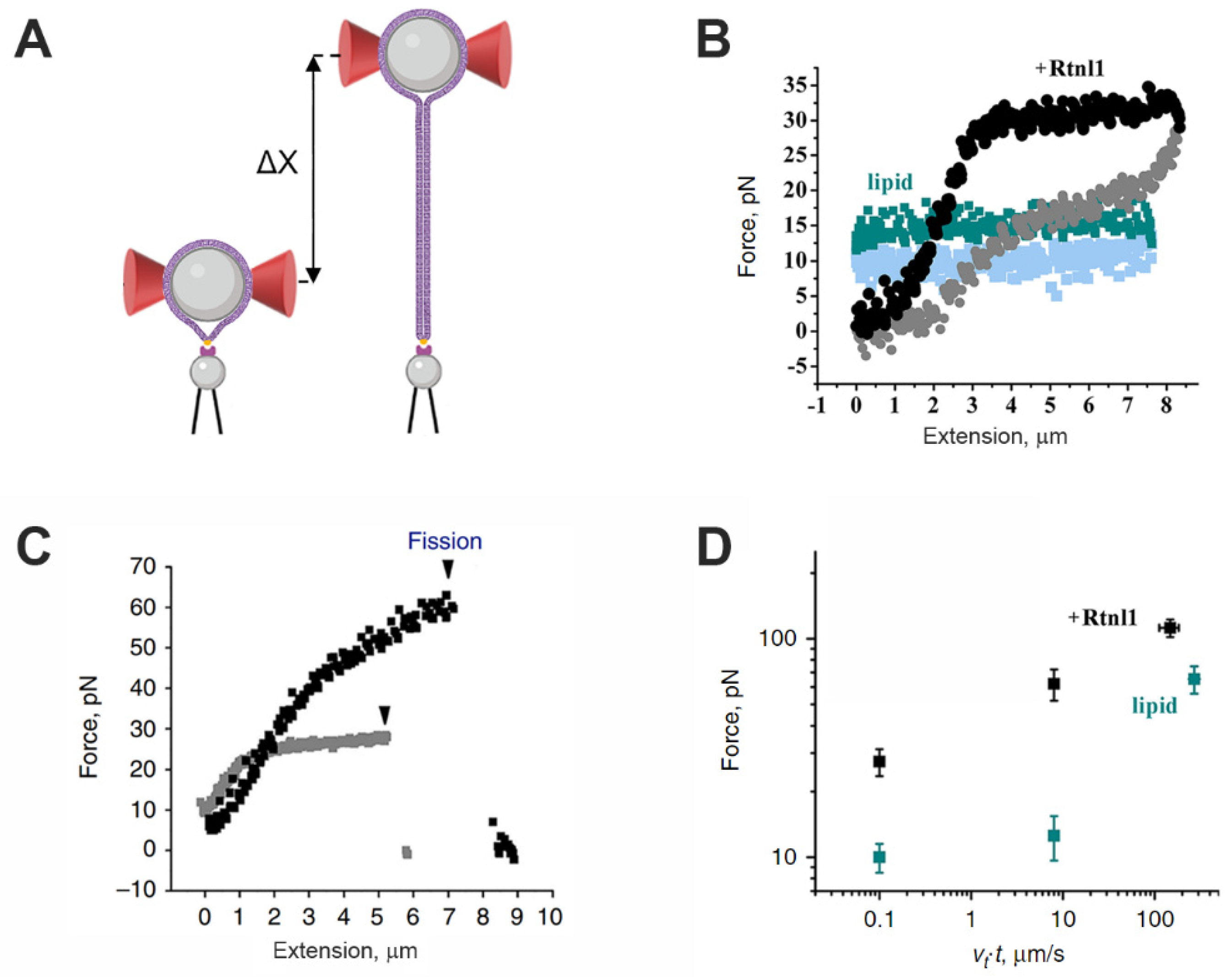
In the last few years, optical tweezers have also proven useful for studying membrane remodeling processes and membrane–protein interactions [10]. These assays typically use membrane model systems, such as free-standing membranes [69], giant plasma membrane vesicles (GPMVs) [70,71,72] and membranes supported on polystyrene or silica beads to study membrane protein and lipid dynamics in near-physiological environments [73,74,75,76,77]. In our lab, in collaboration with the lab of Prof. Vadim Frolov (Iker Basque-UPV), we have used the latter method to study the role of membrane proteins, such as reticulon 1, in membrane remodeling.
The maintenance of the peripheral endoplasmic reticulum (ER) requires active membrane fusion and fission. The ER membrane fusion is mediated by the atlastin family of proteins [78,79], while fission has been proposed to be mediated by the reticulon family of proteins. However, reticulon has also been shown to stabilize the tubular ER branches, which contradicts its apparent scission activity [80,81,82]. We have used our optical tweezers to study the effect of Drosophila reticulon 1 (Rtln1) on single membrane nanotubes (NTs). We generated single NTs in optical tweezers by pulling on a liposome doped with biotinylated lipids with a streptavidin bead (Figure 5A). A single NT presents a characteristic FEC, in which force does not change with extension and the pulling and relaxing cycles are fully reversible (Figure 5B, blue) [83]. The axial force characteristic of each NT measures the tension-driven retraction force of the NT to the lipidic reservoir. However, when the NT was pulled from liposomes containing Rtln1, the FEC showed an increase in the force while pulling and a high hysteresis that alleviated the tensile force when the tube was retracted back (Figure 5B, black). The increase in the force during pulling is related to the constriction of the NT promoted by Rtln, and eventually, this constriction-by-friction promoted by Rtln1 leads to the stochastic fission of the nanotubes, which did not occur in the absence of Rtln1 (Figure 5C). In addition, hysteresis in retraction is an indicator of the stabilization of the tube by the presence of Rtln1. Complementary fluorescence experiments showed that near the reservoir, friction-driven constriction is further enhanced by the sorting of Rtln1 toward the progressively thinning nanotube [84]. Our results demonstrated that Rtln1 combines two different modes of curvature creation: (i) static, which is associated with local membrane bending and accounts for the mechanical stabilization of membrane tubes, and (ii) dynamic, which is associated with the increased viscosity of Rtln1-containing membranes and is responsible for the friction-driven constriction of elongating NTs that leads to their fission. Coupling between these two modes via the curvature-driven sorting of Rtln1 toward the nanotube is critical for fission to occur.
Figure 5. Membrane remodeling studies using optical tweezers. (A) Experimental setup for the study of Rtln1 effect on ER membrane. Proteolipid vesicles containing Rtln1 and a small fraction of biotinylated lipids (0.2%, yellow dots) are deposited on polystyrene beads to form the supported bilayer (purple). The bead covered with lipids was held in the optical trap (red cones). The biotinylated lipids mediated the attachment with a streptavidin−coated bead on top of the micropipette. Membrane nanotubes were pulled from the reservoir−supported membrane by increasing the distance between the trap and the pipette (Δx). (B) Force signal measured during consecutive pulling/relaxing cycles of pure lipids (dark/light blue) and Rtln1 (black/gray) nanotubes. (C) The increase in the axial force during elongation of Rtnl1−nanotubes caused fission, which was detected as an abrupt decrease in the force to 0 pN (arrows). The differences between the black and gray curves are due to the pulling speeds: 0.1 μm/s (gray) and 8 μm/s (black). (D) Dependence of the fission force on the pulling rate for Rtln1−nanotubes (black). Blue squares show the maximal force measured during elongation of pure lipid nanotubes (blue). Panels (B) to (D) reproduced from [84] (Creative Commons license).
Biophysica 02 00047 g005
We are currently using an experimental setup similar to that described in Figure 5A to study the membrane remodeling activities of Dynamin 1 and 2 proteins [85,86].

6. Optical Tweezers to Explore Noncovalent Interactions in Supramolecular Chemistry
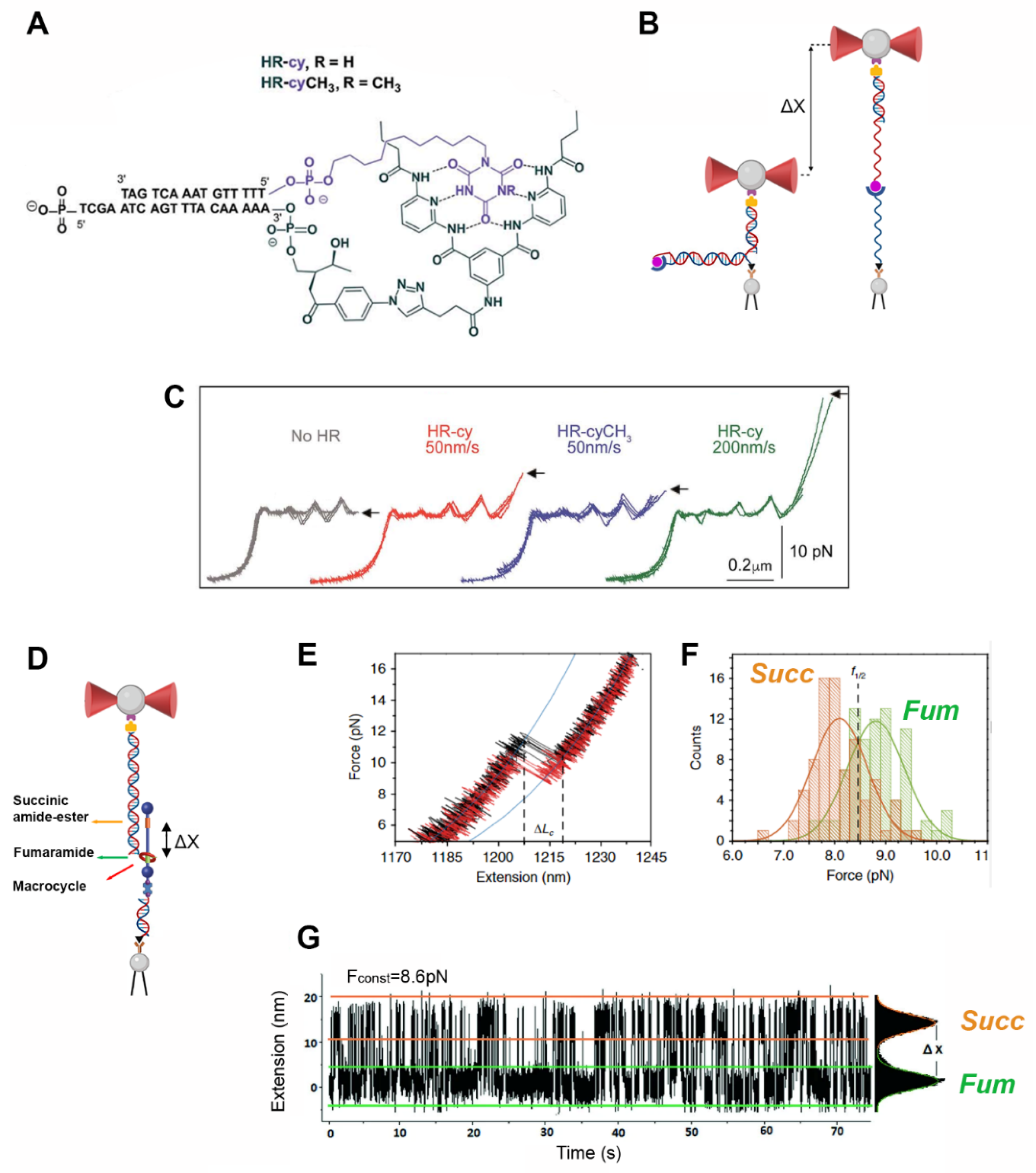
Over the last few decades, researchers working in the field of synthetic molecular machinery (supramolecular chemistry) have shown that supramolecular systems assembled in the lab have the potential to work as authentic molecular motors [18,87]. The operation of these systems, such as that of biological motors, relies on transient and often weak noncovalent interactions. The growing interest in molecular nanotechnology has increased the need for knowledge regarding the dynamics, molecular stability and mechanical strength of noncovalent interactions, especially under biologically compatible aqueous conditions. Atomic force microscopy (AFM) has been particularly successful for the observation of noncovalent interactions in organic solvents [88,89,90]. However, under aqueous conditions, H-bonds are typically weaker than in organic solvents and not amenable to AFM studies. In our lab, we have recently developed new OT-based assays to study, at the single-molecule level, the strength and dynamics of the weak noncovalent interactions that govern the stability and operation of synthetic molecular devices under near-physiological aqueous conditions.
As a starting point, we used our optical tweezers to study the mechanical strength of the H-bonds holding together an individual Hamilton receptor (host) [91] and a cyanuric acid derivative (guest); see Figure 6A [92]. In the optical tweezers, the host–guest system was covalently linked to the 3′- and 5′-termini, respectively, of one end of a DNA hairpin-like construct tethered between two beads (Figure 6B). Pulling the complementary strands of the DNA construct in opposite directions promoted its mechanical unzipping. Importantly, the characteristic force–extension curve of the molecule permitted the unequivocal identification of single attachments between the beads [93] (Figure 6C, gray). After the full unzipping of the DNA construct, the Hamilton receptor and cyanuric acid derivative couple (HR–cy) that close the hairpin is stretched in the axial direction (Figure 6B). This resulted in an increase in force before the disruption of the H-bonds that hold the host–guest system together. The increase in force depended on the nature of the H-bonded host–guest system and on the pulling rate (Figure 6C), as expected for a system under non-equilibrium conditions [94]. Blocking one of the hydrogen-bonding sites resulted in a significant decrease in the rupture force, showing the ability of our method to resolve subtle changes in the mechanical strength of the binding due to the individual H-bonding components. We believe this method can be extended to study other supramolecular assemblies.
Figure 6. Optical tweezers to study noncovalent interactions in synthetic molecular systems. (A) Structure of the Hamilton receptor (black) and cyanuric acid derivative (purple) couple (HR–cy) bound to complementary DNA oligonucleotides. (B) Diagram of the experimental setup depicting the DNA hairpin-like structure closed by the HR-cy couple (left). Pulling the complementary strands of the hairpin in opposite directions promotes the mechanical unzipping of the dsDNA and the application of axial mechanical force to the host–guest couple (right). (C) Representative experiments showing the force–extension curves of DNA constructs without HR-cy (gray) or harboring the HR–cy (red, green) or HR–cyCH3 (blue) couples at their ends. Pulling speeds are shown in nm/s. Arrows indicate the force and position of the rupture events. Panels A, B and C adapted from [92] (Creative Commons license). (D) Experimental setup to study the dynamics of a molecular shuttle. The shuttle is attached to two polystyrene beads through two dsDNA molecules. Load is applied to the system by moving the optical trap away from the micropipette. At constant load, the shuttling dynamics of the macrocycle is inferred by measuring the motions of the bead in the optical trap. (E) Representative pulling (black) and relaxing (red) curves displaying WLC behavior of dsDNA handles (blue line). (F) The intersection of the distributions of the breaking forces at Fum (green Gaussian fit) and Succ (orange Gaussian fit) stations reveals the coexistence force, F1/2. Panels (DF) adapted from [95] (Creative Commons license). (G) Representative trace showing the shuttling dynamics of the macrocycle between the Succ and Fum stations (F1/2= 8.6pN). At the coexistence force, the extension distributions (right) show that the macrocycle spends similar times at each station. Reproduced with permission from [96].
Biophysica 02 00047 g006
In a more elaborate assay, we used our optical tweezers to measure the mechanics and dynamics of individual rotaxanes (or mechanical shuttles) in aqueous conditions [95]. Rotaxane is an interlocked molecule in which a macrocycle is threaded onto a molecular axle and can shuttle reversibly between two (or more) different recognition sites on the axle as a reaction to external stimuli [97,98,99]. These devices are currently of great interest due to their potential applications, from molecular machinery to biomedicine. [100,101,102]. As a model, we used a rotaxane molecule containing an oligoethyleneglycol molecular thread with two binding stations: a fumaramide station (fum) and a succinic amide-ester station (succ) (Figure 6D). The occupancy ratio of the two stations is biased toward fum, as the tetra-amide macrocycle presents a higher affinity for it. To interface the synthetic device with the optical tweezers, a single shuttle was connected between the two functionalized beads using two dsDNA molecules (Figure 6D). The first dsDNA connects the macrocycle to the bead in the optical trap and the second dsDNA connects the end adjacent to the fum station to the other bead. Individual rotaxane–DNA hybrids were subjected to pulling–relaxing cycles. A “jump” is observed in the FEC when the force exceeds the strength of the noncovalent interactions holding the macrocycle, and it travels from the preferred fum station to the succ one (pulling curve, in black Figure 6E) and vice versa (relaxing curve in red, Figure 6E). From the pulling–relaxing cycles, we extracted detailed histograms for the rupture forces at each station (Figure 6F), and the measured forces were comparable to those required to break an equivalent number of H-bonds, according to our previous work [92,95]. From the intersection of the rupture force histograms, we deduced the coexistence force, F1/2, which is the force under which the macrocycle has an equal probability of residing over the fum or succ stations. To study the bi-stability of the system (or the shuttling rates between the two stations), we imposed a constant force on the construct around F1/2. Under these conditions, hundreds of shuttling events of the macrocycle between the two stations were monitored in real time (Figure 5G). We quantified the force-dependent real-time shuttling kinetics of the macrocycle between the two stations and calculated the energy landscape of the system under our experimental conditions [95].
These two seminal works paved the way to exploit the high temporal, spatial and force resolution of optical tweezers to characterize the mechanical properties and dynamics of synthetic molecules. It is noteworthy that in the experimental setups described here, it is possible to change the reaction conditions in situ. This possibility will allow the study of the combined effect of force and other relevant factors, such as the ionic strength, chemical reagents, light and temperature. We hope these methods can open new avenues to investigate the real-time operation of other artificial systems at the single-molecule level.

7. Conclusions and Perspectives

The unique abilities of single-molecule manipulation methods to explore the mechanistic and dynamic processes of biological and synthetic systems have rapidly placed them in an advantageous position to lead the field of in vitro single-molecule biophysics. In combination with biochemical, structural, genetic and synthetic advances, single-molecule manipulation studies will be pivotal in defining and quantifying how biological and synthetic systems work at the molecular level. As in many other research fields, bottom-up in vitro single-molecule approaches, starting from the study of individual components and moving gradually to increasingly complex systems, will set the path to addressing these challenges. However, research in the field of in vitro single-molecule manipulation faces several limitations, such as (1) low throughput: the system under study is interrogated one molecule at a time, which implies that the acquisition of statistically significant results is very time-consuming; (2) inability to detect activities if they are not coupled to conformational changes occurring along the pulling coordinate; and (3) difficulty accurately controlling the reaction temperature. Technological improvements will be pivotal in overcoming the current limitations of single-molecule manipulation techniques and addressing the many challenges ahead. These necessary improvements are currently the subject of intense research. For example, the latest developments in acoustic force spectroscopy [103], magnetic tweezers [104,105] and microfluidic systems [106] are now allowing researchers to multiplex data acquisition, in other words, to obtain increasingly large sets of high-resolution data. The latest generation of hybrid instruments that combine optical trapping (or magnetic manipulation) with fluorescence detection will allow correlating the conformational changes, exchange dynamics and/or stoichiometry of proteins tagged with fluorophores with the real-time kinetics and mechano-chemistry of their reactions [107,108,109,110]. New optical tweezers setups with multiple, freely adjustable optical traps will enable the manipulation of the system under study from several spatial coordinates [111]. Finally, the latest generation of optical tweezers with temperature control systems will help to define the crucial role of temperature in the real-time kinetics and mechano-chemistry of molecular motor operation [8,112]. Ultimately, the characterization and manipulation of biological systems within living cells will require the transfer of single-molecule position and force detection techniques to in vivo conditions. It is foreseeable that fluorescent functionalized nanoparticles could be used as probes and handles for manipulation by optical tweezers inside the cell. However, it is conceivable that non-invasive approaches would be more suitable for the detection of the mechanical motor activity of individual molecules in vivo. In this regard, force probes that change their states as a function of the force applied to them [113,114] and fluorophores that change their emission spectra as a function of mechanical force [115,116] are emerging as good candidates.
In the field of synthetic molecular machinery, taking the step from individual molecules to complex molecular systems, which incorporate several components working in concert, will require exquisite control of the mechanistic and dynamic processes governing their operation at the nanoscale [117]. Single-molecule manipulation techniques provide information about these processes; however, they have not yet had an impact on the field of synthetic molecular motors. To date, atomic force microscopy has been the technique used to interrogate the mechanical strength of synthetic devices at the single-molecule level [118]. However, the force resolution limit of this technique hinders the measurement of the properties of the “weak” noncovalent interactions that rule out the operation of synthetic devices under near-physiological conditions. In this review, we have shown that the combination of a synthetic device with dsDNA handles has allowed us to explore, for the first time, its fast dynamics at the single-molecule level. We speculate that the power of synthetic chemistry to control structure–function relationships at the atomic level and the ability of optical tweezers to manipulate individual systems and provide real-time data will be an ideal combination to address fundamental questions in the field of synthetic molecular machinery.

Author Contributions

Original draft preparation, R.B., M.O.-R., C.R.-P., I.P.G.-A. and B.I.; review and editing, R.B. and B.I.; supervision, R.B. and B.I.; funding acquisition, B.I. All authors have read and agreed to the published version of the manuscript.

Funding

This work was supported by the Spanish Ministry of Economy and Competitiveness [PGC2018-099341-B-I00 to B.I.] and Comunidad de Madrid (NanoMagCOST P2018 INMT-4321 and PEJ2020-AISAL-18484) I.P.G-A was supported by fellowship PRE2019-088885. IMDEA Nanociencia acknowledges support from the Severo Ochoa Program for Centers of Excellence in R&D (CEX2020-001039-S).

Data Availability Statement

The data that support the findings of the studies reported in this review are available from the corresponding author (BI) upon reasonable request.

Conflicts of Interest

The authors declare no conflict of interest.

References

  1. Bustamante, C. In singulo Biochemistry: When Less Is More. Annu. Rev. Biochem. 2008, 77, 45–50. [Google Scholar] [CrossRef] [PubMed]
  2. Bustamante, C.; Chemla, Y.R.; Forde, N.R.; Izhaky, D. Mechanical Processes in Biochemistry. Annu. Rev. Biochem. 2004, 73, 705–748. [Google Scholar] [CrossRef] [PubMed] [Green Version]
  3. Van Mameren, J.; Wuite, G.J.L.; Heller, I. Introduction to Optical Tweezers: Background, System Designs, and Commercial Solutions. In Methods in Molecular Biology; Humana Press: Totowa, NJ, USA, 2011; Volume 783. [Google Scholar] [CrossRef]
  4. Bustamante, C.J.; Chemla, Y.R.; Liu, S.; Wang, M.D. Optical tweezers in single-molecule biophysics. Nat. Rev. Prim. 2021, 1, 25. [Google Scholar] [CrossRef] [PubMed]
  5. Moffitt, J.R.; Chemla, Y.R.; Smith, S.B.; Bustamante, C. Recent Advances in Optical Tweezers. Annu. Rev. Biochem. 2008, 77, 205–228. [Google Scholar] [CrossRef] [PubMed] [Green Version]
  6. Ashkin, A.; Dziedzic, J.M.; Bjorkholm, J.E.; Chu, S. Observation of a single-beam gradient force optical trap for dielectric particles. Opt. Lett. 1986, 11, 288–290. [Google Scholar] [CrossRef] [Green Version]
  7. Smith, S.B.; Cui, Y.; Bustamante, C. [7] Optical-trap force transducer that operates by direct measurement of light momentum. In Methods in Enzymology; Academic Press: New York, NY, USA, 2003; Volume 361. [Google Scholar] [CrossRef]
  8. De Lorenzo, S.; Ribezzi-Crivellari, M.; Arias-Gonzalez, J.R.; Smith, S.B.; Ritort, F. A Temperature-Jump Optical Trap for Single-Molecule Manipulation. Biophys. J. 2015, 108, 2854–2864. [Google Scholar] [CrossRef] [Green Version]
  9. Bocanegra, R.; Plaza, G.I.; Pulido, C.R.; Ibarra, B. DNA replication machinery: Insights from in vitro single-molecule approaches. Comput. Struct. Biotechnol. J. 2021, 19, 2057–2069. [Google Scholar] [CrossRef]
  10. Cheppali, S.K.; Dharan, R.; Sorkin, R. Forces of Change: Optical Tweezers in Membrane Remodeling Studies. J. Membr. Biol. 2022, 255, 677–690. [Google Scholar] [CrossRef]
  11. Rissone, P.; Ritort, F. Nucleic Acid Thermodynamics Derived from Mechanical Unzipping Experiments. Life 2022, 12, 1089. [Google Scholar] [CrossRef]
  12. Bustamante, C.; Alexander, L.; Maciuba, K.; Kaiser, C.M. Single-Molecule Studies of Protein Folding with Optical Tweezers. Annu. Rev. Biochem. 2020, 89, 443–470. [Google Scholar] [CrossRef]
  13. Morin, J.A.; Cao, F.J.; Lázaro, J.M.; Arias-Gonzalez, J.R.; Valpuesta, J.M.; Carrascosa, J.L.; Salas, M.; Ibarra, B. Mechano-chemical kinetics of DNA replication: Identification of the translocation step of a replicative DNA polymerase. Nucleic Acids Res. 2015, 43, 3643–3652. [Google Scholar] [CrossRef] [PubMed] [Green Version]
  14. Castillo, J.P.; Tong, A.B.; Tafoya, S.; Jardine, P.J.; Bustamante, C. A DNA packaging motor inchworms along one strand allowing it to adapt to alternative double-helical structures. Nat. Commun. 2021, 12, 3439. [Google Scholar] [CrossRef] [PubMed]
  15. Gittes, F.; Schmidt, C. Interference model for back-focal-plane displacement detection in optical tweezers. Opt. Lett. 1998, 23, 7–9. [Google Scholar] [CrossRef]
  16. Allersma, M.W.; Gittes, F.; Decastro, M.J.; Stewart, R.J.; Schmidt, C.F. Two-Dimensional Tracking of ncd Motility by Back Focal Plane Interferometry. Biophys. J. 1998, 74, 1074–1085. [Google Scholar] [CrossRef] [PubMed] [Green Version]
  17. Kassem, S.; van Leeuwen, T.; Lubbe, A.S.; Wilson, M.R.; Feringa, B.L.; Leigh, D.A. Artificial molecular motors. Chem. Soc. Rev. 2017, 46, 2592–2621. [Google Scholar] [CrossRef] [PubMed] [Green Version]
  18. Kay, E.R.; Leigh, D.A.; Zerbetto, F. Synthetic Molecular Motors and Mechanical Machines. Angew. Chem. Int. Ed. 2006, 46, 72–191. [Google Scholar] [CrossRef]
  19. Smith, S.B.; Cui, Y.; Bustamante, C. Overstretching B-DNA: The Elastic Response of Individual Double-Stranded and Single-Stranded DNA Molecules. Science 1996, 271, 795–799. [Google Scholar] [CrossRef] [Green Version]
  20. Bosco, A.; Camunas-Soler, J.; Ritort, F. Elastic properties and secondary structure formation of single-stranded DNA at monovalent and divalent salt conditions. Nucleic Acids Res. 2014, 42, 2064–2074. [Google Scholar] [CrossRef]
  21. Wang, M.D.; Yin, H.; Landick, R.; Gelles, J.; Block, S.M. Stretching DNA with optical tweezers. Biophys. J. 1997, 72, 1335–1346. [Google Scholar] [CrossRef] [Green Version]
  22. Shon, M.J.; Rah, S.-H.; Yoon, T.-Y. Submicrometer elasticity of double-stranded DNA revealed by precision force-extension measurements with magnetic tweezers. Sci. Adv. 2019, 5, eaav1697. [Google Scholar] [CrossRef] [Green Version]
  23. Smith, S.B.; Finzi, L.; Bustamante, C. Direct Mechanical Measurements of the Elasticity of Single DNA Molecules by Using Magnetic Beads. Science 1992, 258, 1122–1126. [Google Scholar] [CrossRef]
  24. Camunas-Soler, J.; Ribezzi-Crivellari, M.; Ritort, F. Elastic Properties of Nucleic Acids by Single-Molecule Force Spectroscopy. Annu. Rev. Biophys. 2016, 45, 65–84. [Google Scholar] [CrossRef] [PubMed]
  25. Kratky, O.; Porod, G. Röntgenuntersuchung gelöster Fadenmoleküle. Recl. Trav. Chim. Pays-Bas 1949, 68, 1106–1122. [Google Scholar] [CrossRef]
  26. Landau, L.D.; Lifshitz, E.M. Statistical Physics. Pt. 1, Pt. 2. In Course of Theoretical Physics; Elsevier: Amsterdam, The Netherlands, 1980. [Google Scholar]
  27. Bustamante, C.; Marko, J.F.; Siggia, E.D.; Smith, S. Entropic Elasticity of Lambda-Phage DNA. Science 1994, 265, 1599–1600. [Google Scholar] [CrossRef] [PubMed] [Green Version]
  28. Bustamante, C.; Yan, S. The development of single molecule force spectroscopy: From polymer biophysics to molecular machines. Q. Rev. Biophys. 2022, 55, E9. [Google Scholar] [CrossRef]
  29. Marko, J.F.; Siggia, E.D. Stretching DNA. Macromolecules 1995, 28, 8759–8770. [Google Scholar] [CrossRef]
  30. Baumann, C.G.; Smith, S.B.; Bloomfield, V.A.; Bustamante, C. Ionic effects on the elasticity of single DNA molecules. Proc. Natl. Acad. Sci. USA 1997, 94, 6185–6190. [Google Scholar] [CrossRef] [Green Version]
  31. Odijk, T. Stiff Chains and Filaments under Tension. Macromolecules 1995, 28, 7016–7018. [Google Scholar] [CrossRef]
  32. Herrero-Galán, E.; Fuentes-Perez, M.E.; Carrasco, C.; Valpuesta, J.M.; Carrascosa, J.L.; Moreno-Herrero, F.; Arias-Gonzalez, J.R. Mechanical Identities of RNA and DNA Double Helices Unveiled at the Single-Molecule Level. J. Am. Chem. Soc. 2013, 135, 122–131. [Google Scholar] [CrossRef]
  33. Gross, P.; Laurens, N.; Oddershede, L.B.; Bockelmann, U.; Peterman, E.; Wuite, G.J.L. Quantifying how DNA stretches, melts and changes twist under tension. Nat. Phys. 2011, 7, 731–736. [Google Scholar] [CrossRef]
  34. Vafabakhsh, R.; Ha, T. Extreme Bendability of DNA Less than 100 Base Pairs Long Revealed by Single-Molecule Cyclization. Science 2012, 337, 1097–1101. [Google Scholar] [CrossRef] [PubMed] [Green Version]
  35. Rivetti, C.; Walker, C.; Bustamante, C. Polymer chain statistics and conformational analysis of DNA molecules with bends or sections of different flexibility. J. Mol. Biol. 1998, 280, 41–59. [Google Scholar] [CrossRef] [PubMed] [Green Version]
  36. Hogan, M.; LeGrange, J.; Austin, B. Dependence of DNA helix flexibility on base composition. Nature 1983, 304, 752–754. [Google Scholar] [CrossRef] [PubMed]
  37. Marin-Gonzalez, A.; Vilhena, J.G.; Moreno-Herrero, F.; Perez, R. DNA Crookedness Regulates DNA Mechanical Properties at Short Length Scales. Phys. Rev. Lett. 2019, 122, 048102. [Google Scholar] [CrossRef] [Green Version]
  38. Lankaš, F.; Sponer, J.; Hobza, P.; Langowski, J. Sequence-dependent elastic properties of DNA. J. Mol. Biol. 2000, 299, 695–709. [Google Scholar] [CrossRef] [Green Version]
  39. Marin-Gonzalez, A.; Pastrana, C.L.; Bocanegra, R.; Martín-González, A.; Vilhena, J.G.; Pérez, R.; Ibarra, B.; Aicart-Ramos, C.; Moreno-Herrero, F. Understanding the paradoxical mechanical response of in-phase A-tracts at different force regimes. Nucleic Acids Res. 2020, 48, 5024–5036. [Google Scholar] [CrossRef]
  40. Segal, E.; Widom, J. Poly(dA:dT) tracts: Major determinants of nucleosome organization. Curr. Opin. Struct. Biol. 2009, 19, 65–71. [Google Scholar] [CrossRef] [Green Version]
  41. Viader-Godoy, X.; Pulido, C.R.; Ibarra, B.; Manosas, M.; Ritort, F. Cooperativity-Dependent Folding of Single-Stranded DNA. Phys. Rev. X 2021, 11, 031037. [Google Scholar] [CrossRef]
  42. Montanari, A.; Mézard, M. Hairpin Formation and Elongation of Biomolecules. Phys. Rev. Lett. 2001, 86, 2178–2181. [Google Scholar] [CrossRef] [Green Version]
  43. Bocanegra, R.; Plaza, G.A.I.; Ibarra, B. In vitro single-molecule manipulation studies of viral DNA replication. In The Enzymes; Academic Press: Essen, Germany, 2021. [Google Scholar] [CrossRef]
  44. Joyce, C.M.; Steitz, T.A. Function and structure relationships in dna polymerases. Annu. Rev. Biochem. 1994, 63, 777–822. [Google Scholar] [CrossRef]
  45. Wuite, G.J.; Smith, S.B.; Young, M.; Keller, D.; Bustamante, C. Single-molecule studies of the effect of template tension on T7 DNA polymerase activity. Nature 2000, 404, 103–106. [Google Scholar] [CrossRef]
  46. Ibarra, B.; Chemla, Y.R.; Plyasunov, S.; Smith, S.B.; Lázaro, J.M.; Salas, M.; Bustamante, C. Proofreading dynamics of a processive DNA polymerase. EMBO J. 2009, 28, 2794–2802. [Google Scholar] [CrossRef] [PubMed] [Green Version]
  47. Morin, J.A.; Cao, F.J.; Lázaro, J.M.; Arias-Gonzalez, J.R.; Valpuesta, J.M.; Carrascosa, J.L.; Salas, M.; Ibarra, B. Active DNA unwinding dynamics during processive DNA replication. Proc. Natl. Acad. Sci. USA 2012, 109, 8115–8120. [Google Scholar] [CrossRef] [PubMed] [Green Version]
  48. Maier, B.; Bensimon, D.; Croquette, V. Replication by a single DNA polymerase of a stretched single-stranded DNA. Proc. Natl. Acad. Sci. USA 2000, 97, 12002–12007. [Google Scholar] [CrossRef] [Green Version]
  49. Manosas, M.; Spiering, M.M.; Ding, F.; Bensimon, D.; Allemand, J.-F.; Benkovic, S.J.; Croquette, V. Mechanism of strand displacement synthesis by DNA replicative polymerases. Nucleic Acids Res. 2012, 40, 6174–6186. [Google Scholar] [CrossRef] [PubMed]
  50. Hoekstra, T.P.; Depken, M.; Lin, S.-N.; Cabanas-Danés, J.; Gross, P.; Dame, R.T.; Peterman, E.J.; Wuite, G.J. Switching between Exonucleolysis and Replication by T7 DNA Polymerase Ensures High Fidelity. Biophys. J. 2017, 112, 575–583. [Google Scholar] [CrossRef] [Green Version]
  51. Naufer, M.N.; Murison, D.A.; Rouzina, I.; Beuning, P.J.; Williams, M.C. Single-molecule mechanochemical characterization of E. coli pol III core catalytic activity. Protein Sci. 2017, 26, 1413–1426. [Google Scholar] [CrossRef]
  52. Cerrón, F.; de Lorenzo, S.; Lemishko, K.M.; Ciesielski, G.L.; Kaguni, L.S.; Cao, F.J.; Ibarra, B. Replicative DNA polymerases promote active displacement of SSB proteins during lagging strand synthesis. Nucleic Acids Res. 2019, 47, 5723–5734. [Google Scholar] [CrossRef] [Green Version]
  53. Steitz, T.A. Visualizing polynucleotide polymerase machines at work. EMBO J. 2006, 25, 3458–3468. [Google Scholar] [CrossRef] [Green Version]
  54. Blanco, L.; Bernad, A.; Lazaro, J.M.; Martin, G.; Garmendia, C.; Salas, M. Highly efficient DNA synthesis by the phage Φ29 DNA polymerase. Symmetrical mode of DNA replication. J. Biol. Chem. 1989, 264, 8935–8940. [Google Scholar] [CrossRef]
  55. Kamtekar, S.; Richardson, C.; Salas, M.; Berman, A.; Rodríguez, I.; Steitz, T.; Blanco, L.; de Vega, M.; Lázaro, J. A specific subdomain in φ29 DNA polymerase confers both processivity and strand-displacement capacity. Proc. Natl. Acad. Sci. USA 2005, 102, 6407–6412. [Google Scholar]
  56. Morin, J.A.; Cao, F.J.; Valpuesta, J.M.; Carrascosa, J.L.; Salas, M.; Ibarra, B. Manipulation of single polymerase-DNA complexes: A mechanical view of DNA unwinding during replication. Cell Cycle 2012, 11, 2967–2968. [Google Scholar] [CrossRef] [PubMed] [Green Version]
  57. Nass, M.M.K.; Nass, S. Intramitochondrial fibers with dna characteristics. J. Cell Biol. 1963, 19, 593–611. [Google Scholar] [CrossRef] [PubMed] [Green Version]
  58. Wallace, D.C.; Zheng, X.; Lott, M.T.; Shoffner, J.M.; Hodge, J.A.; Kelley, R.I.; Epstein, C.M.; Hopkins, L.C. Familial mitochondrial encephalomyopathy (MERRF): Genetic, pathophysiological, and biochemical characterization of a mitochondrial DNA disease. Cell 1988, 55, 601–610. [Google Scholar] [CrossRef]
  59. Wallace, D.C.; Singh, G.; Lott, M.T.; Hodge, J.A.; Schurr, T.G.; Lezza, A.M.; Elsas, L.J., II; Nikoskelainen, E.K. Mitochondrial DNA mutation associated with Leber’s hereditary optic neuropathy. Science 1988, 242, 1427–1430. [Google Scholar] [CrossRef]
  60. Holt, I.; Harding, A.E.; Morgan-Hughes, J.A. Mitochondrial DNA polymorphism in mitochondrial myopathy. Qual. Life Res. 1988, 79, 53–57. [Google Scholar] [CrossRef]
  61. Morin, J.A.; Cerrón, F.; Jarillo, J.; Beltran-Heredia, E.; Ciesielski, G.L.; Arias-Gonzalez, J.R.; Kaguni, L.S.; Cao, F.J.; Ibarra, B. DNA synthesis determines the binding mode of the human mitochondrial single-stranded DNA-binding protein. Nucleic Acids Res. 2017, 45, 7237–7248. [Google Scholar] [CrossRef] [Green Version]
  62. Robberson, D.L.; Clayton, D.A. Replication of Mitochondrial DNA in Mouse L Cells and Their Thymidine Kinase-Derivatives: Displacement Replication on a Covalently-Closed Circular Template. Proc. Natl. Acad. Sci. USA 1972, 69, 3810–3814. [Google Scholar] [CrossRef]
  63. Fusté, J.M.; Shi, Y.; Wanrooij, S.; Zhu, X.; Jemt, E.; Persson, Ö.; Sabouri, N.; Gustafsson, C.M.; Falkenberg, M. In Vivo Occupancy of Mitochondrial Single-Stranded DNA Binding Protein Supports the Strand Displacement Mode of DNA Replication. PLoS Genet. 2014, 10, e1004832. [Google Scholar] [CrossRef]
  64. Jarillo, J.; Morín, J.A.; Beltrán-Heredia, E.; Villaluenga, J.P.G.; Ibarra, B.; Cao, F.J. Mechanics, thermodynamics, and kinetics of ligand binding to biopolymers. PLoS ONE 2017, 12, e0174830. [Google Scholar] [CrossRef] [Green Version]
  65. Lohman, T.M.; Ferrari, M.E. Escherichia coli single-stranded DNA-binding protein: Multiple DNA-Binding Modes and Cooperativities. Annu. Rev. Biochem. 1994, 63, 527–570. [Google Scholar] [CrossRef] [PubMed]
  66. Ciesielski, G.L.; Bermek, O.; Rosado-Ruiz, F.A.; Hovde, S.L.; Neitzke, O.J.; Griffith, J.D.; Kaguni, L.S. Mitochondrial Single-stranded DNA-binding Proteins Stimulate the Activity of DNA Polymerase γ by Organization of the Template DNA. J. Biol. Chem. 2015, 290, 28697–28707. [Google Scholar] [CrossRef] [PubMed] [Green Version]
  67. Farr, C.L.; Wang, Y.; Kaguni, L.S. Functional Interactions of Mitochondrial DNA Polymerase and Single-stranded DNA-binding Protein. J. Biol. Chem. 1999, 274, 14779–14785. [Google Scholar] [CrossRef] [PubMed] [Green Version]
  68. Cerrón, F.; Ibarra, B. Measurements of Replication Kinetics of DNA Polymerases on ssDNA Templates Coated with Single-Stranded DNA-Binding Proteins. In Methods in Molecular Biology; Humana Press: Totowa, NJ, USA, 2021. [Google Scholar] [CrossRef]
  69. Dols-Perez, A.; Marin, V.; Amador, G.J.; Kieffer, R.; Tam, D.; Aubin-Tam, M.-E. Artificial Cell Membranes Interfaced with Optical Tweezers: A Versatile Microfluidics Platform for Nanomanipulation and Mechanical Characterization. ACS Appl. Mater. Interfaces 2019, 11, 33620–33627. [Google Scholar] [CrossRef] [PubMed] [Green Version]
  70. Moreno-Pescador, G.; Florentsen, C.D.; Østbye, H.; Sønder, S.L.; Boye, T.L.; Veje, E.L.; Sonne, A.K.; Semsey, S.; Nylandsted, J.; Daniels, R.; et al. Curvature- and Phase-Induced Protein Sorting Quantified in Transfected Cell-Derived Giant Vesicles. ACS Nano 2019, 13, 6689–6701. [Google Scholar] [CrossRef]
  71. Beltrán-Heredia, E.; Tsai, F.-C.; Salinas-Almaguer, S.; Cao, F.J.; Bassereau, P.; Monroy, F. Membrane curvature induces cardiolipin sorting. Commun. Biol. 2019, 2, 225. [Google Scholar] [CrossRef] [Green Version]
  72. Dasgupta, R.; Miettinen, M.S.; Fricke, N.; Lipowsky, R.; Dimova, R. The glycolipid GM1 reshapes asymmetric biomembranes and giant vesicles by curvature generation. Proc. Natl. Acad. Sci. USA 2018, 115, 5756–5761. [Google Scholar] [CrossRef]
  73. Neumann, S.; Pucadyil, T.J.; Schmid, S.L. Analyzing membrane remodeling and fission using supported bilayers with excess membrane reservoir. Nat. Protoc. 2013, 8, 213–222. [Google Scholar] [CrossRef] [Green Version]
  74. Sorkin, R.; Marchetti, M.; Logtenberg, E.; Piontek, M.C.; Kerklingh, E.; Brand, G.; Voleti, R.; Rizo, J.; Roos, W.H.; Groffen, A.J.; et al. Synaptotagmin-1 and Doc2b Exhibit Distinct Membrane-Remodeling Mechanisms. Biophys. J. 2020, 118, 643–656. [Google Scholar] [CrossRef]
  75. Brouwer, I.; Giniatullina, A.; Laurens, N.; van Weering, J.R.T.; Bald, D.; Wuite, G.J.L.; Groffen, A.J. Direct quantitative detection of Doc2b-induced hemifusion in optically trapped membranes. Nat. Commun. 2015, 6, 8387. [Google Scholar] [CrossRef] [Green Version]
  76. Ge, J.; Bian, X.; Ma, L.; Cai, Y.; Li, Y.; Yang, J.; Karatekin, E.; De Camilli, P.; Zhang, Y. Stepwise membrane binding of extended synaptotagmins revealed by optical tweezers. Nat. Chem. Biol. 2022, 18, 313–320. [Google Scholar] [CrossRef] [PubMed]
  77. Ma, L.; Cai, Y.; Li, Y.; Jiao, J.; Wu, Z.; O’Shaughnessy, B.; De Camilli, P.; Karatekin, E.; Zhang, Y. Single-molecule force spectroscopy of protein-membrane interactions. eLife 2017, 6, e30493. [Google Scholar] [CrossRef] [PubMed]
  78. Westrate, L.M.; Lee, J.E.; Prinz, W.A.; Voeltz, G.K. Form Follows Function: The Importance of Endoplasmic Reticulum Shape. Annu. Rev. Biochem. 2015, 84, 791–811. [Google Scholar] [CrossRef] [PubMed]
  79. Orso, G.; Pendin, D.; Liu, S.; Tosetto, J.; Moss, T.J.; Faust, J.E.; Micaroni, M.; Egorova, A.; Martinuzzi, A.; McNew, J.A.; et al. Homotypic fusion of ER membranes requires the dynamin-like GTPase Atlastin. Nature 2009, 460, 978–983. [Google Scholar] [CrossRef] [PubMed]
  80. Wang, S.; Tukachinsky, H.; Romano, F.B.; Rapoport, T.A. Cooperation of the ER-shaping proteins atlastin, lunapark, and reticulons to generate a tubular membrane network. eLife 2016, 5, e18605. [Google Scholar] [CrossRef]
  81. Grumati, P.; Morozzi, G.; Hölper, S.; Mari, M.; Harwardt, M.-L.I.; Yan, R.; Müller, S.; Reggiori, F.; Heilemann, M.; Dikic, I. Full length RTN3 regulates turnover of tubular endoplasmic reticulum via selective autophagy. eLife 2017, 6, e25555. [Google Scholar] [CrossRef]
  82. Khaminets, A.; Heinrich, T.; Mari, M.; Grumati, P.; Huebner, A.K.; Akutsu, M.; Liebmann, L.; Stolz, A.; Nietzsche, S.; Koch, N.; et al. Regulation of endoplasmic reticulum turnover by selective autophagy. Nature 2015, 522, 354–358. [Google Scholar] [CrossRef]
  83. Waugh, R.; Hochmuth, R. Mechanical equilibrium of thick, hollow, liquid membrane cylinders. Biophys. J. 1987, 52, 391–400. [Google Scholar] [CrossRef] [Green Version]
  84. Espadas, J.; Pendin, D.; Bocanegra, R.; Escalada, A.; Misticoni, G.; Trevisan, T.; Del Olmo, A.V.; Montagna, A.; Bova, S.; Ibarra, B.; et al. Dynamic constriction and fission of endoplasmic reticulum membranes by reticulon. Nat. Commun. 2019, 10, 5327. [Google Scholar] [CrossRef] [Green Version]
  85. Ferguson, S.M.; De Camilli, P. Dynamin, a membrane-remodelling GTPase. Nat. Rev. Mol. Cell Biol. 2012, 13, 75–88. [Google Scholar] [CrossRef] [Green Version]
  86. Roux, A.; Koster, G.; Lenz, M.; Sorre, B.; Manneville, J.-B.; Nassoy, P.; Bassereau, P. Membrane curvature controls dynamin polymerization. Proc. Natl. Acad. Sci. USA 2010, 107, 4141–4146. [Google Scholar] [CrossRef] [PubMed] [Green Version]
  87. Vögtle, F.; Stoddart, J.F.; Shibasaki, M. Stimulating Concepts in Chemistry; John Wiley & Sons: Hoboken, NJ, USA, 2000. [Google Scholar] [CrossRef]
  88. Lussis, P.; Svaldo-Lanero, T.; Bertocco, A.; Fustin, C.-A.; Leigh, D.A.; Duwez, A.-S. A single synthetic small molecule that generates force against a load. Nat. Nanotechnol. 2011, 6, 553–557. [Google Scholar] [CrossRef] [PubMed]
  89. Van Quaethem, A.; Lussis, P.; Leigh, D.A.; Duwez, A.-S.; Fustin, C.-A. Probing the mobility of catenane rings in single molecules. Chem. Sci. 2014, 5, 1449–1452. [Google Scholar] [CrossRef]
  90. Brough, B.; Northrop, B.H.; Schmidt, J.J.; Tseng, H.-R.; Houk, K.N.; Stoddart, J.F.; Ho, C.-M. Evaluation of synthetic linear motor-molecule actuation energetics. Proc. Natl. Acad. Sci. USA 2006, 103, 8583–8588. [Google Scholar] [CrossRef] [PubMed] [Green Version]
  91. Chang, S.K.; Hamilton, A.D. Molecular recognition of biologically interesting substrates: Synthesis of an artificial receptor for barbiturates employing six hydrogen bonds. J. Am. Chem. Soc. 1988, 110, 1318–1319. [Google Scholar] [CrossRef]
  92. Naranjo, T.; Cerrón, F.; Nieto-Ortega, B.; Latorre, A.; Somoza, A.; Ibarra, B.; Pérez, E.M. Mechanical measurement of hydrogen bonded host–guest systems under non-equilibrium, near-physiological conditions. Chem. Sci. 2017, 8, 6037–6041. [Google Scholar] [CrossRef] [PubMed]
  93. Essevaz-Roulet, B.; Bockelmann, U.; Heslot, F. Mechanical separation of the complementary strands of DNA. Proc. Natl. Acad. Sci. USA 1997, 94, 11935–11940. [Google Scholar] [CrossRef] [Green Version]
  94. Wang, J.; Kouznetsova, T.B.; Craig, S.L. Single-Molecule Observation of a Mechanically Activated Cis-to-Trans Cyclopropane Isomerization. J. Am. Chem. Soc. 2016, 138, 10410–10412. [Google Scholar] [CrossRef]
  95. Naranjo, T.; Lemishko, K.M.; De Lorenzo, S.; Somoza, A.; Ritort, F.; Pérez, E.M.; Ibarra, B. Dynamics of individual molecular shuttles under mechanical force. Nat. Commun. 2018, 9, 4512. [Google Scholar] [CrossRef] [Green Version]
  96. Lemishko, K.M.; Naranjo, T.; Pérez, E.M.; Ibarra, B. Five Minutes in the Life of a Molecular Shuttle: Near-Equilibrium Measurements of Shuttling Dynamics Using Optical Tweezers. In Building and Probing Small for Mechanics. Advances in Atom and Single Molecule Machines; Springer: Cham, Switzerland, 2020. [Google Scholar] [CrossRef]
  97. Berná, J.; Bottari, G.; Leigh, D.A.; Pérez, E.M. Amide-based molecular shuttles (2001–2006). Pure Appl. Chem. 2007, 79, 39–54. [Google Scholar] [CrossRef]
  98. Yang, W.; Li, Y.; Liu, H.; Chi, L.; Li, Y. Design and Assembly of Rotaxane-Based Molecular Switches and Machines. Small 2012, 8, 504–516. [Google Scholar] [CrossRef] [PubMed]
  99. Credi, A.; Silvi, S.; Venturi, M. Light-Operated Machines Based on Threaded Molecular Structures. In Topics in Current Chemistry; Springer: Berlin, Germany, 2014; Volume 354. [Google Scholar] [CrossRef]
  100. Dvornikovs, V.; House, B.E.; Kaetzel, M.; Dedman, J.R.; Smithrud, D.B. Host−[2] Rotaxanes as Cellular Transport Agents. J. Am. Chem. Soc. 2003, 125, 8290–8301. [Google Scholar] [CrossRef] [PubMed]
  101. Nishimura, D.; Takashima, Y.; Aoki, H.; Takahashi, T.; Yamaguchi, H.; Ito, S.; Harada, A. Single-Molecule Imaging of Rotaxanes Immobilized on Glass Substrates: Observation of Rotary Movement. Angew. Chem. Int. Ed. 2008, 47, 6077–6079. [Google Scholar] [CrossRef]
  102. Van Dongen, S.F.M.; Cantekin, S.; Elemans, J.A.A.W.; Rowan, A.E.; Nolte, R.J.M. Functional interlocked systems. Chem. Soc. Rev. 2014, 43, 99–122. [Google Scholar] [CrossRef] [PubMed]
  103. Sitters, G.; Kamsma, D.; Thalhammer, G.; Ritsch-Marte, M.; Peterman, E.J.G.; Wuite, G.J.L. Acoustic force spectroscopy. Nat. Methods 2014, 12, 47–50. [Google Scholar] [CrossRef] [PubMed]
  104. Kriegel, F.; Vanderlinden, W.; Nicolaus, T.; Kardinal, A.; Lipfert, J. Measuring Single-Molecule Twist and Torque in Multiplexed Magnetic Tweezers. In Methods in Molecular Biology; Humana Press: Totowa, NJ, USA, 2018. [Google Scholar] [CrossRef]
  105. Agarwal, R.; Duderstadt, K.E. Multiplex flow magnetic tweezers reveal rare enzymatic events with single molecule precision. Nat. Commun. 2020, 11, 4714. [Google Scholar] [CrossRef]
  106. Granéli, A.; Yeykal, C.C.; Prasad, T.K.; Greene, E.C. Organized Arrays of Individual DNA Molecules Tethered to Supported Lipid Bilayers. Langmuir 2006, 22, 292–299. [Google Scholar] [CrossRef]
  107. Whitley, K.; Comstock, M.; Chemla, Y. High-Resolution Optical Tweezers Combined with Single-Molecule Confocal Microscopy. In Methods in Enzymology; Academic Press: New York, NY, USA, 2017. [Google Scholar] [CrossRef] [Green Version]
  108. Whitley, K.D.; Comstock, M.J.; Chemla, Y.R. High-Resolution “Fleezers”: Dual-Trap Optical Tweezers Combined with Single-Molecule Fluorescence Detection. In Methods in Molecular Biology; Humana Press: Totowa, NJ, USA, 2017; Volume 1486. [Google Scholar] [CrossRef] [Green Version]
  109. Long, X.; Parks, J.W.; Stone, M.D. Integrated magnetic tweezers and single-molecule FRET for investigating the mechanical properties of nucleic acid. Methods 2016, 105, 16–25. [Google Scholar] [CrossRef] [Green Version]
  110. Heller, I.; Sitters, G.; Broekmans, O.D.; Farge, G.; Menges, C.; Wende, W.; Hell, S.W.; Peterman, E.; Wuite, G.J.L. STED nanoscopy combined with optical tweezers reveals protein dynamics on densely covered DNA. Nat. Methods 2013, 10, 910–916. [Google Scholar] [CrossRef] [Green Version]
  111. Heller, I.; Laurens, N.; Vorselen, D.; Broekmans, O.D.; Biebricher, A.S.; King, G.A.; Brouwer, I.; Wuite, G.J.L.; Peterman, E.J.G. Versatile quadruple-trap optical tweezers for dual DNA experiments. In Methods in Molecular Biology; Humana Press: Totowa, NJ, USA, 2017; Volume 1486, pp. 257–272. [Google Scholar] [CrossRef]
  112. Rico-Pasto, M.; Zaltron, A.; Davis, S.J.; Frutos, S.; Ritort, F. Molten globule–like transition state of protein barnase measured with calorimetric force spectroscopy. Proc. Natl. Acad. Sci. USA 2022, 119, e2112382119. [Google Scholar] [CrossRef] [PubMed]
  113. Liu, Y.; Galior, K.; Ma, V.P.-Y.; Salaita, K. Molecular Tension Probes for Imaging Forces at the Cell Surface. Acc. Chem. Res. 2017, 50, 2915–2924. [Google Scholar] [CrossRef]
  114. Wang, X.; Ha, T. Defining Single Molecular Forces Required to Activate Integrin and Notch Signaling. Science 2013, 340, 991–994. [Google Scholar] [CrossRef] [Green Version]
  115. Dufrêne, Y.F.; Evans, E.; Engel, A.; Helenius, J.; Gaub, H.E.; Müller, D.J. Five challenges to bringing single-molecule force spectroscopy into living cells. Nat. Methods 2011, 8, 123–127. [Google Scholar] [CrossRef]
  116. Oddershede, L.B. Force probing of individual molecules inside the living cell is now a reality. Nat. Chem. Biol. 2012, 8, 879–886. [Google Scholar] [CrossRef] [PubMed]
  117. Feringa, B.L. The Art of Building Small: From Molecular Switches to Molecular Motors. J. Org. Chem. 2007, 72, 6635–6652. [Google Scholar] [CrossRef] [PubMed] [Green Version]
  118. Cheng, B.; Cui, S. Supramolecular Chemistry and Mechanochemistry of Macromolecules: Recent Advances by Single-Molecule Force Spectroscopy. In Topics in Current Chemistry; Springer: Berlin, Germany, 2015. [Google Scholar] [CrossRef]
Figure 1. Optical tweezers setup. (A) To form a stable optical trap, a near-infrared laser beam is tightly focused by a high-numerical-aperture objective (Lens) to create a large spatial gradient of light intensity. Reflected rays of light lead to a net force (FS) that pushes the bead along the direction of propagation of light. Refracted rays generate a momentum change and reactive force (FG) that pull the bead toward the focus. Adapted with permission from [4]. (B) Schematic of a dual-beam optical tweezers setup. Two high-numerical-aperture objectives (light gray) focus two counter-propagating lasers, A (green) and B (yellow), inside a flow cell to form two optical traps superimposed in the same spatial position so that they effectively function as a single trap. Piezo actuators control the position of each laser. To monitor the optical trap position, beam-splitters divert a small percentage of the incoming light of each laser to position-sensitive detectors or PSDs (dark gray). The light leaving each trap is sent to a different PSD to measure forces (Force Detection A and B) [7]. A CCD camera (purple) and a blue LED light (blue line) allow visualization of the interior of the flow cell. (C) An idealized lateral view of our flow cell, in which a DNA molecule is attached between two micron-sized beads, one in the optical trap (red cones) and the other on top of a micropipette (white). (B,C) Adapted with permission from [8].
Biophysica 02 00047 g001
Publisher’s Note: MDPI stays neutral with regard to jurisdictional claims in published maps and institutional affiliations.