Review

Supramolecular-Covalent Peptides Self-Assembly: From Design to Regenerative Medicine and Beyond

by
Raffaele Pugliese
Neuromuscular Omnicentre Lab (NeMO Lab), ASST GOM Niguarda Cà Granda Hospital, 20162 Milan, Italy
Biophysica 2022, 2(4), 324-339; https://doi.org/10.3390/biophysica2040030
Submission received: 29 August 2022 / Revised: 30 September 2022 / Accepted: 4 October 2022 / Published: 11 October 2022
(This article belongs to the Special Issue State-of-the-Art Biophysics in Italy)

Abstract

:
The field of supramolecular peptides self-assembly has undergone outstanding growth since the early 1990s after the serendipitously discovery by Shuguang Zhang of an ionic self-complementary peptide as a repeating segment in a yeast protein. From then on, the field expanded at an accelerating pace and these self-assembled materials have become an integral part of a broad plethora of designer supramolecular nanomaterials useful for different applications ranging from 3D tissue cell cultures, regenerative medicine, up to optoelectronics. However, the supramolecular peptide based-nanomaterials available thus far for regenerative medicine still lack the dynamic complexity found in the biological structures that mediate regeneration. Indeed, self-assembling peptide (SAPs) suffer from poor mechanical stability, losing mechanical properties at low strains. Just like the extracellular matrix (ECM) of living systems, the chemical structure of the SAP-biomaterials should concurrently contain non-covalent and covalent bonds, bringing, respectively, infinite and finite lifetimes of interactions to obtain a reversibly dynamic matrix. In this review, will be highlighted the major advantages and current limitations of SAP-based biomaterials, and it will be discussed the most widely used strategies for precisely tune their mechanical properties (stiffness, resilience, strain-failure, stress resistance), describing recent and promising approaches in tissue engineering, regenerative medicine, and beyond.

1. Introduction

The field of supramolecular peptides self-assembly has undergone outstanding growth since the early 1990s when a self-assembling peptide (SAP) as a repeating segment in a yeast protein was serendipitously discovered by Shuguang Zhang [1,2]. From then on, the field expanded at an accelerating pace and these self-assembled materials have become an integral part of a broad plethora of designer nanomaterials [3,4,5,6] (Figure 1). In the decades that followed, others spread this concept to a wide range of peptides (different in natural and non-natural amino acid sequence, length, hydrophilic and hydrophobic residues, secondary and tertiary structures, forming nanostructures), thereby creating functional supramolecular materials useful for different applications including three-dimensional tissue cell cultures of primary cells and stem cells [7], three-dimensional tissue printing [8], sustained releases of small molecules, growth factors, monoclonal antibodies, and siRNA [9,10,11], regenerative medicine, as well as tissue engineering [12,13,14,15,16], stabilization of membrane proteins (i.e., G-protein coupled receptors, and photosystem I) for designing light harvesting nanobiodevices [17,18,19], actuators for optics and fluidics [20], antibiotics to combat drug resistance [21,22,23,24], vaccines against viruses [25,26,27,28].
We can imagine SAP-molecules like “Lego bricks” with pegs (hydrophilic charged residues) and holes (hydrophobic residues) arranged in a precise manner that can be programmed to assemble in well-defined nanostructures at the molecular level (Figure 2A). Such nanostructures are formed mainly through non-covalent interactions (notably hydrogen bonds, electrostatic interactions, hydrophobic interactions, van der Waals interactions, π-π overlap, and water-mediated hydrogen bonds) between building blocks and involve molecular recognition to induce nucleation and growth [29]. Although these bonds are relatively insignificant in isolation, collectively they become strong and govern the structural conformation of all biological macromolecules [30]. The dynamic and reversible nature of the non-covalent interactions formed throughout the process of supramolecular peptides self-assembly allowed to spatially and temporally control their molecular architectures giving rise to a rich diversity of nano- and micro-structures, including tubes, fibers, rods, tapes, films, and vesicles (Figure 2B). This is particularly relevant for aromatic peptide sequences [31]. Further, the molecular organization can be influenced by external stimuli, such as pH, temperature, and ionic strength. These stimuli may serve to modify the physic-chemical properties and characteristics of the formed supramolecular peptides. Given this reversibility, it is not surprising that supramolecular peptides offer a number of appealing properties including the potential for self-healing, recyclability, and stimuli responsiveness. Additionally, they are able to easily incorporate bioactive cues (i.e., RGDS, IKVAV, and DGEA) or other bio-molecular structures such as nucleic acids, fatty acids, glycans, and growth factors (i.e., VEGF, TGF-β1, BDNF, IGF-1, FGF-2) to form biomimetic supramolecular scaffolds [32].
For all these reasons it has been almost physiological that the SAPs have become a leading strategy to develop regenerative therapies over the past 30 years, since they offered the opportunity to create structures similar to those found in living systems that combine order and dynamics through the reversibility of non-covalent intermolecular bonds. This is why they have been widely tested as fillers, hemostat solutions, wound healers or injectable scaffolds for the regeneration of dental pulp [33,34], cartilage [35,36], bone [37,38], spinal cord injury [39,40], traumatic brain injury [41], and infarcted heart [42,43]. SAP-scaffolds created microenvironments stimulating endogenous regeneration decreasing harmful immune response and were also capable of spatially guiding regenerating tissues. For regenerative medicine this led to a prominent step forward, as there was a strong need to develop nanomaterials that signal cells effectively, instruct cells to adhere, migrate, proliferate and even differentiate, deliver or bind bioactive agents, but at the same time, can biodegrade safely after fulfilling their function.
In the last few decades, numerous SAP based-hydrogels have been used in clinical applications. For instance, RADA16 SAP is sold with the name of PuraStat® as hemostatic agent for surgery, in particular for bleeding from small blood vessels, oozing from capillaries of the parenchyma of solid organs and for delayed bleeding following gastrointestinal endoscopic submucosal dissection in the colon. Chiral-SAPs as d-EAK16 (Sciobio®) are commercialized for use in accelerated wound healing, myocardial infarction repair, uterine repair, cartilage and bone repair. Nafujia® is another SAP used in the clinic for treating patients with chronic diabetic foot ulcer. Readers interested in a more comprehensive overview of SAP scaffolds used in the clinic should consult Zhang’s work [44]. In addition, some polyaromatic SAPs display relaxometric behavior useful for magnetic resonance imaging (MRI) technique since they can be exploiting as nanostructured contrast agents [45]. As reported by Accardo and colleagues [46,47,48,49], thanks to the biocompatible nature of SAPs, these appear to be particularly promising for overcoming the problem of contrast agents in terms of dose reduction and toxicity reduction, thus giving greater safety and improved pharmacokinetic profiles.
However, the supramolecular peptide based-nanomaterials as well as the co-aggregation and co-assembly peptides (CAPs) available thus far for regenerative medicine still lack the dynamic complexity found in the biological structures that mediate regeneration. Indeed, SAPs and CAPs suffer from poor mechanical stability, losing mechanical properties at low strains. This is mainly arising from the non-covalent interactions within supramolecular assemblies. As a matter of fact, most of the SAP-based therapies focused their efforts on the biochemical composition of the target tissue to be regenerated, while the mechanical properties are still out-of-reach. It is therefore necessary that the scaffold mechanical properties and nanoarchitecture should be finely tuned in accordance with the targeted tissue and to achieve the precise control of cellular behavior. Just like the extracellular matrix (ECM) of living systems, the chemical structure of the SAP-biomaterials should concurrently contain non-covalent and covalent bonds, bringing, respectively, infinite and finite lifetimes of interactions to obtain a reversibly dynamic matrix.
In this review, will be highlighted the major advantages and current limitations of SAP-based biomaterials, and it will be discussed the most widely used strategies for precisely tune their mechanical properties (stiffness, resilience, strain-failure, stress resistance), describing recent and promising approaches in tissue engineering, regenerative medicine, optoelectronics, and beyond.

2. Lysine Knots as a Molecular Fastener to Tune Supramolecular Peptides Stiffness

Tuning of SAP biomechanics is usually achieved by changing their concentration when in solution or altering their assembling triggering stimuli (e.g., pH, temperature, solvents, or ionic strength), thus usually altering their self-assembly kinetics, pore size, final confirmation of entangled nanostructures, and the density of bioactive cues (if any was present) tethered to the SAP backbone. A minimalistic strategy to precisely regulate the stiffness of SAPs in a range of interest while maintaining other biologically important variables such as matrix morphology, cross-linking density, and bioactive cues constant can be to use of branched peptides as “molecular connectors” among self-assembled nanostructures made of linear SAPs [50] (cross-linking strategies for designer SAPs are listed in Table 1; SAPs chemical structures are reported in Table 2). Pugliese and co-workers have developed multiple ramifications of the widely studied Ac-LDLKLDLKLDLK-CONH2 peptide (named LDLK12 for its amino acid composition and peptide length) [50], connected with one or multiple “lysine knots”, made of the symmetric double-capping of Nα,Nε-di-Fmoc-Lysine (Fmoc = 9-fluorenylmethyloxy-carbonyl). The self-assembling branches of LDLK12 and the ‘‘lysine-knots” were interposed with one glycine each to (1) avoid formation of hydrophobic patches causing solubility issues, and to (2) ensure sufficient flexibility allowing for branches integration within multiple self-assembled nanostructures of linear LDLK12. These branched peptides alone do not have an appealing self-assembling propensity but can be mixed (at different molar ratio) with linear SAPs before self-assembling in order to have them intermingled with different β-sheets of linear SAPs after gelation. This simple strategy causes a manifold increase of the stiffness of the assembled supramolecular hydrogels (proportional to the number of branches), without affecting SAP propensity to form β-sheet but instead, further stimulates their secondary structure rearrangements. Indeed, such branched peptides readily integrate into the assembled aggregates providing ‘‘molecular joints” among otherwise weakly paired β-structures, significantly tune the scaffold storage modulus (G’) from 50 to 10,000 Pa (which spans the range of G’ found in human central nervous system tissues) [51]. It should be also noted that, contrary to what may be expected, the increase in stiffness due to the LDLK12-branches does not affect the typical injectability and shear-thinning properties of SAPs [52]. This is because the tuning of mechanical properties has not been given by the presence of covalent bonds within the assembled nanostructures, but by additional transient intermolecular non-covalent interactions. Hence, the viscosity of such self-assembling branched-SAPs can decrease and get back close to original values, respectively, during and after injection: a suitable property for minimally invasive approaches in surgery.
These interesting features allowed Gelain’s group to develop a biomechanically robust 3D hydrogel construct comprising synthetic linear LDLK12-based multifunctionalized SAPs and branched peptides (dubbed HydroSAP) embedded with densely seeded human neural stem cells (hNSC) [39,53], which enables the formation of functional 3D neuronal networks in vitro, while when implanted in a sub-acute hemisection model of spinal cord injury (SCI), it reduces astrogliosis leading to significant improvements in axon regeneration and motor functional recovery [39].
Another alternative strategy for tuning SAP biomechanics has been developed by Stupp laboratory. Stupp and his team have enriched the scientific community with different classes of peptide amphiphiles (PAs) that can self-assemble into high aspect ratio nanofibers that resemble a fibrous ECM [54]. To precisely tune hydrogels stiffness they reported the effect of adding positively charged oligo-L-lysines with variable length (Kn, n = 4–120) to negatively charged PA nanofibers consisting of a hydrophobic palmitoyl tail, six β-sheet forming amino acids, and three glutamic acid residues (C16-V3A3E3) [55]. When solutions of Kn are added to a PA solution, they polymerize on the PA filaments by means of electrostatic interactions resulting in more mechanically robust gels. Indeed, the G’ of these supramolecular hydrogels increased linearly with respect to n at a rate of 10.5 Pa per lysine residue, with values ranging from 18.6 Pa (for K3) to 1276 Pa (for K120). Because the interaction between PA and Kn is non-covalent, Stupp and co-workers also reported the possibility of conjugating the fibronectin-mimetic peptide RGDS to Kn molecules in order to obtain a tunable biomimetic hydrogel with an extremely narrow range of G’ moduli found in healthy central nervous system. It is worth noting that this precise strategy to control G’ in bulk PA gels using peptide gelators of variable length can significantly affect survival, neurite growth, and tyrosine hydroxylase-positive in human induced pluripotent stem cells (iPSC)-derived dopaminergic neurons, thus offering a novel tool for the treatment of Parkinson’s disease, characterized by a progressive death of dopaminergic neurons within the brain.
Given the enormous potential of PAs as biomimetic nanostructures for regenerative medicine, Stupp laboratory also reported the use of covalent bond formation among 1,3-diene-palmitoyl-V3A3K3 monomers, as a mechanism to tune the length and cytotoxicity of supramolecular cationic assemblies [56]. The 1,3-diene-palmitic-acid is able to form covalent bonds by radical cross-linking with neighboring 1,3-dienes in the assembly. To create covalent bonds within the PA nanofibers, the system is irradiated with UV-light at 365 nm in the presence of 2,2-dimethoxy-2-phenylacetophenone as a radical initiator. During photoirradiation, the PA fibers will be partially cross-linked, and the PA solution will contain a mixture of monomers and oligomers that can co-assemble. As the photoirradiation time increases, the oligomer content as well as the repeated length of the oligomers will increase, thus leading to the formation of longer supramolecular fibers.
The importance of this system was demonstrated using in vitro experiments: the authors investigated the cytotoxicity of PA nanofibers—exposed to different photoirradiation times—against myoblast cells, demonstrating that cell viability increased with an increase in fiber length, because this latter ensures that the ends of the fibers do not damage the lipid bilayers.
All these findings introduce new opportunities to design novel structures and properties based on the ability of “lysine knots” and/or cross-linked monomers in supramolecular peptide assemblies to equilibrate into biomechanically favorable structures and at the same time biocompatible for regenerative medicine applications.
Table 1. Cross-linking strategies of various designer SAPs.
Self-Assembly PeptidesCross-LinkerAmino Acid
Residues for Cross-Linking
Storage ModulusRefs.
Ac-LDLKLDLKLDLK-CONH2 (LDLK12)
LDLK12 functionalized with KLPGWSG, FAQRVPP
Multiple ramification of LDLK12-50–10,000 Pa[50]
C16-V3A3E3Oligo-L-Lysines-18–1276 Pa[55]
1,3-diene-palmitoyl-V3A3K31,3-diene-palmitic-acid1,3-diene-[56]
Collagen Mimetic Peptides (CMPs)EDC/HOBtLysine-Aspartic acid
Lysine-Glutamic acid
-[57]
LDLK12EDC/Sulfo-NHSLysine-Aspartic acid1–2.2 kPa[58]
Lauryl-VVAGKK-AmGlutaraldehydeAmine group (Lysine, Arginine)105 Pa[59]
Ac-CGGLKLKLKLKLKLKGGC-CONH2
Ac-CGGCGGLKLKLKLKLKLKGGCGGC-CONH2
Ac-CGGCGGCGGLKLKLKLKLKLKGGCGGCGGC-CONH2
Ac-CGGCGGCGGCGGLKLKLKLKLKLKGGCGGCGGCGGC-CONH2
Sulfo-SMCCLysine-Cysteine6–840 kPa[60,61]
FYFCFYFNH4HCO3Cysteine-Cysteine3360 Pa[62]
Fmoc-FFF
LDLK12
LDLK12 functionalized with KLPGWSG, FAQRVPP, SSLVND
Branched-LDLK12
Biotin-GGGPFSSTKT
Biotin-GGGAFSSTKT
Biotin-GGGAFASTKT
Biotin-GGGPFASTKT
Biotin-GGGAFASAKA
Ac-WGGGAFASTKT
Ac-WGGGAFSSTKT
GenipinLysine-Lysine5 kPa–0.2 MPa[63,64,65]
VKVKVKVKVDPPTKVYVKVKV-NH2Frémy’s saltTyrosine-Tyrosine25,470 Pa[66]
Fmoc-FFY, Fmoc-FFGGGY
Ac-YYGGGLDLKLDLKLDLK-CONH2
Ru(bpy)3Cl2Tyrosine-Tyrosine26–106 kPa[67,68]
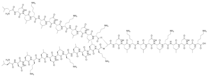
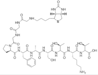
Table 2. Chemical structures of various designer SAPs.
SAP SequenceChemical Structures
Ac-LDLKLDLKLDLKBiophysica 02 00030 i001
FAQRVPPGGGLDLKLDLKLDLKBiophysica 02 00030 i002
KLPGWSGGGGLDLKLDLKLDLKBiophysica 02 00030 i003
Branched-LDLKLDLKLDLKBiophysica 02 00030 i004
SSLVNDGGGLDLKLDLKLDLKBiophysica 02 00030 i005
Biotin-GGGPFSSTKTBiophysica 02 00030 i006
Biotin-GGGAFSSTKTBiophysica 02 00030 i007
Biotin-GGGAFASTKTBiophysica 02 00030 i008
Biotin-GGGPFASTKTBiophysica 02 00030 i009
Ac-WGGGAFASTKTBiophysica 02 00030 i010
Ac-WGGGAFSSTKTBiophysica 02 00030 i011
Biotin-GGGAFASAKABiophysica 02 00030 i012
Fmoc-FFBiophysica 02 00030 i013
FYFCFYFBiophysica 02 00030 i014
C16-V3A3E3Biophysica 02 00030 i015
1,3-diene-palmitoyl-V3A3K3Biophysica 02 00030 i016

3. Covalent Capture by Using Lysine-Aspartic Acid Pairs in Collagen Mimetic Peptides and Self-Assembling Peptides
Although the self-assembly of peptides driven by non-covalent interactions is a powerful method for creating complex nanostructures with both fascinating properties and applications, sometimes these interactions are not enough to ensure greater toughness and stability of the formed supramolecular hydrogels. To overcome these issues, it is necessary to turn to the self-assembly process, followed by the covalent stabilization of the formed nanostructures; the non-covalent self-assembly first controls the nanostructures formation, while subsequent covalent bond formation stabilizes such structures. This technique is called covalent capture [69]. The success of this process requires that the reaction conditions must not disturb the self-assembly phenomenon, and the covalent capture must not negatively influence the scaffold’s nano-architecture.
Hartgerink and his team reported the use of covalent capture of collagen mimetic peptides (CMPs), through the formation of isopeptide bonds between lysine and either aspartate or glutamate, using carboxylate activating reagents 1-ethyl-3-(3-(dimethylamino)propyl) carbodiimide (EDC) and Hydroxybenzotriazole (HOBt) [57]. The choice of these amino acids is not accidental, as they serve two purposes: firstly, they direct self-assembly by allowing the composition and control of the register within the triple helix of CMPs, and subsequently they can be covalently linked (as EDC/HOBt catalyzes the formation of amide bonds between the primary carboxyl and amino groups), thus fixing the assembled structure without disturbing the triple helix conformation. Using this approach, Hartgerink et al. demonstrated that after self-assembly of the CMPs, the covalent bond formation between lysine and glutamate is faster and higher-yielding reaction than lysine with aspartate. Further, the isopeptide bond between lysine and glutamate increases the thermal stability, improves the refolding capabilities, and enhances the triple helical structure of CMP scaffolds. On the contrary, the covalent capture of triple helices with lysine-aspartate bonds occurs slower and does not enhance the helical structure. Through these covalent capture comparisons between lysine and aspartate or glutamate, the authors explored a powerful mechanism for stabilizing the three-dimensional structure of supramolecular collagen-based biomaterials useful for biomedical applications.
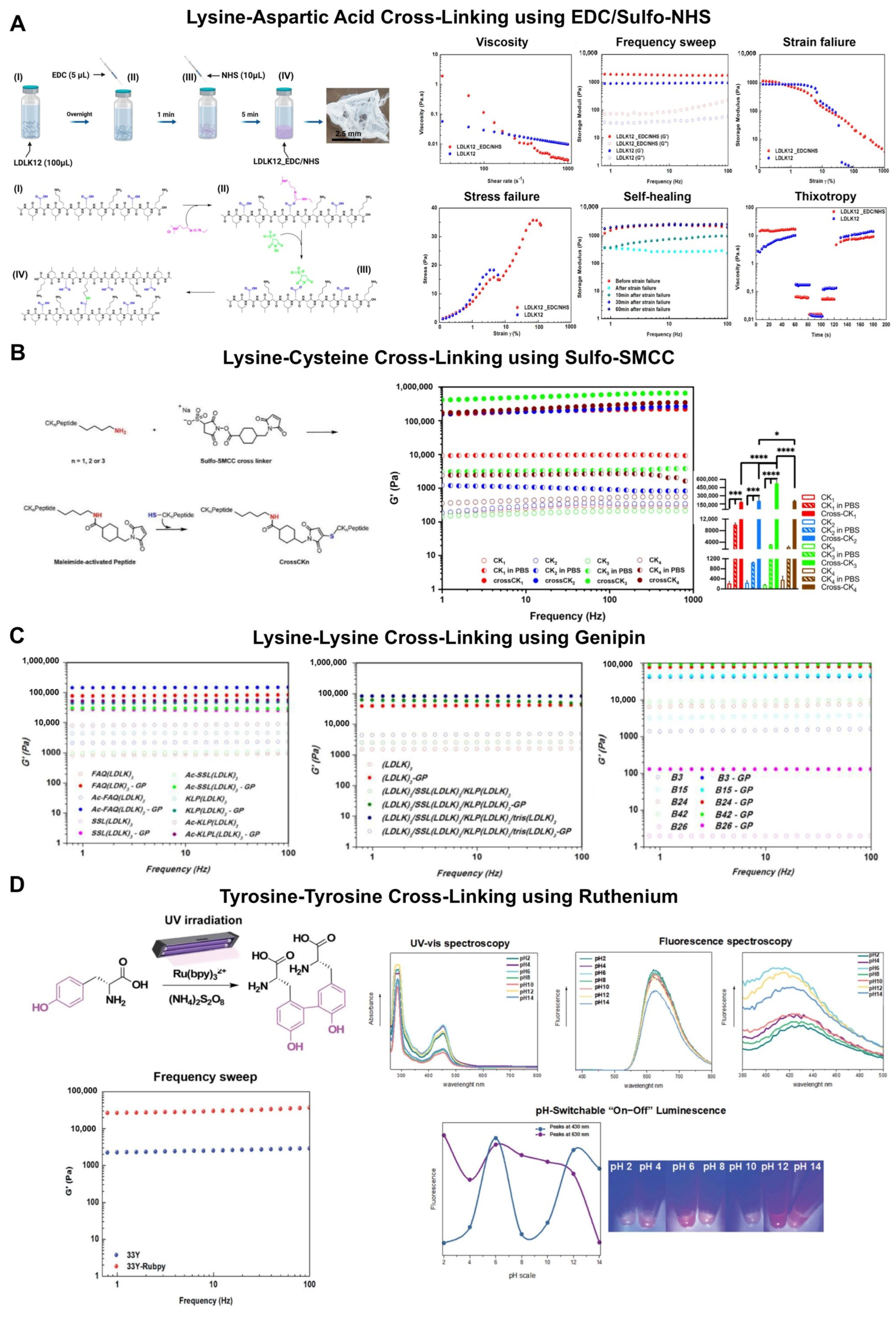
By using a similar approach, Pugliese and Gelain, reported the design of covalent capture of SAP-hydrogel using a one-pot in situ gelation system, based on EDC/N-hydroxysulfosuccinimide (sulfo-NHS) coupling, to readily cross-link LDLK12 molecules, in order to tune its biomechanics without affecting the spontaneous formation of β-sheet-containing nanofilaments [58].
The EDC/sulfo-NHS coupling does not drastically increase the elastic shear modulus compared to the LDLK12 standard. However, the strain–sweep experiments demonstrated a wide linear viscoelastic regime (LVR) of the cross-linked LDLK12 with an unusual bi-modal failure at 5% and 60% of strain, respectively, that had never been observed before in SAP-based biomaterials, which usually show failure at 4–8% of strain. Additionally, this covalent capture gives spontaneous self-healing propensity of the LDLK12 scaffold. After a large strain failure of 1000%, the mechanical properties of the cross-linked LDLK12 return to its original values (Figure 3A). A possible explanation for this feature may be the simultaneous presence of non-covalent and covalent bonds, which led to the formation of a dynamic supramolecular system capable of self-healing without the need of external stimuli. This strategy can be also useful as an alternative approach to incorporate bioactive cues or fluorescent peptides after self-assembly on pre-formed SAP-nanostructures, in order to add at the hydrogel surfaces multiple functions (e.g., for sensing, adhesion, or cell signaling).
Overall, the covalent capture methodology is an effective approach to create highly stable supramolecular peptide-hydrogels, while maintaining their desirable structural, biomechanical, and biocompatibility properties.
Figure 3. (A) Schematic illustration and chemical reaction of the one-pot and in situ LDLK12 cross-linking based on EDC/sulfo-NHS coupling, and their rheological characterization to evaluate the effect of cross-linking on mechanical features. Reproduced with permission from Ref. [52]; (B) Scheme of reaction mechanism of Sulfo-SMCC cross-linking with CKn SAPs, and their G’ values on frequency sweep experiments. * p < 0.01, *** p = 0.001 and **** p < 0.0001 indicate the significance regarding stiffness of materials analyzed. Reproduced with permission from Ref. [56]; (C) Dynamic frequency sweep of functionalized LDLK12 peptides, different branched SAPs, and BMHP1-derived SAPs, with (full dots) and without (empty dots) genipin cross-linking, recorded as a function of angular frequency (0.1–100 Hz) at a fixed strain of 1%. Reproduced with permission from Ref. [61]; (D) Schematic of ruthenium-complex-catalyzed conversion of tyrosine to dityrosine upon light irradiation; storage modulus of not cross-linked (in blue) and photo-cross-linked (in red) peptides, and “off-on-off-on” luminescence switchable from acid to basic pH of the photo-cross-linked 33Y SAP. Reproduced with permission from Ref. [70].
Biophysica 02 00030 g003

4. Chemically Cross-Linked Self-Assembling Peptide Scaffolds

For SAP hydrogels to truly be effective in stiffer tissues or in bioelectronics devices, they must somehow be able to overcome their inherent mechanical weakness. One-way to enhance the mechanical strength of an SAP hydrogel is to reinforce it with chemical cross-linkers either homobifunctional or heterobifunctional. However, although chemical cross-linking of SAP hydrogels can be a viable alternative to altering its mechanical properties versus increasing peptide concentration, it is necessary to use cross-linking agents that do not interfere with the final biocompatibility and functionality of the scaffold. For instance, Mohammad Aref Khalily et al., reported a dynamic covalent cross-linking approach based on glutaraldehyde able to tune the viscoelastic properties and enhance the mechanical stability of Lauryl-VVAGKK-Am PA gels (G’ higher than 105 Pa) [59]. The authors performed an amplitude sweep test displaying the relationship between the storage modulus and strain amplitudes of cross-linked PA-gels. They highlighted that when the limit of LVR is exceeded, the PA-gels show a transition from linear to nonlinear viscoelastic behavior, and the limiting strain amplitude of glutaraldehyde cross-linked samples is approximately six times higher than that of PA controls, thus demonstrating that chemical cross-linking can support the increased resistance to deformation and elasticity of the resulting PA networks. Despite this approach being simple and straightforward—since cross-linking takes place between amine and aldehyde groups in the aqueous medium—it is notorious that glutaraldehyde is toxic, and therefore may limit the translational potential of this strategy in humans.
To increase mechanical strength and processability of SAP hydrogels, Pugliese et al., used sulfo-SMCC, an amine-to-sulfhydryl cross-linker with an N-hydroxysuccinimide ester and maleimide reactive group to further stabilize the self-assembled hydrogels of the Ac-CGGLKLKLKLKLKLKGGC-CONH2 peptide (named CK) [60]. Applying the cross-linking reaction to same concentration solutions of CK, it was possible to increase its mechanical strength over 100-fold, thus obtaining G’ values ranging from 6 kPa (with 0.45 mM sulfo-SMCC) to 170 kPa (with 10 mM sulfo-SMCC). In addition, sulfo-SMCC cross-linking leads to an increase in stress-failure (3 kPa) with two-orders-of-magnitude difference compared to not cross-linked CK peptide (0.05 kPa) that can be attributed to the efficient formation of chemical cross-links in addition to the standard weak intermolecular interactions present in soft self-assembled hydrogels.
Finally, the authors tested the viability and differentiation of hNSCs on the cross-linked scaffolds to determine whether the good standard biocompatibility of SAPs (and thus the potential for regenerative medicine applications) was affected by the sulfo-SMCC cross-linking reaction. The viability and differentiation tests provided encouraging data similar to those obtained with standard non-functionalized SAPs seeded in 2D and 3D conditions [71], with an important difference: increments in storage modulus seems to be less effective in influencing NSC differentiation. This is likely because the extensive cross-linking in CK peptide altered the total net positive charge of the hydrogel, thereby partially counterbalancing the potential increase of glial cells usually found in stiff substrates.
Unfortunately, the beneficial effects of the increase in rigidity and stress-failure of such cross-linking strategy are somewhat negated by the exhaustion of free amino and sulfhydryl groups, as well as by their correct exposure in the aqueous environment, which makes them accessible to the cross-linking reaction. However, Gelain’s group moved significant steps forward in the field of chemical cross-linked SAPs towards the goal of stiff peptidic materials suited for the regeneration of several tissues [61]. Novel CKn peptides (where n = 1–4 is the number of cysteine groups) were designed and characterized to boost the Sulfo-SMCC mediated cross-linking reaction, where they reached G’ values of ~500 kPa (Figure 3B). Furthermore, they reported an additional orthogonal cross-linking by using genipin is also effective for allowing top remarkable G’ values of 840 kPa, thus bringing SAP constructs closer to stiffness’s of cardiac tissue, osteoid and skin.
The boost effect of cysteine residue mediated SAP cross-linking was reported by Accardo et.al, who inserted a cysteine residue in the middle of the primary sequence of an aromatic peptide hydrogelator (FY)3 [62]. The authors reported that after cross-linking of cysteine residues using ammonium bicarbonate (NH4HCO3) for achieving the air oxidation of cysteine residues with consequential formation of disulphide bonds, the FYFCFYF hydrogel underwent to an improvement of the G’ modulus (3360 Pa) compared to the parent (FY)3 (970 Pa). This strategy demonstrates that the introduction of a cross-linkable cysteine residue in the middle of the aromatic peptide hydrogelator does not hamper the gelification process. On the contrary, the presence of cysteine residues generates a stiffer hydrogel, potentially useful for bone tissue regeneration.
Recently Genipin, a natural compound, found in Gardenia jasminoides fruit extracts, was found to play a key role in SAPs stiffness tuning. So far, it has been used to cross-link hydrogel based on gelatin and fibrin [72,73,74], because of its low cytotoxicity (thousands of times less toxic than glutaraldehyde). Genipin is commonly used in Chinese medicine, herbal medicine, and as a food dye. Moreover, it has been reported that the presence of Genipin may favor cell adhesion to artificial matrices [70]. For the above reasons, the use of Genipin in the preparation of new materials for biomedical applications is highly attractive.
Chronopoulou et al., reported the use of Genipin to cross-link Fmoc-FFF based hydrogels with different chirality [63]. The mechanical characterization showed that the L isomer provides a firmer hydrogel than D when treated with Genipin; this may reflect a different rate of the cross-linking reaction and a distinct microscopic organization of the reaction products. In another effort, Chronopoulou et al., demonstrated how the mechanical properties of the Fmoc-tripeptide hydrogel were enhanced using a growing concentration of Genipin (0.5, 1, and 5 mM) [64]. The elastic G’ modulus of the cross-linked hydrogels obtained with Genipin 0.5, 1, and 5 mM was, respectively, of 5 kPa, 12 kPa, and 50 kPa. These results prove that the robustness of the hydrogels is enhanced proportionally to the amount of Genipin used, due to a higher cross-linking degree. Moreover, scanning electron microscopy (SEM) investigations have shown that the cross-linked Fmoc-FFF hydrogels are made of highly entangled and interconnected nanofibers that create a three-dimensional architecture suitable for their use as a drug delivery system. Indeed, the authors observed the controlled release (100 h) of naproxen, a non-steroidal anti-inflammatory drug, correlated with the amount of cross-linker.
In addition, Pugliese et al., demonstrated how the Genipin cross-linking can be adopted with a number of different lysine-containing SAPs (linear, mixtures, branched, biotinylated, functionalized) to produce nanofibrous networks with increased mechanical properties (G′ ≥ 0.2 MPa) and thermostability (≥100 °C) while maintaining their native secondary structure and nanoarchitecture, both critical for many biomedical applications (e.g., biomimetic scaffolds, drug release, etc.) [65] (Figure 3C). The authors also pointed out that Genipin cross-linking deeply changes the optical properties of SAPs, giving rise to absorption and fluorescence bands in the visible spectral range, and the pump probe experiments show changes in relaxation kinetics of cross-linked hydrogels, paving the way to novel optoelectronic and photonic applications of cross-linked SAPs.
Overall, the aforementioned chemical cross-linking approaches are potentially useful for different type of SAPs and may be a precious tool to better tailor the biomechanics of SAP-nanostructured scaffolds, providing them with novel resilience, stress-stiffening, and optoelectronic properties to suit the needs of different applications in biomedicine, tissue engineering, and beyond.

5. Tyrosine Cross-Linking Boost the Mechanical Rigidity of Self-Assembling Peptide Scaffolds

The tyrosine cross-linking draws considerable interest because of its distinctive properties such as outstanding elasticity and intrinsic fluorescence [75]. Furthermore, dityrosine bonds are a key component of many natural materials in their native tissues: indeed tyrosine cross-links stabilize proteins in numerous structural tissues [76]. Different research groups explored the cross-linking of tyrosine to further expand the arsenal of chemical cross-linking strategies in tyrosine-containing self-assembly peptides.
For instance, Fichman and Schneider reported that potassium nitrosodisulfonate (named Frémy’s salt) can be used to dramatically increase the mechanical rigidity of hydrogels formed by tyrosine-containing self-assembling β-hairpin peptides (VKVKVKVKVDPPTKVYVKVKV-NH2) [66]. Frémy’s salt is an inorganic salt and long-lived free radical that is known to oxidize phenols. When Frémy’s salt is added to pre-formed peptide gels, it converts tyrosine residues to o-quinones that can subsequently react with amines present within the lysine side chains of the assembled peptide bundles. The rheological studies showed that G’ values of cross-linked gels were about eight times higher than G’ values of not cross-linked gels (25,470 vs. 2932 Pa, respectively). Lastly, the authors evaluated the cytocombatibility of Frémy’s salt-treated hydrogels toward 2D cell growth of human dermal fibroblasts, demonstrating that Frémy’s salt can be added to the gel to increase its rigidity and simply removed before plating cells on the top surface of peptide material, thus avoiding cytotoxic effects on cell growth.
Wang and co-workers, reported the use of a photo-cross-linking approach to improve the mechanical stability of short SAPs, namely Fmoc-FFY and Fmoc-FFGGGY, based on the ruthenium complex (Ru(bpy)3Cl2)-catalyzed conversion of tyrosine to di-tyrosine that occurs upon light irradiation [67]. Such photo-cross-linking strategy enhanced the mechanical stability of the tyrosine-containing hydrogel by 104-fold with a G’ modulus of 100 kPa. Further, the authors reported that the reinforcement of the hydrogel through photo-cross-linking can be achieved within 2 min thanks to the fast reaction kinetics.
Drawing inspiration from natural living systems, Pugliese et al., developed and validated a photo-cross-linking approach based on ruthenium for the in situ cross-linking of a tyrosine-containing LDLK12 peptide (Ac-YYGGGLDLKLDLKLDLK, dubbed 33Y) [68]. By positioning the tyrosine reactive sites outside the self-assembling backbone of the 33Y peptide sequence, it was possible to prevent interference between the self-assembling and photo-cross- linking processes. As a result, it was demonstrated that ruthenium specifically reacts with 33Y pre-assembled nanostructures enhancing the mechanical stability of the hydrogel with a storage modulus of 26 kPa (Figure 3D). Another intriguing observation ascribable to the direct electron transfer between the ruthenium complex and SAP molecules was that the fluorescence of the photo-cross-linked SAP “turns off” (in acid solution) and “turns on” (in basic solution), generating a pH-switchable on–off system, which could be contribute to new applications of SAP-based biomaterials in biomedical imaging, pH sensing, photonics, soft electronics, and 3D bioprinting.
Furthermore, Lee and co-workers systematically incorporated multiple tyrosine units into peptides of various lengths (2–7 amino acids) as a design element for the construction of a short-peptide library to investigate the impact of tyrosine residues on self-assembly [77]. Among these, the YYACAYY it turned out to be an interesting sequence since can assemble into nano-sheets in solution. The researchers calculated the elastic modulus of the peptide sheet using finite element analysis and nano-indentation, showing that the measured elastic modulus was 8.4 Gpa, which indicates that the peptide sheet is stiffer than cancellous bones.

6. Conclusions

Since the discovery of the first SAP, which later led to the development of hydrogel-based materials, this field has grown dramatically over the past 30 years, and some of these designer supramolecular nanomaterials have become commercial products, have entered the clinical practice or clinical trials, thus bringing benefit to the society. As a matter of fact, an increasingly considerable number of researchers are dedicating their careers to the field of SAPs, trying to push this nanotechnology further and further.
However, as reported in this review, the assembly into well-defined nanostructures at the molecular level of such SAPs occurs mainly through non-covalent interactions. This represented both a blessing and a curse: on the one hand, the dynamic and reversible nature of the non-covalent interactions formed during the self-assembly process made it possible to spatially and temporally control their molecular architectures giving rise to a rich diversity of nano and microstructures, on the other hand, given the weak non-covalent bonds involved in the self-assembly phenomenon, most SAPs exhibited modest mechanical properties, making them fragile and unsuitable for the regeneration of elastic and stiff tissues. That is way, SAP-based hydrogels have been mainly used as fillers, hemostat solutions, wound healers or injectable scaffolds for the regeneration of soft tissues like dental pulp, brain, spinal cord, and cartilage, to name a few. As discussed, different strategies of supramolecular-covalent peptides self-assembly could open new paths to fill this gap and to precisely regulate their stiffness in a range of interest while maintaining other biologically important variables such as matrix morphology, cross-linking density, and bioactive cues constant useful for tissue regeneration. Indeed, different research groups are working on rationally designed the SAP molecules to avoid crosstalk between self-assembly and cross-linking, so that the chemical structure of the SAP-biomaterials should concurrently contain non-covalent and covalent bonds, bringing, respectively, infinite and finite lifetimes of interactions to obtain a reversibly dynamic matrix as the ECM of living systems.
The future developments of cross-linked SAPs will certainly broaden the range of SAP applications like never before, both in tissue engineering and regenerative medicine, up to optoelectronics and beyond, thus becoming competitive and comparable to long-chain polymers.

Funding

This research received no external funding.

Data Availability Statement

Not applicable.

Conflicts of Interest

The authors declare no conflict of interest.

References

  1. Zhang, S. Self-assembling peptides: From a discovery in a yeast protein to diverse uses and beyond. Protein Sci. 2020, 29, 2281–2303. [Google Scholar] [CrossRef]
  2. Zhang, S.; Holmes, T.; Lockshin, C.; Rich, A. Spontaneous assembly of a self-complementary oligopeptide to form a stable macroscopic membrane. Proc. Natl. Acad. Sci. USA 1993, 90, 3334–3338. [Google Scholar] [CrossRef] [Green Version]
  3. Pugliese, R.; Gelain, F. Peptidic biomaterials: From self-assembling to regenerative medicine. Trends Biotechnol. 2017, 35, 145–158. [Google Scholar] [CrossRef] [PubMed]
  4. Gelain, F.; Luo, Z.; Zhang, S. Self-assembling peptide EAK16 and RADA16 nanofiber scaffold hydrogel. Chem. Rev. 2020, 120, 13434–13460. [Google Scholar] [CrossRef] [PubMed]
  5. Zhao, X.; Zhang, S. Designer self-assembling peptide materials. Macromol. Biosci. 2007, 7, 13–22. [Google Scholar] [CrossRef] [PubMed]
  6. La Manna, S.; Di Natale, C.; Onesto, V.; Marasco, D. Self-assembling peptides: From design to biomedical applications. Int. J. Mol. Sci. 2021, 22, 12662. [Google Scholar] [CrossRef]
  7. Hauser, C.A.; Zhang, S. Designer self-assembling peptide nanofiber biological materials. Chem. Soc. Rev. 2010, 39, 2780–2790. [Google Scholar] [CrossRef] [PubMed]
  8. Rauf, S.; Susapto, H.H.; Kahin, K.; Alshehri, S.; Abdelrahman, S.; Lam, J.H.; Asad, S.; Jadhav, S.; Sundaramurthi, D.; Gao, X.; et al. Self-assembling tetrameric peptides allow in situ 3D bioprinting under physiological conditions. J. Mater. Chem. B 2021, 9, 1069–1081. [Google Scholar] [CrossRef]
  9. Raspa, A.; Carminati, L.; Pugliese, R.; Fontana, F.; Gelain, F. Self-assembling peptide hydrogels for the stabilization and sustained release of active Chondroitinase ABC in vitro and in spinal cord injuries. J. Control. Release 2021, 330, 1208–1219. [Google Scholar] [CrossRef] [PubMed]
  10. Zeng, H.; Johnson, M.E.; Oldenhuis, N.J.; Tiambeng, T.N.; Guan, Z. Structure-based design of dendritic peptide bolaamphiphiles for siRNA delivery. ACS Cent. Sci. 2015, 1, 303–312. [Google Scholar] [CrossRef] [PubMed]
  11. Wang, H.; Feng, Z.; Xu, B. Supramolecular assemblies of peptides or nucleopeptides for gene delivery. Theranostics 2019, 9, 3213–3222. [Google Scholar] [CrossRef] [PubMed]
  12. Raspa, A.; Gelain, F. Mimicking extracellular matrix via engineered nanostructured biomaterials for neural repair. Curr. Neuropharmacol. 2021, 19, 2110–2124. [Google Scholar] [CrossRef] [PubMed]
  13. Maude, S.; Ingham, E.; Aggeli, A. Biomimetic self-assembling peptides as scaffolds for soft tissue engineering. Nanomedicine 2013, 8, 823–847. [Google Scholar] [CrossRef] [PubMed]
  14. Koutsopoulos, S. Self-assembling peptide nanofiber hydrogels in tissue engineering and regenerative medicine: Progress, design guidelines, and applications. J. Biomed. Mater. Res. A 2016, 104, 1002–1016. [Google Scholar] [CrossRef]
  15. Tatman, P.D.; Muhonen, E.G.; Wickers, S.T.; Gee, A.O.; Kim, E.S.; Kim, D.H. Self-assembling peptides for stem cell and tissue engineering. Biomater. Sci. 2016, 4, 543–554. [Google Scholar] [CrossRef] [PubMed] [Green Version]
  16. Tian, A.; Xue, J.; Sun, N. Advantages of self-assembled nano peptide hydrogels in biological tissue engineering. Curr. Protein Pept. Sci. 2022, 23, 395–401. [Google Scholar] [CrossRef]
  17. Yeh, J.I.; Du, S.; Tortajada, A.; Paulo, J.; Zhang, S. Peptergents: Peptide detergents that improve stability and functionality of a membrane protein, glycerol-3-phosphate dehydrogenase. Biochemistry 2005, 44, 16912–16919. [Google Scholar] [CrossRef] [PubMed] [Green Version]
  18. Kiley, P.; Zhao, X.; Vaughn, M.; Baldo, M.A.; Bruce, B.D.; Zhang, S. Self-assembling peptide detergents stabilize isolated photosystem I on a dry surface for an extended time. PLoS Biol. 2005, 3, e230. [Google Scholar] [CrossRef] [PubMed] [Green Version]
  19. Zhao, X.; Nagai, Y.; Reeves, P.J.; Kiley, P.; Khorana, H.G.; Zhang, S. Designer short peptide surfactants stabilize G protein-coupled receptor bovine rhodopsin. Proc. Natl. Acad. Sci. USA 2006, 103, 17707–17712. [Google Scholar] [CrossRef] [PubMed] [Green Version]
  20. Chan, K.H.; Tay, J.J.J. Advancement of peptide nanobiotechnology via emerging microfluidic technology. Micromachines 2019, 10, 627. [Google Scholar] [CrossRef]
  21. McCloskey, A.P.; Gilmore, B.F.; Laverty, G. Evolution of antimicrobial peptides to self-assembled peptides for biomaterial applications. Pathogens 2014, 3, 791–821. [Google Scholar] [CrossRef] [PubMed] [Green Version]
  22. Lombardi, L.; Falanga, A.; Del Genio, V.; Galdiero, S. A new hope: Self-assembling peptides with antimicrobial activity. Pharmaceutics 2019, 11, 166. [Google Scholar] [CrossRef] [PubMed] [Green Version]
  23. Zou, P.; Chen, W.T.; Sun, T.; Gao, Y.; Li, L.L.; Wang, H. Recent advances: Peptides and self-assembled peptide-nanosystems for antimicrobial therapy and diagnosis. Biomater. Sci. 2020, 8, 4975–4996. [Google Scholar] [CrossRef] [PubMed]
  24. Chen, C.; Li, G.; Cui, X.; Chen, J.; Yu, Q.; Zong, C.; Zhao, Y.; Xu, M.; Zhou, S.; Xu, H. Mechanistic investigation of a self-assembling peptide against Escherichia coli. Langmuir 2020, 36, 9800–9809. [Google Scholar] [CrossRef] [PubMed]
  25. Negahdaripour, M.; Golkar, N.; Hajighahramani, N.; Kianpour, S.; Nezafat, N.; Ghasemi, Y. Harnessing self-assembled peptide nanoparticles in epitope vaccine design. Biotechnol. Adv. 2017, 35, 575–596. [Google Scholar] [CrossRef]
  26. Zottig, X.; Al-Halifa, S.; Cote-Cyr, M.; Calzas, C.; Le Goffic, R.; Chevalier, C.; Archambault, D.; Bourgault, S. Self-assembled peptide nanorod vaccine confers protection against influenza A virus. Biomaterials 2021, 269, 120672. [Google Scholar] [CrossRef] [PubMed]
  27. Sun, T.; Han, H.; Hudalla, G.A.; Wen, Y.; Pompano, R.R.; Collier, J.H. Thermal stability of self-assembled peptide vaccine materials. Acta Biomater. 2016, 30, 62–71. [Google Scholar] [CrossRef] [Green Version]
  28. Abudula, T.; Bhatt, K.; Eggermont, L.J.; O’Hare, N.; Memic, A.; Bencherif, S.A. Supramolecular self-assembled peptide-based vaccines: Current state and future perspectives. Front. Chem. 2020, 8, 598160. [Google Scholar] [CrossRef]
  29. Yokoi, H.; Kinoshita, T.; Zhang, S. Dynamic reassembly of peptide RADA16 nanofiber scaffold. Proc. Natl. Acad. Sci. USA 2005, 102, 8414–8419. [Google Scholar] [CrossRef] [Green Version]
  30. Zhang, S. Fabrication of novel biomaterials through molecular self-assembly. Nat. Biotechnol. 2003, 21, 1171–1178. [Google Scholar] [CrossRef]
  31. Zhang, J.; Wang, Y.; Rodriguez, B.J.; Yang, R.; Yu, B.; Mei, D.; Li, J.; Tao, K.; Gazit, E. Microfabrication of peptide self-assemblies: Inspired by nature towards applications. Chem. Soc. Rev. 2022, 51, 6936–6947. [Google Scholar] [CrossRef] [PubMed]
  32. Levin, A.; Hakala, T.A.; Schnaider, L.; Bernardes, G.J.; Gazit, E.; Knowles, T.P. Biomimetic peptide self-assembly for functional materials. Nat. Rev. Chem. 2020, 4, 615–634. [Google Scholar] [CrossRef]
  33. Galler, K.M.; Hartgerink, J.D.; Cavender, A.C.; Schmalz, G.; D’Souza, R.N. A customized self-assembling peptide hydrogel for dental pulp tissue engineering. Tissue Eng. Part A 2012, 18, 176–184. [Google Scholar] [CrossRef] [PubMed] [Green Version]
  34. Afami, M.E.; El Karim, I.; About, I.; Krasnodembskaya, A.D.; Laverty, G.; Lundy, F.T. Multicomponent peptide hydrogels as an innovative platform for cell-based tissue engineering in the dental pulp. Pharmaceutics 2021, 13, 1575. [Google Scholar] [CrossRef] [PubMed]
  35. Kisiday, J.; Jin, M.; Kurz, B.; Hung, H.; Semino, C.; Zhang, S.; Grodzinsky, A.J. Self-assembling peptide hydrogel fosters chondrocyte extracellular matrix production and cell division: Implications for cartilage tissue repair. Proc. Natl. Acad. Sci. USA 2002, 99, 9996–10001. [Google Scholar] [CrossRef] [PubMed] [Green Version]
  36. He, B.; Yuan, X.; Zhou, A.; Zhang, H.; Jiang, D. Designer functionalised self-assembling peptide nanofibre scaffolds for cartilage tissue engineering. Expert Rev. Mol. Med. 2014, 16, e12. [Google Scholar] [CrossRef]
  37. Semino, C.E. Self-assembling peptides: From bio-inspired materials to bone regeneration. J. Dent. Res. 2008, 87, 606–616. [Google Scholar] [CrossRef]
  38. Mari-Buye, N.; Luque, T.; Navajas, D.; Semino, C.E. Development of a three-dimensional bone-like construct in a soft self-assembling peptide matrix. Tissue Eng. Part A 2013, 19, 870–881. [Google Scholar] [CrossRef] [Green Version]
  39. Marchini, A.; Raspa, A.; Pugliese, R.; El Malek, M.A.; Pastori, V.; Lecchi, M.; Vescovi, A.L.; Gelain, F. Multifunctionalized hydrogels foster hNSC maturation in 3D cultures and neural regeneration in spinal cord injuries. Proc. Natl. Acad. Sci. USA 2019, 116, 7483–7492. [Google Scholar] [CrossRef] [Green Version]
  40. Tran, K.A.; Partyka, P.P.; Jin, Y.; Bouyer, J.; Fischer, I.; Galie, P.A. Vascularization of self-assembled peptide scaffolds for spinal cord injury repair. Acta Biomater. 2020, 104, 76–84. [Google Scholar] [CrossRef]
  41. Sahab Negah, S.; Oliazadeh, P.; Jahanbazi Jahan-Abad, A.; Eshaghabadi, A.; Samini, F.; Ghasemi, S.; Asghari, A.; Gorji, A. Transplantation of human meningioma stem cells loaded on a self-assembling peptide nanoscaffold containing IKVAV improves traumatic brain injury in rats. Acta Biomater. 2019, 92, 132–144. [Google Scholar] [CrossRef] [PubMed]
  42. Kim, J.H.; Jung, Y.; Kim, S.H.; Sun, K.; Choi, J.; Kim, H.C.; Park, Y.; Kim, S.H. The enhancement of mature vessel formation and cardiac function in infarcted hearts using dual growth factor delivery with self-assembling peptides. Biomaterials 2011, 32, 6080–6088. [Google Scholar] [CrossRef] [PubMed]
  43. French, K.M.; Somasuntharam, I.; Davis, M.E. Self-assembling peptide-based delivery of therapeutics for myocardial infarction. Adv. Drug Deliv. Rev. 2016, 96, 40–53. [Google Scholar] [CrossRef] [PubMed]
  44. Gelain, F.; Luo, Z.; Rioult, M.; Zhang, S. Self-assembling peptide scaffolds in the clinic. NPJ Regen. Med. 2021, 6, 9. [Google Scholar] [CrossRef]
  45. Gallo, E.; Rosa, E.; Diaferia, C.; Rossi, F.; Tesauro, D.; Accardo, A. Systematic overview of soft materials as a novel frontier for MRI contrast agents. RSC Adv. 2020, 10, 27064–27080. [Google Scholar] [CrossRef]
  46. Gallo, E.; Diaferia, C.; Gregorio, E.D.; Morelli, G.; Gianolio, E.; Accardo, A. Peptide-based soft hydrogels modified with gadolinium complexes as MRI contrast agents. Pharmaceuticals 2020, 13, 19. [Google Scholar] [CrossRef] [Green Version]
  47. Diaferia, C.; Gianolio, E.; Accardo, A. Peptide-based building blocks as structural elements for supramolecular Gd-containing MRI contrast agents. J. Pept. Sci. 2019, 25, e3157. [Google Scholar] [CrossRef]
  48. Diaferia, C.; Gianolio, E.; Accardo, A.; Morelli, G. Gadolinium containing telechelic PEG-polymers end-capped by di-phenylalanine motives as potential supramolecular MRI contrast agents. J. Pept. Sci. 2017, 23, 122–130. [Google Scholar] [CrossRef]
  49. Diaferia, C.; Gianolio, E.; Palladino, P.; Arena, F.; Boffa, C.; Morelli, G.; Accardo, A. Peptide materials obtained by aggregation of polyphenylalanine conjugates as gadolinium-based magnetic resonance imaging contrast agents. Adv. Funct. Mater. 2015, 25, 7003–7016. [Google Scholar] [CrossRef]
  50. Pugliese, R.; Fontana, F.; Marchini, A.; Gelain, F. Branched peptides integrate into self-assembled nanostructures and enhance biomechanics of peptidic hydrogels. Acta Biomater. 2018, 66, 258–271. [Google Scholar] [CrossRef]
  51. Antonovaite, N.; Beekmans, S.V.; Hol, E.M.; Wadman, W.J.; Iannuzzi, D. Regional variations in stiffness in live mouse brain tissue determined by depth-controlled indentation mapping. Sci. Rep. 2018, 8, 12517. [Google Scholar] [CrossRef] [PubMed] [Green Version]
  52. Silva, D.; Natalello, A.; Sanii, B.; Vasita, R.; Saracino, G.; Zuckermann, R.N.; Doglia, S.M.; Gelain, F. Synthesis and characterization of designed BMHP1-derived self-assembling peptides for tissue engineering applications. Nanoscale 2013, 5, 704–718. [Google Scholar] [CrossRef] [PubMed]
  53. Marchini, A.; Favoino, C.; Gelain, F. Multi-functionalized self-assembling peptides as reproducible 3D cell culture systems enabling differentiation and survival of various human neural stem cell lines. Front. Neurosci. 2020, 14, 413. [Google Scholar] [CrossRef] [PubMed]
  54. Hendricks, M.P.; Sato, K.; Palmer, L.C.; Stupp, S.I. Supramolecular assembly of peptide amphiphiles. Acc. Chem. Res. 2017, 50, 2440–2448. [Google Scholar] [CrossRef]
  55. Godbe, J.M.; Freeman, R.; Burbulla, L.F.; Lewis, J.; Krainc, D.; Stupp, S.I. Gelator length precisely tunes supramolecular hydrogel stiffness and neuronal phenotype in 3D culture. ACS Biomater. Sci. Eng. 2020, 6, 1196–1207. [Google Scholar] [CrossRef] [PubMed]
  56. Sato, K.; Ji, W.; Palmer, L.C.; Weber, B.; Barz, M.; Stupp, S.I. Programmable assembly of peptide amphiphile via noncovalent-to-covalent bond conversion. J. Am. Chem. Soc. 2017, 139, 8995–9000. [Google Scholar] [CrossRef] [Green Version]
  57. Hulgan, S.A.H.; Jalan, A.A.; Li, I.C.; Walker, D.R.; Miller, M.D.; Kosgei, A.J.; Xu, W.; Phillips, G.N., Jr.; Hartgerink, J.D. Covalent capture of collagen triple helices using lysine-aspartate and lysine-glutamate pairs. Biomacromolecules 2020, 21, 3772–3781. [Google Scholar] [CrossRef] [PubMed]
  58. Pugliese, R.; Gelain, F. Cross-linked self-assembling peptides and their post-assembly functionalization via one-pot and in situ gelation system. Int. J. Mol. Sci. 2020, 21, 4261. [Google Scholar] [CrossRef]
  59. Khalily, M.A.; Goktas, M.; Guler, M.O. Tuning viscoelastic properties of supramolecular peptide gels via dynamic covalent crosslinking. Org. Biomol. Chem. 2015, 13, 1983–1987. [Google Scholar] [CrossRef] [Green Version]
  60. Pugliese, R.; Marchini, A.; Saracino, G.A.A.; Zuckermann, R.N. Cross-linked self-assembling peptide scaffolds. Nano Res. 2018, 11, 586–602. [Google Scholar] [CrossRef]
  61. Ciulla, M.G.; Pugliese, R.; Gelain, F. Boosted cross-linking and characterization of high-performing self-assembling peptides. Nanomaterials 2022, 12, 320. [Google Scholar] [CrossRef] [PubMed]
  62. Diaferia, C.; Rosa, E.; Balasco, N.; Sibillano, T.; Morelli, G.; Giannini, C.; Vitagliano, L.; Accardo, A. The introduction of a cysteine residue modulates the mechanical properties of aromatic-based solid aggregates and self-supporting hydrogels. Chemistry 2021, 27, 14886–14898. [Google Scholar] [CrossRef] [PubMed]
  63. Chronopoulou, L.; Margheritelli, S.; Toumia, Y.; Paradossi, G.; Bordi, F.; Sennato, S.; Palocci, C. Biosynthesis and characterization of cross-linked fmoc peptide-based hydrogels for drug delivery applications. Gels 2015, 1, 179–193. [Google Scholar] [CrossRef] [PubMed] [Green Version]
  64. Chronopoulou, L.; Toumia, Y.; Cerroni, B.; Pandolfi, D.; Paradossi, G.; Palocci, C. Biofabrication of genipin-crosslinked peptide hydrogels and their use in the controlled delivery of naproxen. New Biotechnol. 2017, 37, 138–143. [Google Scholar] [CrossRef] [PubMed]
  65. Pugliese, R.; Moretti, L.; Maiuri, M.; Romanazzi, T.; Cerullo, G.; Gelain, F. Superior mechanical and optical properties of a heterogeneous library of cross-linked biomimetic self-assembling peptides. Mater. Des. 2020, 194, 108901. [Google Scholar] [CrossRef]
  66. Fichman, G.; Schneider, J.P. Utilizing fremy’s salt to increase the mechanical rigidity of supramolecular peptide-based gel networks. Front. Bioeng. Biotechnol. 2020, 8, 594258. [Google Scholar] [CrossRef]
  67. Ding, Y.; Li, Y.; Qin, M.; Cao, Y.; Wang, W. Photo-cross-linking approach to engineering small tyrosine-containing peptide hydrogels with enhanced mechanical stability. Langmuir 2013, 29, 13299–13306. [Google Scholar] [CrossRef]
  68. Pugliese, R.; Montuori, M.; Gelain, F. Bioinspired photo-crosslinkable self-assembling peptides with pH-switchable “on–off” luminescence. Nanoscale Adv. 2022, 4, 447–456. [Google Scholar] [CrossRef] [PubMed]
  69. Prins, L.J.; Scrimin, P. Covalent capture: Merging covalent and noncovalent synthesis. Angew. Chem. Int. Ed. 2009, 48, 2288–2306. [Google Scholar] [CrossRef]
  70. Montemurro, F.; De Maria, C.; Orsi, G.; Ghezzi, L.; Tine, M.R.; Vozzi, G. Genipin diffusion and reaction into a gelatin matrix for tissue engineering applications. J. Biomed. Mater. Res. Part B Appl. Biomater. 2017, 105, 473–480. [Google Scholar] [CrossRef]
  71. Gelain, F.; Bottai, D.; Vescovi, A.; Zhang, S. Designer self-assembling peptide nanofiber scaffolds for adult mouse neural stem cell 3-dimensional cultures. PLoS ONE 2006, 1, e119. [Google Scholar] [CrossRef] [PubMed] [Green Version]
  72. Campos, F.; Bonhome-Espinosa, A.B.; Vizcaino, G.; Rodriguez, I.A.; Duran-Herrera, D.; Lopez-Lopez, M.T.; Sanchez-Montesinos, I.; Alaminos, M.; Sanchez-Quevedo, M.C.; Carriel, V. Generation of genipin cross-linked fibrin-agarose hydrogel tissue-like models for tissue engineering applications. Biomed. Mater. 2018, 13, 025021. [Google Scholar] [CrossRef] [PubMed]
  73. Schek, R.M.; Michalek, A.J.; Iatridis, J.C. Genipin-crosslinked fibrin hydrogels as a potential adhesive to augment intervertebral disc annulus repair. Eur. Cells Mater. 2011, 21, 373–383. [Google Scholar] [CrossRef] [PubMed]
  74. Dare, E.V.; Griffith, M.; Poitras, P.; Kaupp, J.A.; Waldman, S.D.; Carlsson, D.J.; Dervin, G.; Mayoux, C.; Hincke, M.T. Genipin cross-linked fibrin hydrogels for in vitro human articular cartilage tissue-engineered regeneration. Cells Tissues Organs 2009, 190, 313–325. [Google Scholar] [CrossRef] [PubMed]
  75. Jones, L.H.; Narayanan, A.; Hett, E.C. Understanding and applying tyrosine biochemical diversity. Mol. Biosyst. 2014, 10, 952–969. [Google Scholar] [CrossRef] [PubMed]
  76. Partlow, B.P.; Applegate, M.B.; Omenetto, F.G.; Kaplan, D.L. Dityrosine cross-linking in designing biomaterials. ACS Biomater. Sci. Eng. 2016, 2, 2108–2121. [Google Scholar] [CrossRef]
  77. Jang, H.S.; Lee, J.H.; Park, Y.S.; Kim, Y.O.; Park, J.; Yang, T.Y.; Jin, K.; Lee, J.; Park, S.; You, J.M.; et al. Tyrosine-mediated two-dimensional peptide assembly and its role as a bio-inspired catalytic scaffold. Nat. Commun. 2014, 5, 3665. [Google Scholar] [CrossRef] [PubMed]
Figure 1. Self-assembly peptide-based nanomaterials history and milestones.
Biophysica 02 00030 g001
Figure 2. Self-assembling peptide molecules as “Lego bricks”. (A) Hierarchical structures formation of SAPs, formed mainly through non-covalent interactions (notably hydrogen bonds, electrostatic interactions, hydrophobic interactions, van der Waals interactions, π-π overlap, and water-mediated hydrogen bonds) between building blocks involving molecular recognition for inducing nucleation and nanostructures growth; (B) Typical molecular architectures (i.e., tubes, fibers, and vesicles) formed throughout the process of supramolecular peptides self-assembly.
Biophysica 02 00030 g002
Publisher’s Note: MDPI stays neutral with regard to jurisdictional claims in published maps and institutional affiliations.