Amyloid-Mediated Mechanisms of Membrane Disruption
Abstract
1. Introduction
2. Aβ Peptides
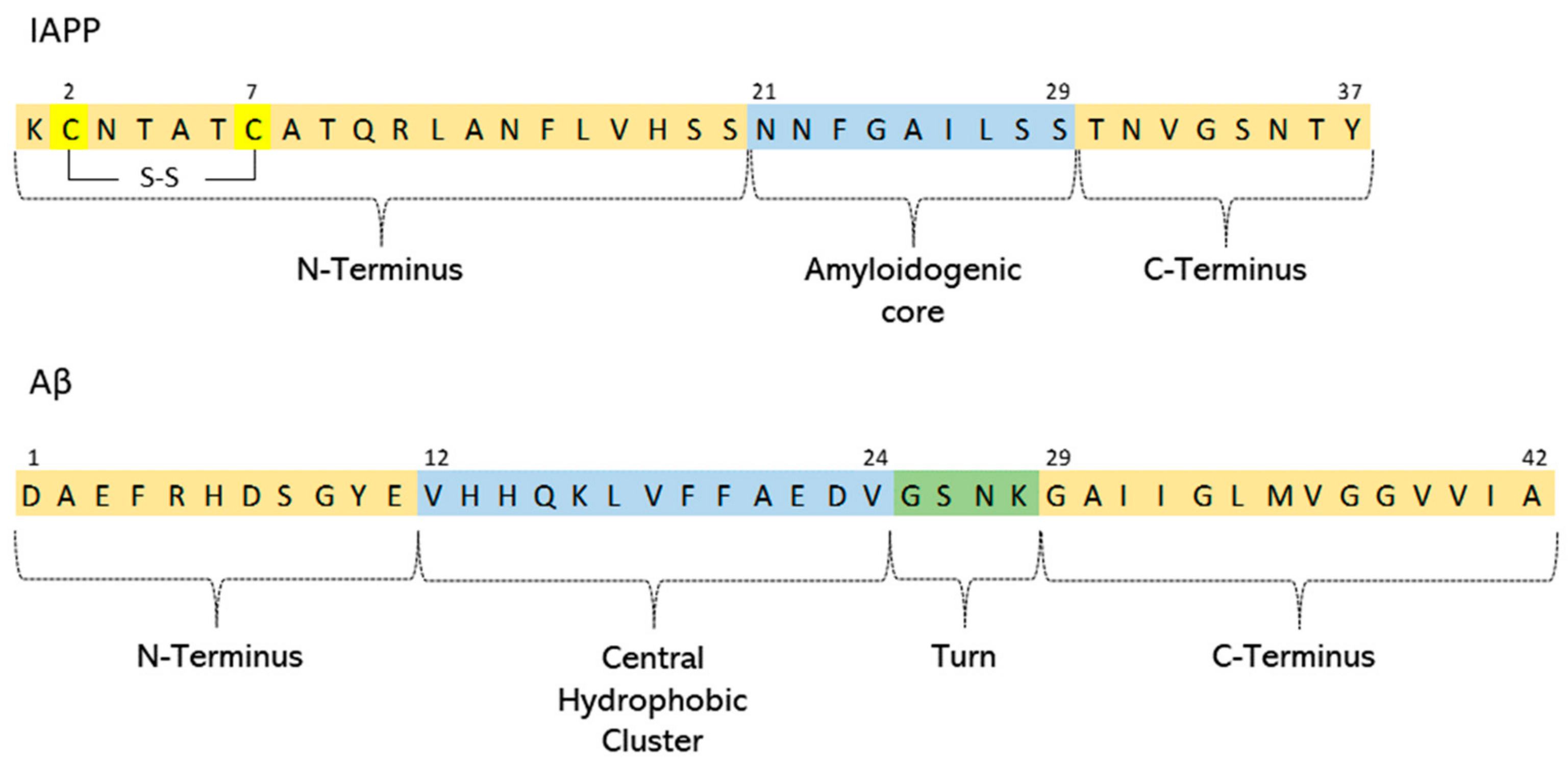
3. Islet Amyloid Polypeptide (IAPP)
4. Prion Protein
5. Biophysical Studies of Amyloid–Membrane Interactions
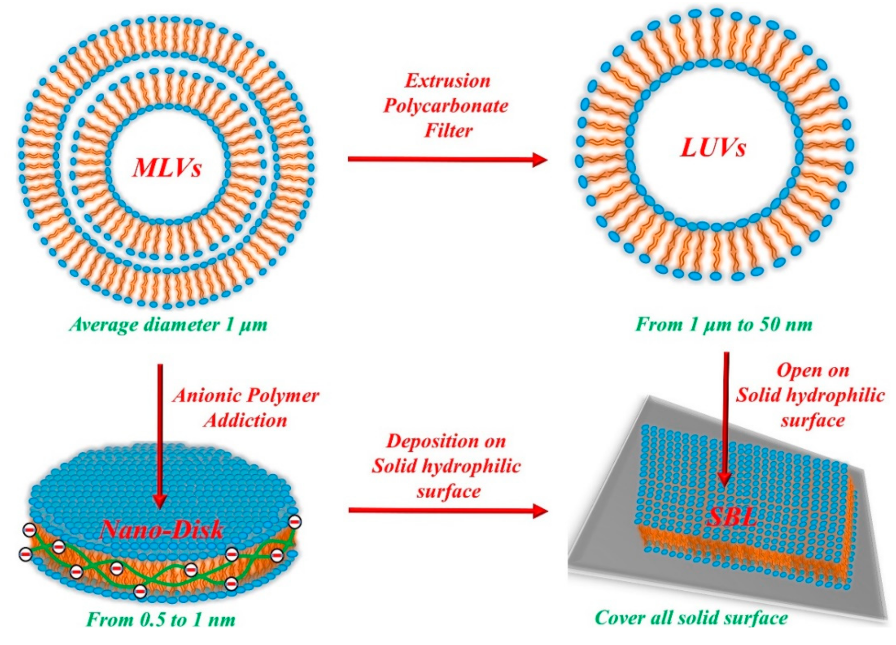
5.1. Model Membranes
5.2. Mechanisms of Amyloid-Mediated Membrane Damage
- (i)
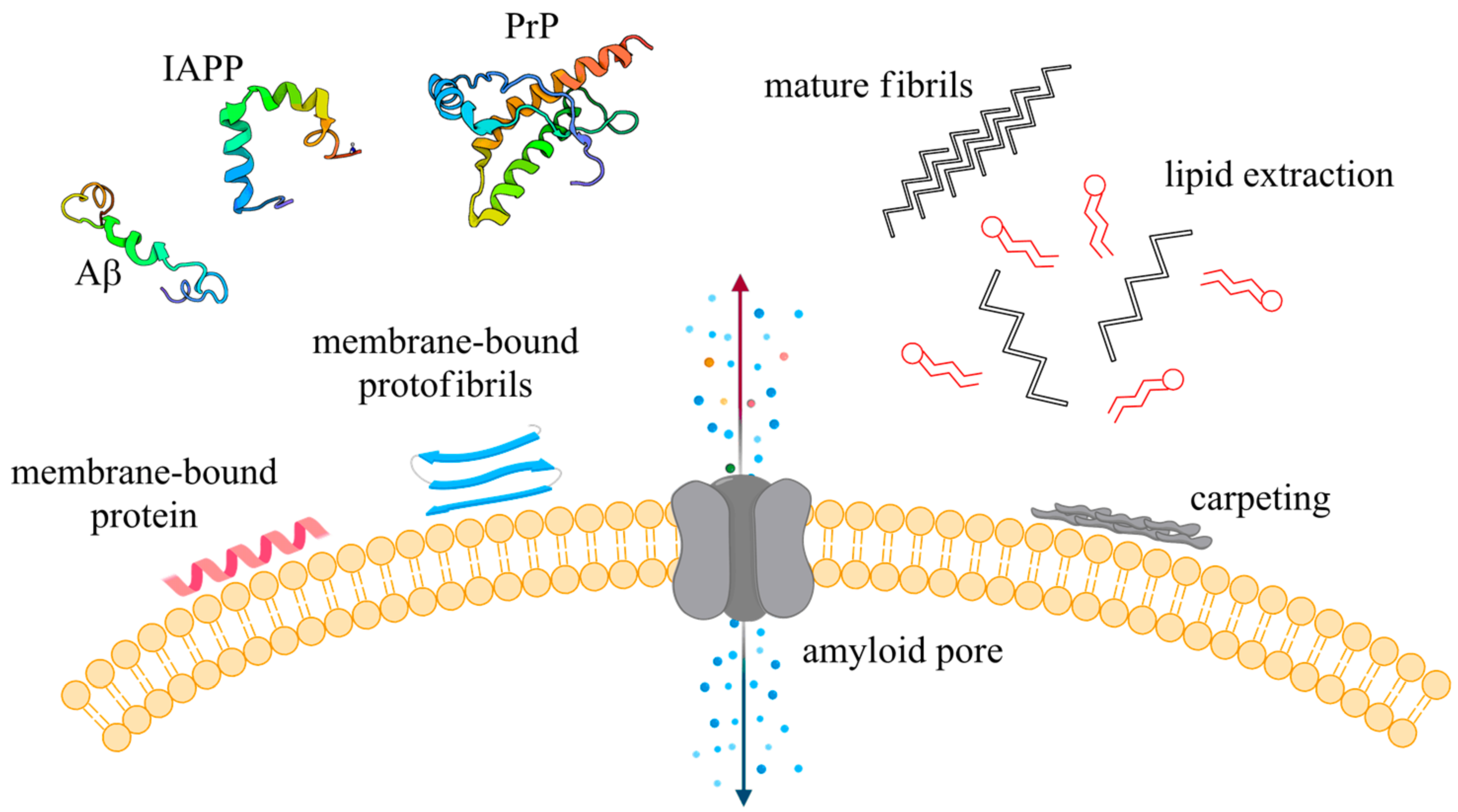
- Generation of stable transmembrane protein pores. The interaction of amyloidogenic protein, in its monomeric or oligomeric forms, with the membrane lipid bilayer led to the formation of pores which act as non-specific ion channels. It was reported that Aβ peptide, after interaction with lipid membranes, can form calcium-permeable channels that were suggested to induce cell death [147]. Consistently with this “channel hypothesis”, formation of calcium channels by Aβ depends on the presence of anionic lipids and is favored by acidic solutions. Moreover, it was also demonstrated that calcium permeable channels are reversibly blocked by zinc ions and small molecules like Congo Red [148]. It has also been reported that IAPP oligomers are able to form pore-like structures in the membrane resulting in pro-apoptotic Ca2+ dysregulation [149,150]. Of note, similar mechanisms have been reported for α-synuclein and PrP oligomers [151,152].
- (ii)
- Membrane destabilization via a “carpet model”. According to this model, the interaction of prefibrillar species with the lipid bilayer surface results in an asymmetric pressure between both layers. Relaxation of this pressure, proximal or distal to the protein, is accompanied by leakage of small molecule, leading to membrane damage [153]. Carpeting could lead to the detergent like mechanism of membrane disruption.
- (iii)
- Removal of lipid components from the bilayer by a detergent-like mechanism. The asymmetric pressure generated by peptides carpeting of membrane surface could lead to the removal of lipid from one or both the leaflets of the membrane. Removal of the outer leaflet may result in a transient membrane thinning, allowing the leakage of small molecules. Alternatively, removal from both leaflets results in the formation of a hole.
6. Membrane-Bound Amyloids
6.1. Aβ/Membrane Complexes
6.2. IAPP–Membrane Interactions
6.3. Prion–Membrane Interactions
7. Conclusions
Author Contributions
Funding
Data Availability Statement
Conflicts of Interest
References
- Stelzmann, R.A.; Norman Schnitzlein, H.; Reed Murtagh, F. Über Eine Eigenartige Erkankung Der Hirnrinde (An English Translation of Alzheimer’s 1907 Paper). Clin. Anat. 1995, 8, 429–431. [Google Scholar] [CrossRef] [PubMed]
- Glenner, G.G.; Wong, C.W. Alzheimer’s Disease: Initial Report of the Purification and Characterization of a Novel Cerebrovascular Amyloid Protein. Biochem. Biophys. Res. Commun. 1984, 120, 885–890, reprinted in Biochem. Biophys. Res. Commun. 2012, 425, 534–539. [Google Scholar] [CrossRef]
- Lee, V.M.; Balin, B.J.; Otvos, L.; Trojanowski, J.Q. A68: A Major Subunit of Paired Helical Filaments and Derivatized Forms of Normal Tau. Science 1991, 251, 675–678. [Google Scholar] [CrossRef] [PubMed]
- Selkoe, D.J.; Hardy, J. The Amyloid Hypothesis of Alzheimer’s Disease at 25 Years. EMBO Mol. Med. 2016, 8, 595–608. [Google Scholar] [CrossRef] [PubMed]
- Chiti, F.; Dobson, C.M. Protein Misfolding, Functional Amyloid, and Human Disease. Annu. Rev. Biochem. 2006, 75, 333–366. [Google Scholar] [CrossRef] [PubMed]
- Wimo, A.; Prince, M. World Alzheimer Report 2010: The Global Economic Impact of Dementia; Alzheimer’s Disease International: London, UK, 2010. [Google Scholar]
- Chiti, F.; Dobson, C.M. Protein Misfolding, Amyloid Formation, and Human Disease: A Summary of Progress Over the Last Decade. Annu. Rev. Biochem. 2017, 86, 27–68. [Google Scholar] [CrossRef] [PubMed]
- Word health Organization. Towards a Dementia Plan: A WHO Guide; Licence: CC BY-NC-SA 3.0 IGO; World Health Organization: Geneva, Switzerland, 2018. [Google Scholar]
- Word Health Organization. 2015 Alzheimer’s Disease Facts and Figures. Alzheimers Dement. 2015, 11, 332–384. [Google Scholar] [CrossRef]
- IDF Diabetes Atlas. 2019. Available online: www.diabetesatlas.org (accessed on 11 December 2019).
- Aguzzi, A.; O’Connor, T. Protein Aggregation Diseases: Pathogenicity and Therapeutic Perspectives. Nat. Rev. Drug Discov. 2010, 9, 237–248. [Google Scholar] [CrossRef]
- Lansbury, P.T. Structural Neurology: Are Seeds at the Root of Neuronal Degeneration? Neuron 1997, 19, 1151–1154. [Google Scholar] [CrossRef][Green Version]
- Pauli, G. Tissue Safety in View of CJD and Variant CJD. Cell Tissue Bank 2005, 6, 191–200. [Google Scholar] [CrossRef]
- Harman, J.L.; Silva, C.J. Bovine Spongiform Encephalopathy. J. Am. Vet. Med. Assoc. 2009, 234, 59–72. [Google Scholar] [CrossRef]
- Miller, M.W.; Williams, E.S.; McCarty, C.W.; Spraker, T.R.; Kreeger, T.J.; Larsen, C.T.; Thorne, E.T. Epizootiology of Chronic Wasting Disease in Free-Ranging Cervids in Colorado and Wyoming. J. Wildl. Dis. 2000, 36, 676–690. [Google Scholar] [CrossRef]
- Angers, R.C.; Kang, H.-E.; Napier, D.; Browning, S.; Seward, T.; Mathiason, C.; Balachandran, A.; McKenzie, D.; Castilla, J.; Soto, C.; et al. Prion Strain Mutation Determined by Prion Protein Conformational Compatibility and Primary Structure. Science 2010, 328, 1154–1158. [Google Scholar] [CrossRef]
- Horby, P. Variant Creutzfeldt-Jakob Disease: An Unfolding Epidemic of Misfolded Proteins. J. Paediatr. Child Health 2002, 38, 539–542. [Google Scholar] [CrossRef]
- Ironside, J.W. The Spectrum of Safety: Variant Creutzfeldt-Jakob Disease in the United Kingdom. Semin. Hematol. 2003, 40, 16–22. [Google Scholar] [CrossRef]
- Holman, R.C.; Belay, E.D.; Christensen, K.Y.; Maddox, R.A.; Minino, A.M.; Folkema, A.M.; Haberling, D.L.; Hammett, T.A.; Kochanek, K.D.; Sejvar, J.J.; et al. Human Prion Diseases in the United States. PLoS ONE 2010, 5, e8521. [Google Scholar] [CrossRef]
- Hartl, F.U. Protein Misfolding Diseases. Annu. Rev. Biochem. 2017, 86, 21–26. [Google Scholar] [CrossRef]
- Brandt, R.; Léger, J.; Lee, G. Interaction of Tau with the Neural Plasma Membrane Mediated by Tau’s Amino-Terminal Projection Domain. J. Cell Biol. 1995, 131, 1327–1340. [Google Scholar] [CrossRef]
- Schmidt, M.; Sachse, C.; Richter, W.; Xu, C.; Fändrich, M.; Grigorieff, N. Comparison of Alzheimer Aβ(1–40) and Aβ(1–42) Amyloid Fibrils Reveals Similar Protofilament Structures. Proc. Natl. Acad. Sci. USA 2009, 106, 19813–19818. [Google Scholar] [CrossRef]
- Mori, H.; Takio, K.; Ogawara, M.; Selkoe, D.J. Mass Spectrometry of Purified Amyloid Beta Protein in Alzheimer’s Disease. J. Biol. Chem. 1992, 267, 17082–17086. [Google Scholar] [CrossRef]
- Näslund, J.; Schierhorn, A.; Hellman, U.; Lannfelt, L.; Roses, A.D.; Tjernberg, L.O.; Silberring, J.; Gandy, S.E.; Winblad, B.; Greengard, P. Relative Abundance of Alzheimer A Beta Amyloid Peptide Variants in Alzheimer Disease and Normal Aging. Proc. Natl. Acad. Sci. USA 1994, 91, 8378–8382. [Google Scholar] [CrossRef]
- Goldgaber, D.; Lerman, M.I.; McBride, O.W.; Saffiotti, U.; Gajdusek, D.C. Characterization and Chromosomal Localization of a CDNA Encoding Brain Amyloid of Alzheimer’s Disease. Science 1987, 235, 877–880. [Google Scholar] [CrossRef]
- Selkoe, D.J. Normal and Abnormal Biology of the Beta-Amyloid Precursor Protein. Annu. Rev. Neurosci. 1994, 17, 489–517. [Google Scholar] [CrossRef]
- Masters, C.L.; Simms, G.; Weinman, N.A.; Multhaup, G.; McDonald, B.L.; Beyreuther, K. Amyloid Plaque Core Protein in Alzheimer Disease and Down Syndrome. Proc. Natl. Acad. Sci. USA 1985, 82, 4245–4249. [Google Scholar] [CrossRef]
- Kang, J.; Lemaire, H.G.; Unterbeck, A.; Salbaum, J.M.; Masters, C.L.; Grzeschik, K.H.; Multhaup, G.; Beyreuther, K.; Müller-Hill, B. The Precursor of Alzheimer’s Disease Amyloid A4 Protein Resembles a Cell-Surface Receptor. Nature 1987, 325, 733–736. [Google Scholar] [CrossRef]
- Buoso, E.; Lanni, C.; Schettini, G.; Govoni, S.; Racchi, M. β-Amyloid Precursor Protein Metabolism: Focus on the Functions and Degradation of Its Intracellular Domain. Pharmacol. Res. 2010, 62, 308–317. [Google Scholar] [CrossRef]
- Sisodia, S.S. Beta-Amyloid Precursor Protein Cleavage by a Membrane-Bound Protease. Proc. Natl. Acad. Sci. USA 1992, 89, 6075–6079. [Google Scholar] [CrossRef]
- Haass, C.; Hung, A.Y.; Schlossmacher, M.G.; Teplow, D.B.; Selkoe, D.J. Beta-Amyloid Peptide and a 3-KDa Fragment Are Derived by Distinct Cellular Mechanisms. J. Biol. Chem. 1993, 268, 3021–3024. [Google Scholar] [CrossRef]
- Koo, E.H.; Squazzo, S.L. Evidence That Production and Release of Amyloid Beta-Protein Involves the Endocytic Pathway. J. Biol. Chem. 1994, 269, 17386–17389. [Google Scholar] [CrossRef]
- Thornton, E.; Vink, R.; Blumbergs, P.C.; Van Den Heuvel, C. Soluble Amyloid Precursor Protein Alpha Reduces Neuronal Injury and Improves Functional Outcome Following Diffuse Traumatic Brain Injury in Rats. Brain Res. 2006, 1094, 38–46. [Google Scholar] [CrossRef]
- Reid, P.C.; Urano, Y.; Kodama, T.; Hamakubo, T. Alzheimer’s Disease: Cholesterol, Membrane Rafts, Isoprenoids and Statins. J. Cell. Mol. Med. 2007, 11, 383–392. [Google Scholar] [CrossRef] [PubMed]
- Morley, J.E.; Farr, S.A. Hormesis and Amyloid-β Protein: Physiology or Pathology? J. Alzheimers Dis. 2012, 29, 487–492. [Google Scholar] [CrossRef] [PubMed]
- Hillen, H. The Beta Amyloid Dysfunction (BAD) Hypothesis for Alzheimer’s Disease. Front. Neurosci. 2019, 13. [Google Scholar] [CrossRef] [PubMed]
- Grasso, G.; Giuffrida, M.L.; Rizzarelli, E. Metallostasis and Amyloid β-Degrading Enzymes. Metallomics 2012, 4, 937–949. [Google Scholar] [CrossRef]
- Leissring, M.A. The AβCs of Aβ-Cleaving Proteases. J. Biol. Chem. 2008, 283, 29645–29649. [Google Scholar] [CrossRef]
- Grasso, G.; Bonnet, S. Metal Complexes and Metalloproteases: Targeting Conformational Diseases. Metallomics 2014, 6, 1346–1357. [Google Scholar] [CrossRef]
- Glenner, G.G. Amyloid Beta Protein and the Basis for Alzheimer’s Disease. Prog. Clin. Biol. Res. 1989, 317, 857–868. [Google Scholar]
- Hardy, J.A.; Higgins, G.A. Alzheimer’s Disease: The Amyloid Cascade Hypothesis. Science 1992, 256, 184–185. [Google Scholar] [CrossRef]
- Hardy, J.; Selkoe, D.J. The Amyloid Hypothesis of Alzheimer’s Disease: Progress and Problems on the Road to Therapeutics. Science 2002, 297, 353–356. [Google Scholar] [CrossRef]
- Lewis, J.; Dickson, D.W.; Lin, W.L.; Chisholm, L.; Corral, A.; Jones, G.; Yen, S.H.; Sahara, N.; Skipper, L.; Yager, D.; et al. Enhanced Neurofibrillary Degeneration in Transgenic Mice Expressing Mutant Tau and APP. Science 2001, 293, 1487–1491. [Google Scholar] [CrossRef]
- Götz, J.; Chen, F.; van Dorpe, J.; Nitsch, R.M. Formation of Neurofibrillary Tangles in P301l Tau Transgenic Mice Induced by Abeta 42 Fibrils. Science 2001, 293, 1491–1495. [Google Scholar] [CrossRef]
- Oddo, S.; Billings, L.; Kesslak, J.P.; Cribbs, D.H.; LaFerla, F.M. Abeta Immunotherapy Leads to Clearance of Early, but Not Late, Hyperphosphorylated Tau Aggregates via the Proteasome. Neuron 2004, 43, 321–332. [Google Scholar] [CrossRef]
- Bellia, F.; Lanza, V.; García-Viñuales, S.; Ahmed, I.M.M.; Pietropaolo, A.; Iacobucci, C.; Malgieri, G.; D’Abrosca, G.; Fattorusso, R.; Nicoletti, V.G.; et al. Ubiquitin Binds the Amyloid β Peptide and Interferes with Its Clearance Pathways. Chem. Sci. 2019, 10, 2732–2742. [Google Scholar] [CrossRef]
- Loo, D.T.; Copani, A.; Pike, C.J.; Whittemore, E.R.; Walencewicz, A.J.; Cotman, C.W. Apoptosis Is Induced by Beta-Amyloid in Cultured Central Nervous System Neurons. Proc. Natl. Acad. Sci. USA 1993, 90, 7951–7955. [Google Scholar] [CrossRef]
- García-Viñuales, S.; Ahmed, R.; Sciacca, M.F.M.; Lanza, V.; Giuffrida, M.L.; Zimbone, S.; Romanucci, V.; Zarrelli, A.; Bongiorno, C.; Spinella, N.; et al. Trehalose Conjugates of Silybin as Prodrugs for Targeting Toxic Aβ Aggregates. ACS Chem. Neurosci. 2020, 11, 2566–2576. [Google Scholar] [CrossRef]
- Lanza, V.; Milardi, D.; Pappalardo, G.; Di Natale, G. Repurposing of Copper(II)-Chelating Drugs for the Treatment of Neurodegenerative Diseases. Curr. Med. Chem. 2018, 25, 525–539. [Google Scholar] [CrossRef]
- Romanucci, V.; García-Viñuales, S.; Tempra, C.; Bernini, R.; Zarrelli, A.; Lolicato, F.; Milardi, D.; Di Fabio, G. Modulating Aβ Aggregation by Tyrosol-Based Ligands: The Crucial Role of the Catechol Moiety. Biophys. Chem. 2020, 265, 106434. [Google Scholar] [CrossRef]
- Barrow, C.J.; Yasuda, A.; Kenny, P.T.M.; Zagorski, M.G. Solution Conformations and Aggregational Properties of Synthetic Amyloid β-Peptides of Alzheimer’s Disease. Analysis of Circular Dichroism Spectra. J. Mol. Biol. 1992, 225, 1075–1093. [Google Scholar] [CrossRef]
- Protein Aggregation and Neurodegenerative Disease. Nature Medicine. Available online: https://www.nature.com/articles/nm1066 (accessed on 20 January 2021).
- Walsh, D.M.; Hartley, D.M.; Kusumoto, Y.; Fezoui, Y.; Condron, M.M.; Lomakin, A.; Benedek, G.B.; Selkoe, D.J.; Teplow, D.B. Amyloid Beta-Protein Fibrillogenesis. Structure and Biological Activity of Protofibrillar Intermediates. J. Biol. Chem. 1999, 274, 25945–25952. [Google Scholar] [CrossRef]
- Jarrett, J.T.; Berger, E.P.; Lansbury, P.T. The Carboxy Terminus of the β Amyloid Protein Is Critical for the Seeding of Amyloid Formation: Implications for the Pathogenesis of Alzheimer’s Disease. Biochemistry 1993, 32, 4693–4697. [Google Scholar] [CrossRef]
- Grimm, M.O.W.; Mett, J.; Grimm, H.S.; Hartmann, T. APP Function and Lipids: A Bidirectional Link. Front. Mol. Neurosci. 2017, 10. [Google Scholar] [CrossRef]
- Chimon, S.; Shaibat, M.A.; Jones, C.R.; Calero, D.C.; Aizezi, B.; Ishii, Y. Evidence of Fibril-like β-Sheet Structures in a Neurotoxic Amyloid Intermediate of Alzheimer’s β-Amyloid. Nat. Struct. Mol. Biol. 2007, 14, 1157–1164. [Google Scholar] [CrossRef]
- Zhang, X.-X.; Pan, Y.-H.; Huang, Y.-M.; Zhao, H.-L. Neuroendocrine Hormone Amylin in Diabetes. World J. Diabetes 2016, 7, 189–197. [Google Scholar] [CrossRef]
- Press, M.; Jung, T.; König, J.; Grune, T.; Höhn, A. Protein Aggregates and Proteostasis in Aging: Amylin and β-Cell Function. Mech. Ageing Dev. 2019, 177, 46–54. [Google Scholar] [CrossRef] [PubMed]
- Marzban, L.; Trigo-Gonzalez, G.; Zhu, X.; Rhodes, C.J.; Halban, P.A.; Steiner, D.F. Role of Beta-Cell Prohormone Convertase (PC)1/3 in Processing of pro-Islet Amyloid Polypeptide. Diabetes 2004, 53, 141–148. [Google Scholar] [CrossRef] [PubMed]
- Marzban, L.; Soukhatcheva, G.; Verchere, C.B. Role of Carboxypeptidase E in Processing of Pro-Islet Amyloid Polypeptide in Beta-Cells. Endocrinology 2005, 146, 1808–1817. [Google Scholar] [CrossRef] [PubMed]
- Visa, M.; Alcarraz-Vizán, G.; Montane, J.; Cadavez, L.; Castaño, C.; Villanueva-Peñacarrillo, M.L.; Servitja, J.-M.; Novials, A. Islet Amyloid Polypeptide Exerts a Novel Autocrine Action in β-Cell Signaling and Proliferation. FASEB J. 2015, 29, 2970–2979. [Google Scholar] [CrossRef] [PubMed]
- Rushing, P.A.; Hagan, M.M.; Seeley, R.J.; Lutz, T.A.; Woods, S.C. Amylin: A Novel Action in the Brain to Reduce Body Weight. Endocrinology 2000, 141, 850–853. [Google Scholar] [CrossRef]
- Paulsson, J.F.; Andersson, A.; Westermark, P.; Westermark, G.T. Intracellular amyloid-like deposits contain unprocessed pro islet amyloid polypeptide (proIAPP) in beta-cells of transgenic mice overexpressing human IAPP and transplanted human islets. Diabetologia 2006, 49, 1237–1246. [Google Scholar] [CrossRef]
- Akter, R.; Cao, P.; Noor, H.; Ridgway, Z.; Tu, L.-H.; Wang, H.; Wong, A.G.; Zhang, X.; Abedini, A.; Schmidt, A.M.; et al. Islet Amyloid Polypeptide: Structure, Function, and Pathophysiology. J. Diabetes Res. 2015, 2016, 2798269. [Google Scholar] [CrossRef]
- Milardi, D.; Gazit, E.; Radford, S.E.; Xu, Y.; Gallardo, R.U.; Caflisch, A.; Westermark, G.T.; Westermark, P.; La Rosa, C.; Ramamoorthy, A. Proteostasis of Islet Amyloid Polypeptide: A Molecular Perspective of Risk Factors and Protective Strategies for Type II Diabetes. Chem. Rev. 2021, 121, 1845–1893. [Google Scholar] [CrossRef]
- Westermark, P.; Engström, U.; Johnson, K.H.; Westermark, G.T.; Betsholtz, C. Islet Amyloid Polypeptide: Pinpointing Amino Acid Residues Linked to Amyloid Fibril Formation. Proc. Natl. Acad. Sci. USA 1990, 87, 5036–5040. [Google Scholar] [CrossRef]
- Charge, S.B.; Koning, E.J.; Clark, A. Effect of PH and Insulin on Fibrillogenesis of Islet Amyloid Polypeptide in Vitro. Biochemistry 1995, 34, 14588–14593. [Google Scholar] [CrossRef]
- Makin, O.S.; Serpell, L.C. Structural Characterisation of Islet Amyloid Polypeptide Fibrils. J. Mol. Biol. 2004, 335, 1279–1288. [Google Scholar] [CrossRef]
- Westermark, G.T.; Westermark, P.; Berne, C.; Korsgren, O. Widespread Amyloid Deposition in Transplanted Human Pancreatic Islets. N. Engl. J. Med. 2008, 359, 997–999. [Google Scholar] [CrossRef]
- Westermark, G.T.; Gebre-Medhin, S.; Steiner, D.F.; Westermark, P. Islet Amyloid Development in a Mouse Strain Lacking Endogenous Islet Amyloid Polypeptide (IAPP) but Expressing Human IAPP. Mol. Med. 2000, 6, 998–1007. [Google Scholar] [CrossRef]
- Liang, G.; Zhao, J.; Yu, X.; Zheng, J. Comparative Molecular Dynamics Study of Human Islet Amyloid Polypeptide (IAPP) and Rat IAPP Oligomers. Biochemistry 2013, 52, 1089–1100. [Google Scholar] [CrossRef]
- Cao, P.; Meng, F.; Abedini, A.; Raleigh, D.P. The Ability of Rodent Islet Amyloid Polypeptide to Inhibit Amyloid Formation by Human Islet Amyloid Polypeptide Has Important Implications for the Mechanism of Amyloid Formation and the Design of Inhibitors. Biochemistry 2010, 49, 872–881. [Google Scholar] [CrossRef]
- Chiu, C.; Singh, S.; de Pablo, J.J. Effect of Proline Mutations on the Monomer Conformations of Amylin. Biophys. J. 2013, 105, 1227–1235. [Google Scholar] [CrossRef]
- Nilsson, M.R.; Raleigh, D.P. Analysis of Amylin Cleavage Products Provides New Insights into the Amyloidogenic Region of Human Amylin, Edited by P.E. Wright. J. Mol. Biol. 1999, 294, 1375–1385. [Google Scholar] [CrossRef]
- Khemtémourian, L.; Engel, M.F.M.; Liskamp, R.M.J.; Höppener, J.W.M.; Killian, J.A. The N-Terminal Fragment of Human Islet Amyloid Polypeptide Is Non-Fibrillogenic in the Presence of Membranes and Does Not Cause Leakage of Bilayers of Physiologically Relevant Lipid Composition. Biochim. Biophys. Acta Biomembr. 2010, 1798, 1805–1811. [Google Scholar] [CrossRef]
- Nanga, R.P.R.; Brender, J.R.; Xu, J.; Veglia, G.; Ramamoorthy, A. Structures of Rat and Human Islet Amyloid Polypeptide IAPP(1-19) in Micelles by NMR Spectroscopy. Biochemistry 2008, 47, 12689–12697. [Google Scholar] [CrossRef]
- Gilead, S.; Gazit, E. The Role of the 14–20 Domain of the Islet Amyloid Polypeptide in Amyloid Formation. Exp. Diabetes Res. 2008. [Google Scholar] [CrossRef]
- Bemporad, F.; Calloni, G.; Campioni, S.; Plakoutsi, G.; Taddei, N.; Chiti, F. Sequence and Structural Determinants of Amyloid Fibril Formation. Acc. Chem. Res. 2006, 39, 620–627. [Google Scholar] [CrossRef]
- Jaikaran, E.T.; Higham, C.E.; Serpell, L.C.; Zurdo, J.; Gross, M.; Clark, A.; Fraser, P.E. Identification of a Novel Human Islet Amyloid Polypeptide β-Sheet Domain and Factors Influencing Fibrillogenesis. J. Am. Chem. Soc. 2001, 308, 515–525. [Google Scholar] [CrossRef]
- Röder, C.; Kupreichyk, T.; Gremer, L.; Schäfer, L.U.; Pothula, K.R.; Ravelli, R.B.G.; Willbold, D.; Hoyer, W.; Schröder, G.F. Cryo-EM Structure of Islet Amyloid Polypeptide Fibrils Reveals Similarities with Amyloid-β Fibrils. Nat. Struct. Mol. Biol. 2020, 27, 660–667. [Google Scholar] [CrossRef]
- Cao, Q.; Boyer, D.R.; Sawaya, M.R.; Ge, P.; Eisenberg, D.S. Cryo-EM Structure and Inhibitor Design of Human IAPP (Amylin) Fibrils. Nat. Struct Mol. Biol. 2020, 27, 653–659. [Google Scholar] [CrossRef]
- Luca, S.; Yau, W.-M.; Leapman, R.; Tycko, R. Peptide Conformation and Supramolecular Organization in Amylin Fibrils: Constraints from Solid-State NMR. Biochemistry 2007, 46, 13505–13522. [Google Scholar] [CrossRef]
- Jaikaran, E.T.; Clark, A. Islet amyloid and type 2 diabetes: From molecular misfolding to islet pathophysiology. Biochim. Biophys. Acta 2001, 1537, 179–203. [Google Scholar] [CrossRef]
- Brender, J.R.; Salamekh, S.; Ramamoorthy, A. Membrane Disruption and Early Events in the Aggregation of the Diabetes Related Peptide IAPP from a Molecular Perspective. Acc. Chem. Res. 2012, 45, 454–462. [Google Scholar] [CrossRef] [PubMed]
- Janson, J.; Ashley, R.H.; Harrison, D.; McIntyre, S.; Butler, P.C. The Mechanism of Islet Amyloid Polypeptide Toxicity Is Membrane Disruption by Intermediate-Sized Toxic Amyloid Particles. Diabetes 1999, 48, 491–498. [Google Scholar] [CrossRef] [PubMed]
- Kumar, S.; Schlamadinger, D.E.; Brown, M.A.; Dunn, J.M.; Mercado, B.; Hebda, J.A.; Saraogi, I.; Rhoades, E.; Hamilton, A.D.; Miranker, A.D. Islet Amyloid-Induced Cell Death and Bilayer Integrity Loss Share a Molecular Origin Targetable with Oligopyridylamide-Based α-Helical Mimetics. Chem. Biol. 2015, 22, 369–378. [Google Scholar] [CrossRef] [PubMed]
- Kyung-Hoon Lee, A.Z.; Raleigh, D. Amyloidogenicity and Cytotoxicity of Des-Lys-1 Human Amylin Provides Insight into Amylin Self-Assembly and Highlights the Difficulties of Defining Amyloidogenicity. Protein Eng. Des. Sel. PEDS 2019, 32, 87–93. [Google Scholar]
- Lee, K.-H.; Noh, D.; Zhyvoloup, A.; Raleigh, D. Analysis of Prairie Vole Amylin Reveals the Importance of the N-Terminus and Residue 22 in Amyloidogenicity and Cytotoxicity. Biochemistry 2020, 59, 471–478. [Google Scholar] [CrossRef]
- Ridgway, Z.; Lee, K.-H.; Zhyvoloup, A.; Wong, A.; Eldrid, C.; Hannaberry, E.; Thalassinos, K.; Abedini, A.; Raleigh, D.P. Analysis of Baboon IAPP Provides Insight into Amyloidogenicity and Cytotoxicity of Human IAPP. Biophys. J. 2020, 118, 1142–1151. [Google Scholar] [CrossRef]
- Jha, S.; Snell, J.M.; Sheftic, S.R.; Patil, S.M.; Daniels, S.B.; Kolling, F.W.; Alexandrescu, A.T. PH Dependence of Amylin Fibrillization. Biochemistry 2014, 53, 300–310. [Google Scholar] [CrossRef]
- Green, J.; Goldsbury, C.; Mini, T.; Sunderji, S.; Frey, P.; Kistler, J.; Cooper, G.; Aebi, U. Full-Length Rat Amylin Forms Fibrils Following Substitution of Single Residues from Human Amylin. J. Mol. Biol. 2003, 326, 1147–1156. [Google Scholar] [CrossRef]
- Khemtemourian, L.; Guillemain, G.; Foufelle, F.; Killian, J.A. Residue Specific Effects of Human Islet Polypeptide Amyloid on Self-Assembly and on Cell Toxicity. Biochimie 2017, 142, 22–30. [Google Scholar] [CrossRef]
- Hoffmann, A.R.F.; Saravanan, M.S.; Lequin, O.; Killian, J.A.; Khemtemourian, L. A Single Mutation on the Human Amyloid Polypeptide Modulates Fibril Growth and Affects the Mechanism of Amyloid-Induced Membrane Damage. Biochim. Biophys. Acta (BBA) Biomembr. 2018, 1860, 1783–1792. [Google Scholar] [CrossRef]
- Milardi, D.; Sciacca, M.F.M.; Pappalardo, M.; Grasso, D.M.; La Rosa, C. The Role of Aromatic Side-Chains in Amyloid Growth and Membrane Interaction of the Islet Amyloid Polypeptide Fragment LANFLVH. Eur. Biophys. J. 2011, 40, 1–12. [Google Scholar] [CrossRef]
- Doran, T.; Kamens, A.; Byrnes, N.; Nilsson, B. Role of amino acid hydrophobicity, aromaticity, and molecular volume on IAPP(20-29) amyloid self-assembly. Proteins 2012, 80, 1053–1065. [Google Scholar] [CrossRef]
- Azriel, R.; Gazit, E. Analysis of the Minimal Amyloid-Forming Fragment of the Islet Amyloid Polypeptide an Experimental Support for the Key Role of the Phenylalanine Residue in Amyloid Formation. J. Biol. Chem. 2001, 276, 34156–34161. [Google Scholar] [CrossRef]
- Tu, L.-H.; Raleigh, D.P. Role of Aromatic Interactions in Amyloid Formation by Islet Amyloid Polypeptide. Biochemistry 2013, 52, 333–342. [Google Scholar] [CrossRef]
- Marek, P.; Abedini, A.; Song, B.; Kanungo, M.; Johnson, M.E.; Gupta, R.; Zaman, W.; Wong, S.S.; Raleigh, D.P. Aromatic Interactions Are Not Required for Amyloid Fibril Formation by Islet Amyloid Polypeptide but Do Influence the Rate of Fibril Formation and Fibril Morphology. Biochemistry 2007, 46, 3255–3261. [Google Scholar] [CrossRef]
- Gazit, E. A Possible Role for π-Stacking in the Self-Assembly of Amyloid Fibrils. FASEB J. 2002, 16, 77–83. [Google Scholar] [CrossRef]
- Padrick, S.B.; Miranker, A.D. Islet Amyloid Polypeptide: Identification of Long-Range Contacts and Local Order on the Fibrillogenesis Pathway. J. Biol. Chem. 2001, 308, 783–794. [Google Scholar]
- Abedini, A.; Plesner, A.; Cao, P.; Ridgway, Z.; Zhang, J.; Tu, L.-H.; Middleton, C.T.; Chao, B.; Sartori, D.J.; Meng, F.; et al. Time-Resolved Studies Define the Nature of Toxic IAPP Intermediates, Providing Insight for Anti-Amyloidosis Therapeutics. eLife 2016, 5, e12977. [Google Scholar] [CrossRef]
- Abedini, A.; Raleigh, D.P. A Critical Assessment of the Role of Helical Intermediates in Amyloid Formation by Natively Unfolded Proteins and Polypeptides. Protein Eng. Des. Sel. 2009, 22, 453–459. [Google Scholar] [CrossRef]
- Koo, B.W.; Miranker, A.D. Contribution of the Intrinsic Disulfide to the Assembly Mechanism of Islet Amyloid. Protein Sci. 2005, 14, 231–239. [Google Scholar] [CrossRef]
- Ridgway, Z.; Zhang, X.; Wong, A.G.; Abedini, A.; Schmidt, A.M.; Raleigh, D.P. Analysis of the Role of the Conserved Disulfide in Amyloid Formation by Human Islet Amyloid Polypeptide in Homogeneous and Heterogeneous Environments. Biochemistry 2018, 57, 3065–3074. [Google Scholar] [CrossRef]
- Rodriguez Camargo, D.C.; Tripsianes, K.; Buday, K.; Franko, A.; Göbl, C.; Hartlmüller, C.; Sarkar, R.; Aichler, M.; Mettenleiter, G.; Schulz, M.; et al. The Redox Environment Triggers Conformational Changes and Aggregation of HIAPP in Type II Diabetes. Sci. Rep. 2017, 7, 44041. [Google Scholar] [CrossRef]
- Anguiano, M.; Nowak, R.J.; Lansbury, P.T. Protofibrillar islet amyloid polypeptide permeabilizes synthetic vesicles by a pore-like mechanism that may be relevant to type II diabetes. Biochemistry 2002, 41, 11338–11343. [Google Scholar] [CrossRef] [PubMed]
- Sasahara, K. Membrane-Mediated Amyloid Deposition of Human Islet Amyloid Polypeptide. Biophys. Rev. 2018, 10, 453–462. [Google Scholar] [CrossRef][Green Version]
- Porat, Y.; Kolusheva, S.; Jelinek, R.; Gazit, E. The Human Islet Amyloid Polypeptide Forms Transient Membrane-Active Prefibrillar Assemblies. Biochemistry 2003, 42, 10971–10977. [Google Scholar] [CrossRef] [PubMed]
- Colby, D.W.; Prusiner, S.B. Prions. Cold Spring Harb. Perspect. Biol. 2011, 3, a006833. [Google Scholar] [CrossRef]
- Abskharon, R.; Wang, F.; Wohlkonig, A.; Ruan, J.; Soror, S.; Giachin, G.; Pardon, E.; Zou, W.; Legname, G.; Ma, J.; et al. Structural Evidence for the Critical Role of the Prion Protein Hydrophobic Region in Forming an Infectious Prion. PLoS Pathog. 2019, 15, e1008139. [Google Scholar] [CrossRef] [PubMed]
- Pappalardo, M.; Milardi, D.; Grasso, D.; La Rosa, C. Steered Molecular Dynamics Studies Reveal Different Unfolding Pathways of Prions from Mammalian and Non-Mammalian Species. New J. Chem. 2007, 31, 901–905. [Google Scholar] [CrossRef]
- Zhang, J.; Zhang, Y. Molecular Dynamics Studies on 3D Structures of the Hydrophobic Region PrP(109-136). Acta Biochim. Biophys. Sin. 2013, 45, 509–519. [Google Scholar] [CrossRef][Green Version]
- Garnier, J.; Osguthorpe, D.J.; Robson, B. Analysis of the Accuracy and Implications of Simple Methods for Predicting the Secondary Structure of Globular Proteins. J. Mol. Biol. 1978, 120, 97–120. [Google Scholar] [CrossRef]
- Riek, R.; Wider, G.; Billeter, M.; Hornemann, S.; Glockshuber, R.; Wuthrich, K. Prion Protein NMR Structure and Familial Human Spongiform Encephalopathies. Proc. Natl. Acad. Sci. USA 1998, 95, 11667–11672. [Google Scholar] [CrossRef]
- Donne, D.G.; Viles, J.H.; Groth, D.; Mehlhorn, I.; James, T.L.; Cohen, F.E.; Prusiner, S.B.; Wright, P.E.; Dyson, H.J. Structure of the Recombinant Full-Length Hamster Prion Protein PrP(29-231): The N Terminus Is Highly Flexible. Proc. Natl. Acad. Sci. USA 1997, 94, 13452–13457. [Google Scholar] [CrossRef]
- Liu, H.; Farr-Jones, S.; Ulyanov, N.B.; Llinas, M.; Marqusee, S.; Groth, D.; Cohen, F.E.; Prusiner, S.B.; James, T.L. Solution Structure of Syrian Hamster Prion Protein RPrP(90−231). Biochemistry 1999, 38, 5362–5377. [Google Scholar] [CrossRef]
- Riek, R.; Hornemann, S.; Wider, G.; Glockshuber, R.; Wüthrich, K. NMR Characterization of the Full-Length Recombinant Murine Prion Protein, m PrP(23-231). FEBS Lett. 1997, 413, 282–288. [Google Scholar] [CrossRef]
- Zahn, R.; Liu, A.; Luhrs, T.; Riek, R.; von Schroetter, C.; Lopez Garcia, F.; Billeter, M.; Calzolai, L.; Wider, G.; Wuthrich, K. NMR Solution Structure of the Human Prion Protein. Proc. Natl. Acad. Sci. USA 2000, 97, 145–150. [Google Scholar] [CrossRef]
- Cho, K.R.; Huang, Y.; Yu, S.; Yin, S.; Plomp, M.; Qiu, S.R.; Lakshminarayanan, R.; Moradian-Oldak, J.; Sy, M.-S.; De Yoreo, J.J. A Multistage Pathway for Human Prion Protein Aggregation in Vitro: From Multimeric Seeds to β-Oligomers and Nonfibrillar Structures. J. Am. Chem. Soc. 2011, 133, 8586–8593. [Google Scholar] [CrossRef]
- Bocharova, O.V.; Breydo, L.; Parfenov, A.S.; Salnikov, V.V.; Baskakov, I.V. In Vitro Conversion of Full-Length Mammalian Prion Protein Produces Amyloid Form with Physical Properties of PrPSc. J. Mol. Biol. 2005, 346, 645–659. [Google Scholar] [CrossRef]
- Baskakov, I.V.; Bocharova, O.V. In Vitro Conversion of Mammalian Prion Protein into Amyloid Fibrils Displays Unusual Features †. Biochemistry 2005, 44, 2339–2348. [Google Scholar] [CrossRef]
- Safar, J.; Wille, H.; Itri, V.; Groth, D.; Serban, H.; Torchia, M.; Cohen, F.E.; Prusiner, S.B. Eight Prion Strains Have PrP Sc Molecules with Different Conformations. Nat. Med. 1998, 4, 1157–1165. [Google Scholar] [CrossRef]
- Singer, S.J.; Nicolson, G.L. The Fluid Mosaic Model of the Structure of Cell Membranes. Science 1972, 175, 720–731. [Google Scholar] [CrossRef]
- Kim, S.; Jacobs, R.E.; White, S.H. Preparation of Multilamellar Vesicles of Defined Size-Distribution by Solvent-Spherule Evaporation. Biochim. Biophys. Acta (BBA) Biomembr. 1985, 812, 793–801. [Google Scholar] [CrossRef]
- Van Meer, G.; Voelker, D.R.; Feigenson, G.W. Membrane Lipids: Where They Are and How They Behave. Nat. Rev. Mol. Cell Biol. 2008, 9, 112–124. [Google Scholar] [CrossRef] [PubMed]
- Raudino, A.; Zuccarello, F.; La Rosa, C.; Buemi, G. Thermal Expansion and Compressibility Coefficients of Phospholipid Vesicles: Experimental Determination and Theoretical Modeling. J. Phys. Chem. 1990, 94, 4217–4223. [Google Scholar] [CrossRef]
- Wiener, M.C.; White, S.H. Fluid Bilayer Structure Determination by the Combined Use of X-Ray and Neutron Diffraction. II. “Composition-Space” Refinement Method. Biophys. J. 1991, 59, 174–185. [Google Scholar] [CrossRef]
- Schäfer, H.; Mädler, B.; Sternin, E. Determination of Orientational Order Parameters from 2H NMR Spectra of Magnetically Partially Oriented Lipid Bilayers. Biophys. J. 1998, 74, 1007–1014. [Google Scholar] [CrossRef]
- Sachse, J.-H.; King, M.D.; Marsh, D. ESR Determination of Lipid Translational Diffusion Coefficients at Low Spin-Label Concentrations in Biological Membranes, Using Exchange Broadening, Exchange Narrowing, and Dipole-Dipole Interactions. J. Magn. Reson. (1969) 1987, 71, 385–404. [Google Scholar] [CrossRef]
- Heberle, F.A.; Buboltz, J.T.; Stringer, D.; Feigenson, G.W. Fluorescence Methods to Detect Phase Boundaries in Lipid Bilayer Mixtures. Biochim. Biophys. Acta (BBA) Mol. Cell Res. 2005, 1746, 186–192. [Google Scholar] [CrossRef]
- Marsh, D. Equation of State for Phospholipid Self-Assembly. Biophys. J. 2016, 110, 188–196. [Google Scholar] [CrossRef]
- Drazenovic, J.; Wang, H.; Roth, K.; Zhang, J.; Ahmed, S.; Chen, Y.; Bothun, G.; Wunder, S.L. Effect of Lamellarity and Size on Calorimetric Phase Transitions in Single Component Phosphatidylcholine Vesicles. Biochim. Biophys. Acta (BBA) Biomembr. 2015, 1848, 532–543. [Google Scholar] [CrossRef]
- Moscho, A.; Orwar, O.; Chiu, D.T.; Modi, B.P.; Zare, R.N. Rapid Preparation of Giant Unilamellar Vesicles. Proc. Natl. Acad. Sci. USA 1996, 93, 11443–11447. [Google Scholar] [CrossRef]
- Patil, Y.P.; Ahluwalia, A.K.; Jadhav, S. Isolation of Giant Unilamellar Vesicles from Electroformed Vesicle Suspensions and Their Extrusion through Nano-Pores. Chem. Phys. Chem. Phys. Lipids 2013, 167–168, 1–8. [Google Scholar] [CrossRef]
- Lichtenberg, D.; Freire, E.; Schmidt, C.F.; Barenholz, Y.; Felgner, P.L.; Thompson, T.E. Effect of Surface Curvature on Stability, Thermodynamic Behavior, and Osmotic Activity of Dipalmitoylphosphatidylcholine Single Lamellar Vesicles. Biochemistry 1981, 20, 3462–3467. [Google Scholar] [CrossRef]
- Keller, C.A.; Kasemo, B. Surface Specific Kinetics of Lipid Vesicle Adsorption Measured with a Quartz Crystal Microbalance. Biophys. J. 1998, 75, 1397–1402. [Google Scholar] [CrossRef]
- Cho, N.-J.; Frank, C.W.; Kasemo, B.; Höök, F. Quartz Crystal Microbalance with Dissipation Monitoring of Supported Lipid Bilayers on Various Substrates. Nat. Protoc. 2010, 5, 1096–1106. [Google Scholar] [CrossRef]
- Scalisi, S.; Sciacca, M.F.M.; Zhavnerko, G.; Grasso, D.M.; Marletta, G.; La Rosa, C. Self-Assembling Pathway of HiApp Fibrils within Lipid Bilayers. ChemBioChem 2010, 11, 1856–1859. [Google Scholar] [CrossRef]
- Castellana, E.T.; Cremer, P.S. Solid Supported Lipid Bilayers: From Biophysical Studies to Sensor Design. Surf. Sci. Rep. 2006, 61, 429–444. [Google Scholar] [CrossRef]
- Knowles, T.J.; Finka, R.; Smith, C.; Lin, Y.-P.; Dafforn, T.; Overduin, M. Membrane Proteins Solubilized Intact in Lipid Containing Nanoparticles Bounded by Styrene Maleic Acid Copolymer. J. Am. Chem. Soc. 2009, 131, 7484–7485. [Google Scholar] [CrossRef]
- Hardin, N.Z.; Kocman, V.; Mauro, G.M.D.; Ravula, T.; Ramamoorthy, A. Metal-Chelated Polymer Nanodiscs for NMR Studies. Angew. Chem. Int. Ed. 2019, 58, 17246–17250. [Google Scholar] [CrossRef]
- Hardin, N.Z.; Ravula, T.; Mauro, G.D.; Ramamoorthy, A. Hydrophobic Functionalization of Polyacrylic Acid as a Versatile Platform for the Development of Polymer Lipid Nanodisks. Small 2019, 15, 1804813. [Google Scholar] [CrossRef]
- Xue, M.; Cheng, L.; Faustino, I.; Guo, W.; Marrink, S.J. Molecular Mechanism of Lipid Nanodisk Formation by Styrene-Maleic Acid Copolymers. Biophys. J. 2018, 115, 494–502. [Google Scholar] [CrossRef]
- Perrin, R.J.; Woods, W.S.; Clayton, D.F.; George, J.M. Interaction of Human α-Synuclein and Parkinson’s Disease Variants with Phospholipids: Structural Analysis Using Site-Directed Mutagenesis. J. Biol. Chem. 2000, 275, 34393–34398. [Google Scholar] [CrossRef]
- Eliezer, D.; Kutluay, E.; Bussell, R.; Browne, G. Conformational Properties of α-Synuclein in Its Free and Lipid-Associated States. J. Mol. Biol. 2001, 307, 1061–1073. [Google Scholar] [CrossRef]
- Cheng, B.; Gong, H.; Xiao, H.; Petersen, R.B.; Zheng, L.; Huang, K. Inhibiting Toxic Aggregation of Amyloidogenic Proteins: A Therapeutic Strategy for Protein Misfolding Diseases. Biochim. Biophys. Acta (BBA) Gen. Subj. 2013, 1830, 4860–4871. [Google Scholar] [CrossRef]
- Pollard, H.B.; Rojas, E.; Arispe, N. A New Hypothesis for the Mechanism of Amyloid Toxicity, Based on the Calcium Channel Activity of Amyloid β Protein (AβP) in Phospholipid Bilayer Membranes. Ann. N. Y. Acad. Sci. 1993, 695, 165–168. [Google Scholar] [CrossRef]
- Hirakura, Y.; Lin, M.-C.; Kagan, B.L. Alzheimer Amyloid Aβ1–42 Channels: Effects of Solvent, PH, and Congo Red. J. Neurosci. Res. 1999, 57, 458–466. [Google Scholar] [CrossRef]
- Huang, C.; Gurlo, T.; Haataja, L.; Costes, S.; Daval, M.; Ryazantsev, S.; Wu, X.; Butler, A.E.; Butler, P.C. Calcium-Activated Calpain-2 Is a Mediator of Beta Cell Dysfunction and Apoptosis in Type 2 Diabetes. J. Biol. Chem. 2010, 285, 339–348. [Google Scholar] [CrossRef] [PubMed]
- Mirzabekov, T.A.; Lin, M.; Kagan, B.L. Pore Formation by the Cytotoxic Islet Amyloid Peptide Amylin. J. Biol. Chem. 1996, 271, 1988–1992. [Google Scholar] [CrossRef] [PubMed]
- Hirakura, Y.; Yiu, W.W.; Yamamoto, A.; Kagan, B.L. Amyloid Peptide Channels: Blockade by Zinc and Inhibition by Congo Red (Amyloid Channel Block). Amyloid 2000, 7, 194–199. [Google Scholar] [CrossRef] [PubMed]
- Quist, A.; Doudevski, I.; Lin, H.; Azimova, R.; Ng, D.; Frangione, B.; Kagan, B.; Ghiso, J.; Lal, R. Amyloid Ion Channels: A Common Structural Link for Protein-Misfolding Disease. Proc. Natl. Acad. Sci. USA 2005, 102, 10427–10432. [Google Scholar] [CrossRef]
- Hebda, J.A.; Miranker, A.D. The Interplay of Catalysis and Toxicity by Amyloid Intermediates on Lipid Bilayers: Insights from Type II Diabetes. Annu. Rev. Biophys. 2009, 38, 125–152. [Google Scholar] [CrossRef]
- Sciacca, M.F.M.; Kotler, S.A.; Brender, J.R.; Chen, J.; Lee, D.; Ramamoorthy, A. Two-Step Mechanism of Membrane Disruption by Aβ through Membrane Fragmentation and Pore Formation. Biophys. J. 2012, 103, 702–710. [Google Scholar] [CrossRef]
- Sciacca, M.F.M.; Brender, J.R.; Lee, D.-K.; Ramamoorthy, A. Phosphatidylethanolamine Enhances Amyloid Fiber-Dependent Membrane Fragmentation. Biochemistry 2012, 51, 7676–7684. [Google Scholar] [CrossRef]
- Kakio, A.; Nishimoto, S.; Yanagisawa, K.; Kozutsumi, Y.; Matsuzaki, K. Interactions of Amyloid β-Protein with Various Gangliosides in Raft-Like Membranes: Importance of GM1 Ganglioside-Bound Form as an Endogenous Seed for Alzheimer Amyloid. Biochemistry 2002, 41, 7385–7390. [Google Scholar] [CrossRef]
- Ikeda, K.; Matsuzaki, K. Driving Force of Binding of Amyloid β-Protein to Lipid Bilayers. Biochem. Biophys. Res. Commun. 2008, 370, 525–529. [Google Scholar] [CrossRef]
- Arispe, N.; Rojas, E.; Pollard, H.B. Alzheimer Disease Amyloid Beta Protein Forms Calcium Channels in Bilayer Membranes: Blockade by Tromethamine and Aluminum. Proc. Natl. Acad. Sci. USA 1993, 90, 567–571. [Google Scholar] [CrossRef]
- Lau, T.-L.; Ambroggio, E.E.; Tew, D.J.; Cappai, R.; Masters, C.L.; Fidelio, G.D.; Barnham, K.J.; Separovic, F. Amyloid-β Peptide Disruption of Lipid Membranes and the Effect of Metal Ions. J. Mol. Biol. 2006, 356, 759–770. [Google Scholar] [CrossRef]
- Sciacca, M.F.M.; Lolicato, F.; Di Mauro, G.; Milardi, D.; D’Urso, L.; Satriano, C.; Ramamoorthy, A.; La Rosa, C. The Role of Cholesterol in Driving IAPP-Membrane Interactions. Biophys. J. 2016, 111, 140–151. [Google Scholar] [CrossRef]
- Tamamizu-Kato, S.; Kosaraju, M.G.; Kato, H.; Raussens, V.; Ruysschaert, J.-M.; Narayanaswami, V. Calcium-Triggered Membrane Interaction of the α-Synuclein Acidic Tail. Biochemistry 2006, 45, 10947–10956. [Google Scholar] [CrossRef]
- Sciacca, M.F.M.; Pappalardo, M.; Milardi, D.; Grasso, D.M.; La Rosa, C. Calcium-Activated Membrane Interaction of the Islet Amyloid Polypeptide: Implications in the Pathogenesis of Type II Diabetes Mellitus. Arch. Biochem. Biophys. 2008, 477, 291–298. [Google Scholar] [CrossRef]
- Sciacca, M.F.M.; Milardi, D.; Messina, G.M.L.; Marletta, G.; Brender, J.R.; Ramamoorthy, A.; La Rosa, C. Cations as Switches of Amyloid-Mediated Membrane Disruption Mechanisms: Calcium and IAPP. Biophys. J. 2013, 104, 173–184. [Google Scholar] [CrossRef]
- Scollo, F.; Tempra, C.; Lolicato, F.; Sciacca, M.F.M.; Raudino, A.; Milardi, D.; La Rosa, C. Phospholipids Critical Micellar Concentrations Trigger Different Mechanisms of Intrinsically Disordered Proteins Interaction with Model Membranes. J. Phys. Chem. Lett. 2018, 9, 5125–5129. [Google Scholar] [CrossRef]
- Sciacca, M.F.; Lolicato, F.; Tempra, C.; Scollo, F.; Sahoo, B.R.; Watson, M.D.; García-Viñuales, S.; Milardi, D.; Raudino, A.; Lee, J.C.; et al. Lipid-Chaperone Hypothesis: A Common Molecular Mechanism of Membrane Disruption by Intrinsically Disordered Proteins. ACS Chem. Neurosci. 2020, 11, 4336–4350. [Google Scholar] [CrossRef]
- Drolle, E.; Negoda, A.; Hammond, K.; Pavlov, E.; Leonenko, Z. Changes in Lipid Membranes May Trigger Amyloid Toxicity in Alzheimer’s Disease. PLoS ONE 2017. [Google Scholar] [CrossRef] [PubMed]
- Niu, Z.; Zhang, Z.; Zhao, W.; Yang, J. Interactions between Amyloid β Peptide and Lipid Membranes. Biochim. Biophys. Acta Biomembr. 2018, 1860, 1663–1669. [Google Scholar] [CrossRef] [PubMed]
- Peters, I.; Igbavboa, U.; Schütt, T.; Haidari, S.; Hartig, U.; Rosello, X.; Böttner, S.; Copanaki, E.; Deller, T.; Kögel, D.; et al. The Interaction of Beta-Amyloid Protein with Cellular Membranes Stimulates Its Own Production. Biochim. Biophys. Acta Biomembr. 2009, 1788, 964–972. [Google Scholar] [CrossRef] [PubMed]
- Korshavn, K.J.; Satriano, C.; Lin, Y.; Zhang, R.; Dulchavsky, M.; Bhunia, A.; Ivanova, M.I.; Lee, Y.-H.; La Rosa, C.; Lim, M.H.; et al. Reduced Lipid Bilayer Thickness Regulates the Aggregation and Cytotoxicity of Amyloid-β. J. Biol. Chem. 2017, 292, 4638–4650. [Google Scholar] [CrossRef]
- Niu, Z.; Zhao, W.; Zhang, Z.; Xiao, F.; Tang, X.; Yang, J. The Molecular Structure of Alzheimer β-Amyloid Fibrils Formed in the Presence of Phospholipid Vesicles. Angew. Chem. Int. Ed. 2014, 53, 9294–9297. [Google Scholar] [CrossRef]
- Cazzaniga, E.; Bulbarelli, A.; Lonati, E.; Orlando, A.; Re, F.; Gregori, M.; Masserini, M. Abeta Peptide Toxicity Is Reduced after Treatments Decreasing Phosphatidylethanolamine Content in Differentiated Neuroblastoma Cells. Neurochem. Res. 2011, 36, 863–869. [Google Scholar] [CrossRef]
- Pannuzzo, M.; Milardi, D.; Raudino, A.; Karttunen, M.; La Rosa, C. Analytical Model and Multiscale Simulations of Aβ Peptide Aggregation in Lipid Membranes: Towards a Unifying Description of Conformational Transitions, Oligomerization and Membrane Damage. Phys. Chem. Chem. Phys. 2013, 15, 8940–8951. [Google Scholar] [CrossRef]
- Habchi, J.; Chia, S.; Galvagnion, C.; Michaels, T.C.T.; Bellaiche, M.M.J.; Ruggeri, F.S.; Sanguanini, M.; Idini, I.; Kumita, J.R.; Sparr, E.; et al. Cholesterol Catalyses Aβ42 Aggregation through a Heterogeneous Nucleation Pathway in the Presence of Lipid Membranes. Nat. Chem. 2018, 10, 673–683. [Google Scholar] [CrossRef]
- Matsuzaki, K. How Do Membranes Initiate Alzheimer’s Disease? Formation of Toxic Amyloid Fibrils by the Amyloid β-Protein on Ganglioside Clusters. Acc. Chem. Res. 2014, 47, 2397–2404. [Google Scholar] [CrossRef]
- Okada, T.; Ikeda, K.; Wakabayashi, M.; Ogawa, M.; Matsuzaki, K. Formation of Toxic Abeta(1-40) Fibrils on GM1 Ganglioside-Containing Membranes Mimicking Lipid Rafts: Polymorphisms in Abeta(1-40) Fibrils. J. Mol. Biol. 2008, 382, 1066–1074. [Google Scholar] [CrossRef]
- Sani, M.-A.; Gehman, J.D.; Separovic, F. Lipid Matrix Plays a Role in Abeta Fibril Kinetics and Morphology. FEBS Lett. 2011, 585, 749–754. [Google Scholar] [CrossRef]
- Sciacca, M.F.M.; Monaco, I.; La Rosa, C.; Milardi, D. The Active Role of Ca2+ Ions in Aβ-Mediated Membrane Damage. Chem. Commun. 2018, 54, 3629–3631. [Google Scholar] [CrossRef]
- Lambert, M.P.; Barlow, A.K.; Chromy, B.A.; Edwards, C.; Freed, R.; Liosatos, M.; Morgan, T.E.; Rozovsky, I.; Trommer, B.; Viola, K.L.; et al. Diffusible, Nonfibrillar Ligands Derived from Aβ1–42 Are Potent Central Nervous System Neurotoxins. Proc. Natl. Acad. Sci. USA 1998, 95, 6448–6453. [Google Scholar] [CrossRef]
- Shankar, G.M.; Li, S.; Mehta, T.H.; Garcia-Munoz, A.; Shepardson, N.E.; Smith, I.; Brett, F.M.; Farrell, M.A.; Rowan, M.J.; Lemere, C.A.; et al. Amyloid-β Protein Dimers Isolated Directly from Alzheimer’s Brains Impair Synaptic Plasticity and Memory. Nat. Med. 2008, 14, 837–842. [Google Scholar] [CrossRef]
- Townsend, M.; Shankar, G.M.; Mehta, T.; Walsh, D.M.; Selkoe, D.J. Effects of Secreted Oligomers of Amyloid Beta-Protein on Hippocampal Synaptic Plasticity: A Potent Role for Trimers. J. Physiol. 2006, 572, 477–492. [Google Scholar] [CrossRef]
- Müller-Schiffmann, A.; Herring, A.; Abdel-Hafiz, L.; Chepkova, A.N.; Schäble, S.; Wedel, D.; Horn, A.H.C.; Sticht, H.; de Souza Silva, M.A.; Gottmann, K.; et al. Amyloid-β Dimers in the Absence of Plaque Pathology Impair Learning and Synaptic Plasticity. Brain 2016, 139, 509–525. [Google Scholar] [CrossRef]
- Ono, K.; Condron, M.M.; Teplow, D.B. Structure–Neurotoxicity Relationships of Amyloid β-Protein Oligomers. Proc. Natl. Acad. Sci. USA 2009, 106, 14745–14750. [Google Scholar] [CrossRef]
- Bernstein, S.L.; Dupuis, N.F.; Lazo, N.D.; Wyttenbach, T.; Condron, M.M.; Bitan, G.; Teplow, D.B.; Shea, J.-E.; Ruotolo, B.T.; Robinson, C.V.; et al. Amyloid-β Protein Oligomerization and the Importance of Tetramers and Dodecamers in the Aetiology of Alzheimer’s Disease. Nat. Chem. 2009, 1, 326–331. [Google Scholar] [CrossRef]
- Bitan, G.; Kirkitadze, M.D.; Lomakin, A.; Vollers, S.S.; Benedek, G.B.; Teplow, D.B. Amyloid β-Protein (Aβ) Assembly: Aβ40 and Aβ42 Oligomerize through Distinct Pathways. Proc. Natl. Acad. Sci. USA 2003, 100, 330–335. [Google Scholar] [CrossRef]
- O’Nuallain, B.; Freir, D.B.; Nicoll, A.J.; Risse, E.; Ferguson, N.; Herron, C.E.; Collinge, J.; Walsh, D.M. Amyloid Beta-Protein Dimers Rapidly Form Stable Synaptotoxic Protofibrils. J. Neurosci. 2010, 30, 14411–14419. [Google Scholar] [CrossRef]
- Lesné, S.; Koh, M.T.; Kotilinek, L.; Kayed, R.; Glabe, C.G.; Yang, A.; Gallagher, M.; Ashe, K.H. A Specific Amyloid-Beta Protein Assembly in the Brain Impairs Memory. Nature 2006, 440, 352–357. [Google Scholar] [CrossRef]
- Economou, N.J.; Giammona, M.J.; Do, T.D.; Zheng, X.; Teplow, D.B.; Buratto, S.K.; Bowers, M.T. Amyloid β-Protein Assembly and Alzheimer’s Disease: Dodecamers of Aβ42, but Not of Aβ40, Seed Fibril Formation. J. Am. Chem. Soc. 2016, 138, 1772–1775. [Google Scholar] [CrossRef]
- Gong, Y.; Chang, L.; Viola, K.L.; Lacor, P.N.; Lambert, M.P.; Finch, C.E.; Krafft, G.A.; Klein, W.L. Alzheimer’s Disease-Affected Brain: Presence of Oligomeric Aβ Ligands (ADDLs) Suggests a Molecular Basis for Reversible Memory Loss. Proc. Natl. Acad. Sci. USA 2003, 100, 10417–10422. [Google Scholar] [CrossRef]
- Kumar, A.; Paslay, L.C.; Lyons, D.; Morgan, S.E.; Correia, J.J.; Rangachari, V. Specific Soluble Oligomers of Amyloid-β Peptide Undergo Replication and Form Non-Fibrillar Aggregates in Interfacial Environments. J. Biol. Chem. 2012, 287, 21253–21264. [Google Scholar] [CrossRef]
- Breydo, L.; Uversky, V.N. Structural, Morphological, and Functional Diversity of Amyloid Oligomers. FEBS Lett. 2015, 589, 2640–2648. [Google Scholar] [CrossRef]
- Kokubo, H.; Kayed, R.; Glabe, C.G.; Staufenbiel, M.; Saido, T.C.; Iwata, N.; Yamaguchi, H. Amyloid Beta Annular Protofibrils in Cell Processes and Synapses Accumulate with Aging and Alzheimer-Associated Genetic Modification. Int. J. Alzheimers Dis. 2009. [Google Scholar] [CrossRef]
- Kayed, R.; Pensalfini, A.; Margol, L.; Sokolov, Y.; Sarsoza, F.; Head, E.; Hall, J.; Glabe, C. Annular Protofibrils Are a Structurally and Functionally Distinct Type of Amyloid Oligomer. J. Biol. Chem. 2009, 284, 4230–4237. [Google Scholar] [CrossRef] [PubMed]
- Xu, W.; Wei, G.; Su, H.; Nordenskiöld, L.; Mu, Y. Effects of Cholesterol on Pore Formation in Lipid Bilayers Induced by Human Islet Amyloid Polypeptide Fragments: A Coarse-Grained Molecular Dynamics Study. Phys. Rev. E 2011, 84, 051922. [Google Scholar] [CrossRef] [PubMed]
- Caillon, L.; Lequin, O.; Khemtémourian, L. Evaluation of Membrane Models and Their Composition for Islet Amyloid Polypeptide-Membrane Aggregation. Biochim. Biophys. Acta (BBA) Biomembr. 2013, 1828, 2091–2098. [Google Scholar] [CrossRef] [PubMed]
- Caillon, L.; Duma, L.; Lequin, O.; Khemtemourian, L. Cholesterol Modulates the Interaction of the Islet Amyloid Polypeptide with Membranes. Mol. Membr. Biol. 2014, 31, 239–249. [Google Scholar] [CrossRef]
- Wakabayashi, M.; Matsuzaki, K. Ganglioside-Induced Amyloid Formation by Human Islet Amyloid Polypeptide in Lipid Rafts. FEBS Lett. 2009, 583, 2854–2858. [Google Scholar] [CrossRef]
- Lozano, M.M.; Hovis, J.S.; Moss, F.R.; Boxer, S.G. Dynamic Reorganization and Correlation among Lipid Raft Components. J. Am. Chem. Soc. 2016, 138, 9996–10001. [Google Scholar] [CrossRef]
- Zhang, X.; St. Clair, J.R.; London, E.; Raleigh, D.P. Islet Amyloid Polypeptide Membrane Interactions: Effects of Membrane Composition. Biochemistry 2017, 56, 376–390. [Google Scholar] [CrossRef]
- Ribeiro, D.; Horvath, I.; Heath, N.; Hicks, R.; Forslöw, A.; Wittung-Stafshede, P. Extracellular Vesicles from Human Pancreatic Islets Suppress Human Islet Amyloid Polypeptide Amyloid Formation. Proc. Natl. Acad. Sci. USA 2017. [Google Scholar] [CrossRef]
- Christensen, M.H.; Schiott, B. Membrane Interactions of IAPP. Biophys. J. 2019, 116, 491. [Google Scholar] [CrossRef]
- Qiao, Q. Formation of Alpha-Helical and Beta-Sheet Structures in Membrane-Bound Human IAPP Monomer and the Resulting Membrane Deformation. Phys. Chem. Chem. Phys. 2019, 21, 20239–20251. [Google Scholar] [CrossRef]
- Alves, N.A.; Frigori, R.B. In Silico Comparative Study of Human and Porcine Amylin. J. Phys. Chem. B 2018, 122, 10714–10721. [Google Scholar] [CrossRef]
- Su, X. All-Atom Structure Ensembles of Islet Amyloid Polypeptides Determined by Enhanced Sampling and Experiment Data Restraints. Proteins-Struct. Funct. Bioinform. 2019, 87, 541–550. [Google Scholar] [CrossRef]
- Hu, P.P.; Huang, C.Z. Prion Protein: Structural Features and Related Toxicity. Acta Biochim. Biophys. Sin. 2013, 45, 435–441. [Google Scholar] [CrossRef][Green Version]
- Kagan, B.L. Membrane Pores in the Pathogenesis of Neurodegenerative Disease. In Progress in Molecular Biology and Translational Science; Elsevier: Amsterdam, The Netherlands, 2012; Volume 107, pp. 295–325. ISBN 978-0-12-385883-2. [Google Scholar]
- Ambadi Thody, S.; Mathew, M.K.; Udgaonkar, J.B. Mechanism of Aggregation and Membrane Interactions of Mammalian Prion Protein. Biochim. Biophys. Acta (BBA) Biomembr. 2018, 1860, 1927–1935. [Google Scholar] [CrossRef] [PubMed]
- Paulis, D.; Maras, B.; Schininà, M.E.; di Francesco, L.; Principe, S.; Galeno, R.; Abdel-Haq, H.; Cardone, F.; Florio, T.; Pocchiari, M.; et al. The Pathological Prion Protein Forms Ionic Conductance in Lipid Bilayer. Neurochem. Int. 2011, 59, 168–174. [Google Scholar] [CrossRef]
- Sonkina, S.; Tukhfatullina, I.I.; Benseny-Cases, N.; Ionov, M.; Bryszewska, M.; Salakhutdinov, B.A.; Cladera, J. Interaction of the Prion Protein Fragment PrP 185-206 with Biological Membranes: Effect on Membrane Permeability. J. Pept. Sci. 2010, 16, 342–348. [Google Scholar] [CrossRef] [PubMed]
- Grasso, D.; Milardi, D.; Guantieri, V.; La Rosa, C.; Rizzarelli, E. Interaction of Prion Peptide PrP 180-193 with DPPC Model Membranes: A Thermodynamic Study. New J. Chem. 2003, 27, 359–364. [Google Scholar] [CrossRef]
- Pappalardo, M.; Milardi, D.; La Rosa, C.; Zannoni, C.; Rizzarelli, E.; Grasso, D. A Molecular Dynamics Study on the Conformational Stability of PrP 180–193 Helix II Prion Fragment. Chem. Phys. Lett. 2004, 390, 511–516. [Google Scholar] [CrossRef]
- Grasso, D.; Milardi, D.; La Rosa, C.; Rizzarelli, E. The Different Role of Cu++ and Zn++ Ions in Affecting the Interaction of Prion Peptide PrP106-126 with Model Membranes. Chem. Commun. 2004, 246–247. [Google Scholar] [CrossRef] [PubMed]
- Di Natale, G.; Pappalardo, G.; Milardi, D.; Sciacca, M.F.M.; Attanasio, F.; La Mendola, D.; Rizzarelli, E. Membrane Interactions and Conformational Preferences of Human and Avian Prion N-Terminal Tandem Repeats: The Role of Copper(II) Ions, PH, and Membrane Mimicking Environments. J. Phys. Chem. B 2010, 114, 13830–13838. [Google Scholar] [CrossRef] [PubMed]
- Sanghera, N.; Swann, M.J.; Ronan, G.; Pinheiro, T.J.T. Insight into Early Events in the Aggregation of the Prion Protein on Lipid Membranes. Biochim. Biophys. Acta (BBA) Biomembr. 2009, 1788, 2245–2251. [Google Scholar] [CrossRef][Green Version]
- Critchley, P.; Kazlauskaite, J.; Eason, R.; Pinheiro, T.J.T. Binding of Prion Proteins to Lipid Membranes. Biochem. Biophys. Res. Commun. 2004, 313, 559–567. [Google Scholar] [CrossRef]


| Protein | Disease |
|---|---|
| Amyloid-β peptide (Aβ) | Alzheimer’s disease |
| Islet amyloid polypeptide (IAPP) | Type 2 diabetes mellitus |
| α-Synuclein (αs) | Parkinson’s disease |
| Prion protein (PrP) | Prion diseases |
| Transthyretin (TTR) | Senile systemic amyloidosis |
| Serum amyloid A (SAA) | AA amyloidosis |
Publisher’s Note: MDPI stays neutral with regard to jurisdictional claims in published maps and institutional affiliations. |
© 2021 by the authors. Licensee MDPI, Basel, Switzerland. This article is an open access article distributed under the terms and conditions of the Creative Commons Attribution (CC BY) license (https://creativecommons.org/licenses/by/4.0/).
Share and Cite
Sciacca, M.F.M.; La Rosa, C.; Milardi, D. Amyloid-Mediated Mechanisms of Membrane Disruption. Biophysica 2021, 1, 137-156. https://doi.org/10.3390/biophysica1020011
Sciacca MFM, La Rosa C, Milardi D. Amyloid-Mediated Mechanisms of Membrane Disruption. Biophysica. 2021; 1(2):137-156. https://doi.org/10.3390/biophysica1020011
Chicago/Turabian StyleSciacca, Michele F. M., Carmelo La Rosa, and Danilo Milardi. 2021. "Amyloid-Mediated Mechanisms of Membrane Disruption" Biophysica 1, no. 2: 137-156. https://doi.org/10.3390/biophysica1020011
APA StyleSciacca, M. F. M., La Rosa, C., & Milardi, D. (2021). Amyloid-Mediated Mechanisms of Membrane Disruption. Biophysica, 1(2), 137-156. https://doi.org/10.3390/biophysica1020011


