Article

Screening of Azo-Dye-Degrading Bacteria from Textile Industry Wastewater-Activated Sludge

by
Grazielly Maria Didier de Vasconcelos
1,
Isabela Karina Della-Flora
1,2,
Maikon Kelbert
1,
Lidiane Maria de Andrade
3,
Débora de Oliveira
1,
Selene Maria de Arruda Guelli Ulson de Souza
1,
Antônio Augusto Ulson de Souza
1 and
Cristiano José de Andrade
1,*
1
Department of Chemical Engineering and Food Engineering, Federal University of Santa Catarina, Florianópolis 88040-900, SC, Brazil
2
Institute of Biomaterials, Department of Materials Science and Engineering, University of Erlangen-Nuremberg, Cauerstrasse 6, 91058 Erlangen, Germany
3
Dempster MS Lab, Chemical Engineering Department of Polytechnic School of University of São Paulo (USP), R. Do Lago, 250–Butantã, São Paulo 05338-110, SP, Brazil
*
Author to whom correspondence should be addressed.
Eng 2024, 5(1), 116-132; https://doi.org/10.3390/eng5010008
Submission received: 16 October 2023 / Revised: 21 December 2023 / Accepted: 3 January 2024 / Published: 10 January 2024

Abstract

:
This study investigates the biodegradation of Reactive Red 141 (RR 141), an azo dye prevalent in the textile industry, by bacteria isolated from activated sludge in a textile effluent treatment plant. RR 141, characterized by nitrogen–nitrogen double bonds (-N=N-), contributes to environmental issues when improperly disposed of in textile effluents, leading to reduced oxygen levels in water bodies, diminished sunlight penetration, and the formation of potentially carcinogenic and mutagenic aromatic amines. This research focuses on identifying bacteria from activated sludge with the potential to decolorize RR 141. Microbiological identification employs MALDI-TOF-MS, known for its precision and rapid identification of environmental bacteria, enhancing treatment efficiency. Results highlight Bacillus thuringiensis and Kosakonia radicincitans as the most promising strains for RR 141 decolorization. Analysis of micro-organisms in activated sludge and database exploration suggests a correlation between these strains and the decolorization process. It is worth noting that this is the first report on the potential use of K. radicincitans for azo dye decolorization. Three distinct culture media—BHI, MSG, and MS—were assessed to investigate their impact on RR 141 decolorization. Notably, BHI and MSG media, incorporating a carbon source, facilitated the bacterial growth of both tested species (B. thuringiensis and K. radicincitans), a phenomenon absent in the MS medium. This observation suggests that the bacteria exhibit limited capability to utilize RR 141 dye as a carbon source, pointing towards the influence of the culture medium on the discoloration process. The study evaluates performance kinetics, decolorization capacity through UV-VIS spectrophotometry, potential degradation pathways via HPLC-MS analysis, phytotoxicity, and enzymatic activity identification. B. thuringiensis and K. radicincitans exhibit potential in decolorizing RR141, with 38% and 26% removal individually in 120 h. As a consortium, they achieved 36% removal in 12 h, primarily through biosorption rather than biodegradation, as indicated by HPLC-MS analyses. In conclusion, the research emphasizes the importance of exploring bacteria from activated sludge to optimize azo dye degradation in textile effluents. B. thuringiensis and K. radicincitans emerge as promising candidates for bioremediation, and the application of MALDI-TOF-MS proves invaluable for rapid and precise bacteria identification.

1. Introduction

The global textile trade is expected to grow by 4.4% between 2021 and 2028 [1]. This continuous growth in textile products relates to a wastewater production rate above 130 L per kg of produced fabric [2]. The diversity and recalcitrance of contaminants are associated with the toxicity of some substances, such as dyes, resulting in an effluent with high biological oxygen demand (BOD), chemical oxygen demand (COD), intensive color, and salinity [3,4]. Azo dyes are recalcitrant and highly water-soluble compounds widely used by the textile industry. They are chemically composed of nitrogen-to-nitrogen double bonds (-N=N-) [5]. The incorrect disposal of textile effluents impacts the environment holistically, for instance, reducing the oxygen concentration in water bodies as well as the sunlight penetration, with a consequent reduction in the photosynthetic activity of aquatic algae and plants, the formation of aromatic amines—potentially carcinogenic and mutagenic, among others [6,7]. An example is Reactive Red 141 (RR 141), an azo dye commonly used in the textile industry [8]. It is a compound with high stability and limited biodegradability, presenting a challenge in its remediation processes [8].
Usually, textile industry effluent treatment is composed of physical–chemical steps (coagulation/flocculation and decantation) followed by biological processes, mainly activated sludge [9]. In this context, the microbiome of activated sludge from wastewater treatment plants is being explored to identify isolated species with dye decolorization potential [7,10,11]. These environments indicate the presence of bacteria groups that have significantly adapted to a diverse range of azo dyes, thereby supporting the possibility of discovering novel and more effective bacteria for azo dye decolorization. [12]. Exploring activated sludges as a microbial consortia is a vital perspective to optimize biodegradation. Also, studying isolated strains from activated sludges enables the elucidation of biodegradation pathways. MALDI-TOF-MS (matrix-assisted laser desorption/ionization—time of flight—mass spectrometry) is highlighted as a promising methodology for activated sludge microbiome identification with consequent treatment-yield enhancement [2]. MALDI-TOF has high accuracy and precision for identifying environmental bacteria, thereby improving time and cost savings [13,14]. With enhanced reference databases, it should be routinely applied in environmental studies [13]. The technique demonstrates high accuracy in identifying various bacterium types, aiding rapid bacterial identification in diverse environmental settings [13,15].
Therefore, this study aimed to isolate and identify bacterial strains from activated sludge of a treatment textile wastewater plant capable of decolorizing RR-141 (Reactive Red 141) azo dye. The two most promising strains were used to investigate kinetic performance, decolorization ability through UV-VIS spectrophotometry, possible pathways via HPLC-MS (High Performance Liquid Chromatography—Mass Spectrometry) analysis, phytotoxicity, and enzyme activity identification. The scanning electron microscope of the activated sludge sample was also executed.

2. Materials and Methods

2.1. Chemicals

Reactive Red 141 (RR-141/RHE7B), acetonitrile (CH3CN, Sigma-Aldrich, St. Louis, MO, USA), brain heart infusion (BHI, KASVI, Milan, Italy), dibasic potassium phosphate (K2HPO4, VETEC, anhydrous, Duque de Caxias, Brazil), ethanol (C2H6O, Sigma-Aldrich, St. Louis, MO, USA), magnesium sulfate (MgSO4⋅7H2O, NUCLEAR, Angra dos Reis, Brazil), sodium chloride (NaCl, VETEC, Duque de Caxias, Brazil), formic acid (CH2O2, BIOTEC, São Paulo, Brazil), α-cyano-4-hydroxycinnamic acid (C10H7NO3, Sigma-Aldrich, St. Louis, MO, USA), trifluoroacetic acid (CF3COOH, ÊXODO CIENTÍFICA, Sumaré, Brazil), barium chloride (BaCl2·2H2O, DINÂMICA, São Paulo, Brazil), methanol (CH3OH, UV-IR-HPLC-HPLC isocratic Panreac, Castellar del Vallés, Spain), calcium chloride (CaCl2·2H2O, NEON, São Paulo, Brazil), magnesium sulfate (MgSO4·7H2O, VETEC, Duque de Caxias, Brazil), sodium bicarbonate (NaHCO3, NUCLEAR, Angra dos Reis, Brazil), potassium chloride (KCl, LAFAN, São Paulo, Brazil), dipotassium phosphate (K2HPO4, Sigma-Aldrich, São Paulo, Brazil), monopotassium phosphate (KH2PO4, NUCLEAR, Angra dos Reis, Brazil), ammonium sulfate ((NH4)2SO4, NUCLEAR, Angra dos Reis, Brazil), and glucose (C6H12O6, MERCK, São Paulo, Brazil) were used as chemicals, without any pre-treatment.

2.2. Bacterial Isolation

Activated sludge from the secondary settling tank of a textile industry treatment plant located in the city of Blumenau, Brazil was collected and transported to the laboratory at room temperature. The solid fraction of an activated sludge sample was separated from the liquid fraction through centrifugation (10,000× g for 5 min). Homogenized biological sludge (4 g L−1) was added in a 37 g L−1 of Brain Heart Infusion (BHI) solution and incubated at 30 °C and 150 rpm for 24 h. Later, the samples were diluted in a 0.85% NaCl solution, employing a dilution factor of 10−6. From this solution, 1 mL was spread in BHI agar and grown at 30 °C for 24 h in an incubator. The different colonies were transferred to BHI agar tubes and grown at 30 °C for 24 h. After, the isolated bacteria were maintained on BHI agar slants and 1.5 mL microcentrifuge tubes using a cryopreservative liquid (BHI with glycerol, 2:8) and preserved at −20 °C for further assays.

2.3. Microbiome Identification

The incubated cells (Petri plate—BHI at 30 °C for 24 h) were transferred, using a pipette tip, to a 1.5 mL screw-cap extraction tube (Eppendorf, Hamburg, Germany) and wholly mixed with 0.3 mL of double-distilled water. Absolute ethanol (0.9 mL) was added and cautiously mixed, and the tubes were centrifuged for 2 min at 20,000× g. The supernatant was rejected. The precipitate was air-dried and mixed thoroughly with 50 μL of formic acid (70%) and 50 μL of acetonitrile. The mixture was submitted to centrifugation (20,000× g, 2 min). The supernatant (1 μL) was dried at room temperature on a ground steel MALDI target plate. The samples received an extra layer of 2 μL of a saturated solution of α-cyano-4-hydroxycinnamic acid in 50% acetonitrile and 2.5% trifluoroacetic acid and were dried at room temperature [16]. An UltrafleXtreme MALDI-TOF mass spectrometer (Bruker Daltonics, Bremen, Germany) performed the mass spectrometry analysis on the linear positive ion mode. Mass spectra were obtained in a range from 2 to 20 kDa with ions generated via the irradiation of a smart beam using a frequency of 2000 Hz, PIE 100 ns, 7 kV lens [17]. The voltages were 25 and 23 kV for the first and second ion sources, respectively.
MALDI Biotyper CA System software (Bruker Daltonics, Bremen, Germany) was used to identify bacteria with cut-off values higher than 1.7 for species identification [18]. The values labeled as “score” in Table 1 signify the resemblance of the identified species to the database employed in the identification technique. A score > 2.3 implies a “highly probable identification”, while a score between 2 and 2.299 indicates a “confident identification of the genus and probable identification of the species”. Scores falling between 1.7 and 1.999 suggest a “probable identification of the genus”, and a score < 1.7 denotes an “unreliable identification” [19]. Lastly, the micro-organisms identified were compared with several databases (DOAJ, JSTOR, Science Direct, Scopus, Springer, and Google Scholar) to prospect potential azo-dye-degrading strains, and the used keywords were “strain name” AND “decolorization”.
The dominant micro-organisms within the biological sludge, harnessed as a microbial reservoir for dye remediation, are documented in Table 1. Additionally, the documental database used to identify scientific trends in the context of biological dye remediation is shown in Table 1. The most abundant strain in the activated sludge is Bacillus thuringiensis, which presents a moderate number of reports associated with decolorization than E. coli and B. cereus species (widely used). Moreover, certain identified bacteria, such as Kosakonia sp., display subtle yet discernible correlations with the decolorization process.
Acinetobacter baumannii, Klebsiella oxytoca, and Escherichia coli are gram-negative bacteria that are widely related to dye decolorization. A. baumannii aerobically decolorized two textile azo dyes—Reactive Blue and Reactive Black 5—with 90% and 87% efficiency after 48 h [20] and were also tested to decolorize Reactive black 5, Reactive blue 19, Reactive red 120, and Reactive Red 198 reaching yields above 96% [21,22]. K. oxytoca promoted the highest decolorization potential of 69.68% for vat brown dye [23] and achieved simultaneous decolorization (83.8% within 24 h) and biohydrogen production (2.47 mL h−1) [24]. E. coli was used to biodegrade methylene blue [25]. The authors reported 92.9% of dye removal. E. coli spp. can also be applied simultaneously with other micro-organisms, such as Pseudomonas putida [26], Enterobacter asburiae, E. ludwigii, and B. thuringiensis, with an excellent yield of over 96% [27].

2.4. Prospection of Potential Azo-Dye-Degrading Bacteria in Solid and Liquid Mediums

The azo dye decolorization potential in a solid medium was evaluated using the streak plate method. Preculture broth (100 μL) of each culture was streaked on a solid medium composed of BHI (37 g L−1) and RR-141 (60 mg L−1) and incubated at 30 °C for 168 h [28].
Three distinct growth mediums were employed for the investigation conducted in the liquid environment: the first medium consisted of BHI (37 g L−1); the second medium, referred to as mineral salt media (MS), was composed of the following components per liter: NaCl (5 g), MgSO4·7H2O (0.1 g), K2HPO4 (10 mg), KH2PO4 (1 g), and (NH4)2SO4 (2 g). For the third cultivation medium, the same composition as MS was used, but glucose (3 g L−1) was added as a carbon source. This last medium will be referred to as MSG. All liquid mediums had the addition of the dye RR-141 (30 mg L−1). The liquid culture mediums were inoculated with 10% (v/v) of each bacteria strain (approximately 1 × 10⁹ cells mL−1) and incubated at 30 °C and 100 rpm for 7 days.
Following the incubation period, each culture sample was subjected to centrifugation at 10,000× g for 10 min. The resulting supernatant was then analyzed using a UV/Vis spectrometer (Femto Cirrus 80, São Paulo, Brazil) with measurements taken at a wavelength of 516 nm. The extent of color removal was quantified using the equation defined as Equation (1):
D e c o l o r i z a t i o n % = A B S 0 A B S f A B S 0 × 100
where ABSf is the sample absorbance after 7 days and ABS0 is the initial system absorbance. Two strains (B. thuringiensis and K. radicincitans) that revealed the highest decolorizing potential in all assays were selected for the subsequent assays.

2.5. Evaluation of Carbon Sources on the Kinetic Degradation of Azo Dye

Batch assays were performed to confirm the azo-dye-decolorizing capability of the selected strains. The decolorization rate and their performance as isolated strains or a consortium were performed. The consortium was standardized in the same work volume, considering 10% (v/v) of inoculum (CFU (Colony formed unit) ≃ 1 × 10⁹ cells mL−1 to each bacteria species). The decolorization kinetics were evaluated in three culture media: BHI, MS, and MSG (composition described in Section 2.4). All liquid mediums had the addition of the dye RR-141 (30 mg L−1).
The pH, microbial growth (assessed via optical density at 600 nm using a UV-Vis spectrophotometer), and decolorization (quantified at 516 nm using a UV-Vis spectrophotometer as outlined in Section 2.4) were monitored over 120 h.

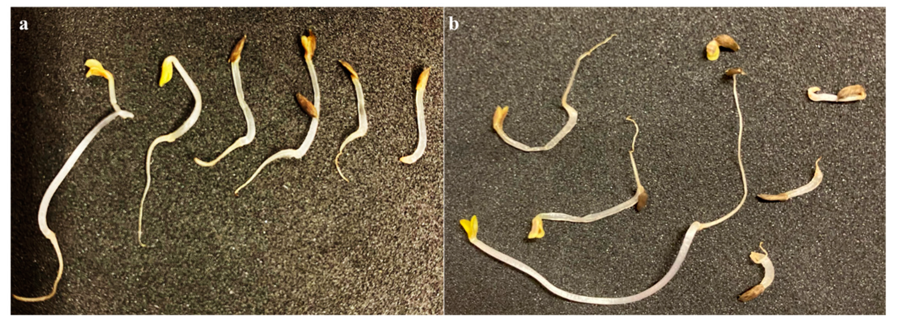
2.6. Phytotoxicity Assay

Due to the rapid and uniform germination, Lactuca sativa seeds were used as standard assays [29]. Before inoculation, the seeds were cleaned, and the surface was sterilized using 99% ethanol solution for 5 min and then washed several times using sterilized distilled water. The assays were performed on sterile Petri dishes (Ø 90 mm) covered with qualitative filter paper (Unifil®, Curitiba, Brazil, 80 g m−2). In each plate, 3 mL of the solution to be tested were added, and 10 seeds were equally spaced on the filter paper. Tap water (TW) was a positive control [30]. The Petri dishes were sealed and incubated (TECNAL TE-371, type BOD, Piracicaba, Brazil) at 30 °C. The germination and growth rates were analyzed daily for 7 days. The experiment was carried out in duplicate.

2.7. Detection of Azo Dye

The azo dye was detected using an HPLC coupled to mass spectrometry detection (HPLC–MS) equipped via a C18 column (Shimpack XR-ODS 50 x 2.0 mm I.D.). The samples were prepared via precipitation with BaCl2 followed by filtration. The samples were eluted at a flow rate of 0.05 L.min−1 and monitored at 370 nm. The eluents A (ultrapure water containing 1% formic acid) and B (methanol) served as mobile phases in an isocratic mode (30% A and 70% B). Nitrogen was used as the nebulizing gas (1.50 L.min−1), heated sheath gas, and drying gas (3 L.min−1, 250 °C).

2.8. Enzymatic Azo Dye Degradation

Isolated colonies of each bacterial culture were inoculated (needle) in Petri dishes containing BHI agar and RR-141 (60 mg L−1) and then incubated at 30 °C for 24 h. The differences in the halo formation diameters were calculated considering the total and CFU diameter differences. The diameters resulted from two perpendicular axis measurement averages [31].

3. Results and Discussion

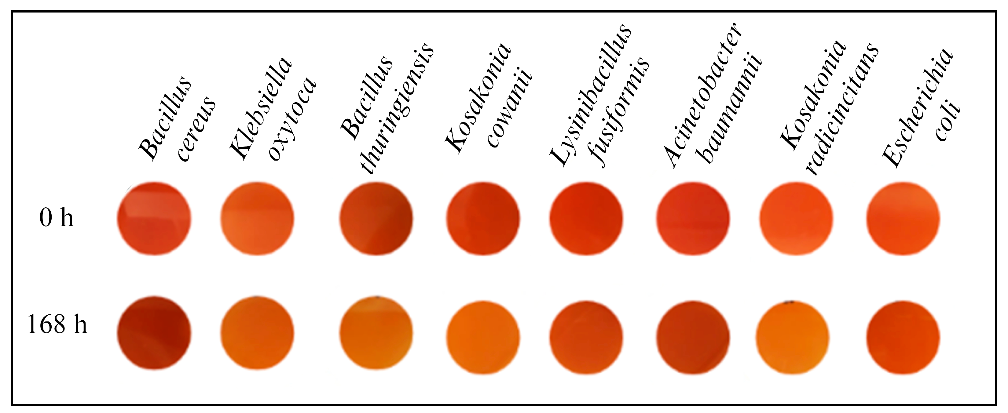
3.1. Screening of Dye Decolourization in Solid and Liquid Mediums

In biological processes, the bioavailability of enzymes interferes directly with the dye transformation, which can be performed extracellularly and intracellularly. Nevertheless, the most effective strategy involves extracellular degradation [32]. Since azo dyes have complex structures, their diffusion through cell membranes is hampered. Therefore, the assays performed in the solid medium are strictly related to degradation by extracellular enzymes. The visual analysis of Petri plates (Figure 1) indicated that K. oxytoca, B. thuringiensis, K. cowanii, and K. radicincitans colonies have the highest potential for decolorizing RR-141 (60 mg L−1) after 168 h of incubation at 30 °C. Moreover, the decolorization occurred primarily within 48 h of incubation.
A similar trend was observed by Kiayi et al. (2019) [28], where the solid-plate test promoted total decolorization of carmoisine (50 mg L−1) within 4 days by S. cerevisiae colonies, with no visual changes in the fifth and sixth days.
Biofilm-producing bacteria is an important factor since biofilm is an excellent means to retain micro-organisms and improve their performance in environmental biotechnologies [33,34]. Proteins and carbohydrates from EPS allow binds between the microbial biomasses and substrates, favoring their activities [33,34]. In this context, it is worth noting that the diffusion effects are essential to reach the high yields of biodegradability. Considering SEM micrographs of activated sludge (Appendix A, Figure A1), no biofilm was produced as an alternative to enhance the degradation process; the dye diffusion to the micro-organism is hampered in this experiment.
For the tests in a liquid medium, BHI and MSG media aimed to elucidate the optimal pathway for strain performance. This was achieved by evaluating their behavior in a nutrient-rich and opaque medium (BHI) and a less enriched and more translucid medium (MSG).
The analysis of the results confirmed B. thuringiensis (Figure 2) as the most promising dye-degrading species. It also indicated K. radicincitans, B. cereus, and A. baumannii as potential strains for RR-141 degradation.
Hence, building upon the outcomes of the solid and liquid mediums and considering the frequency of reports associated with each species (as depicted in Table 1), B. thuringiensis and K. radicincitans were chosen as candidates for subsequent investigations.

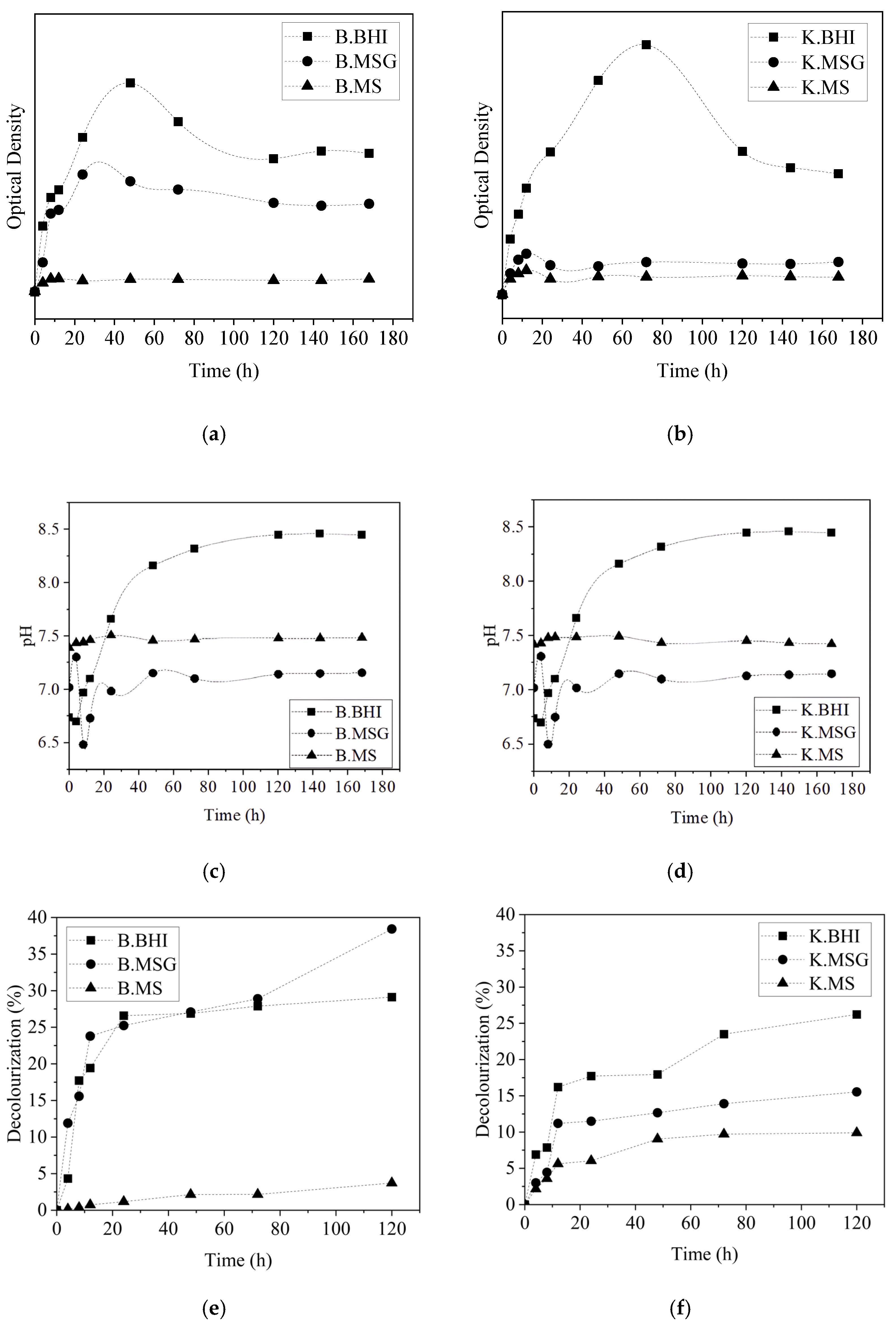
3.2. Effect of Carbon Sources and Dye-Decolorizing Kinetics by B. thuringiensis or K. radicincitans

The potential correlation between the carbon source provided by the culture medium and its impact on bacterial dye decolorization capacity was explored. To investigate this, the decolorization of the dye was examined while concurrently monitoring microbial growth and pH variation (Figure 3). This investigation was conducted using three distinct culture media (BHI, MS, and MSG), each within the context of the two chosen bacterial strains, B. thuringiensis and K. radicincitans.
Bacterial growth was more significant in the BHI medium, followed by MSG, for both strains (Figure 3a,b). No substantial changes in the optical density were identified in the MS medium (absence of carbon source, except azo dye RR-141), which could indicate the lack of ability of the strains to use the dye as a primary carbon source (Figure 4). Most micro-organisms are not capable of utilizing dyes as a carbon source for growth and require a carbohydrate source, such as peptone (present in BHI medium) or glucose (present in the MSG medium) [35].
In the system with the BHI medium, a more pronounced alkalinization of the medium is observed over time than in the MSG system (Figure 3c,d). In the MS system, where no bacterial growth was observed, there were no significant changes in pH. The decolorization efficiency of the azo dye is significantly influenced by pH. This could be attributed to the transportation of dye molecules across the cell membrane, a step recognized as the limiting factor in the decolorization process [35]. The optimal pH for decolorizing azo dyes varies according to the bacterial species but is often reported to be between 6 and 10 [35].
The decolorization of the azo dye RR-141 is depicted in Figure 3e,f. The B. thuringiensis strain performed better in MSG (38%), while K. radicincitans achieved the highest yield in the BHI medium (26%). This behavior could be linked to how these bacteria perform under specific conditions, including carbon and nutrient sources and pH levels.
The discoloration observed in this experiment may result from two combined or isolated mechanisms: (i) The process of biosorption and bioaccumulation of the azo dye by the bacterial biomass; (ii) The biodegradation of the azo dye through the bacteria’s metabolic/enzymatic machinery [36].
In the first case, the dye is removed from the liquid medium and stored inside the bacterial cell, but the azo dye does not undergo any chemical modification. This mechanism is especially interesting for applications in wastewater treatment processes contaminated by dyes when the objective is to reuse the dye, which can be separated from the effluent through the biosorption process.
On the other hand, in the biodegradation process, the azo dye is biodegraded into secondary metabolites or completely mineralized to H2O and CO2 [37]. The biodegradation process carried out by bacteria is generally an enzymatic process [36,38]. Enzymes such as azoreductaze, laccase, peroxidase, and phenoloxidase have been reported in the biodegradation processes of azo dyes [36,39,40,41,42]. Enzyme action comprehension and identification at each metabolism stage are essential to improve the degradation process and are indicated for further studies.

3.3. Enzymatic Azo Dye Degradation

The test in a solid medium was carried out to investigate the presence of enzymes in the azo dye decolorization process [43]. B. thuringiensis and K. radicincitans showed, after 24 h, a halo formation represented by a more translucid area surrounding the CFU that could be associated with extracellular enzyme production [31]. Considering the halo’s measurement (Table 2), K. radicincitans presented the best results, which indicate good development (Figure 4, OD graphic) and consequent enzymatic activity. In addition, the cell growth on dye-supplemented BHI agar medium with white colonies aspect can be associated with dye-decolorizing potential. Since cell mat coloring results from dye biosorption, maintaining the original mat color indicates biodegradation via an enzymatic process [44].
Oxidative and reductive enzymes are vital to azo dye biodegradation [45]. Azo dye remediation intermediated by enzymes can be intra or extracellular. However, the high complexity of azo dyes hampers their diffusion through cell membranes, so the preferable route is via enzyme release in the extracellular environment [46]. Microbial strains used in the decolorization process must have efficient enzymes and a transport system to permit the absorption of dyes in cells [45]. Enzymes improve the reductive cleavage of azo bonds, producing intermediate metabolites posteriorly degraded by aerobic or anaerobic mechanisms [47].
Azoreductases, laccases, and peroxidases are enzymes often related to azo dye discoloration. The action of azoreductase occurs through reduction mechanisms mediated by a flavoprotein in the microbial electron transport chain [48], which converts azo dyes into colorless products (aromatic amines) [47]. The laccases act either via direct or indirect oxidation [49] through an unspecific free radical mechanism that results in phenolic products and prevents the formation of aromatic amines [50]. The peroxidase mechanism comprises the oxidation of the phenolic group and the production of a radical close to the azo bonds [51].

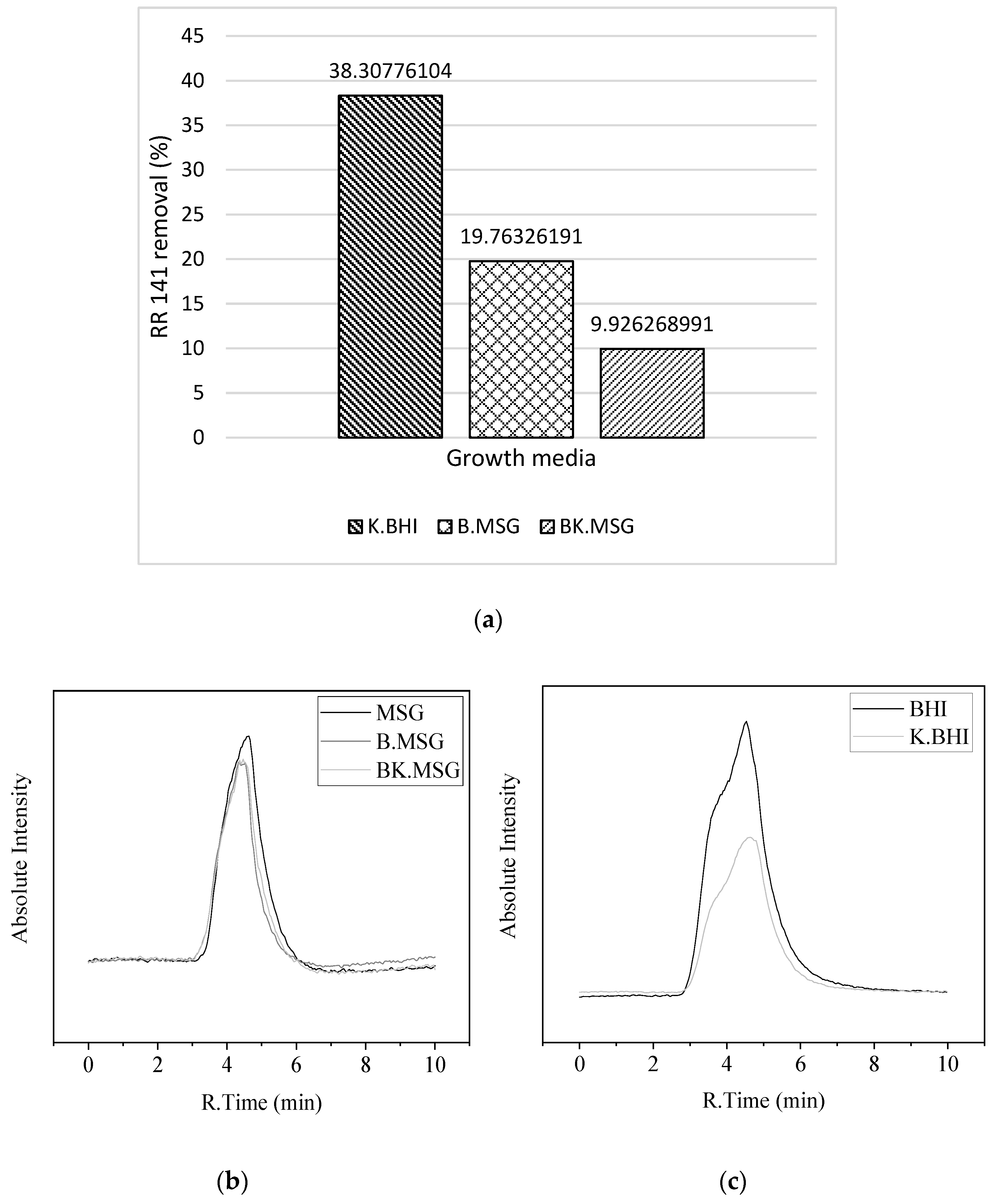
3.4. Dye-Decolorizing Kinetics by B. thuringiensis and K. radicincitans as a Consortium

Degradation of azo dyes is frequently accomplished by employing microbial consortia. The cooperative metabolic interactions within these microbial communities contribute to a more extensive biodegradation and mineralization process. In this study, the consortium evaluation revealed a good development in both media, with MSG promoting better conditions for a higher decolorization yield (Figure 5). Compared to the isolated kinetic study, the decolorization process achieved better results in a minor period, with approximately 36% of decolorization yielded within 12 h, associated with a synergic effect.
Some microbial consortia had achieved yields and objectives that no individual strain could successfully reach in biodegradation studies [52,53,54]. Additionally, mixed culture evaluations are more similar to practical situations since aseptic conditions can add more cost and higher stability over environmental stress such as composition, pH, or temperature variations. The synergistic action can occur via different pathways: (i) A micro-organism causes dye biotransformation, rendering it more reachable to another organism that would not be able to act on this dye in its original state; (ii) Only the micro-organism promotes some solution decolorization via the modification of the chromophore. However, the complete degradation is not achieved, and the metabolic products may have a toxic nature, as in an anaerobic reduction of azo dyes. Once another micro-organism takes this unwished metabolite as a nutrient source, the complete degradation, leading to carbon dioxide, ammonia, and water, can be achieved (only by mixed populations). This mineralization reaction ensures that no potentially harmful degradation products are released into the environment [32].
It is not easy to reproduce and effectively interpret the results when using mixed cultures because it only provides a wide view of what is happening in the system; it is difficult to identify and quantify individual culture growth and it hampers the elucidation of the degradation mechanism [2]. For these reasons, the application and deep comprehension of color removal via single bacterial cultures are essential since they promote a more straightforward interpretation of experimental observations and reproducibility [55]. Comparing the two carbon sources tested, better discoloration was obtained in the MSG medium (40%) than in the BHI medium (18%). Studies on the decolorization of azo dyes have reported higher removal rates, but this removal may depend on the dye dosage [56]. Eslami et al. (2019) [56] showed a 98% removal of the RR198 dye by a bacterial consortium (Enterococcus faecalis and Klebsiella variicola) at concentrations of 10–25 mg L−1. Although, the removal efficiency was reduced at higher contractions (50, 75, and 100 mg L−1) (55.62%, 25.82%, and 15.42%, respectively). This indicates that the dye can be toxic to bacteria at higher concentrations and reduce the decolorization efficiency. B. thuringiensis and K. radicincitans may perform better in decolorizing R141 at lower concentrations than those used in this study (30 mg L−1). This hypothesis can be investigated in future studies.

3.5. Biodegradation Analysis

HPLC-MS analysis was conducted to verify the reduction in dye quantity within both individual bacterial systems and the consortium. This assessment was conducted in the culture medium where the most significant discoloration occurred. Specifically, we examined the removal of dye by K. radicincitans in BHI medium (K.BHI) and by B. thuringiensis in MSG medium (B.MSG) and evaluated the removal of dye in MSG medium by the bacterial consortium (BK.MSG).
The chromatograms of dye solutions at the initial and after each studied media treatment were obtained (Figure 5). The HPLC profile of the RR-141 solution in both media exhibited a single peak at retention times of 4.53 and 4.63 min to BHI and MSG, respectively (matrix effect—chromatography). The assay results of the solution after treatment exposed peaks corresponding to RR 141 with reduced intensity. The highest dye removal was observed in the K.BHI system, reaching 38%, followed by the B.MSG system with 19%, and the BK.MSG bacterial consortium system achieved a 10% reduction (Figure 5a).
While UV-Vis analysis (Section 3.2 and Section 3.4) and HPLC-MS measurements revealed a decrease in dye concentration following the biological treatments, no new peaks were observed in the chromatograms. The emergence of new peaks in chromatograms after biological treatment typically signifies the formation of byproducts resulting from bacterial metabolism, a common occurrence during the biodegradation process of RR 141 [57,58,59]. The absence of new peaks in the chromatograms of treated solutions contributes to a possible adsorption strand via the biomass generated [60,61,62]. Once adsorption significantly influences the decolorization phenomena, another speculation can be made about the effect of culture media and biomass composition and adsorption.
Table 3 summarizes the percentages of decolorization and removal of the RR 141 dye, measured via UV-Vis spectroscopy and HPLC techniques. While both methods aim to comprehend the process of dye decolorization, their outcomes may not necessarily align or exhibit the same trend. The optimal decolorization condition for B. thuringiensis, identified through UV-Vis spectroscopy analysis, demonstrated a 38% reduction, whereas HPLC analysis only indicated a 19% decolorization. Likewise, the bacterial consortium exhibited an optimal 40% decolorization via UV-Vis spectroscopy, while HPLC analysis revealed a lower 10% decolorization. In contrast, for K. radicincitans, HPLC analysis indicated a higher optimal decolorization (38%) than UV-Vis analysis (26%).
The decrease in UV-Vis spectroscopy absorbance is commonly misconstrued as dye biodegradation, primarily indicating decolorization rather than the actual degradation of the dye molecule [63]. UV-Vis spectroscopy, relying on a single wavelength, may misinterpret dye concentration in solutions due to factors like intermediate formation, overall solution changes, and pH-dependent dye peaks [63]. Because of this, UV-Vis responses cannot be directly compared with responses from techniques such as HPLC; they must complement each other. Therefore, UV-Vis analysis can be used to assess discoloration and HPLC to investigate the mechanisms of discoloration and the removal percentage [64].

3.6. Phytotoxicity Assay

A phytotoxicity study was carried out to evaluate the toxic or non-toxic nature of the dye. The analysis of phytotoxicity (Figure 6) revealed the toxic nature of Reactive Red 141 to the Lactuca sativa seeds. The germination rate was lower with Reactive Red 141 (60%, i.e., six of ten seeds germinated, on average) compared to tap water (80%, i.e., eight of ten seeds germinated, on average).
These findings align with previous studies highlighting the phytotoxic of azo dyes. An investigation by Sompark et al. (2021) revealed that the germination rate of mung bean seeds exposed to RR 141 at a concentration of 0.5 g/L was 62.50%, in contrast to the 100% germination observed when the seeds were subjected solely to distilled water treatment [65].
RR 141 was assessed for its impact on the germination of beetroot, cabbage, and tomato seeds, resulting in notably reduced germination rates of 36.7%, 40.0%, and 33.3%, respectively [59]. In contrast, when these identical seed varieties were germinated in a control group with pure water, a significantly higher germination rate exceeding 85% was observed [59].
The Reactive Red 141 is significantly inhibitory for the plants’ germination, indicating the presence of phytotoxic compounds. Azo dyes and some metabolites present carcinogenic, toxic, and mutagenic properties to the environment and humankind [66]. Bacterial azo dye degradation comprises the cleavage of azo bonds and the intermediates breakdown. However, three mechanism routes promote the carcinogenic activation of azo dyes: (i) Direct oxidation of the azo linkage to diazonium salts with highly reactive electrophilic behavior; (ii) Oxidation of azo dyes in the presence of structures formed by free aromatic amine groups; (iii) Reduction and cleavage of the azo bond with consequent formation of aromatic amines [67,68].

4. Conclusions

This study explores the potential of various bacteria for azo dye remediation in industrial wastewater. Bacillus thuringiensis and Kosakonia radicincitans show promise in decolorizing the azo dye RR141, achieving around 38% and 26% decolorization in 120 h, respectively. When used together as a bacterial consortium, they remove 36% of the dye in 12 h. The dye removal analyses conducted via HPLC-MS suggest that the primary removal mechanisms are associated with the biosorption of the dye into the bacterial biomass rather than its biodegradation. The study also investigates the impact of carbon sources and pH on decolorization, emphasizing the importance of nutrient-rich media. It suggests that microbial consortia can lead to more efficient decolorization but underscores the need to study individual bacterial cultures to understand their capabilities and mechanisms. Overall, the research offers valuable insights into bioremediation, highlighting the potential of specific bacterial strains and microbial consortia for addressing dye pollution in industrial wastewater. Further studies in this area could lead to more efficient and eco-friendly solutions.

Author Contributions

Conceptualization, G.M.D.d.V. and C.J.d.A.; methodology, G.M.D.d.V. and C.J.d.A.; formal analysis, G.M.D.d.V. and L.M.d.A.; investigation, G.M.D.d.V.; Vasconcelos, I.K.D.-F. and L.M.d.A.; writing—original draft preparation, G.M.D.d.V. and I.K.D.-F.; writing—review and editing, C.J.d.A., D.d.O., S.M.d.A.G.U.d.S., A.A.U.d.S. and M.K.; supervision, C.J.d.A. All authors have read and agreed to the published version of the manuscript.

Funding

This research was funded by Conselho Nacional de Desenvolvimento Científico e Tecnológico (CNPq 133874/2019-2) and by Fundação de Amparo a Pesquisa e Inovação do Estado de Santa Catarina (FAPESC).

Institutional Review Board Statement

Not applicable.

Informed Consent Statement

Not applicable.

Data Availability Statement

Data are contained within the article.

Conflicts of Interest

The authors declare no conflicts of interest. The funders had no role in the study’s design; in the collection, analyses, or interpretation of data; in the writing of the manuscript; or in the decision to publish the results.

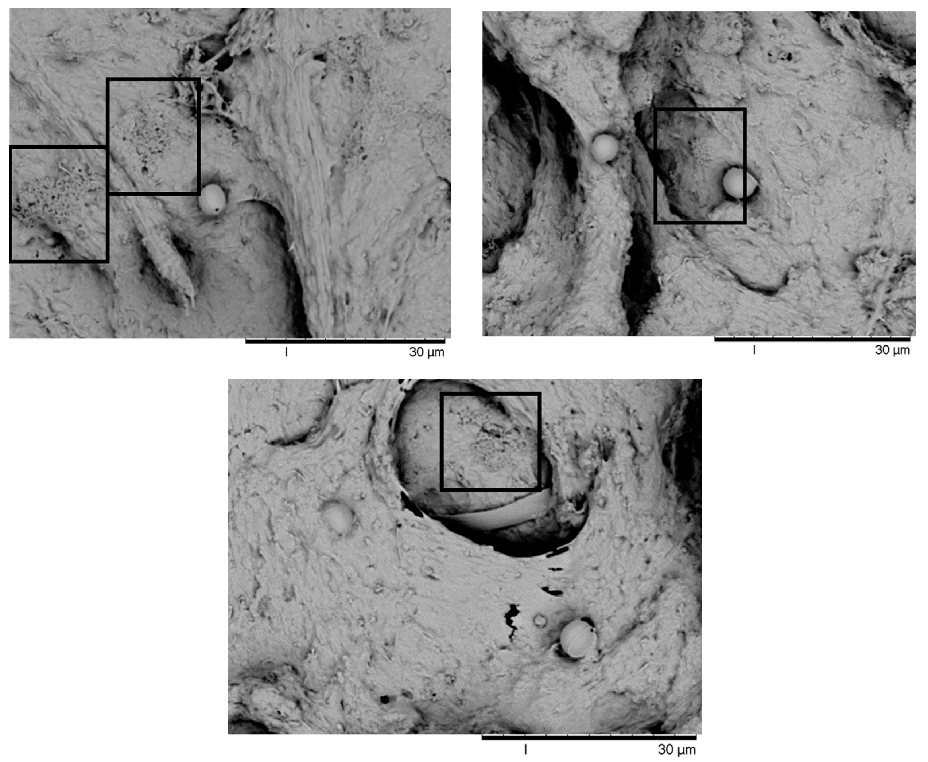
Appendix A. Scanning Electron Microscope (SEM)

The analysis of SEM micrographs of activated sludge indicated the presence of a biofilm matrix, very likely to beflocculating bacteria. Some essential characteristics are related to the structure and function of biofilm, for example, organic compounds such as extracellular polymeric substances (EPS), which play a significant role in modifying the surface (charge and hydrophobicity) to give suitable conditions for bacterial connection [21,22,23]. Large amounts of EPS and different bacterial species can be found in biofilms [24]. EPS is defined as a complex combination of high-molecular-weight microbial biopolymers. Its composition is based on humic substances, lipids, polysaccharides, proteins, and uranic acids. Its liquid anionic composition enables the effective sequestration of positively charged species, such as some dyes [25]. There are two primary forms of EPS: as a capsule covalently bounded to the cell surface or as slime polysaccharides roughly associated with the cell surface, as detected in Figure A1.
Figure A1. Scanning electron microscope of the activated sludge—biofilm formation.
Eng 05 00008 g0a1

References

  1. Grand View Research Textile Market Size, Share & Trends Analysis Report by Raw Material (Wool, Chemical, Silk), by Product (Natural Fibers, Polyester), by Application (Household, Technical), by Region, and Segment Forecasts, 2021–2028; Grand View Research: San Francisco, CA, USA, 2021.
  2. de Vasconcelos, G.M.D.; Mulinari, J.; Souza, S.M.d.A.G.U.d.; de Souza, A.A.U.; de Oliveira, D.; de Andrade, C.J. Biodegradation of Azo Dye-Containing Wastewater by Activated Sludge: A Critical Review. World J. Microbiol. Biotechnol. 2021, 37, 101. [Google Scholar] [CrossRef] [PubMed]
  3. Lade, H.S.; Waghmode, T.R.; Kadam, A.A.; Govindwar, S.P. Enhanced Biodegradation and Detoxification of Disperse Azo Dye Rubine GFL and Textile Industry Effluent by Defined Fungal-Bacterial Consortium. Int. Biodeterior. Biodegrad. 2012, 72, 94–107. [Google Scholar] [CrossRef]
  4. Bilińska, L.; Gmurek, M.; Ledakowicz, S. Comparison between Industrial and Simulated Textile Wastewater Treatment by AOPs—Biodegradability, Toxicity and Cost Assessment. Chem. Eng. J. 2016, 306, 550–559. [Google Scholar] [CrossRef]
  5. Paździor, K.; Bilińska, L.; Ledakowicz, S.; Pa, K.; Bili, L.; Paździor, K.; Bilińska, L.; Ledakowicz, S. A Review of the Existing and Emerging Technologies in the Combination of AOPs and Biological Processes in Industrial Textile Wastewater Treatment. Chem. Eng. J. 2019, 376. [Google Scholar] [CrossRef]
  6. Paździor, K.; Wrębiak, J.; Klepacz-Smółka, A.; Gmurek, M.; Bilińska, L.; Kos, L.; Sójka-Ledakowicz, J.; Ledakowicz, S. Influence of Ozonation and Biodegradation on Toxicity of Industrial Textile Wastewater. J. Environ. Manag. 2017, 195, 166–173. [Google Scholar] [CrossRef] [PubMed]
  7. Zhu, Y.; Xu, J.; Cao, X.; Cheng, Y.; Zhu, T. Characterization of Functional Microbial Communities Involved in Diazo Dyes Decolorization and Mineralization Stages. Int. Biodeterior. Biodegrad. 2018, 132, 166–177. [Google Scholar] [CrossRef]
  8. Rodrigues, F.K.; Salau, N.P.G.; Dotto, G.L. New Insights about Reactive Red 141 Adsorption onto Multi–Walled Carbon Nanotubes Using Statistical Physics Coupled with Van Der Waals Equation. Sep. Purif. Technol. 2019, 224, 290–294. [Google Scholar] [CrossRef]
  9. Manekar, P.; Patkar, G.; Aswale, P.; Mahure, M.; Nandy, T. Detoxifying of High Strength Textile Effluent through Chemical and Bio-Oxidation Processes. Bioresour. Technol. 2014, 157, 44–51. [Google Scholar] [CrossRef]
  10. Zhang, B.; Xu, X.; Zhu, L. Activated Sludge Bacterial Communities of Typical Wastewater Treatment Plants: Distinct Genera Identification and Metabolic Potential Differential Analysis. AMB Express 2018, 8, 184. [Google Scholar] [CrossRef]
  11. Li, W.; Mu, B.; Yang, Y. Feasibility of Industrial-Scale Treatment of Dye Wastewater via Bio-Adsorption Technology. Bioresour. Technol. 2019, 277, 157–170. [Google Scholar] [CrossRef]
  12. Meerbergen, K.; Willems, K.A.; Dewil, R.; Van Impe, J.; Appels, L.; Lievens, B. Isolation and Screening of Bacterial Isolates from Wastewater Treatment Plants to Decolorize Azo Dyes. J. Biosci. Bioeng. 2018, 125, 448–456. [Google Scholar] [CrossRef] [PubMed]
  13. Ashfaq, M.Y.; Da’na, D.A.; Al-Ghouti, M.A. Application of MALDI-TOF MS for Identification of Environmental Bacteria: A Review. J. Environ. Manag. 2022, 305, 114359. [Google Scholar] [CrossRef] [PubMed]
  14. Avanzi, I.R.; Gracioso, L.H.; Baltazar, M.d.P.G.; Karolski, B.; Perpetuo, E.A.; Nascimento, C.A.O.D. Rapid Bacteria Identification from Environmental Mining Samples Using MALDI-TOF MS Analysis. Environ. Sci. Pollut. Res. 2017, 24, 3717–3726. [Google Scholar] [CrossRef] [PubMed]
  15. Abdel Samad, R.; Al Disi, Z.; Mohammad Ashfaq, M.Y.; Wahib, S.M.; Zouari, N. The Use of Principle Component Analysis and MALDI-TOF MS for the Differentiation of Mineral Forming: Virgibacillus and Bacillus Species Isolated from Sabkhas. RSC Adv. 2020, 10, 14606–14616. [Google Scholar] [CrossRef] [PubMed]
  16. Marklein, G.; Josten, M.; Klanke, U.; Müller, E.; Horré, R.; Maier, T.; Wenzel, T.; Kostrzewa, M.; Bierbaum, G.; Hoerauf, A.; et al. Matrix-Assisted Laser Desorption Ionization-Time of Flight Mass Spectrometry for Fast and Reliable Identification of Clinical Yeast Isolates. J. Clin. Microbiol. 2009, 47, 2912–2917. [Google Scholar] [CrossRef]
  17. Alves, L.A.C.; Souza, R.C.; da Silva, T.M.C.; Watanabe, A.; Dias, M.; Mendes, M.A.; Ciamponi, A.L. Identification of Microorganisms in Biofluids of Individuals with Periodontitis and Chronic Kidney Disease Using Matrix-Assisted Laser Desorption/Ionization Time-of-Flight Mass Spectrometry. Rapid Commun. Mass. Spectrom. 2016, 30, 1228–1232. [Google Scholar] [CrossRef]
  18. Han, H.W.; Chang, H.C.; Hunag, A.H.; Chang, T.C. Optimization of the Score Cutoff Value for Routine Identification of Staphylococcus Species by Matrix-Assisted Laser Desorption Ionization-Time-of-Flight Mass Spectrometry. Diagn. Microbiol. Infect. Dis. 2015, 83, 349–354. [Google Scholar] [CrossRef]
  19. Normand, A.C.; Cassagne, C.; Gautier, M.; Becker, P.; Ranque, S.; Hendrickx, M.; Piarroux, R. Decision Criteria for MALDI-TOF MS-Based Identification of Filamentous Fungi Using Commercial and in-House Reference Databases. BMC Microbiol. 2017, 17, 1–17. [Google Scholar] [CrossRef]
  20. Sreedharan, V.; Saha, P.; Rao, K.V.B. Dye Degradation Potential of Acinetobacter Baumannii Strain VITVB against Commercial Azo Dyes. Bioremediation J. 2021, 25, 347–368. [Google Scholar] [CrossRef]
  21. Unnikrishnan, S.; Khan, M.H.; Ramalingam, K. Dye-Tolerant Marine Acinetobacter Baumannii-Mediated Biodegradation of Reactive Red. Water Sci. Eng. 2018, 11, 265–275. [Google Scholar] [CrossRef]
  22. Ameenudeen, S.; Unnikrishnan, S.; Ramalingam, K. Statistical Optimization for the Efficacious Degradation of Reactive Azo Dyes Using Acinetobacter Baumannii JC359. J. Environ. Manag. 2021, 279, 111512. [Google Scholar] [CrossRef] [PubMed]
  23. Adebajo, S.; Balogun, S.; Akintokun, A. Decolourization of Vat Dyes by Bacterial Isolates Recovered from Local Textile Mills in Southwest, Nigeria. Microbiol. Res. J. Int. 2017, 18, 1–8. [Google Scholar] [CrossRef] [PubMed]
  24. Yu, L.; Cao, M.; Wang, P.; Wang, S.; Yue, Y.; Yuan, W.; Qiao, W.; Wang, F.; Song, X. Simultaneous Decolorization and Biohydrogen Production from Xylose by Klebsiella Oxytoca GS-4-08 in the Presence of Azo Dyes with Sulfonate and Carboxyl Groups. Appl. Environ. Microbiol. 2017, 83, 1–13. [Google Scholar] [CrossRef] [PubMed]
  25. Balraj, B.; Hussain, Z.; King, P. Experimental Study on Non Sporulating Escherichia Coli Bacteria in Removing Methylene Blue. Int. J. Pharma Bio Sci. 2016, 7, B629–B637. [Google Scholar]
  26. Hilda Josephine, S.; Sekar, A.S.S. A Comparative Study of Biodegradation of Textile Azo Dyes by Escherichia Coli and Pseudomonas Putida. Nat. Environ. Pollut. Technol. 2014, 13, 417–420. [Google Scholar]
  27. Haque, M.M.; Haque, M.A.; Mosharaf, M.K.; Marcus, P.K. Novel Bacterial Biofilm Consortia That Degrade and Detoxify the Carcinogenic Diazo Dye Congo Red. Arch. Microbiol. 2021, 203, 643–654. [Google Scholar] [CrossRef]
  28. Kiayi, Z.; Lotfabad, T.B.; Heidarinasab, A.; Shahcheraghi, F. Microbial Degradation of Azo Dye Carmoisine in Aqueous Medium Using Saccharomyces Cerevisiae ATCC 9763. J. Hazard. Mater. 2019, 373, 608–619. [Google Scholar] [CrossRef]
  29. dos Santos, F.E.; Carvalho, M.S.S.; Silveira, G.L.; Correa, F.F.; Cardoso, M.d.G.; Andrade-Vieira, L.F.; Vilela, L.R. Phytotoxicity and Cytogenotoxicity of Hydroalcoholic Extracts from Solanum Muricatum Ait. and Solanum Betaceum Cav. (Solanaceae) in the Plant Model Lactuca Sativa. Environ. Sci. Pollut. Res. 2019, 26, 27558–27568. [Google Scholar] [CrossRef]
  30. Peduto, T.A.G.; de Jesus, T.A.; Kohatsu, M.Y. Sensibilidade de Diferentes Sementes Em Ensaio de Fitotoxicidade. Rev. Bras. De Ciência Tecnol. E Inovação 2019, 4, 200. [Google Scholar] [CrossRef]
  31. Barros, F.F.C.; Simiqueli, A.P.R.; de Andrade, C.J.; Pastore, G.M. Production of Enzymes from Agroindustrial Wastes by Biosurfactant-Producing Strains of Bacillus Subtilis. Biotechnol. Res. Int. 2013, 2013, 103960. [Google Scholar] [CrossRef]
  32. Kandelbauer, A.; Guebitz, G.M. Bioremediation for the Decolorization of Textile Dyes-A Review. Environ. Chem. Green. Chem. Pollut. Ecosyst. 2005, 269–288. [Google Scholar] [CrossRef]
  33. Haque, M.M.; Haque, M.A.; Mosharaf, M.K.; Rahman, A.; Islam, M.S.; Nahar, K.; Molla, A.H. Enhanced Biofilm-Mediated Degradation of Carcinogenic and Mutagenic Azo Dye by Novel Bacteria Isolated from Tannery Wastewater. J. Environ. Chem. Eng. 2023, 11. [Google Scholar] [CrossRef]
  34. Haque, M.M.; Hossen, M.N.; Rahman, A.; Roy, J.; Talukder, M.R.; Ahmed, M.; Ahiduzzaman, M.; Haque, M.A. Decolorization, Degradation and Detoxification of Mutagenic Dye Methyl Orange by Novel Biofilm Producing Plant Growth-Promoting Rhizobacteria. Chemosphere 2024, 346. [Google Scholar] [CrossRef]
  35. Khan, R.; Bhawana, P.; Fulekar, M.H. Microbial Decolorization and Degradation of Synthetic Dyes: A Review. Rev. Environ. Sci. Biotechnol. 2013, 12, 75–97. [Google Scholar] [CrossRef]
  36. Kapoor, R.T.; Danish, M.; Singh, R.S.; Rafatullah, M.; Abdul, A.K. Exploiting Microbial Biomass in Treating Azo Dyes Contaminated Wastewater: Mechanism of Degradation and Factors Affecting Microbial Efficiency. J. Water Process Eng. 2021, 43, 102255. [Google Scholar] [CrossRef]
  37. Bal, G.; Thakur, A. Distinct Approaches of Removal of Dyes from Wastewater: A Review. In Proceedings of the Materials Today: Proceedings; Elsevier Ltd.: Amsterdam, The Netherlands, 2021; Volume 50, pp. 1575–1579. [Google Scholar]
  38. Saratale, G.; Kalme, S.; Bhosale, S.; Govindwar, S. Biodegradation of Kerosene by Aspergillus Ochraceus NCIM-1146. J. Basic. Microbiol. 2007, 47, 400–405. [Google Scholar] [CrossRef] [PubMed]
  39. Shah, B.; Jain, K.; Jiyani, H.; Mohan, V.; Madamwar, D. Microaerophilic Symmetric Reductive Cleavage of Reactive Azo Dye—Remazole Brilliant Violet 5R by Consortium VIE6: Community Synergism. Appl. Biochem. Biotechnol. 2016, 180, 1029–1042. [Google Scholar] [CrossRef]
  40. Mullai, P.; Yogeswari, M.K.; Vishali, S.; Tejas Namboodiri, M.M.; Gebrewold, B.D.; Rene, E.R.; Pakshirajan, K. Aerobic Treatment of Effluents From Textile Industry. In Current Developments in Biotechnology and Bioengineering; Elsevier: Amsterdam, The Netherlands, 2017; pp. 3–34. ISBN 9780444636768. [Google Scholar]
  41. Chandanshive, V.V.; Kadam, S.K.; Khandare, R.V.; Kurade, M.B.; Jeon, B.H.; Jadhav, J.P.; Govindwar, S.P. In Situ Phytoremediation of Dyes from Textile Wastewater Using Garden Ornamental Plants, Effect on Soil Quality and Plant Growth. Chemosphere 2018, 210, 968–976. [Google Scholar] [CrossRef]
  42. Chen, Y.; Feng, L.; Li, H.; Wang, Y.; Chen, G.; Zhang, Q. Biodegradation and Detoxification of Direct Black G Textile Dye by a Newly Isolated Thermophilic Microflora. Bioresour. Technol. 2018, 250, 650–657. [Google Scholar] [CrossRef]
  43. Zhang, C.; Chen, H.; Xue, G.; Liu, Y.; Chen, S.; Jia, C. A Critical Review of the Aniline Transformation Fate in Azo Dye Wastewater Treatment. J. Clean. Prod. 2021, 321. [Google Scholar] [CrossRef]
  44. Karim, M.E.; Dhar, K.; Hossain, M.T. Decolorization of Textile Reactive Dyes by Bacterial Monoculture and Consortium Screened from Textile Dyeing Effluent. J. Genet. Eng. Biotechnol. 2018, 16, 375–380. [Google Scholar] [CrossRef] [PubMed]
  45. Mahmood, S.; Khalid, A.; Arshad, M.; Mahmood, T.; Crowley, D.E. Detoxification of Azo Dyes by Bacterial Oxidoreductase Enzymes. Crit. Rev. Biotechnol. 2016, 36, 639–651. [Google Scholar] [CrossRef] [PubMed]
  46. Sari, I.P.; Simarani, K. Decolorization of Selected Azo Dye by Lysinibacillus Fusiformis W1B6: Biodegradation Optimization, Isotherm, and Kinetic Study Biosorption Mechanism. Adsorpt. Sci. Technol. 2019, 37, 492–508. [Google Scholar] [CrossRef]
  47. Pandey, A.; Singh, P.; Iyengar, L. Bacterial Decolorization and Degradation of Azo Dyes. Int. Biodeterior. Biodegrad. 2007, 59, 73–84. [Google Scholar] [CrossRef]
  48. Misal, S.A.; Gawai, K.R. Azoreductase: A Key Player of Xenobiotic Metabolism. Bioresour. Bioprocess. 2018, 5. [Google Scholar] [CrossRef]
  49. Khlifi, R.; Belbahri, L.; Woodward, S.; Ellouz, M.; Dhouib, A.; Sayadi, S.; Mechichi, T. Decolourization and Detoxification of Textile Industry Wastewater by the Laccase-Mediator System. J. Hazard. Mater. 2010, 175, 802–808. [Google Scholar] [CrossRef]
  50. Wong, Y.; Yu, J. Laccase-Catalyzed Decolorization of Synthetic Dyes. Water Res. 1999, 33, 3512–3520. [Google Scholar] [CrossRef]
  51. Chivukula, M.; Renganathan, V. Phenolic Azo Dye Oxidation by Laccase from Pyricularia Oryzae. Appl. Environ. Microbiol. 1995, 61, 4374–4377. [Google Scholar] [CrossRef]
  52. Neifar, M.; Sghaier, I.; Guembri, M.; Chouchane, H.; Mosbah, A.; Ouzari, H.I.; Jaouani, A.; Cherif, A. Recent Advances in Textile Wastewater Treatment Using Microbial Consortia. J. Text. Eng. Fash. Technol. 2019, 5, 134–146. [Google Scholar] [CrossRef]
  53. Shindhal, T.; Rakholiya, P.; Varjani, S.; Pandey, A.; Ngo, H.H.; Guo, W.; Ng, H.Y.; Taherzadeh, M.J. A Critical Review on Advances in the Practices and Perspectives for the Treatment of Dye Industry Wastewater. Bioengineered 2021, 12, 70–87. [Google Scholar] [CrossRef]
  54. Samuchiwal, S.; Gola, D.; Malik, A. Decolourization of Textile Effluent Using Native Microbial Consortium Enriched from Textile Industry Effluent. J. Hazard. Mater. 2021, 402, 123835. [Google Scholar] [CrossRef] [PubMed]
  55. Thakur, J.K.; Paul, S.; Dureja, P.; Annapurna, K.; Padaria, J.C.; Gopal, M. Degradation of Sulphonated Azo Dye Red HE7B by Bacillus Sp. and Elucidation of Degradative Pathways. Curr. Microbiol. 2014, 69, 183–191. [Google Scholar] [CrossRef] [PubMed]
  56. Eslami, H.; Shariatifar, A.; Rafiee, E.; Shiranian, M.; Salehi, F.; Hosseini, S.S.; Eslami, G.; Ghanbari, R.; Ebrahimi, A.A. Decolorization and Biodegradation of Reactive Red 198 Azo Dye by a New Enterococcus Faecalis–Klebsiella Variicola Bacterial Consortium Isolated from Textile Wastewater Sludge. World J. Microbiol. Biotechnol. 2019, 35. [Google Scholar] [CrossRef] [PubMed]
  57. Al-Tohamy, R.; Ali, S.S.; Xie, R.; Schagerl, M.; Khalil, M.A.; Sun, J. Decolorization of Reactive Azo Dye Using Novel Halotolerant Yeast Consortium HYC and Proposed Degradation Pathway. Ecotoxicol. Environ. Saf. 2023, 263. [Google Scholar] [CrossRef] [PubMed]
  58. Khandare, S.D.; Teotia, N.; Kumar, M.; Diyora, P.; Chaudhary, D.R. Biodegradation and Decolorization of Trypan Blue Azo Dye by Marine Bacteria Vibrio Sp. JM-17. Biocatal. Agric. Biotechnol. 2023, 51. [Google Scholar] [CrossRef]
  59. Tizazu, S.; Tesfaye, G.; Wang, A.; Guadie, A.; Andualem, B. Microbial Diversity, Transformation and Toxicity of Azo Dye Biodegradation Using Thermo-Alkaliphilic Microbial Consortia. Heliyon 2023, 9. [Google Scholar] [CrossRef] [PubMed]
  60. Pearce, C.I.; Lloyd, J.R.; Guthrie, J.T. The Removal of Colour from Textile Wastewater Using Whole Bacterial Cells: A Review. Dye. Pigment. 2003, 58, 179–196. [Google Scholar] [CrossRef]
  61. Rodrigues, C.S.D.; Madeira, L.M.; Boaventura, R.A.R. Synthetic Textile Dyeing Wastewater Treatment by Integration of Advanced Oxidation and Biological Processes—Performance Analysis with Costs Reduction. J. Environ. Chem. Eng. 2014, 2, 1027–1039. [Google Scholar] [CrossRef]
  62. Siddiqui, S.I.; Fatima, B.; Tara, N.; Rathi, G.; Chaudhry, S.A. Recent Advances in Remediation of Synthetic Dyes from Wastewaters Using Sustainable and Low-Cost Adsorbents; Elsevier Ltd.: Amsterdam, The Netherlands, 2018; ISBN 9780081024911. [Google Scholar]
  63. Foster, J.E.; Adamovsky, G.; Gucker, S.N.; Blankson, I.M. A Comparative Study of the Time-Resolved Decomposition of Methylene Blue Dye under the Action of a Nanosecond Repetitively Pulsed Dbd Plasma Jet Using Liquid Chromatography and Spectrophotometry. IEEE Trans. Plasma Sci. 2013, 41, 503–512. [Google Scholar] [CrossRef]
  64. Ali, H. Biodegradation of Synthetic Dyes—A Review. Water Air Soil. Pollut. 2010, 213, 251–273. [Google Scholar] [CrossRef]
  65. Sompark, C.; Singkhonrat, J.; Sakkayawong, N. Biotransformation of Reactive Red 141 by Paenibacillus Terrigena KKW2-005 and Examination of Product Toxicity. J. Microbiol. Biotechnol. 2021, 31, 967–977. [Google Scholar] [CrossRef] [PubMed]
  66. Kalme, S.; Ghodake, G.; Govindwar, S. Red HE7B Degradation Using Desulfonation by Pseudomonas Desmolyticum NCIM 2112. Int Biodeterior Biodegrad. 2007, 60, 327–333. [Google Scholar] [CrossRef]
  67. Chung, K.T. Mutagenicity and Carcinogenicity of Aromatic Amines Metabolically Produced from Azo Dyes. J. Environ. Sci. Health C Environ. Carcinog. Ecotoxicol. Rev. 2000, 18, 51–74. [Google Scholar] [CrossRef]
  68. Brown, M.A.; De Vito, S.C. Predicting Azo Dye Toxicity. Crit. Rev. Environ. Sci. Technol. 1993, 23, 249–324. [Google Scholar] [CrossRef]
Figure 1. Decolorization potential study in the solid medium at 0 h and 168 h.
Eng 05 00008 g001
Figure 2. Decolorization yields of each isolated culture in BHI and MSG media.
Eng 05 00008 g002
Figure 3. Temporal evolution of bacterial growth ((a) B. thuringiensis; (b) K. radicincitans), pH variation ((c) B. thuringiensis; (d) K. radicincitans), and RR-141 decolorization ((e) B. thuringiensis; (f) K. radicincitans) in three culture media (BHI, MSG, and MS).
Eng 05 00008 g003
Figure 4. Temporal evolution of bacterial growth (a), pH variation (b), and decolorization rate of the consortia system in BHI and MSG media over 120 h, with pictures of the initial and end states of both medium (c).
Eng 05 00008 g004
Figure 5. Removal percentage of RR 141 dye for the tests K.BHI 1, B.MSG 2, and BK.MSG 3 (a) along with their respective chromatograms (MSG (b) and BHI (c)). 1 K. radicincitans in BHI medium; 2 B. thuringiensis in MSG medium; 3 the consortium in MSG medium.
Eng 05 00008 g005
Figure 6. Phytotoxicity experiments: (a) In RR-141 solution; (b) Tap water.
Eng 05 00008 g006
Table 1. Micro-organisms identified in the biological sludge and its association studies with decolorization detected using different journal directories.
StrainScoreDOAJ *JSTOR *Science Direct *Scopus *Springer *Google Scholar *
Bacillus cereus2.160192733511,600
Klebsiella oxytoca1.74008157605
Bacillus thuringiensis2.21013121121190
Kosakonia cowanii1.960010111
Lysinibacillus fusiformis1.81004117198
Acinetobacter baumannii2.160010143676
Kosakonia radicincitans1.86000007
Escherichia coli1.757131829102817,200
* Journal directories.
Table 2. Measurements of the colony-forming unit and its halo for each bacteria species in the enzyme activity assay.
Bacillus thuringiensisKosakonia radicincitans
Øcolony (cm)0.250 ± 0.0560.549 ± 0.031
Øcolony + halo (cm)0.339 ± 0.0520.675 ± 0.051
Øhalo (cm)0.091 ± 0.0170.126 ± 0.031
Table 3. Percentages of decolorization (UV-Vis spectroscopy analysis) and removal (HPLC analysis) of RR 141 in liquid media.
Micro-OrganismsDecolorization Percentage of RR 141 DyeRemoval Percentage of RR 141 Dye
BHIMSGMSBHIMSG
B. thuringiensis29%38%4%-19%
K. radicincitans26%15%10%38%-
Consortium18%40%--10%
Disclaimer/Publisher’s Note: The statements, opinions and data contained in all publications are solely those of the individual author(s) and contributor(s) and not of MDPI and/or the editor(s). MDPI and/or the editor(s) disclaim responsibility for any injury to people or property resulting from any ideas, methods, instructions or products referred to in the content.

