Article

Minimally Invasive Subcutaneous Adipose Tissue Biopsy in a Nonhuman Primate Model: Approach and Outcomes

by
Cheyenna M. Espinoza
1,
Cole Myers
1,
Scott H. Oppler
1,
Laura Hocum Stone
1,
Davis Seelig
2,3,
Parthasarathy Rangarajan
1,4,
Sabarinathan Ramachandran
1,4 and
Melanie L. Graham
1,5,*
1
Department of Surgery, University of Minnesota, Minneapolis, MN 55455, USA
2
Department of Veterinary Clinical Sciences, University of Minnesota, St. Paul, MN 55108, USA
3
Department of Comparative Pathology Shared Resource, Masonic Cancer Center, University of Minnesota, Minneapolis, MN 55455, USA
4
Schulze Diabetes Institute, University of Minnesota, Minneapolis, MN 55455, USA
5
Department of Veterinary Population Medicine, University of Minnesota, St. Paul, MN 55108, USA
*
Author to whom correspondence should be addressed.
Surgeries 2025, 6(4), 106; https://doi.org/10.3390/surgeries6040106
Submission received: 25 October 2025 / Revised: 22 November 2025 / Accepted: 24 November 2025 / Published: 26 November 2025

Abstract

Background/Objectives: Adipose tissue (AT) plays significant roles in energy storage, metabolite signaling, and immunomodulation. The understanding of its underlying mechanisms of dysregulation can provide insight into complex disease processes through analysis with histology, flow cytometry, metabolomics, and proteomics. Tissue sampling in the clinical setting has largely shifted towards minimally invasive approaches to improve factors such as patient satisfaction, post-operative recovery, and procedure length. In contrast, preclinical animal models continue to rely on more invasive methods until refined, minimally invasive techniques are developed and systematically assessed. To improve animal welfare and enhance clinical translatability, there is a critical need to reverse translate these approaches into animal models. Methods: Our study evaluated the feasibility and performance of a commercially available vacuum-assisted biopsy (VAB) device for AT sampling in a preclinical nonhuman primate (NHP) model. Six rhesus NHPs successfully underwent three serial AT biopsies with a VAB device (n = 18). Results: All animals recovered without any serious or unexpected adverse events. The amount of adipose tissue collected per biopsy (0.5–2.7 g) was proportional to the number of individual tracks. Isolation of the stromal vascular fraction (SVF) from a subset of samples (n = 6) yielded 0.41 ± 0.12 × 10⁶ cells/g of tissue. Conclusions: The minimally invasive VAB technique is a safe and reliable method of AT collection in NHPs. This feasibility study demonstrated adequate volumes of tissue cores that are suitable for typical, downstream research applications including immunologic studies and pathology, while improving animal welfare.

1. Introduction

Adipose tissue (AT) dysfunction is central to many of the mechanisms of chronic inflammation in obesity as “unhealthy” AT phenotypes exacerbate the imbalance between immune cell regulation, cytokine secretion, and systemic inflammation [1,2,3,4]. In healthy individuals, AT acts as a lipid energy reservoir and its expansion occurs by adipogenesis from precursor cells with limited ectopic storage of fat outside of visceral and subcutaneous tissue deposits [4]. With chronic excess caloric exposure and subsequent development of obesity, mechanisms can shift from healthy adipogenesis to dysfunctional adipocyte hypertrophy and ectopic storage [4,5]. While AT dysfunction in obesity is apparent, there remain gaps in our current knowledge of how surgical and medical obesity interventions impact the existing dysfunction in the AT microenvironment. Metabolic surgery remains more effective in reducing obesity-related complications compared to lifestyle and intensive medical therapy alone, with benefits persisting even in the setting of weight regain [6,7,8,9,10]. New medical therapies targeting incretins, such as glucagon-like peptide-1 receptor agonists, have shown promise in combating the global epidemic of obesity [11,12]. As these treatments modulate systemic metabolic and inflammatory pathways, understanding the AT environment is increasingly important for characterizing treatment-induced changes in tissue structure, function, and immunology that may influence clinical outcomes [6,7,8,9,10,11,12,13].
AT contains several cell populations of mechanistic interest, including adipocytes and the stromal vascular fraction (SVF), a heterogenous population of immune, endothelial, perivascular, and adipocyte-derived stem cells involved in tissue remodeling and metabolic regulation [5,14]. SVF analysis has several clinical implications ranging from potential therapeutic applications in regenerative medicine to investigating the immune landscape of AT in diverse medical and surgical contexts [5]. In a preclinical model of vertical sleeve gastrectomy in obese nonhuman primates (NHPs), surgical intervention suppressed tissue resident macrophage populations within the SVF of visceral AT compared to sham-operated controls, indicating immune remodeling of the AT microenvironment [15]. Additionally, AT can provide further insights into disease processes through proteomic analysis with distinct proteomic signatures observed in the visceral and subcutaneous AT compartments during obesity. This suggests compartment-specific dysregulation in obesity [13].
AT sampling methods range from more invasive and open surgical techniques to less invasive, percutaneous approaches including fine needle aspiration (FNA), core needle biopsies (CNBs), vacuum-assisted biopsies (VABs), and liposuction retrieval. Choosing an appropriate sampling technique requires consideration of factors such as the intended downstream analysis, required tissue volume, cell viability and kinetics, procedure duration, post-operative recovery, cosmetic outcomes, operator expertise, and any concurrent procedures [16,17,18,19]. In clinical practice, these techniques are broadly used; for example, percutaneous methods have largely replaced open surgery in the diagnosis of breast and soft tissue masses [16,20,21]. Liposuction is commonly used in cosmetic procedures for body contouring and served as an opportunistic source of AT specific cell populations, such as AT-derived mesenchymal cells [17].
Open techniques for AT biopsy involve tissue collection through a surgical incision which yields larger samples, but typically requires closure with sutures [15,22]. These open approaches for soft tissue sampling are associated with higher rates of complications and longer recovery times compared to less invasive methods [19,23]. When concurrent surgical procedures are performed near the AT compartment of interest, the biopsy can be opportunistically obtained through the existing surgical incision, avoiding additional incisions [15,24]. In comparison, FNA is a minimally invasive technique performed under direct visualization or ultrasound guidance. It involves repeatedly inserting a needle into target tissue while aspirating cells with a syringe. The collected cells are deposited as a smear onto a slide for pathologic examination. Limitations of FNA include smaller sample sizes and loss of tissue architecture, limiting analysis to cytology [25]. CNB is similar to FNA but uses a spring-loaded cutting needle to obtain a core of tissue. This preserves tissue architecture while remaining less invasive than open biopsy [25,26]. Liposuction techniques utilize hollow cannulas combined with either mechanical or ultrasonic disruption to aspirate large volumes of liquid AT under vacuum [17].
VAB was developed to address some of the limitations encountered with large CNB of breast masses [27]. After needle insertion, a vacuum applies negative pressure to draw tissue into the biopsy channel before the cutting mechanism is deployed [27]. This technique overcomes the sample size limitations of both FNA and CNB, allowing for larger, more intact tissue samples to be obtained [21,26,28]. Many different commercial VAB devices are available varying in needle size, vacuum strength, and cutting mechanisms to suit specific tissue types and clinical applications, including EleVation™ (BD, Franklin Lakes, NJ, USA), ATEC® Sapphire™ (Hologic Inc., Bedford, MA, USA), EnCor® Enspire™ (BARD GmbH, Karlsruhe, Germany), Vacora™ (BARD GmbH, Karlsruhe, Germany), and Celero® (Hologic Inc., Bedford, MA, USA) [25]. However, the larger tissue samples obtained with VAB come with a slightly increased risk for bleeding when compared to FNA or CNB [26].
Traditionally, investigators have adopted open surgical techniques for AT biopsies in preclinical NHP research settings as well as in other animal models [2,15,29,30,31]. However, the choice of biopsy technique should be tailored to the goals of the study, with consideration of alternative minimally invasive approaches when appropriate. Minimally invasive biopsy techniques that yield modest but high-quality representative tissue samples are well suited for downstream histologic and “omics” analyses in preclinical research. By minimizing disruption to the local tissue environment, these techniques reduce potential confounding effects, enabling more accurate assessment of tissue biology while also improving procedure duration, recovery time, and pain control [20,23]. This approach enables serial sampling within the same individual, often not achievable with open surgical techniques due to greater tissue disruption and prolonged recovery. By allowing each individual to serve as its own control, serial sampling significantly reduces the number of animals needed, supporting the “reduction” principle of the 3Rs framework and enhancing the statistical power of preclinical studies. These advantages highlight the importance of validating minimally invasive biopsy methods in preclinical research. Moreover, prioritizing animal welfare not only aligns with clinical practices but also reinforces scientific rigor and strengthens the reliability of research outcomes.
This study aims to evaluate the feasibility and performance of a minimally invasive AT biopsy technique in a NHP model as a reliable method for obtaining AT suitable for downstream analysis. Specifically, we assess tissue yield, histological quality, the efficiency of immunocyte isolation, and incidence of procedure-related adverse events.

2. Materials and Methods

All animal procedures were approved by the University of Minnesota Institutional Animal Care and Use Committee (IACUC) and conducted in compliance with the Animal Welfare Act, adhered to the principles stated in the NIH Guide for Care and Use of Laboratory Animals [32], and were implemented and reported in compliance with the ARRIVE guidelines [33].

2.1. Animal Demographics

Six spontaneously obese male rhesus macaques underwent minimally invasive subcutaneous AT biopsies between October 2024 and June 2025 as a part of an unrelated obesity intervention study. Animals had an average age of 9.9 years and weight of 15.4 kg at the time of AT biopsy (Table 1). The three biopsies were performed sequentially, with 23 weeks between the first and second procedures and 8 weeks between the second and third.

2.2. Site Selection and Biopsy

Subcutaneous adipose tissue was obtained from the abdominal region. Biopsies were obtained using the BD EleVationTM Breast Biopsy Driver (EVDriver, Becton, Dickinson and Company, Franklin Lakes, NJ, USA) with the compatible 10G BD EleVationTM Probe (EV10, Becton, Dickinson and Company, Franklin Lakes, NJ, USA). This is an untethered, battery-powered vacuum-assisted system that operates cordless. It contains a collection cannister, allowing multiple core biopsies to be obtained during a single biopsy procedure without needing to retrieve each specimen separately.

2.3. Post Biopsy Care

Animal behavior and clinical status were evaluated at least twice daily. All animals were trained to cooperate with examination, blood collection, and general husbandry activities as part of the behavioral management program [34,35]. Animals were provided water ad libitum and fed a standardized diet of 2055c Envigo Harlan Teklad Nonhuman Primate diet and supplemented with additional food enrichment consisting of fresh fruits and vegetables. Room temperature was maintained at 20–26.7 °C, humidity was maintained at 30–70%, and lights were programmed to a 12 h-on, 12 h-off circadian light cycle with 30 min dawn/dusk intervals. Veterinary rounds and weights were performed weekly for routine evaluation, and full veterinary exams and bloodwork were performed at least annually. All animals participated in a behavioral management program that included enrichment activities, social interaction, and structured training sessions. This encourages species typical behaviors, engagement with routine laboratory and husbandry procedures, and provides a rotating variety of appetitive, sensory, cognitive, physical, and social enrichment opportunities. All animals enrolled in this study were offered equal access and time for exercise and identical enrichment activities.

2.4. Tissue Processing for Immunohistochemistry and Adipocyte Morphology

AT cores were fixed in 10% formalin and paraffin embedded for immunohistochemistry. H&E and IBA-1 staining was performed as previously described by our group [15]. For IBA-1 staining, we used polyclonal goat anti-IBA-1 antibody (polyclonal, 1:1000, catalog #ab107159, Abcam, Cambridge, MA, USA). IBA-1-stained slides were digitized at 40× using the Motic EasyScan Digital Slide Scanner and corresponding Motic EasyScan Application Version 1.2.2.184 (Motic, Richmond, BC, Canada). Representative images were captured using Aperio ImageScope viewing software 12.4.6 (Leica Biosystems Imaging, Vista, CA, USA). All tissue processing and analysis was completed with blinding to animal outcomes.

2.5. Tissue Processing for Stromal Vascular Fraction Component Analysis

Isolation of immunocytes from the SVF occurred as previously described by our group with minor modifications herein [15]. AT samples were placed in cRPMI medium and processed on the same day within 4 h following collection. AT was minced and digested in cRPMI with 1% collagenase A (Millipore Sigma Cat. #10103578001, Burlington, MA, USA) for 10–30 min at 37 °C depending on volume of tissue. Tissue was dissociated by manually straining through a 250 μm mesh cell strainer (Corning, Tewksbury, MA, USA) and then passed through a 100 μm filter (Corning, Tewksbury, MA, USA) via centrifugation. The resulting cell pellet was resuspended in cRPMI and then processed via Ficoll-Paque density gradient (Cytiva Cat. #45-001-750, Marlborough, MA, USA). The resulting cell layer and pellet were collected, washed, and treated with ACK lysis buffer to remove red blood cells, if present. The final cell suspension was assessed for yield using a Countess II automated cell counter (Thermofisher Scientific, Carlsbad, CA, USA).

2.6. Outcomes and Endpoints

The primary outcome was to assess the feasibility and reproducibility of a minimally invasive subcutaneous fat biopsy technique in NHPs. Procedural success was defined as the ability to obtain AT with minimal invasiveness, performed under sedation without general anesthesia, and without the need for surgical intervention. AT samples obtained at baseline, prior to any weight modification, were processed to evaluate the isolation of stromal vascular fraction suitable for flow cytometric analysis. While cell isolation and flow cytometry were conducted on samples obtained post-weight manipulation, these data are beyond the scope of the present methods-focused paper and are not reported here. Sample adequacy was determined based on visible tissue volume and successful cell isolation from pre-intervention biopsies. Procedure duration, measured from needle entry site creation to application of topical skin adhesive, was recorded to evaluate efficiency. Additionally, repeat sampling was performed to assess whether the technique could be used in longitudinal studies without compromising animal welfare or sample integrity. These endpoints supported the evaluation of this method as a practical and minimally invasive approach for AT sampling in translational research.

2.7. Statistical Analysis

Values are reported as mean ± standard error of the mean (SEM) unless otherwise specified. Statistical analysis was performed using Prism version 10.5.0 (GraphPad Software, San Diego, CA, USA). Secondary outcomes of pre-intervention biopsies were further assessed using linear regression to assess for relationships between study variables including (i) use of additional tracks on total AT mass obtained per biopsy, (ii) age and immunocyte yield, and (iii) animal weight and immunocyte yield. Regression results are reported as estimated slopes, 95% confidence intervals, and p-values. Significance level of 0.05 was utilized during the analysis.

3. Results

3.1. Site and Biopsy Approach

The biopsy needle entry site was lateral and inferior to the umbilicus as depicted in Figure 1 with the biopsy track intended to extend inferiorly and medially through the subcutaneous AT. The intended tracks did not cross the midline of the abdomen. This strategy allows for serial biopsies to be performed at distinct sites, on opposite sides of the abdomen, ensuring that each sample reflects the native tissue environment rather than healing or wound-related changes.

3.2. Positioning and Preparation

Animals were fasted to avoid regurgitation or aspiration during the procedure. From their home enclosure, animals presented their hind limb on cue which allowed for intramuscular (IM) administration of 12 mg/kg ketamine and 0.1 mg/kg midazolam for sedation. Following sedation, 0.01 mg/kg buprenorphine was administered IM for analgesia. They were positioned supine, and hair was clipped from the abdomen. A chlorhexidine based surgical scrub was then applied, and the abdomen draped with sterile towels.

3.3. Biopsy Technique

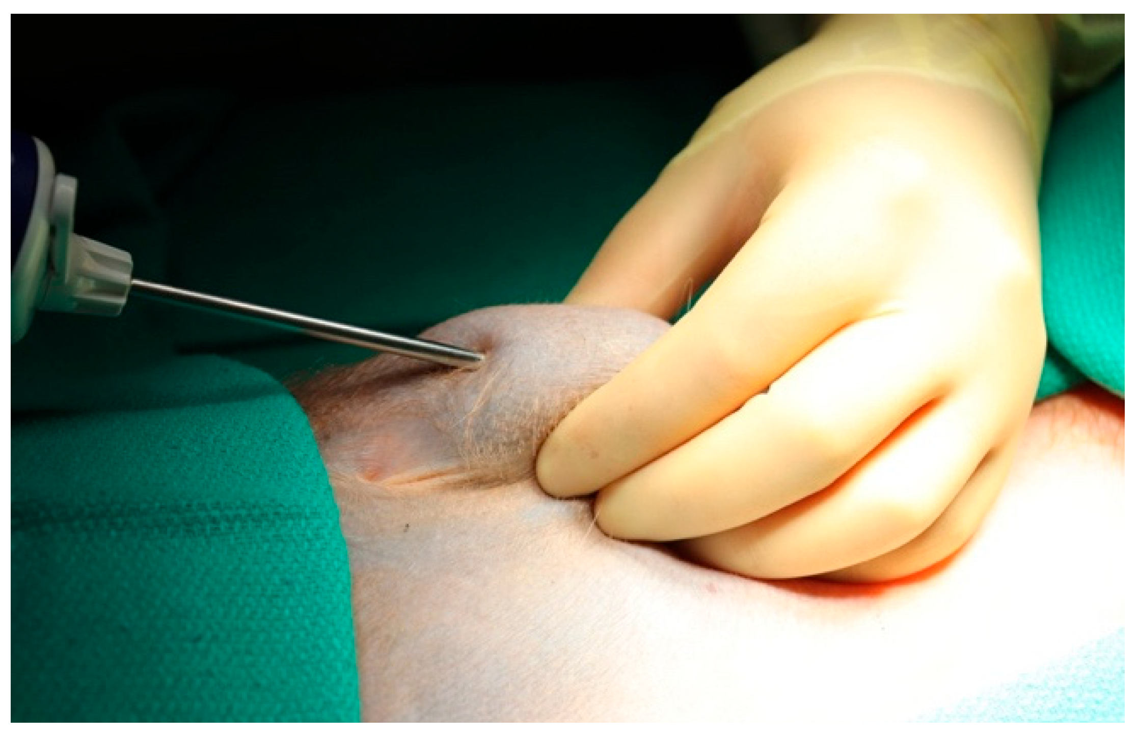
The procedure was performed with aseptic technique. The intended biopsy entry site and intended track were anesthetized with 1% lidocaine. A skin nick was made with an 18-gauge needle to prevent the dulling of the biopsy needle. Prior to insertion of the needle, the subcutaneous tissue was lifted off the abdominal fascia as depicted in Figure 2. The needle probe was then inserted with its cutting cannula facing upwards, taking care to leave adequate subcutaneous tissue both above and below the needle to account for the vacuum component of the biopsy system. This facilitated safe needle insertion without inadvertent injury to the abdominal fascia or skin.
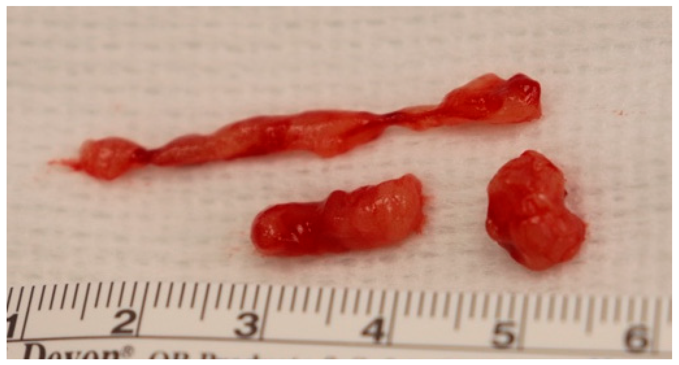
Once the needle was correctly positioned, the vacuum and cutting mechanism were deployed, and AT core was suctioned into the collection chamber. Within the same tract, the needle was then rotated approximately 70–90 degrees and another core was collected. This was repeated in a clockwise pattern until samples are taken at approximately 2′, 4′, 7′, and 10′ o’clock positions. The process was then repeated with the needle reinserted into a new track. The core samples were collected from the cannister for processing and storage. Figure 3 demonstrates a representative successful sample collected with the BD EleVationTM System.
After completion of tissue collection, manual pressure was held on the abdominal wall for a minimum of 5 min. The needle entry site was evaluated for hemostasis and then topical surgical glue was placed to close the needle insertion site.

3.4. Post-Operative Care

The animal was observed for recovery from sedation and directly monitored until they were alert, able to perch, and no longer ataxic. The abdominal site was evaluated twice daily as apart of routine care until ecchymosis resolved and the site was fully healed.

3.5. Special Considerations

(1)
When using the biopsy device, special attention should be made to the angle of the biopsy device. When positioned near compliant structures, such as skin, activation of the vacuum may inadvertently pull these tissues into the device, resulting unintended excision.
(2)
In regions of interest with limited fat, ultrasound guidance may be used to confirm appropriate needle placement, above the fascial plane and below the skin.
(3)
When applying manual pressure after the procedure, pressure should be maintained along the entire needle track and rather than only the entry site. Because AT is vascular, minor oozing may occur post-tissue collection; additional or repeated pressure may be applied as needed to achieve hemostasis.

3.6. Biopsy Results

This minimally invasive subcutaneous AT biopsy technique was successfully performed in 6/6 NHPs, each undergoing three consecutive AT biopsies. The primary endpoint was achieved in 100% of procedures (n = 18) with the successful acquisition of AT using this minimally invasive technique. All animals recovered uneventfully from sedation and resumed normal activities without restriction on the same day. Mean procedure duration was 11.4 ± 1.1 min, with a range of 3 to 23 min (Table 2). Peri-incisional ecchymosis, expected due to the high vascularity of AT, resolved on average of 7.5 ± 1.3 days post-operatively for all biopsies.
Three adverse events were reported across all eighteen procedures. One animal developed a subcutaneous hematoma post-operatively which was identified incidentally on a routine veterinary examination on post-operative day 15. The hematoma was monitored closely until resolution and did not require any further interventions. During two separate biopsy events, the skin was inadvertently drawn into the biopsy device by the vacuum and cut during the automated firing the VAB device due to technical error. One laceration required a single suture repair while the other was managed conservatively and the defect reapproximated with surgical glue. Resolution of ecchymosis for these two adverse events ranged within 4–8 days.
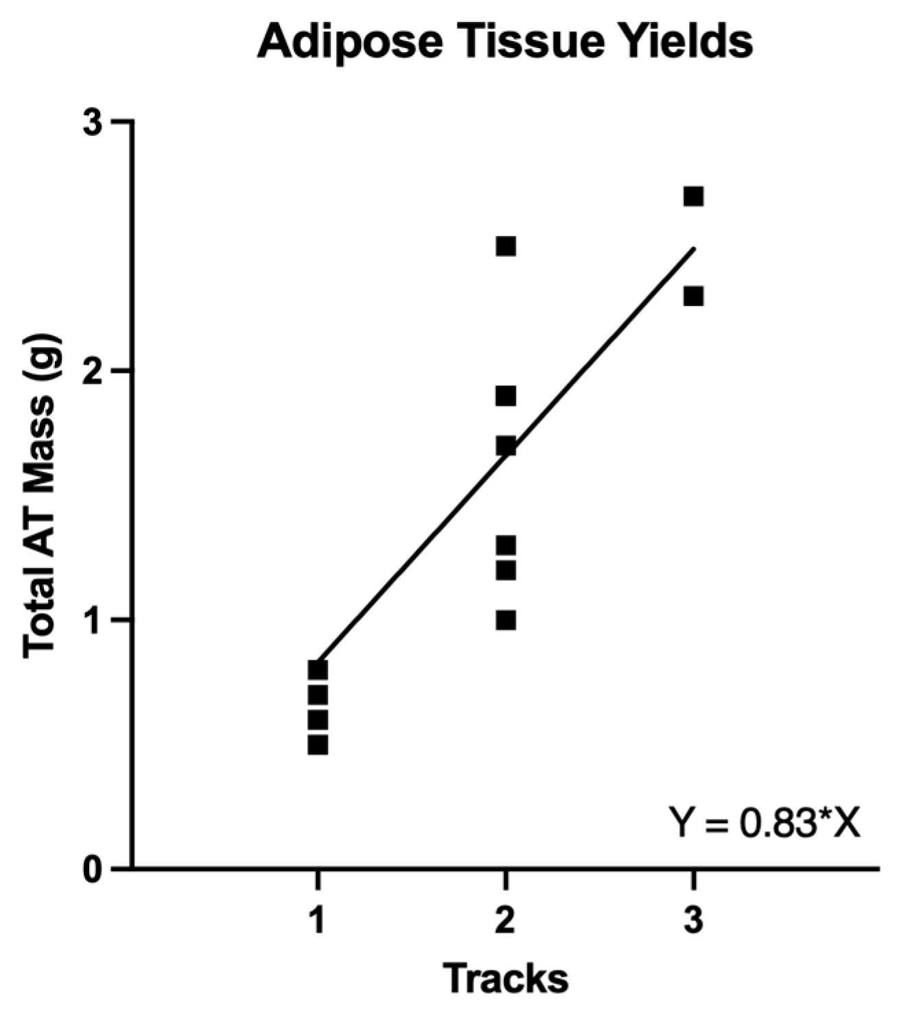
Sampling for each biopsy occurred along 1–3 separate needle tracks with four core biopsies collected per track. The total AT mass retrieved per sampling event ranged within 0.5–2.7 g, with an average total yield of 1.44 ± 0.2 g per biospy. Total AT mass was grouped based on the number of separate needle tracks used in each sampling event and is summarized in Table 3. The average mass obtained increased from 0.62 to 1.7 to 2.6 g of AT with the addition of an additional track. Incremental yields were assessed with simple linear regression as depicted in Figure 4, and each additional track used during the procedure increased the total yield by 0.83 grams (95% CI [0.74, 0.92], p < 0.001).
The pre-intervention biopsies (n = 6) underwent further processing as they were not subject to potential confounders from obesity interventions utilized in an unrelated study. The stromal vascular fraction was isolated from each AT sample in this cohort and immunocyte counts were obtained. Total immunocyte yield ranged within 0.47–1.83 × 10⁶ cells per each sampling event with an average of 0.41 ± 0.12 × 10⁶ cells/g of AT.
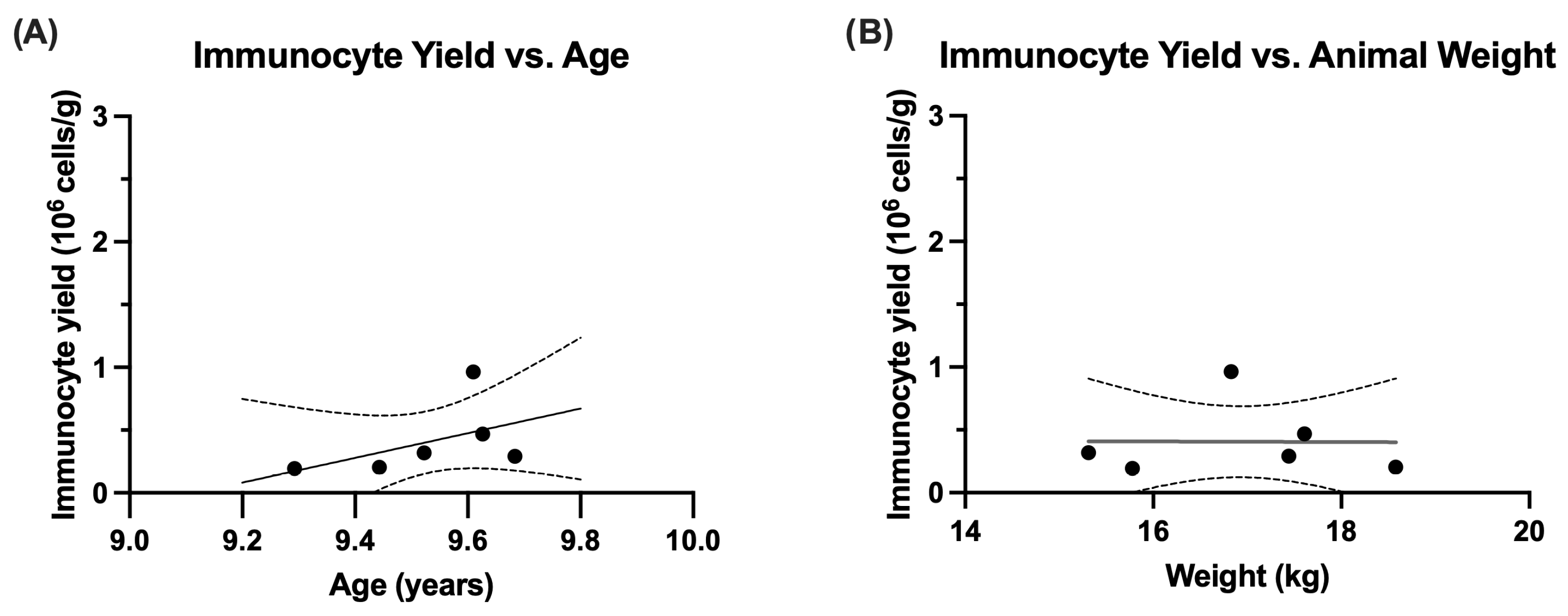
The relationships between immunocyte yield per gram of AT and demographics including age and body weight were further examined to investigate any potential confounding influences. Linear regressions were conducted on the pre-intervention biopsies (n = 6) to assess the relationship between immunocyte yield per AT gram and animal weight at the time of the biopsy (Figure 5A) and age (Figure 5B). No significant linear relationships were observed with animal weight (slope of −0.002; 95%CI [−0.334, 0.329], p = 0.986) or animal age (slope of 0.983; 95% CI [−1.465, 3.431], p = 0.327) in our small obese NHP cohort.
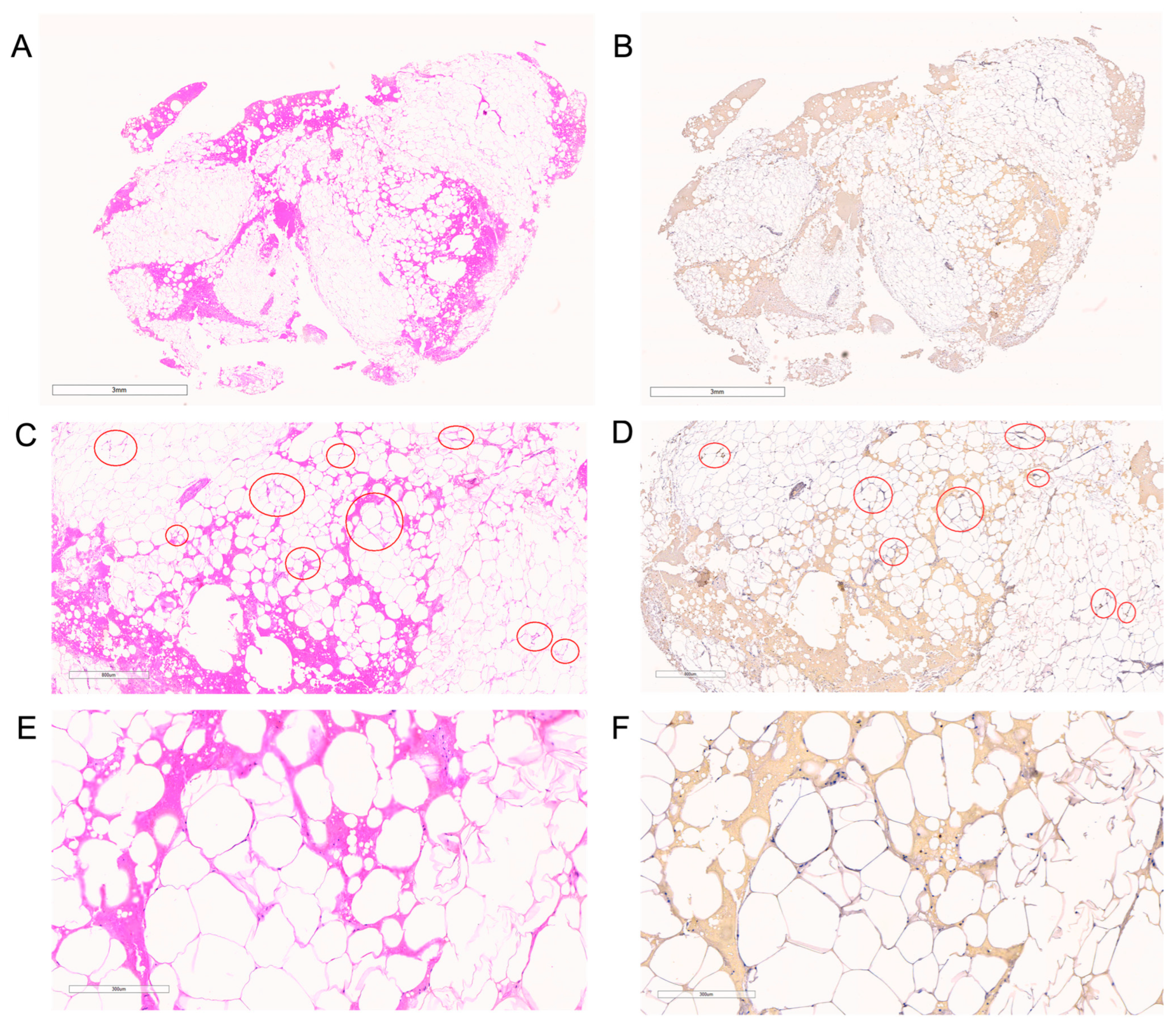
Formalin fixed samples were sent for histological examination including hematoxylin and eosin (H&E) for general architecture examination and ionized calcium-binding adaptor molecule 1 (IBA-1), a macrophage-specific marker. Figure 6 depicts a representative biopsy core obtained from the same animal with increasing magnitude of magnification stained with both H&E and IBA-1. Crown-like structures, a hallmark of obesity-related architecture changes, were widely distributed through our samples as depicted in Figure 6C,D and at a higher magnitude in Figure 6E,F.

4. Discussion

The AT microenvironment consists of multiple cell types, including mature adipocytes and components of the SVF which contains immune cells, endothelial cells, pericytes, and adipocyte-derived stem cells [5,14]. The diversity of cells contributes to its functions beyond energy storage, including cell signaling via adipokine and cytokine secretion, immunomodulation by adipose-derived stem cells, and regulation of immune cell phenotypes [5,36]. In obesity, dysregulation of AT plays a central role in the development of insulin resistance, leading to changes in tissue architecture and shifts in both immune cell populations and phenotypes [1,4,36,37,38,39]. The long-term consequences of the observed AT changes in obesity remain poorly understood and further research is needed to determine how various weight loss interventions may reverse or exacerbate these dysfunctional changes. Contemporary approaches to investigating immunomodulation include histologic and morphologic analysis, flow cytometry, metabolomics, transcriptomics, and proteomics [5,15,22,24,40]. Each of these analytical approaches requires specific considerations for tissue collection to ensure sample suitability, which in turn influences the choice of the various AT biopsy techniques. Key considerations include the quantity of tissue required, target cell population, necessity of preserving cell architecture, and efficacy of the isolation methods [18,41].
VAB systems, such as the BD EleVationTM Breast Biopsy Driver, have been used extensively for minimally invasive soft tissue sampling in multiple disciplines, including for the identification of breast pathologies in oncologic literature [25]. Adoption of these biopsy systems has widely replaced open surgical methods clinically as they produce large core biopsies that retain tissue architecture for histologic examination compared to other sampling methods like FNA [26]. Our study successfully reverse translates the use of a VAB system as a minimally invasive alternative for AT sampling method in a NHP model, balancing the need for sufficient tissue volumes for future analyses with the prioritization of animal welfare. Each gram of AT produced an average of 0.41 ± 0.12 × 10⁶ rhesus immune cells from the SVF. This was comparable to reported SVF isolation techniques in human populations, which have demonstrated isolation of 0.43–0.7 × 10⁶ cells/gram of AT from abdominal subcutaneous AT through either open resection or liposuction techniques [17,22]. Our technique provides sufficient volume of tissue for hypothesis-driven downstream analysis utilizing advanced technology such as flow cytometry, single cell whole transcriptome/T cell receptor analysis, and RNA sequencing [5,15,22,24,40,42]. Our study examined various potential influences on immunocyte yield, finding no significant association with age or weight. However, the modest cohort of obese NHPs exhibited limited variability in these factors, as age and weight were intentionally controlled by design to reduce confounding variables that could affect the interpretation of obesity-related outcomes. Further investigation is warranted to assess the generalizability to a larger and more heterogenous group of NHPs.
Research laboratories include personnel with diverse backgrounds and varying levels of surgical training. This VAB method for subcutaneous AT sampling requires minimal training and can be performed by individuals without extensive formal surgical training. The device enables multiple samples to be collected from a single needle insertion site by applying standardized negative pressure, preserving tissue architecture. Unlike open procedures, this minimally invasive technique eliminated the need for an incision, suture material for closure, or electrocautery. Additionally, real-time visualization of tissue collection through the device’s transparent cannister allows the operator to estimate the number of additional firings of the device needed to obtain the target volume of AT.
While there is an overall simplicity to using the VAB device, there are several technical considerations which should be considered when using in a preclinical model for AT sampling. The use in smaller animals with decreased volume of subcutaneous AT may pose an increased risk for inadvertent injuries to skin, so careful placement of the needle in the middle of the sample or use of a smaller gauge needle should be considered. Avoidance of positioning the opening of the biopsy channel on the BD Driver directly towards the skin in the 12 o’clock position and careful angling of the device when in operation can reduce this risk. In our cohort, we observed one instance of a post-operative hematoma detected during routine veterinary examination on post-operative day 15. The hematoma resolved spontaneously, did not affect the animal’s daily activities, and required no further clinical intervention. Monitoring for post-procedural bleeding should be routine in all AT biopsy methods given the highly vascularized nature of AT [5,41]. Open techniques of soft tissue sampling have increased risk of adverse bleeding events and often will use electrocautery to address sources of intra-procedure bleeding [15,23]. Our minimally invasive technique does not utilize cautery, and bleeding can be controlled successfully with manual pressure along the needle track after removing the biopsy device for a minimum of 5 mi. Importantly, the low complication rate observed in this study and the nature of the events are consistent with, and in some cases more favorable than, what is reported in clinical VAB experience.
Our findings demonstrate the VAB system can reliably obtain sufficient AT from NHPs for a range of downstream analyses including proteomics, metabolomics, transcriptomics, or flow cytometry. It should be noted that some minimally invasive biopsy techniques may alter the growth kinetics of SVF cells during culture and expansion [17]. While our study did not evaluate this effect, given our focus on non-expansion-based analyses, this consideration should inform biopsy method selection and warrants further investigation if cell expansion is planned.
Significant challenges remain in unraveling the complex mechanisms underlying AT function and its dysregulation across various disease states. Animal models play a crucial role in providing mechanistic insights into AT biology. Among these, NHP models are particularly valuable, as they help minimize confounding factors, such as variability in disease severity, comorbidities, medication use, diet, activity levels, recall bias, and noncompliance, which are difficult to control for in clinical investigations [43]. They closely model human anatomy and physiology and can be used to answer complex system-based questions. Their use in the fields of obesity, metabolism, transplantation, and immunology has led to critical scientific advancements [15,43]. The refinement of investigation techniques in preclinical models is critical to improving animal welfare [44]. Noninvasive methods of tissue sampling are used routinely in clinical settings and should be considered when appropriate, especially in instances where sedation is necessary or multiple consecutive examinations or procedures will be performed. Replacement of open surgical procedures with minimally invasive techniques has several advantages. They have been shown to have shorter procedural lengths, less sedation, improved pain control, and earlier return to normal activities [20,23]. Removal of unintended stress can also limit potential confounders of advanced assessments, which is critical when examining influences on immunometabolism and improves clinical translation to human studies [44,45].

5. Conclusions

Minimally invasive AT sampling in NHP preclinical models is both safe and effective. This approach provides sufficient tissue for investigating complex mechanisms of AT dysfunction across disease states while simultaneously enhancing animal welfare.

Author Contributions

Conceptualization: C.M.E., C.M., S.H.O., L.H.S., S.R. and M.L.G. Formal analysis: C.M.E., C.M., S.H.O. and M.L.G. Funding acquisition: S.R. and M.L.G. Investigation: C.M.E., C.M., S.H.O., L.H.S., P.R., D.S., S.R. and M.L.G. Methodology: C.M.E., C.M., S.H.O., L.H.S., S.R. and M.L.G. Project administration: M.L.G. Resources: S.R. and M.L.G. Supervision: D.S., S.R. and M.L.G. Validation: C.M.E., C.M., S.H.O., L.H.S., S.R. and M.L.G. Visualization: C.M.E., C.M., S.H.O. and L.H.S. Writing—original draft preparation: C.M.E. Writing—review and editing: C.M.E., C.M., S.H.O., L.H.S., P.R., D.S., S.R. and M.L.G. All authors have read and agreed to the published version of the manuscript.

Funding

This research was funded by University of Minnesota Institute for Diabetes, Obesity, and Metabolism Pilot & Feasibility Grant Program. This study was partially supported by the Goodale Chair Endowment, which provided key staffing and animal resources under the direction of Principal Investigator Dr. Melanie Graham at the University of Minnesota.

Institutional Review Board Statement

All animal use was approved by the University of Minnesota Institutional Animal Care and Use Committee (protocol number 2112-39642A, approved 23 February 2022 & protocol number 2407-42224A, approved 10 February 2024), in compliance with the Animal Welfare Act, and adhered to the principles stated in the Guide for the Care and Use of Laboratory Animals, 8th edition and the USDA Animal Care Blue Book.

Informed Consent Statement

Not applicable.

Data Availability Statement

The raw data supporting the conclusions of this article will be made available by the authors, without undue reservation.

Acknowledgments

The authors gratefully acknowledge the excellent and expert contribution of the University of Minnesota’s Preclinical Research Center team, especially Melanie N, Niewinski, Sierra D. Palmer, Sydney N. Phu, Mikayla Chavis, Paige Elyea, Laura Hickel, Jody Janecek, Lucas Mutch, Liam Keffer, Mellani Lubuag, Brenna Mulhollam, Tenzin Sopa, Jade Wilder, Westley Timmerman, Carley Hill, Samantha Troff, Noah Deutmeyer, Zi Xuan Wong, Natalie Fransen, and the students for husbandry and clinical care of our animals. Pathology images were captured using Aperio ImageScope viewing software 12.4.6 (Leica Biosystems Imaging, Vista, CA, USA). The graphical abstract (https://BioRender.com/v6wzw1m, accessed on 11 August 2025) and Figure 1 (https://BioRender.com/8fa5ycp, accessed on 11 August 2025) were created in BioRender.

Conflicts of Interest

The authors declare no conflicts of interest.

References

  1. Christ, A.; Lauterbach, M.; Latz, E. Western Diet and the Immune System: An Inflammatory Connection. Immunity 2019, 51, 794–811. [Google Scholar] [CrossRef] [PubMed]
  2. Cottam, M.A.; Caslin, H.L.; Winn, N.C.; Hasty, A.H. Multiomics reveals persistence of obesity-associated immune cell phenotypes in adipose tissue during weight loss and weight regain in mice. Nat. Commun. 2022, 13, 2950. [Google Scholar] [CrossRef]
  3. Lee, Y.-S.; Jun, H.-S. Anti-Inflammatory Effects of GLP-1-Based Therapies beyond Glucose Control. Mediat. Inflamm. 2016, 2016, 3094642. [Google Scholar] [CrossRef]
  4. Longo, M.; Zatterale, F.; Naderi, J.; Parrillo, L.; Formisano, P.; Raciti, G.A.; Beguinot, F.; Miele, C. Adipose Tissue Dysfunction as Determinant of Obesity-Associated Metabolic Complications. Int. J. Mol. Sci. 2019, 20, 2358. [Google Scholar] [CrossRef]
  5. Bradley, D.; Deng, T.; Shantaram, D.; Hsueh, W.A. Orchestration of the Adipose Tissue Immune Landscape by Adipocytes. Annu. Rev. Physiol. 2024, 86, 199–223. [Google Scholar] [CrossRef] [PubMed]
  6. Ikramuddin, S.; Korner, J.; Lee, W.J.; Thomas, A.J.; Connett, J.E.; Bantle, J.P.; Leslie, D.B.; Wang, Q.; Inabnet, W.B., 3rd; Jeffery, R.W.; et al. Lifestyle Intervention and Medical Management With vs Without Roux-en-Y Gastric Bypass and Control of Hemoglobin A1c, LDL Cholesterol, and Systolic Blood Pressure at 5 Years in the Diabetes Surgery Study. JAMA 2018, 319, 266–278. [Google Scholar] [CrossRef] [PubMed]
  7. Pipek, L.Z.; Moraes, W.A.F.; Nobetani, R.M.; Cortez, V.S.; Condi, A.S.; Taba, J.V.; Nascimento, R.F.V.; Suzuki, M.O.; do Nascimento, F.S.; de Mattos, V.C.; et al. Surgery is associated with better long-term outcomes than pharmacological treatment for obesity: A systematic review and meta-analysis. Sci. Rep. 2024, 14, 9521. [Google Scholar] [CrossRef]
  8. Adams, T.D.; Davidson, L.E.; Litwin, S.E.; Kim, J.; Kolotkin, R.L.; Nanjee, M.N.; Gutierrez, J.M.; Frogley, S.J.; Ibele, A.R.; Brinton, E.A.; et al. Weight and Metabolic Outcomes 12 Years after Gastric Bypass. N. Engl. J. Med. 2017, 377, 1143–1155. [Google Scholar] [CrossRef]
  9. Stenberg, E.; Cao, Y.; Ottosson, J.; Hedberg, S.; Naslund, E. Glycaemic and weight effects of metabolic surgery or semaglutide in diabetes dosage for patients with type 2 diabetes. Diabetes Obes. Metab. 2024, 26, 5812–5818. [Google Scholar] [CrossRef]
  10. Ghanem, O.M.; Abi Mosleh, K.; Kerbage, A.; Lu, L.; Hage, K.; Abu Dayyeh, B.K. Continued Diabetes Remission Despite Weight Recurrence: Gastric Bypass Long-Term Metabolic Benefit. J. Am. Coll. Surg. 2024, 238, 862–871. [Google Scholar] [CrossRef]
  11. Rubino, D.; Abrahamsson, N.; Davies, M.; Hesse, D.; Greenway, F.L.; Jensen, C.; Lingvay, I.; Mosenzon, O.; Rosenstock, J.; Rubio, M.A.; et al. Effect of Continued Weekly Subcutaneous Semaglutide vs Placebo on Weight Loss Maintenance in Adults With Overweight or Obesity: The STEP 4 Randomized Clinical Trial. JAMA 2021, 325, 1414–1425. [Google Scholar] [CrossRef]
  12. Wilding, J.P.H.; Batterham, R.L.; Davies, M.; Van Gaal, L.F.; Kandler, K.; Konakli, K.; Lingvay, I.; McGowan, B.M.; Oral, T.K.; Rosenstock, J.; et al. Weight regain and cardiometabolic effects after withdrawal of semaglutide: The STEP 1 trial extension. Diabetes Obes. Metab. 2022, 24, 1553–1564. [Google Scholar] [CrossRef]
  13. Hruska, P.; Kucera, J.; Pekar, M.; Holeczy, P.; Mazur, M.; Buzga, M.; Kuruczova, D.; Lenart, P.; Fialova Kucerova, J.; Potesil, D.; et al. Proteomic Signatures of Human Visceral and Subcutaneous Adipocytes. J. Clin. Endocrinol. Metab. 2022, 107, 755–775. [Google Scholar] [CrossRef] [PubMed]
  14. Bora, P.; Majumdar, A.S. Adipose tissue-derived stromal vascular fraction in regenerative medicine: A brief review on biology and translation. Stem Cell Res. Ther. 2017, 8, 145. [Google Scholar] [CrossRef]
  15. Nugent, J.L.; Singh, A.; Wirth, K.M.; Oppler, S.H.; Hocum Stone, L.; Janecek, J.L.; Sheka, A.C.; Kizy, S.; Moore, M.E.G.; Staley, C.; et al. A nonhuman primate model of vertical sleeve gastrectomy facilitates mechanistic and translational research in human obesity. iScience 2021, 24, 103421. [Google Scholar] [CrossRef] [PubMed]
  16. Zannis, V.J.; Aliano, K.M. The evolving practice pattern of the breast surgeon with disappearance of open biopsy for nonpalpable lesions. Am. J. Surg. 1998, 176, 525–528. [Google Scholar] [CrossRef] [PubMed]
  17. Oedayrajsingh-Varma, M.J.; van Ham, S.M.; Knippenberg, M.; Helder, M.N.; Klein-Nulend, J.; Schouten, T.E.; Ritt, M.J.; van Milligen, F.J. Adipose tissue-derived mesenchymal stem cell yield and growth characteristics are affected by the tissue-harvesting procedure. Cytotherapy 2006, 8, 166–177. [Google Scholar] [CrossRef]
  18. Alderete, T.L.; Sattler, F.R.; Sheng, X.; Tucci, J.; Mittelman, S.D.; Grant, E.G.; Goran, M.I. A novel biopsy method to increase yield of subcutaneous abdominal adipose tissue. Int. J. Obes. 2015, 39, 183–186. [Google Scholar] [CrossRef]
  19. Bruening, W.; Fontanarosa, J.; Tipton, K.; Treadwell, J.R.; Launders, J.; Schoelles, K. Systematic review: Comparative effectiveness of core-needle and open surgical biopsy to diagnose breast lesions. Ann. Intern. Med. 2010, 152, 238–246. [Google Scholar] [CrossRef]
  20. Bozzini, A.; Cassano, E.; Raciti, D.; Disalvatore, D.; Pala, O.; Vingiani, A.; Renne, G. Analysis of Efficacy and Accuracy of 2 Vacuum-Assisted Breast Biopsy Devices: Mammotome and Elite. Clin. Breast Cancer 2018, 18, e1277–e1282. [Google Scholar] [CrossRef]
  21. Mohr, Z.; Hirche, C.; Klein, T.; Kneif, S.; Hunerbein, M. Vacuum-assisted minimally invasive biopsy of soft-tissue tumors. J. Bone Jt. Surg. Am. 2012, 94, 103–109. [Google Scholar] [CrossRef]
  22. Hagman, D.K.; Kuzma, J.N.; Larson, I.; Foster-Schubert, K.E.; Kuan, L.Y.; Cignarella, A.; Geamanu, E.; Makar, K.W.; Gottlieb, J.R.; Kratz, M. Characterizing and quantifying leukocyte populations in human adipose tissue: Impact of enzymatic tissue processing. J. Immunol. Methods 2012, 386, 50–59. [Google Scholar] [CrossRef]
  23. Teke, M.; Rich, B.S.; Walther, A.; Schwartz, D.; McDuffie, L.A.; Butera, G.; Roach, J.P.; Rothstein, D.H.; Lal, D.R.; Riehle, K.; et al. A Comparison of Commonly Utilized Diagnostic Biopsy Techniques for Pediatric Patients With Cancer: A Systematic Review by the APSA Cancer Committee. J. Pediatr. Surg. 2025, 60, 161893. [Google Scholar] [CrossRef] [PubMed]
  24. Henegar, C.; Tordjman, J.; Achard, V.; Lacasa, D.; Cremer, I.; Guerre-Millo, M.; Poitou, C.; Basdevant, A.; Stich, V.; Viguerie, N.; et al. Adipose tissue transcriptomic signature highlights the pathological relevance of extracellular matrix in human obesity. Genome Biol. 2008, 9, R14. [Google Scholar] [CrossRef]
  25. Katsuta, L.; Fujioka, T.; Kubota, K.; Mori, M.; Yamaga, E.; Yashima, Y.; Sato, A.; Adachi, M.; Ishiba, T.; Oda, G.; et al. Comparison of state-of-the-art biopsy systems for ultrasound-guided breast biopsy using a chicken breast phantom. J. Med. Ultrason. 2024, 51, 627–633. [Google Scholar] [CrossRef]
  26. Nakano, S.; Imawari, Y.; Mibu, A.; Otsuka, M.; Oinuma, T. Differentiating vacuum-assisted breast biopsy from core needle biopsy: Is it necessary? Br. J. Radiol. 2018, 91, 20180250. [Google Scholar] [CrossRef] [PubMed]
  27. Hoorntje, L.E.; Peeters, P.H.; Mali, W.P.; Borel Rinkes, I.H. Vacuum-assisted breast biopsy: A critical review. Eur. J. Cancer 2003, 39, 1676–1683. [Google Scholar] [CrossRef]
  28. Berg, W.A.; Krebs, T.L.; Campassi, C.; Magder, L.S.; Sun, C.C. Evaluation of 14- and 11-gauge directional, vacuum-assisted biopsy probes and 14-gauge biopsy guns in a breast parenchymal model. Radiology 1997, 205, 203–208. [Google Scholar] [CrossRef]
  29. DeStephanis, D.; Long, M.R.; Williams, A.G.; Santiago, M.; Tonkin, J.; Stevens, C.M.; Davis, M.A.; Ruggiero, A.D.; Henstridge, D.C.; Premilovac, D.; et al. Metabolically unhealthy adipose tissue is characterized by reductions in mitochondrial size and function. Obesity 2025, 33, 537–547. [Google Scholar] [CrossRef] [PubMed]
  30. Sharun, K.; Pawde, A.M.; Kumar, R.; Kalaiselvan, E.; Kinjavdekar, P.; Dhama, K.; Pal, A. Standardization and characterization of adipose-derived stromal vascular fraction from New Zealand white rabbits for bone tissue engineering. Vet. World 2021, 14, 508–514. [Google Scholar] [CrossRef]
  31. Jimenez-Gomez, Y.; Mattison, J.A.; Pearson, K.J.; Martin-Montalvo, A.; Palacios, H.H.; Sossong, A.M.; Ward, T.M.; Younts, C.M.; Lewis, K.; Allard, J.S.; et al. Resveratrol improves adipose insulin signaling and reduces the inflammatory response in adipose tissue of rhesus monkeys on high-fat, high-sugar diet. Cell Metab. 2013, 18, 533–545. [Google Scholar] [CrossRef]
  32. Sharples, F.; Anestidou, L.; Beil, K.; Fletcher, C.; Haycraft, R.; Zurlo, J. Guide for the Use and Care of Laboratory Animals. In National Research Council (US) Committee or the Update of the Guide for the Care and Use of Laboratory Animals; National Academies Press: Washington, DC, USA, 2011. [Google Scholar]
  33. Percie du Sert, N.; Hurst, V.; Ahluwalia, A.; Alam, S.; Avey, M.T.; Baker, M.; Browne, W.J.; Clark, A.; Cuthill, I.C.; Dirnagl, U.; et al. The ARRIVE guidelines 2.0: Updated guidelines for reporting animal research. PLoS Biol. 2020, 18, e3000410. [Google Scholar] [CrossRef]
  34. Graham, M.L.; Rieke, E.F.; Mutch, L.A.; Zolondek, E.K.; Faig, A.W.; DuFour, T.A.; Munson, J.W.; Kittredge, J.A.; Schuurman, H.J. Successful implementation of cooperative handling eliminates the need for restraint in a complex non-human primate disease model. J. Med. Primatol. 2012, 41, 89–106. [Google Scholar] [CrossRef]
  35. Palmer, S.; Oppler, S.H.; Graham, M.L. Behavioral management as a coping strategy for managing stressors in primates: The influence of temperament and species. Biology 2022, 11, 423. [Google Scholar] [CrossRef]
  36. Busebee, B.; Ghusn, W.; Cifuentes, L.; Acosta, A. Obesity: A Review of Pathophysiology and Classification. Mayo Clin. Proc. 2023, 98, 1842–1857. [Google Scholar] [CrossRef]
  37. Kumanyika, S.; Dietz, W.H. Solving Population-wide Obesity—Progress and Future Prospects. N. Engl. J. Med. 2020, 383, 2197–2200. [Google Scholar] [CrossRef] [PubMed]
  38. Collaborators, G.B.D.O.; Afshin, A.; Forouzanfar, M.H.; Reitsma, M.B.; Sur, P.; Estep, K.; Lee, A.; Marczak, L.; Mokdad, A.H.; Moradi-Lakeh, M.; et al. Health Effects of Overweight and Obesity in 195 Countries over 25 Years. N. Engl. J. Med. 2017, 377, 13–27. [Google Scholar] [CrossRef] [PubMed]
  39. Park, C.S.; Shastri, N. The Role of T Cells in Obesity-Associated Inflammation and Metabolic Disease. Immune Netw. 2022, 22, e13. [Google Scholar] [CrossRef] [PubMed]
  40. Vijay, J.; Gauthier, M.F.; Biswell, R.L.; Louiselle, D.A.; Johnston, J.J.; Cheung, W.A.; Belden, B.; Pramatarova, A.; Biertho, L.; Gibson, M.; et al. Single-cell analysis of human adipose tissue identifies depot and disease specific cell types. Nat. Metab. 2020, 2, 97–109. [Google Scholar] [CrossRef] [PubMed]
  41. Campbell, K.L.; Makar, K.W.; Kratz, M.; Foster-Schubert, K.E.; McTiernan, A.; Ulrich, C.M. A pilot study of sampling subcutaneous adipose tissue to examine biomarkers of cancer risk. Cancer Prev. Res. 2009, 2, 37–42. [Google Scholar] [CrossRef]
  42. Chung, Y.; Chang, J.Y.; Soedono, S.; Julietta, V.; Joo, E.J.; Kwon, S.H.; Choi, S.I.; Kim, Y.J.; Cho, K.W. Distinct T Cell Subset Profiles and T-Cell Receptor Signatures in Metabolically Unhealthy Obesity. Int. J. Mol. Sci. 2025, 26, 3372. [Google Scholar] [CrossRef] [PubMed]
  43. Havel, P.J.; Kievit, P.; Comuzzie, A.G.; Bremer, A.A. Use and Importance of Nonhuman Primates in Metabolic Disease Research: Current State of the Field. ILAR J. 2017, 58, 251–268. [Google Scholar] [CrossRef] [PubMed]
  44. Graham, M.L.; Prescott, M.J. The multifactorial role of the 3Rs in shifting the harm-benefit analysis in animal models of disease. Eur. J. Pharmacol. 2015, 759, 19–29. [Google Scholar] [CrossRef] [PubMed]
  45. Oppler, S.H.; Palmer, S.D.; Phu, S.N.; Graham, M.L. The Role of Behavioral Management in Enhancing Clinical Care and Efficiency, Minimizing Social Disruption, and Promoting Welfare in Captive Primates. Vet. Sci. 2024, 11, 401. [Google Scholar] [CrossRef]
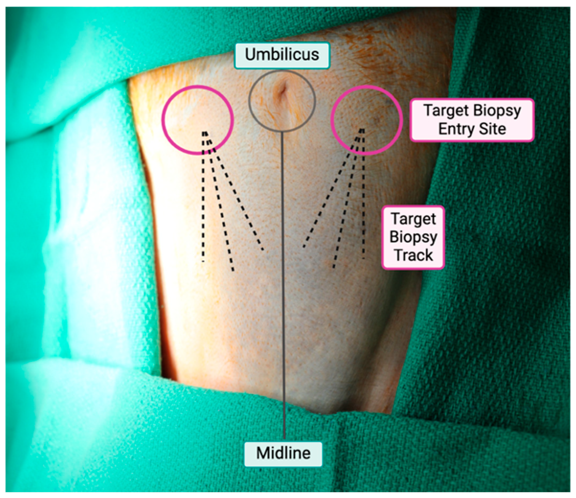
Figure 1. After preparation, the umbilicus and pubic symphysis are palpated to identify the midline. As depicted with the pink circle, the biopsy entry site is intended to be inferior and lateral to the umbilicus. The dashed lines depict the trajectory of the BD EleVation biopsy system needle for 3 separate tracks to maximize adipose tissue collection and minimize risks of bleeding.
Surgeries 06 00106 g001
Figure 2. The subcutaneous tissue is lifted off the abdominal wall during needle insertion.
Surgeries 06 00106 g002
Figure 3. Adipose tissue core biopsies obtained utilizing the BD EleVationTM System with a percutaneous technique. Some variability in the length of cores obtained from fracturing is seen.
Surgeries 06 00106 g003
Figure 4. Total adipose tissue yields were grouped by number of tracks used during a sampling event. The ordinary least squares fit approximates a 0.83 gram increase in yield for every additional tract during the procedure.
Surgeries 06 00106 g004
Figure 5. The immunocyte yield from the stromal vascular fraction was compared to age (A) and weight (B) of the animals. Neither age nor weight were significantly associated with immunocyte yield. The solid line demonstrates the ordinary least squares fit while the dashed lines indicated the 95% condifence interval.
Surgeries 06 00106 g005
Figure 6. (A) Representative AT morphology was assessed by IHC staining including H&E and (B) IBA-1 from an obese nonhuman primate. Utilizing this minimally invasive AT collection technique, we demonstrated preserved tissue architecture. On H&E staining, adipocytes appeared enlarged with characteristic hypertrophy commonly observed in obesity. (C,E) At higher magnification, crown-like structures, annotated with red circles, were widely dispersed throughout the tissue. These structures were further confirmed with IBA-1 staining (D,F), highlighting macrophage infiltration consistent with inflammatory remodeling in obese adipose tissue. (A,B) Magnification 1×, scale bar 3 mm. (C,D) Magnification 3×, scale bar 800 μm. (E,F) Magnification 10×, scale bar 300 μm.
Surgeries 06 00106 g006
Table 1. Baseline characteristics.
Mean ± SEMMedian (Min, Max)
Age (years)9.9 ± 0.079.9 (9.3, 10.3)
Weight (kg)15.4 ± 0.516.1 (11.3, 18.6)
Table 2. Summary of biopsy and adipose processing parameters.
Mean ± SEMMedian (Min, Max)
Biopsy Characteristics (n = 18)
Procedure Time (min)11.4 ± 1.110 (3, 23)
Resolution of Ecchymosis (days)7.5 ± 1.36 (3, 28)
Separate Tracks per Event1.8 ± 0.22 (1, 3)
Total Mass per Event (g)1.44 ± 0.21.5 (0.5, 2.7)
Adipose Processing of Initial Biopsy Set (n = 6)
AT Immunocyte Yield (106 cells/g)0.41 ± 0.120.30 (0.19, 0.96)
Table 3. Total mass obtained.
BiopsiesMean ± SEMMedian (Min, Max)
Single Trackn = 60.62 ± 0.05 g0.6 (0.5, 0.8)
Two Tracksn = 101.7 ± 0.14 g1.9 (1.0, 2.5)
Three Tracksn = 22.6 ± 0.2 g2.5 (2.3, 2.7)
Disclaimer/Publisher’s Note: The statements, opinions and data contained in all publications are solely those of the individual author(s) and contributor(s) and not of MDPI and/or the editor(s). MDPI and/or the editor(s) disclaim responsibility for any injury to people or property resulting from any ideas, methods, instructions or products referred to in the content.