Article

Short-Term Outcomes of a Novel Fascio-Aponeurotic Flap Technique for Ulnar Nerve Instability at the Elbow

1
Dipartimento di Ortopedia, Fondazione Policlinico Universitario Agostino Gemelli IRCSS, 00168 Rome, Italy
2
Department of Orthopaedics, Azienda Ospedaliera San Giovanni Addolorata, 00184 Rome, Italy
3
Department of Clinical Science and Translational Medicine, Section of Orthopaedics and Traumatology, University of Rome “Tor Vergata”, 00133 Rome, Italy
4
Catholic University of the Sacred Heart, 00168 Rome, Italy
5
Galeazzi Orthopaedic Institute, 20157 Milano, Italy
6
Chirurgie de la Main, CHIREC site Delta, 1160 Bruxelles, Belgium
7
Unit of Orthopaedics, “Spedali Civili” Hospital, University of Brescia, 25123 Brescia, Italy
*
Author to whom correspondence should be addressed.
Surgeries 2025, 6(3), 49; https://doi.org/10.3390/surgeries6030049
Submission received: 3 May 2025 / Revised: 4 June 2025 / Accepted: 17 June 2025 / Published: 24 June 2025
(This article belongs to the Section Hand Surgery and Research)

Abstract

Background: Cubital tunnel syndrome is the second most common compressive neuropathy of the upper limb, and it is characterized by ulnar nerve compression at the elbow. Traditional surgical options, including simple decompression and anterior transposition, have limitations in addressing ulnar nerve instability. This study introduces and evaluates the short-term outcomes of a novel surgical technique, the fascio-aponeurotic epicondylar flap (FAEF), for stabilizing the ulnar nerve and managing its instability. Materials and methods: A retrospective study was conducted on ten patients with longstanding cubital tunnel syndrome and confirmed ulnar nerve dislocation or instability. All patients underwent surgical intervention using the FAEF technique, which involves creating a quadrangular fascial flap from the epicondylar fascia to stabilize the ulnar nerve within the retrocondylar groove. Outcomes were assessed using clinical follow-ups, the Michigan Hand Outcomes Questionnaire (MHQ), VAS, and qDASH scores over a 90-day postoperative period. Results: All ten patients experienced complete resolution of neurological symptoms, including paresthesia, pain, and nerve clicking, by the final follow-up. Postoperative recovery was uneventful, with no complications such as infections or hematomas. Grip strength and hand functionality were fully restored, with significant improvements in MHQ scores (mean: 94). Dynamic elbow mobilization initiated on the first postoperative day resulted in full recovery of elbow range of motion. No recurrence of ulnar nerve dislocation was observed. Discussion: The FAEF technique effectively stabilizes the ulnar nerve, alleviates symptoms, and restores function while minimizing risks associated with traditional procedures, such as nerve trauma and elbow instability. By preserving the anatomical integrity of the medial epicondyle and enhancing nerve mobility, this approach represents a less invasive alternative to anterior transposition and medial epicondylectomy. Conclusions: The FAEF technique is a viable and effective surgical option for managing ulnar nerve instability in cubital tunnel syndrome. It offers a less invasive solution with excellent short-term outcomes, making it a promising addition to the surgical armamentarium for this condition. Further studies are warranted to evaluate long-term efficacy and broader applicability.

1. Introduction

Cubital tunnel syndrome, or ulnar nerve compression syndrome at the elbow, is the second most common compressive neuropathy of the upper limb, following carpal tunnel syndrome [1]. The cubital tunnel is an anatomical passageway with a bony floor formed by the medial epicondyle of the humerus and the olecranon of the ulna, and a ligamentous roof created by a fibrous structure called Osborne’s ligament, which may thicken and contribute to nerve compression. Beyond this tunnel, the ulnar nerve continues its path through a fibrous channel between the two heads of the flexor carpi ulnaris (FCU) muscle, both of which are anchored to the medial epicondyle and the olecranon. Thickening of this fibrous band can also exert compressive effects on the nerve [2]. Additionally, some cases involve instability or dislocation of the ulnar nerve from the cubital tunnel during elbow flexion and extension.
Clinically, cubital tunnel syndrome manifests as tingling and numbness in the palmar and ulnar hand regions, particularly affecting the fourth and fifth fingers. Symptoms often worsen at night and can intensify with prolonged elbow flexion. In advanced stages, the condition may lead to muscle weakness and atrophy in the intrinsic hand muscles and flexors of the affected fingers, impairing grip strength, especially in finger adduction and abduction. Chronic cases may result in a deformity known as “benediction hand”, characterized by extension at the metacarpophalangeal joint and flexion at the interphalangeal joints of the fourth and fifth fingers due to muscular imbalance [3].
The etiology of cubital tunnel syndrome can be congenital, involving a narrow canal, or secondary to arthritis or trauma, such as fractures or dislocations that alter anatomical structures. Other contributing factors include repetitive elbow flexion and extension, prolonged elbow flexion (as seen in certain sports or musical instrument use), and exposure to vibrating tools [4].
Diagnosis is mainly clinical and is based on patient history and physical examination. Electrophysiological studies, such as electromyography and electroneurography, can confirm the diagnosis and assess the severity of nerve damage. These tests also help differentiate cubital tunnel syndrome from ulnar nerve entrapment at the wrist (Guyon’s canal syndrome) or dual compression at both the elbow and wrist. Distinguishing features include sensory deficits on the ulnar dorsal hand, muscle testing, nerve conduction velocity, and the Tinel sign at the cubital tunnel [2]. Dynamic ultrasound can provide additional diagnostic insight by visualizing the nerve, assessing any edema or volumetric changes, and detecting subluxation during elbow movement.
Ulnar nerve subluxation or dislocation at the elbow may present with symptoms resembling compressive neuropathy but with normal electromyographic findings. Childress (1975) classified ulnar nerve instability into two types: Type A (subluxation) involves the nerve shifting from the olecranon groove to the medial epicondyle tip with elbow flexion beyond 90°, while Type B (dislocation) describes the nerve completely passing the medial epicondyle during flexion [5]. This condition, more common in younger patients, has a progressive course and typically requires surgical intervention due to its irreversible anatomical nature.
Various surgical approaches have been described for cubital tunnel syndrome, but there is ongoing debate about the optimal technique [2]. Surgical options include simple nerve decompression, decompression with anterior transposition (subcutaneous, submuscular, intramuscular, or subfascial), and medial epicondylectomy [6,7,8,9,10,11]. While anterior transposition and medial epicondylectomy are often used when instability accompanies compression, they carry significant disadvantages, such as increased risk of nerve trauma (contusion, stretching, or strangulation) or elbow destabilization.
In 2023, the authors introduced a novel surgical technique as an alternative to anterior transposition for managing ulnar nerve instability. This approach involves creating a protective arch around the nerve by fashioning a quadrangular flap from the aponeurotic fascia of the epicondylar muscles. A cadaveric study involving 20 elbows confirmed the kinematic feasibility of this technique [12]. This manuscript reports its first clinical application in living patients, along with the short-term recovery outcomes of the initial ten patients who underwent this surgical procedure

2. Materials and Methods

This retrospective study evaluated the short-term clinical outcomes of patients undergoing ulnar nerve instability treatment that employed the fascio-aponeurotic epicondylar flap (FAEF).
The study was conducted in compliance with the principles outlined in the Declaration of Helsinki as part of a thesis dissertation at the Catholic University in Rome. Informed consent was obtained from all participants.
This retrospective study analyzed ten patients (four males and six females; mean age: 41.2 years) presenting with preoperative symptoms of longstanding paresthesia and pain localized to the ulnar nerve territory, exacerbated by elbow flexion. Common clinical findings included reduced hand grip strength and a positive Tinel’s sign at the elbow. Electromyography (EMG) confirmed ulnar neuropathy at the elbow in all cases. Dynamic ultrasound identified ulnar nerve dislocation or instability during elbow flexion–extension, classified according to the Childress criteria, with 6 patients demonstrating Type A instability, 2 with Type B instability, and 2 with prior surgical anterior transposition.
The exclusion criteria included patients with acute trauma or recent elbow injuries unrelated to ulnar nerve pathology; those with systemic conditions such as diabetes mellitus or rheumatoid arthritis known to cause peripheral neuropathy; patients with unstable or incomplete EMG results or inconclusive dynamic ultrasound findings; and individuals with insufficient or incomplete medical records that could impede retrospective data analysis.
Surgical intervention employed the fascio-aponeurotic epicondylar flap (FAEF) technique, as described by R. De Vitis. This procedure involves creating a quadrangular fascial flap from the epicondylar region and securing it to stabilize the ulnar nerve within the retrocondylar groove. All procedures were performed under regional anesthesia using a brachial plexus block. Patients were positioned supine with the affected arm externally rotated and abducted on an arm support. A pneumatic tourniquet inflated to 250 mmHg was applied at the arm’s root to achieve ischemia. A curved skin incision approximately 10 cm long was made over the epitrochlea–olecranon groove to expose the surgical site.
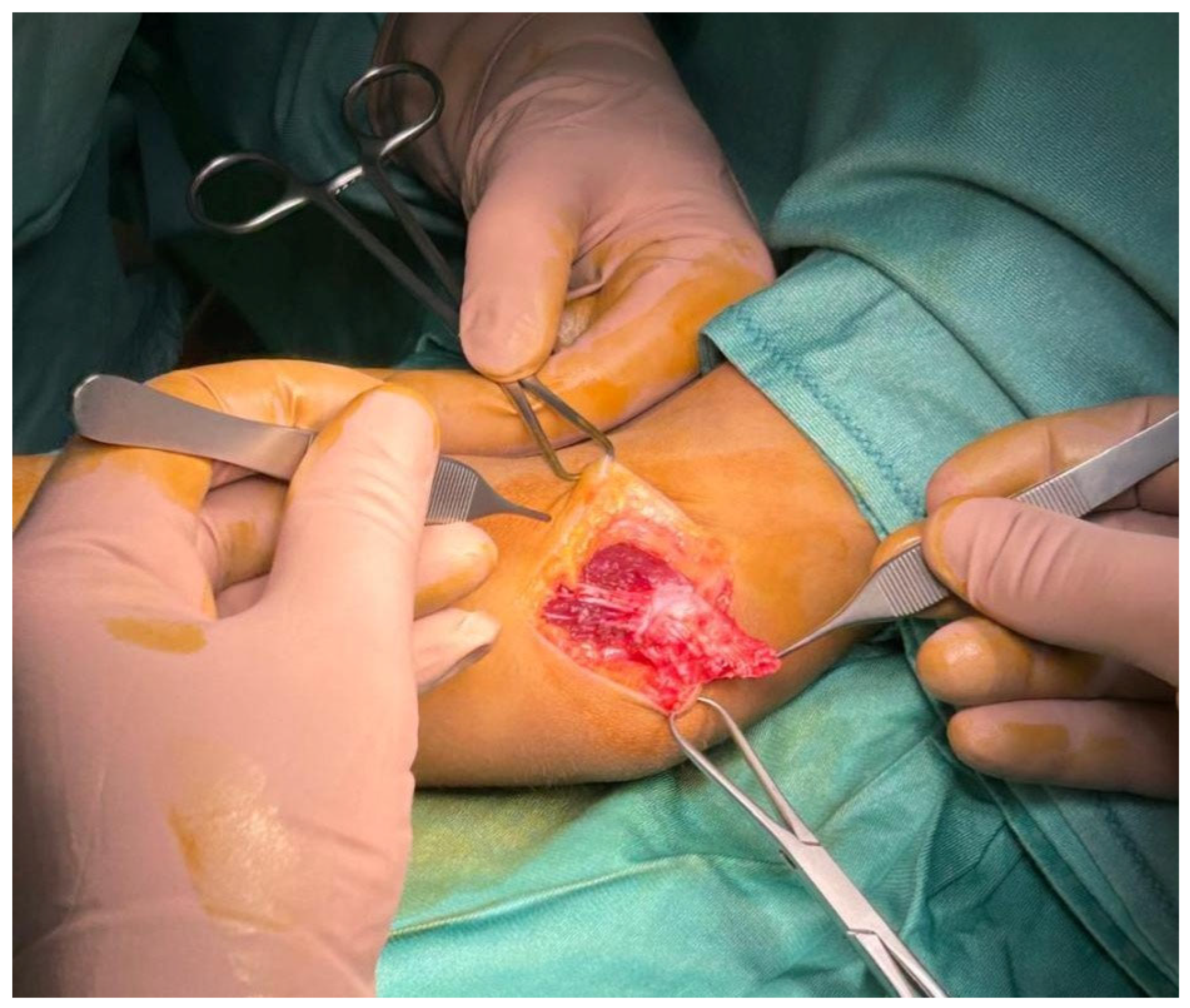
The ulnar nerve was located within the cubital tunnel posterior to the epitrochlea. Surgical exploration confirmed complete ulnar nerve dislocation with passive elbow flexion (Figure 1). Neurolysis was performed, followed by the FAEF procedure. This involved sculpting a quadrangular epicondylar fascial flap and rotating it 180° (Figure 2). Suturing was performed on the olecranic portion of the incised epitrocleo–olecranic ligament in order to stabilize the nerve within the retrocondylar groove, ensuring that excessive compression was avoided (Figure 3). Intraoperative testing confirmed proper nerve gliding during elbow flexion–extension. The surgical site was closed with Donati stitches, and an elastic compression bandage was applied.
Postoperatively, patients began gradual elbow mobilization on the first day, with progressive increases in range of motion after suture removal at 14 days. Clinical follow-up revealed resolution of nerve clicking, both objectively and as reported by patients. Early postoperative evaluations showed mild improvement in sensory function. Follow-up at 30, 60, and 90 days demonstrated complete resolution of ulnar nerve dislocation, disappearance of neurological symptoms, and full return to normal daily activities.
The monitored complications included infection, flap failure accompanied by recurrent symptoms or nerve instability, and nerve adhesions.
Outcomes were assessed using the Michigan Hand Outcomes Questionnaire (MHQ) [13], Visual Analog Scale (VAS), and Quick Disabilities of the Arm, Shoulder, and Hand (qDASH) scores [14], which showed significant improvements in hand function and quality of life.
The paired t-test was used to compare preoperative and postoperative scores for VAS, qDASH, and MHQ. A significance level (α) of 0.05 was chosen.

3. Results

All ten patients included in this study underwent surgical intervention using the fascio-aponeurotic epicondylar flap (FAEF) technique without any intraoperative complications. The postoperative recovery period was uneventful, with no reports of infections, hematomas, or wound healing complications. None of the patients required medical or nutraceutical therapy to resolve or improve neuralgic symptoms during the postoperative period.
At the final follow-up, conducted 90 days postoperatively, no deficits in elbow range of motion were observed. Early mobilization, initiated on the first postoperative day, facilitated the full recovery of elbow function. Clinical evaluations confirmed complete resolution of ulnar nerve dislocation and the absence of nerve clicking, as was both objectively assessed by physicians and subjectively reported by patients.
Neurological symptoms, including paresthesia and pain in the ulnar nerve territory, were fully resolved in all patients by the 90-day follow-up. Grip strength returned to normal, and patients experienced marked improvements in hand functionality. Sensory deficits showed mild improvement early in the recovery period, with complete resolution by the final follow-up.
Functional outcomes were assessed using the Michigan Hand Outcomes Questionnaire (MHQ), Visual Analog Scale (VAS), and QuickDASH (qDASH) scales.
VAS scores: The mean preoperative VAS score of 8.7 (95% CI: 8.4–9.0) decreased to 1.6 (95% CI: 1.3–1.9) postoperatively, with a mean difference of −7.1 (95% CI: −7.4 to −6.8, Cohen’s d = 3.5), indicating a very large effect size and a significant reduction in pain (p < 0.0001).
QuickDASH scores: The mean preoperative QuickDASH score improved from 89.37 (95% CI: 86.2–92.5) to 20.38 (95% CI: 18.1–22.7) postoperatively, with a mean difference of −69.0 (95% CI: −72.5 to −65.5, Cohen’s d = 4.1), reflecting a substantial reduction in functional disability (p < 0.0001).
MHQ scores: The mean MHQ score increased from 31.47% (95% CI: 28.4–34.5) preoperatively to 94.25% (95% CI: 92.1–96.4) postoperatively, with a mean difference of +62.78% (95% CI: +59.5 to +66.0, Cohen’s d = 4.8), demonstrating excellent improvements in hand functionality and patient satisfaction (p < 0.0001).
All results were statistically significant (p < 0.0001) for each metric, demonstrating the effectiveness of the FAEF technique in improving pain relief, functional outcomes, and patient satisfaction.
By the final follow-up, all patients had resumed normal daily activities without limitations. These results highlight the effectiveness of the FAEF technique in stabilizing the ulnar nerve, alleviating neurological symptoms, and restoring full elbow and hand function in patients with ulnar nerve dislocation or instability.
The results are summarized in Table 1.

4. Discussion

The size of the cubital tunnel plays a pivotal role in the development of ulnar nerve compression at the elbow, as it physiologically decreases by approximately 55% during elbow flexion [8,15]. Concurrently, this motion elongates the ulnar nerve by 4–7 mm. This combination of compression and stretching can, in some cases, lead to ischemic damage and inflammation, ultimately impairing nerve function. Non-surgical treatments for ulnar nerve entrapment syndromes have shown limited effectiveness [2,3,4,16,17,18,19]. Elhassan (2007) reported some success with conservative management using NSAIDs and night-time protective splints to limit elbow flexion [20]. However, most studies indicate that medical therapy alone is insufficient [2,3,4,16,17,18,19]. When NSAIDs and immobilization fail, corticosteroid injections or minimally invasive techniques are often considered [2,3,4].
Minimally invasive treatments, such as percutaneous placement of peripheral electrodes, offer straightforward and effective methods for alleviating pain, particularly with intermittent, low-frequency stimulation [21]. However, the success of these techniques heavily depends on the severity of nerve compression. A review of 50 studies involving over 2000 patients with cubital tunnel syndrome found that mild compression often resolves spontaneously, whereas non-invasive techniques are largely ineffective in severe cases [22]. When symptoms become significantly disabling and conservative management fails, surgical intervention becomes necessary, with benefits outweighing the risks of vascular or nerve damage [22,23,24,25,26,27].
The primary objective of surgical treatment is to achieve full nerve decompression and reposition the nerve in an area free from compression or kinking. Common surgical approaches include simple decompression [4,6,7,24,25,26] and decompression accompanied by anterior transposition [9,11,27], which can be performed subcutaneously, submuscularly, intramuscularly, or subfascially. Although medial epicondylectomy is another option, its use has declined in recent years [10]. More recently, minimally invasive surgical techniques, such as endoscopic neurolysis [26] and mini-incision in situ decompressions [27], have emerged as alternatives.
A study by Nabhan (2005) compared simple decompression with subcutaneous anterior transposition in 66 patients, finding no significant differences in outcomes at 3 and 9 months postoperative [28]. This finding supports simple decompression as the less invasive option. However, if ulnar nerve instability is detected after decompression, transposition is strongly indicated. Endoscopic neurolysis, another minimally invasive option, involves using a probe with a micro camera to section the retinaculum and fascia. When subluxation of the ulnar nerve is identified, endoscopic transposition [26,29] or medial epicondylectomy may also be performed [30,31]. A 2005 study reported significant symptom improvement or resolution in most cases following endoscopic neurolysis [32], while a 2021 study of 60 patients documented complete symptom resolution [33]. Despite promising results, the long-term efficacy of these techniques remains uncertain due to limited follow-up studies. Additionally, endoscopic neurolysis is contraindicated in patients with conditions such as elbow deformities, arthritis, or recurrent cubital tunnel syndrome, limiting its applicability.
At present, there is no consensus on the optimal surgical technique for cubital tunnel syndrome, with treatment decisions often based on surgeon preference. Each technique has limitations. For instance, subcutaneous anterior transposition increases the risk of hypersensitivity and nerve damage, while medial epicondylectomy can compromise elbow stability, especially if more than 40% of the medial epicondyle is removed. To mitigate these issues, some surgeons bury the ulnar nerve within or beneath muscle planes during transposition, but this can lead to scar contracture, nerve strangulation, and subsequent neuritis [30].
Subfascial anterior transposition, introduced by Chuang and Treciak in 1998, minimizes scar adhesion and accelerates recovery compared to submuscular or intramuscular methods by requiring less dissection [34]. An evolution of this technique, proposed by Hyun Ho Han in 2014, involves wrapping the ulnar nerve with a fascial flap to improve sliding during elbow flexion–extension movements [1]. In a study of 20 patients with cubital tunnel syndrome, this approach achieved good or excellent outcomes in 95% of cases, with no complications or recurrences. Han’s technique combines the advantages of subfascial transposition with the benefits of placing the ulnar nerve in contact with the healthy epicondylar fascia, reducing adhesion risk and enhancing nerve mobility.
The surgical method described in our study shares these advantages. By covering the ulnar nerve with an intact fascio-aponeurotic flexor-pronator flap, it promotes nerve sliding while preserving the protective anatomy of the medial epicondyle. This approach may also be applicable in primary ulnar nerve instability, offering a less invasive alternative without compromising the possibility of subsequent traditional surgical repair if needed.
In comparing the fascia wrapping technique [1] and the fascio-aponeurotic epicondylar flap (FAEF) technique, both demonstrated high efficacy in stabilizing the ulnar nerve and improving outcomes. The fascia wrapping technique achieved a 95% rate of good or excellent results, with significant motor conduction velocity (MCV) improvements and no complications or recurrences. Similarly, the FAEF technique resulted in 100% symptom resolution, restored grip strength, and significant functional improvements (VAS, qDASH, and MHQ).
The fascia wrapping technique involves loosely rolling the nerve with an elevated fascial flap to reduce adhesion and enhance gliding, while the FAEF technique uses a rotated fascial flap to stabilize the nerve, emphasizing long-term stability by preserving the medial epicondyle’s anatomy. Recovery was faster with the FAEF technique, allowing mobilization on the first postoperative day and full functional recovery within 90 days, compared to 4 weeks for the fascia wrapping technique.
Both techniques showed minimal complications, with neither reporting infections or recurrences. The fascia wrapping study, with 20 patients and a 24-month follow-up, provided more robust long-term data, while the FAEF study, involving 10 patients over 90 days, offered promising short-term results but limited generalizability.
Ulnar nerve instability is a condition that has been reported in very few studies.
A recent review highlighted that many of these studies are influenced by biases related to ulnar nerve subluxation. The review concluded that while both decompression and transposition procedures result in similar symptomatic improvements, transposition is associated with higher complication rates [35].
Despite this, anterior transposition of the ulnar nerve continues to be a widely performed procedure, particularly for cases of ulnar nerve instability and recurrent cubital tunnel syndrome. Ruettermann, in conducting a systematic review of surgical options for recurrent cubital tunnel syndrome, found that only 16 out of 296 studies met the inclusion criteria for treating recurrent cases. Due to significant selection bias and inconsistent outcome measures among the studies, a meta-analysis was deemed impossible. As a result, there is no strong evidence to definitively support anterior transposition of the ulnar nerve over in situ decompression or stabilization in cases of recurrent cubital tunnel syndrome [36].
For idiopathic ulnar nerve entrapment, including severe cases, the current evidence, albeit low to moderate in certainty, suggests that there is little to no difference in functional improvement or surgical complications between simple decompression and decompression with either subcutaneous or submuscular transposition [2].
This study has several limitations, including its retrospective design, the lack of blinding, and a relatively small sample size.
Although these promising results warrant dissemination, the small sample size (n = 10) significantly limits the statistical power and generalizability of the findings. Additionally, the 90-day follow-up period is insufficient to evaluate potential long-term complications. A study with an extended follow-up period is planned, and future research, potentially multicenter and of longer duration, will be conducted to address these limitations.
Despite these limitations, the study’s strength includes the implementation of a surgical technique that led to a higher quality life for all patients.

5. Conclusions

This retrospective study demonstrated that the fascio-aponeurotic epicondylar flap technique is a viable option for addressing primary or secondary ulnar nerve instability following neurolysis. It offers a less invasive alternative to more traditional procedures while maintaining the potential for secondary interventions if necessary. This technique appears suitable for both male and female patients and represents a promising addition to the surgical options for cubital tunnel syndrome.

Author Contributions

Conceptualization, R.D.V. and G.T.; methodology, M.P.; software, G.M.S.; validation, V.C.; formal analysis, M.D.; investigation, A.C.; resources, R.D.V. and E.M.G.; data curation, A.C. and E.M.G.; writing—original draft preparation, A.C. and R.D.V.; writing—review and editing, L.M.; visualization, none; supervision, G.T.; project administration, R.D.V.; funding acquisition, none. All authors have read and agreed to the published version of the manuscript.

Funding

This research received no external funding.

Institutional Review Board Statement

Local Ethical committee review and approval were not required for this study due to the prevailing legislation (Legislative Decree 196/2003) according to Article 110 of the Privacy Code, as amended by Legislative Decree 101/2008.

Informed Consent Statement

Informed consent for surgical treatment and use and publication of clinical data and images was obtained from all participants.

Data Availability Statement

The data presented in this study are available on request from the corresponding author due to privacy restriction.

Conflicts of Interest

The authors declare no conflicts of interest.

References

  1. Han, H.H.; Kang, H.W.; Lee, J.Y.; Jung, S.N. Fascia wrapping technique: A modified method for the treatment of cubital tunnel syndrome. Sci. World J. 2014, 2014, 482702. [Google Scholar] [CrossRef] [PubMed]
  2. Caliandro, P.; La Torre, G.; Padua, R.; Giannini, F.; Padua, L. Treatment for ulnar neuropathy at the elbow. Cochrane Database Syst. Rev. 2016, 11, CD006839. [Google Scholar] [CrossRef]
  3. Vij, N.; Traube, B.; Bisht, R.; Singleton, I.; Cornett, E.M.; Kaye, A.D.; Imani, F.; Mohammadian Erdi, A.; Varrassi, G.; Viswanath, O.; et al. An Update on Treatment Modalities for Ulnar Nerve Entrapment: A Literature Review. Anesthesiol. Pain Med. 2020, 10, e112070. [Google Scholar] [CrossRef] [PubMed]
  4. Staples, J.R.; Calfee, R. Cubital Tunnel Syndrome: Current Concepts. J. Am. Acad. Orthop. Surg. 2017, 25, e215–e224. [Google Scholar] [CrossRef]
  5. Childress, H.M. Recurrent Ulnar-Nerve Dislocation at the Elbow. Clin. Orthop. Relat. Res. 1975, 108, 168–173. [Google Scholar] [CrossRef] [PubMed]
  6. Lavyne, M.H.; Bell, W.O. Simple decompression and occasional microsurgical epineurolysis under local anesthesia as treatment for ulnar neuropathy at the elbow. Neurosurgery 1982, 11, 6–11. [Google Scholar] [CrossRef]
  7. Wilson, D.H.; Krout, R. Surgery of ulnar neuropathy at the elbow: 16 cases treated by decompression without transposition. J. Neurosurg. 1973, 38, 780–785. [Google Scholar] [CrossRef]
  8. Andrews, K.; Rowland, A.; Pranjal, A.; Ebraheim, N. Cubital tunnel syndrome: Anatomy, clinical presentation, and management. J. Orthop. 2018, 15, 832–836, Erratum in J. Orthop. 2020, 23, 275. [Google Scholar] [CrossRef]
  9. Eaton, R.G.; Crowe, J.F.; Parkes, J.C., 3rd. Anterior transposition of the ulnar nerve using a Non-Compressing fasciodermal sling. J. Bone Jt. Surg. Am. 1980, 62, 820–825. [Google Scholar] [CrossRef]
  10. O’Grady, E.E.; Vanat, Q.; Power, D.M.; Tan, S. A systematic review of medial epicondylectomy as a surgical treatment for cubital tunnel syndrome. J. Hand Surg. (Eur. Vol.) 2017, 42, 941–945. [Google Scholar] [CrossRef]
  11. Richmond, J.C.; Southmayd, W.W. Superficial anterior transposition of the ulnar nerve at the elbow for ulnar neuritis. Clin. Orthop. Relat. Res. 1982, 164, 42–44. [Google Scholar] [CrossRef]
  12. D’Orio, M.; Fulchignoni, C.; De Vitis, R.; Passiatore, M.; Taccardo, G.; Marzella, L.; Lazzerini, A. Feasibility of a fascial flap to avoid anterior transposition of unstable Ulnar nerve: A cadaver study. J. Clin. Orthop. Trauma 2023, 42, 102202. [Google Scholar] [CrossRef]
  13. Passiatore, M.; De Vitis, R.; Cilli, V.; Milano, G.; Saccomanno, M.F.; Cotroneo, C.; Brozzini, E.; Vigliarolo, D.; Taccardo, G. The Italian Version of the Michigan Hand Outcomes Questionnaire (MHQ): Translation, Cross-Cultural Adaptation and Validation. J. Hand Surg. (Asian-Pac. Vol.) 2021, 26, 666–683. [Google Scholar] [CrossRef]
  14. Padua, R.; Padua, L.; Ceccarelli, E.; Romanini, E.; Zanoli, G.; Amadio, P.C.; Campi, A. Italian version of the Disability of the Arm, Shoulder and Hand (DASH) questionnaire. Cross-Cultural adaptation and validation. J. Hand Surg. Br. 2003, 28, 179–186. [Google Scholar] [CrossRef]
  15. Pratt, N. Anatomy of nerve entrapment sites in the upper quarter. J. Hand Ther. 2005, 18, 216–229. [Google Scholar] [CrossRef] [PubMed]
  16. Dellon, A.L.; Hament, W.; Gittelshon, A. Nonoperative management of cubital tunnel syndrome: An 8-year prospective study. Neurology 1993, 43, 1673–1677. [Google Scholar] [CrossRef] [PubMed]
  17. D’Orio, M.; De Vitis, R.; Taccardo, G.; Rocchi, L.; Ferrari, F.; Perna, A.; Passiatore, M. Clinical usefulness of nutraceutics with acetyl-L-carnitine, α-lipoic acid, phosphatidylserine, curcumin, C, E and B-group vitamins in patients awaiting for carpal tunnel release during COVID-19 pandemic: A randomized controlled open label prospective study. Acta Biomed. 2023, 94, e2023050. [Google Scholar]
  18. Passiatore, M.; Perna, A.; De-Vitis, R.; Taccardo, G. The Use of Alfa-Lipoic Acid-R (ALA-R) in Patients with Mild-Moderate Carpal Tunnel Syndrome: A Randomised Controlled Open Label Prospective Study. Malays. Orthop. J. 2020, 14, 1–6. [Google Scholar]
  19. Evangelista, M.; Cilli De Vitis, R.; Militerno, A.; Fanfani, F. Ultra-Micronized Palmitoylethanolamide Effects on Sleep-Wake Rhythm and Neuropathic Pain Phenotypes in Patients with Carpal Tunnel Syndrome: An Open-Label, Randomized Controlled Study. CNS Neurol. Disord. Drug Targets 2018, 17, 291–298. [Google Scholar] [CrossRef]
  20. Elhassan, B.; Steinmann, S.P. Entrapment neuropathy of the ulnar nerve. J. Am. Acad. Orthop. Surg. 2007, 15, 672–681. [Google Scholar] [CrossRef]
  21. Deer, T.R.; Jain, S.; Hunter, C.; Chakravarthy, K. Neurostimulation for Intractable Chronic Pain. Brain Sci. 2019, 9, 23. [Google Scholar] [CrossRef]
  22. Dellon, A.L. Review of treatment results for ulnar nerve entrapment at the elbow. J. Hand Surg. Am. 1989, 14, 688–700. [Google Scholar] [CrossRef]
  23. Arle, J.E.; Zager, E.L. Surgical treatment of common entrapment neuropathies in the upper limbs. Muscle Nerve 2000, 23, 1160–1174. [Google Scholar] [CrossRef] [PubMed]
  24. Filippi, R.; Farag, S.; Reisch, R.; Grunert, P.; Böcher-Schwarz, H. Cubital tunnel syndrome. Treatment by decompression without transposition of ulnar nerve. Minim. Invasive Neurosurg. 2002, 45, 164–168. [Google Scholar] [CrossRef] [PubMed]
  25. Huang, J.H.; Samadani, U.; Zager, E.L. Ulnar nerve entrapment neuropathy at the elbow: Simple decompression. Neurosurgery 2004, 55, 1150–1153. [Google Scholar] [CrossRef] [PubMed]
  26. Mirza, A.; Reinhart, M.K.; Bove, J.; Litwa, J. Scope-Assisted release of the cubital tunnel. J. Hand Surg. Am. 2011, 36, 147–151. [Google Scholar] [CrossRef]
  27. Zhang, X.; Dhawan, V.; Yu, Y.; Zhang, G.; Shao, X. A minimally invasive approach for cubital tunnel release and ulnar nerve transposition. Phys. Sportsmed. 2017, 45, 110–113. [Google Scholar] [CrossRef]
  28. Nabhan, A.; Ahlhelm, F.; Kelm, J.; Reith, W.; Schwerdtfeger, K.; Steudel, W.I. Simple decompression or subcutaneous anterior transposition of the ulnar nerve for cubital tunnel syndrome. J. Hand Surg. Br. 2005, 30, 521–524. [Google Scholar] [CrossRef]
  29. Ahcan, U.; Zorman, P. Endoscopic decompression of the ulnar nerve at the elbow. J. Hand Surg. Am. 2007, 32, 1171–1176. [Google Scholar] [CrossRef]
  30. Baek, G.H.; Kwon, B.C.; Chung, M.S. Comparative study between minimal medial epicondylectomy and anterior subcutaneous transposition of the ulnar nerve for cubital tunnel syndrome. J. Shoulder Elb. Surg. 2006, 15, 609–613. [Google Scholar] [CrossRef]
  31. Amako, M.; Nemoto, K.; Kawaguchi, M.; Kato, N.; Arino, H.; Fujikawa, K. Comparison between partial and minimal medial epicondylectomy combined with decompression for the treatment of cubital tunnel syndrome. J. Hand Surg. Am. 2000, 25, 1043–1050. [Google Scholar] [CrossRef] [PubMed]
  32. Porcellini, G.; Paladini, P.; Campi, F.; Merolla, G. Arthroscopic neurolysis of the ulnar nerve at the elbow. Chir. Organi Mov. 2005, 90, 191–200. [Google Scholar] [PubMed]
  33. Lucchina, S.; Fusetti, C.; Guidi, M. Sonographic Follow-Up of Patients with Cubital Tunnel Syndrome Undergoing In Situ Open Neurolysis or Endoscopic Release: The SPECTRE Study. Hand 2021, 16, 385–390. [Google Scholar] [CrossRef] [PubMed]
  34. Chuang, D.C.; Treciak, M.A. Subfascial anterior transposition: A modified method for the treatment of cubital tunnel syndrome (CuTS). Tech. Hand Up. Extrem. Surg. 1998, 2, 178–183. [Google Scholar] [CrossRef] [PubMed]
  35. Clark, D.M.; Piscoya, A.S.; Dunn, J.C.; Nesti, L.J. The impact of Pre-Existing ulnar nerve instability on the surgical treatment of cubital tunnel syndrome: A systematic review. J. Shoulder Elb. Surg. 2020, 29, 2339–2346. [Google Scholar] [CrossRef]
  36. Ruettermann, M. Challenging the dogma: Anterior transposition of the ulnar nerve is indicated in recurrent cubital tunnel syndrome. J. Hand Surg. (Eur. Vol.) 2021, 46, 45–449. [Google Scholar] [CrossRef]
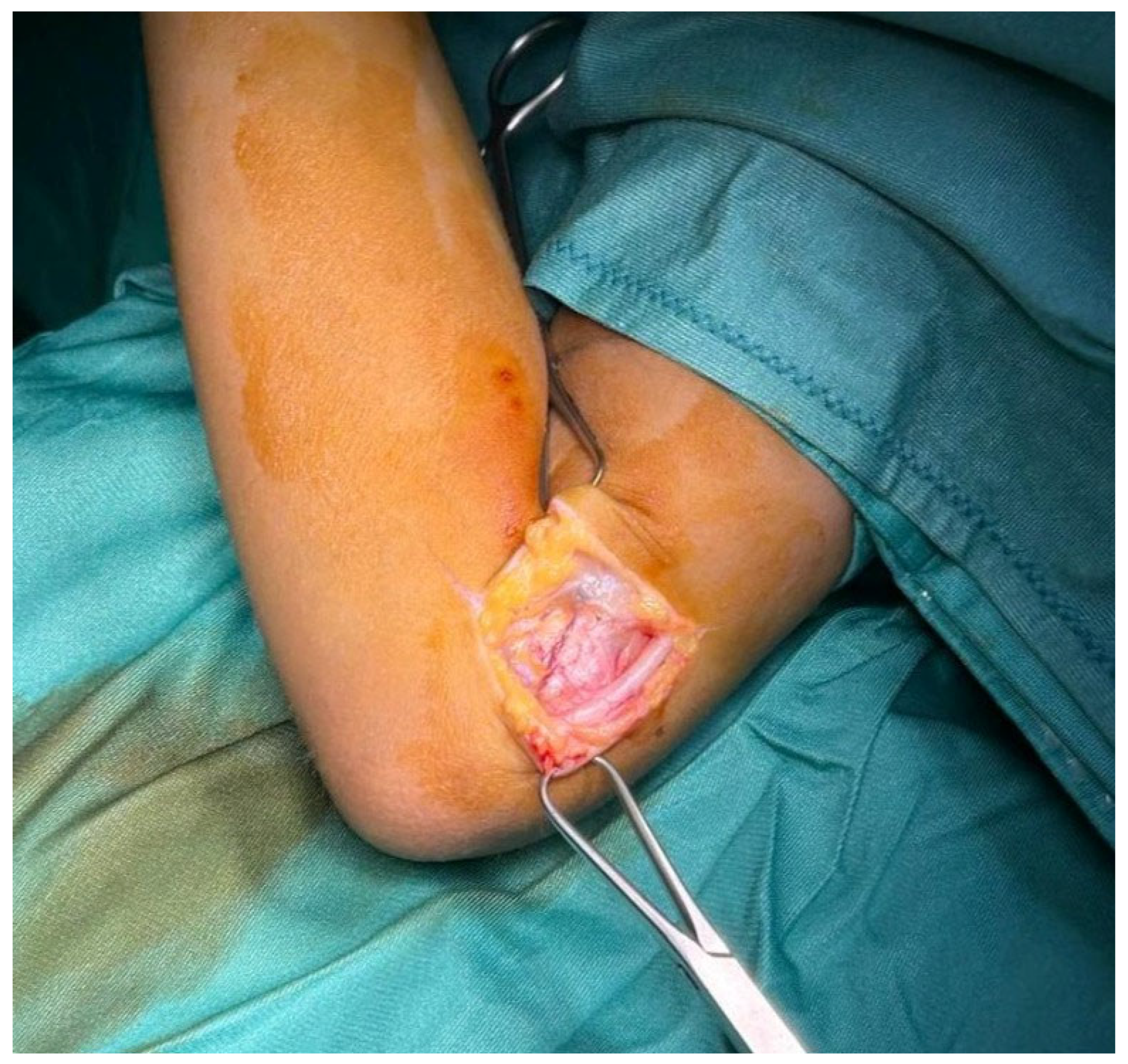
Figure 1. Surgical incision with evidence of a dislocation of the ulnar nerve.
Surgeries 06 00049 g001
Figure 2. Replacement of the ulnar nerve and harvested flap from the aponeurotic portion of the flexor tendon.
Surgeries 06 00049 g002
Figure 3. Ulnar nerve stabilized by the flap sutured on the olecranic portion of the incised epitrocleo–olecranic ligament.
Surgeries 06 00049 g003
Table 1. Fascio-aponeurotic epicondylar flap (FAEF) case series. Abbreviations: VAS (Visual Analog Scale, score range 1–10); qDash (Quick Disability of the Arm, Shoulder, and Hand, score range 0–100); MHQ (Michigan Hand Questionnaire, score range 0–100).
PatientsSexPreoperative
Condition
AgePre-op VASPost-op VASPre-op qDashPost-op qDashPre-op MHQPost-op MHQ
1MDislocation by tumor109.000.0095.2512.5023.1%98.4%
2MType B instability3310.003.0096.5029.5022.2%89.3%
3FPrevious surgical
anterior transposition
2610.001.0092.0014.5021.6%96.4%
4FType A instability308.001.0085.2523.5039.0%91.6%
5FType A instability388.002.0079.5021.2534.7%94.3%
6MType A instability219.001.0098.2513.7528.3%95.7%
7FPrevious surgical
anterior transposition
909.004.0090.7535.2541.8%86.9%
8FType A instability468.001.0085.2519.2546.8%97.6%
9MType B instability558.001.0090.7517.7534.7%98.1%
10FType A instability638.002.0080.2516.5022.5%94.2%
Mean6/4 41.28.701.6089.3720.3831.47%94.25%
Disclaimer/Publisher’s Note: The statements, opinions and data contained in all publications are solely those of the individual author(s) and contributor(s) and not of MDPI and/or the editor(s). MDPI and/or the editor(s) disclaim responsibility for any injury to people or property resulting from any ideas, methods, instructions or products referred to in the content.