Review

Radial Artery Used as Conduit for Coronary Artery Bypass Grafting

1
Department of Cardiac Surgery, Centre Cardiologique du Nord, 93200 Saint-Denis, France
2
Department of Cardiac Surgery, La Pitié Salpetriere Hospital, 75013 Paris, France
*
Author to whom correspondence should be addressed.
Surgeries 2025, 6(1), 6; https://doi.org/10.3390/surgeries6010006
Submission received: 28 November 2024 / Revised: 28 December 2024 / Accepted: 6 January 2025 / Published: 14 January 2025
(This article belongs to the Special Issue Cardiothoracic Surgery)

Abstract

:
It was in 1989 that we first reported on the use of the radial artery (RA) as a secondary arterial graft for coronary artery bypass grafting (CABG). Nevertheless, discrepancies in clinical endpoints between the RA and alternative conduits have been reported in consecutive randomised trials. With over fifty years of accumulated practice in RA bypass grafting, we sought to identify the second-best option for CABG by reviewing the literature. A consistently successful second-best conduit for CABG has been demonstrated using the radial artery. Compared to saphenous vein grafts, the findings indicate improved outcomes and better patency results. Furthermore, it has been demonstrated to be a safe and effective conduit in the territory of the right coronary artery. The lack of available literature and the scarcity of similar case series restrict the application of the gastroepiploic artery. After five decades of utilisation, it can be unequivocally stated that the radial artery is the optimal conduit for coronary bypass surgery following the left internal thoracic artery to the left anterior descending artery.

Graphical Abstract

1. Introduction and Historic Context

Coronary artery bypass grafting (CABG) constitutes a surgical procedure in which the patient’s own arteries or veins are employed as grafts to circumvent obstructed coronary arteries, the result of the accumulation of atherosclerotic plaque. CABG is one of the procedures with the highest frequency of occurrence among major surgical operations, with approximately 400,000 cases conducted annually in the United States. Nevertheless, over the past decade, there has been a notable reduction in the number of CABG procedures performed in the United States. This reduction has occurred despite an ageing population and growing evidence to support the effectiveness and safety of the operation [1,2,3,4,5,6]. This decline has been accompanied by a corresponding increase in percutaneous coronary revascularisation (PCI) procedures.
The utilisation of the radial artery (RA) in CABG surgery represents an evolutionary trajectory in surgical techniques [7]. Despite the fact that the operation was first described almost 50 years ago, it has continued to evolve through the gradual acquisition of knowledge regarding the complex physiological processes involved in its execution and related to the function of arterial grafts [7,8,9,10,11,12].
The radial artery was first used by our team as a bypass graft in CABG operation, as described in the Annals journal [7]. Since then, our centre has performed more than 910 radial artery grafts, with the latest follow-up occurring at 27 years post-surgery [8,9,10,11,12]. Our findings demonstrated that the radial artery graft remained patent and revascularised the left anterior descending artery (LAD) without evidence of graft disease. Additionally, the radial artery served as a second target conduit on the circumflex artery and right coronary artery (RCA) branches at this time point. Five further angiographic evaluations of individuals who had undergone surgery in the early 1970s utilising the RA demonstrated that the RA grafts were fully patent 13 to 18 years following their operation [8,9].
The long-term effectiveness of saphenous-vein grafts (SVGs) in coronary artery bypass surgery has prompted extensive debate regarding the optimal conduit for CABG procedures. This includes the relative merits of bilateral versus single internal-thoracic-artery (LITA) grafts and the radial artery [13,14,15,16,17,18,19,20,21]. The extant evidence and the consensus guidelines indicate that the left internal mammary artery should be used to provide grafting to the left anterior descending coronary artery [22,23,24,25]. The enhanced efficacy observed with the LITA is likely attributable to its greater long-term vascular integrity. A number of investigations have documented significantly diminished longevity associated with the use of saphenous vein grafts, with approximately 75% exhibiting occlusion or notable disease at decade-long follow-up [26]. In comparison, the LITA has been demonstrated to retain vascular integrity rates greater than 90% [27].
There is ongoing debate regarding the optimal second conduit; however, the 2018 ESC/ESCTS guidelines for coronary revascularisation indicate that the radial artery may be used with a class 1A recommendation [2,25,28]. The recent randomised controlled ARTS trial [13,14] found no significant benefit in the use of bilateral internal mammary arteries in comparison to a single artery. In view of the foregoing observations, a comprehensive review of the use of radial arteries was conducted in Table 1.

2. Technical Consideration

The left internal thoracic artery (LITA) and the greater saphenous vein (SV) are the most widely accepted bypass conduits. The utilisation of a LITA graft to the LAD is recognised as an important quality marker in CABG and is linked to better long-term patency rates than SV grafts. It is evident that the clinical outcomes associated with this cohort are superior to those observed in patients who do not receive a LITA graft [1,2,29,30,31,32]. SV grafts are most commonly harvested from the patient’s thigh through small incisions guided by endoscopy [33]. The utilisation of conduits from alternative arterial sites, including the right internal thoracic artery (RITA) and the gastroepiploic artery (GEA), has been the subject of investigation. This investigation has revealed that such conduits exhibit superior patency rates in comparison to those of SV conduits. However, these alternative conduits are not currently employed as standard practice [10,34,35,36].
During the surgical procedure, a comprehensive evaluation of each epicardial coronary artery exhibiting a proximally located obstructive lesion is undertaken via direct external inspection and palpation, with the objective of identifying a distal landing site that is optimally suited for the procedure. The process of suturing the terminal graft is facilitated by the use of magnification, representing the most technologically challenging aspect of the surgical procedure. The distal anastomosis is conducted using 7/0 or 8/0 polypropylene suture, contingent on the wall thickness and the diameter of the designated vessel. In patients who have received the radial artery as a graft, it is imperative to clamp the RA during cardioplegia delivery to avert a steal phenomenon through the graft, which lacks intrinsic valves.
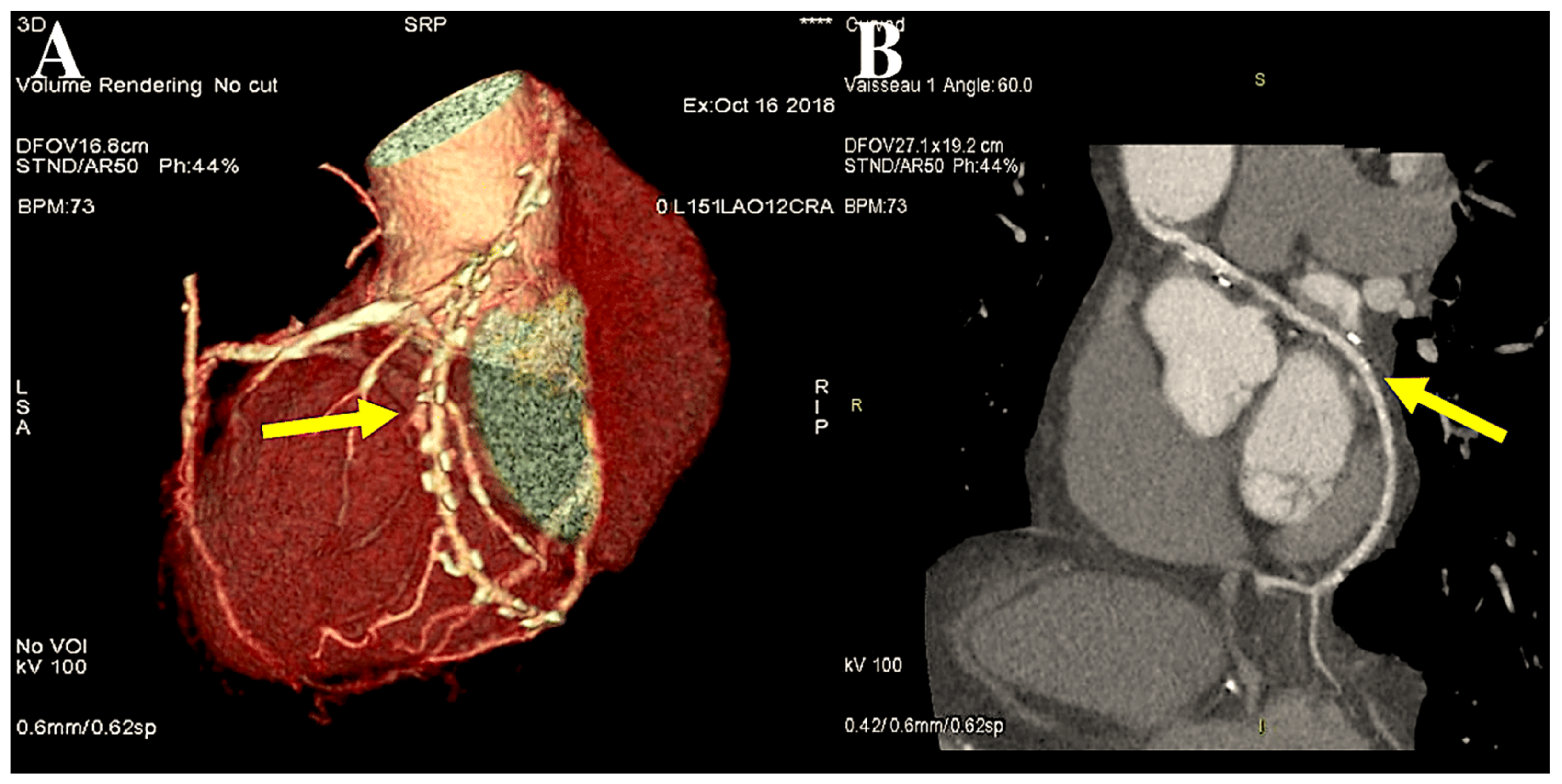
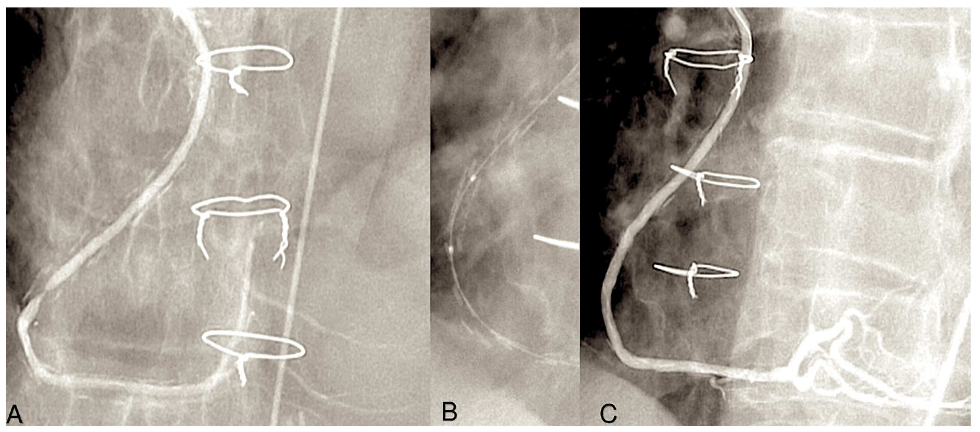
As a general principle, the surgeon advocates proximal anastomosis to the anterior portion of ascending aorta. Proximal anastomoses for individual conduits are achieved through the use of a suture to affix the graft end to side to an aortotomy in the proximal ascending aorta. A circular aperture measuring 3 mm in diameter is created in the aortic wall using a punch. The graft is then secured directly to the aorta using 7/0 Prolene sutures (Figure 1A,B).
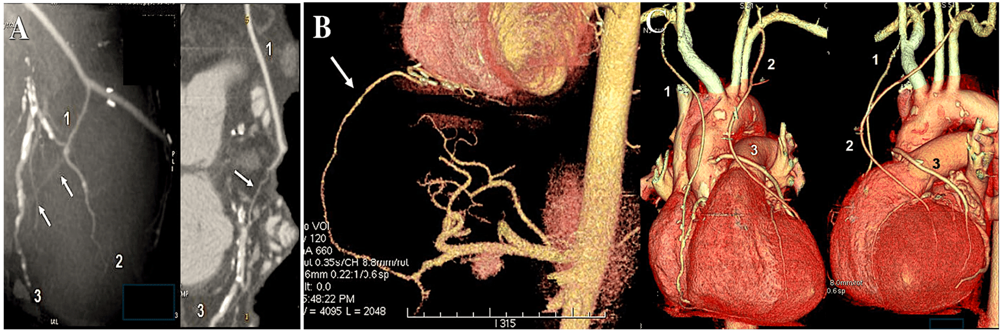
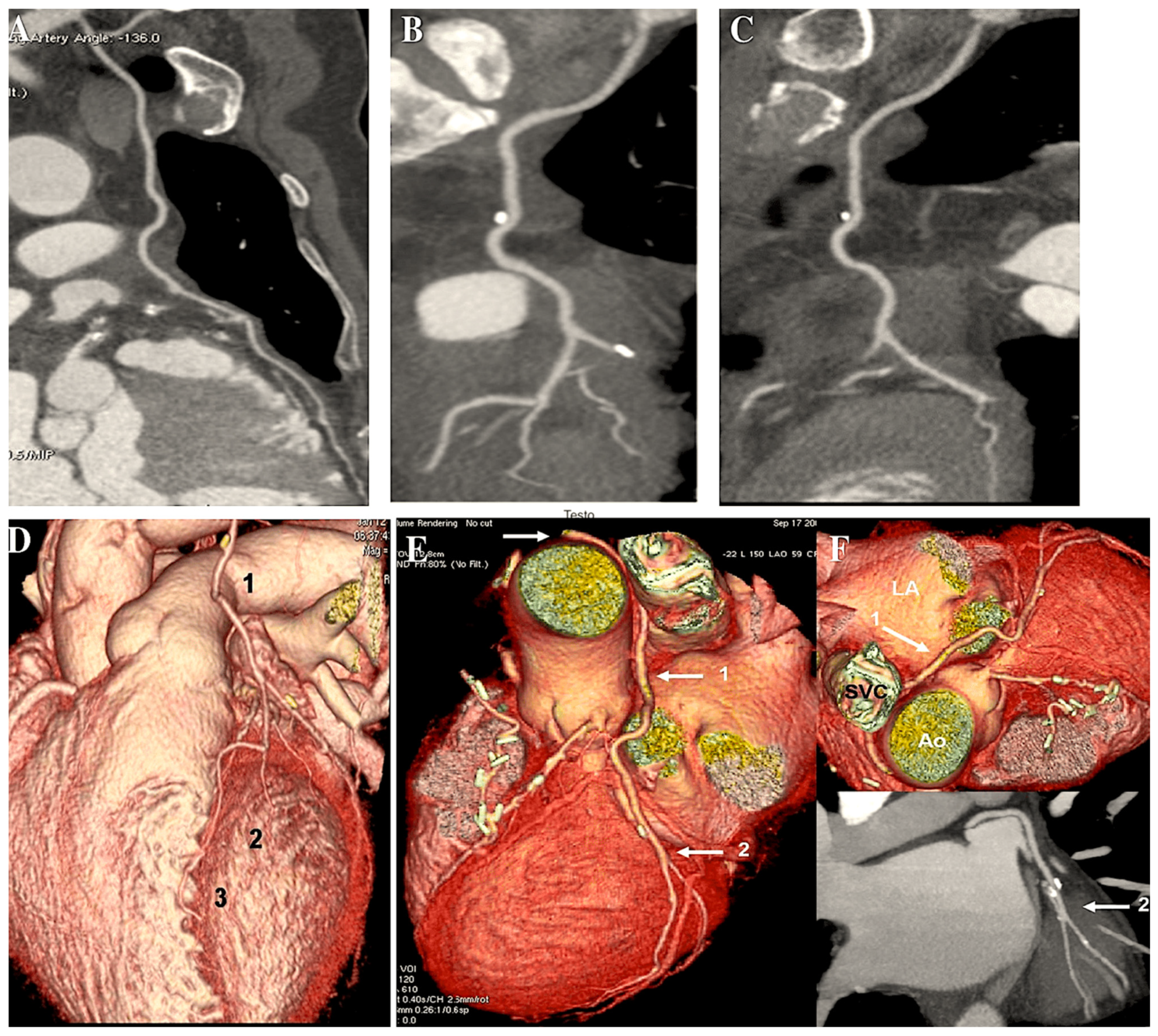
However, in cases of target vessels situated in the proximal obtuse marginal position, the graft utilised for CABG is anastomosed anteriorly or alternatively on the opposite side of the aorta through the transverse sinus (Figure 2A,B).
In the case of in situ arterial grafts (such as a left internal thoracic artery graft), the native arterial inflow is retained, thus necessitating an alternative approach (Figure 3).
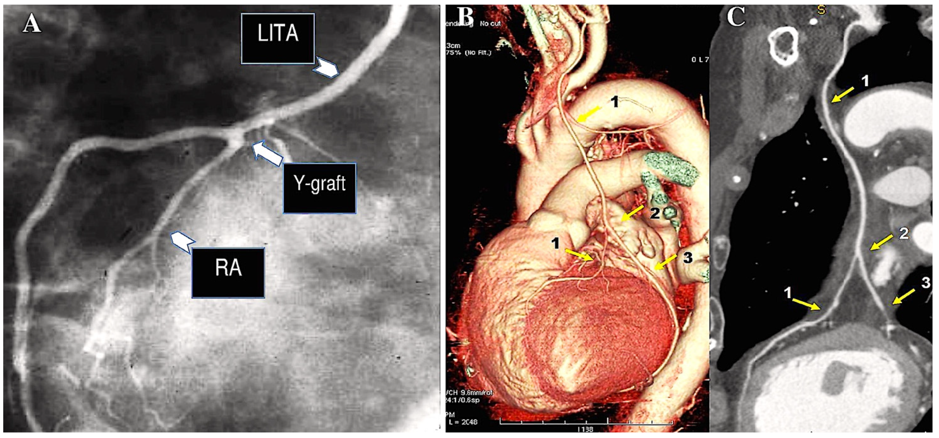
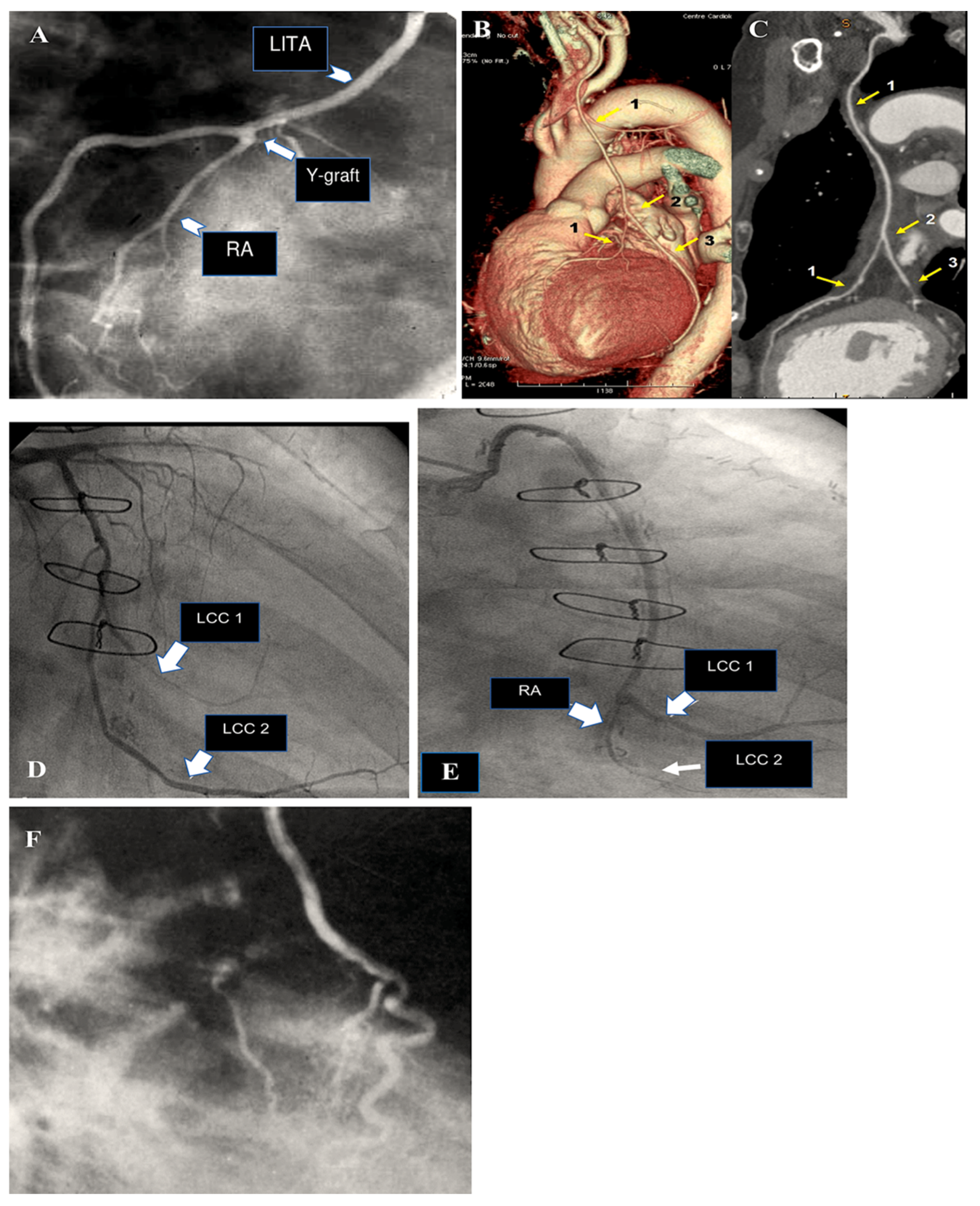
The RA conduit can be employed for sequential revascularisation and sutured as Y-graft to LITA (Figure 4A). Moreover, it is considered to be a prime location for performing proximal anastomosis on an additional RA conduit (Figure 4A). Such conduits may comprise an additional radial artery graft or a free RITA (Figure 4B,C).
The technique of proximal anastomosis of the RA to the LITA was initially described by Calafiore [37,38] and subsequently employed by other surgeons [9,21]. This approach involves the creation of a composite arterial graft, which is technically challenging. In the event of geometric distortion, the composite graft may become dysfunctional. Proximal RA anastomosis can be performed prior to the initiation of extracorporeal circulation. The optimal location for the anastomosis is identified as the first segment of the left ITA graft, located adjacent to the pleura and in close proximity to the pericardial cavity. It is optimal for the suture to be completed in a manner that orientates the two arteries in parallel. Furthermore, it is imperative to meticulously measure the extent of the RA graft as its length has the capacity to exert tension on the IMA pedicle (in the event that it is inadequate) or to cause the deformation of the RA conduit (if it is excessively lengthy). The conventional CABG procedure typically requires between three and four hours to complete. Patients typically remain in the hospital for a period of five to seven days following the procedure, with a recovery period of 6 to 12 weeks post-discharge [39]. It is important to consider certain technical aspects in relation to the radial artery.

2.1. Harvesting and Preparation of the Radial Artery

The left radial artery is selected for preferential harvesting in all individuals, regardless of handedness, for two primary reasons: firstly, the left RA has a higher probability of sparing in the event of transradial angiography, and, secondly, its removal can be conducted concurrently with the left internal thoracic artery dissection. The individual is placed in a position with the arm at an angle of 90° to the longitudinal axis of the body. Arterial blood pressure is monitored by means of a radial artery catheter placed in the opposite arm. Intravenous catheters are not introduced into the arm on the side of the radial artery harvesting as this would result in backflow from the infusion line into the operative area. The incision is made laterally, 2 cm above the wrist, and extends in a medial direction to the line where the elbow meets the wrist. The antebrachial fascia is excised above the vascular pedicle in a manner that avoids injury to the vascular pedicle and the surrounding tissues. In the proximal third of the forearm, the RA courses deep underneath the brachioradialis muscle, which is subsequently displaced laterally in a manner that avoids injury to the muscle and does not require its division. The multitude of collateral vessels are secured with metallic clips, and the vascular pedicle is removed in a single piece with the artery and both satellite veins. The majority of the dissection process can be undertaken without the use of electrically powered cautery devices [7,8,9,10,11].
The presence of anatomical variants of the RA necessitates minor technical adaptations. In rare instances, the location of the distal segment of the vessel may be in close proximity to the antebrachial fascia [40]. This represents a typical site of the vessel’s accessory RA in cases of rare vessel duplication. Nevertheless, the surgical approach does not deviate from the standard procedure. The radial artery may be extracted up to the point at which the brachial artery bifurcates. This is contingent on the number of grafts required and their relative extent, with a view to achieving optimal anastomosis. The potential hazard to the patient is the possibility of injury to the ulnar artery. Indeed, the bifurcation of the brachial artery is typically situated beneath the elbow, with the origin of the ulnar artery located in a deep posterior position, obscured by the muscle belly of the pronator teres. It is of the utmost importance to prevent injury to the vessel in question during the process of dissecting or ligating the RA graft.
Two anatomical indicators provide evidence that the bifurcation of the brachial artery is in proximity. The emergence of the recurrent radial artery, in conjunction with the presence of a dense venous network surrounding the radial artery, serves as an indication of specific conditions. It is advised that a separation of 1 cm be maintained from the point of brachial artery bifurcation. In rare instances, the RA may arise from either the proximal brachial artery or the axillary artery, which accounts for 14% of cases [40]. This anatomical divergence is of clinical consequence as an incision extending beyond the elbow should not be made if the origin of the RA is not identified in that region. The process of dermal regeneration may be affected when the interline of the elbow is divided. Following excision, the RA typically exhibits vasoconstriction and is, therefore, not suitable for immediate use in bypass procedures. It is recommended that the tissue be preserved initially in a solution comprising blood, heparin, and papaverine. The catheter is then inserted into the proximal end of the radial artery using a 16-gauge Cathlon catheter, followed by hydrostatic dilation at a low pressure using the same mixture. This enables the surgeon to completely release the constricting spasm and verify the bleeding control of the minor collateral branches. Numerous studies have demonstrated that this technique does not lead to the structural impairment of the vessel wall, particularly the intima [41]. An alternative approach that is equally effective involves disconnecting the distal end of the radial artery following complete heparinisation while preserving the proximal end connected to the brachial artery. The radial artery is then maintained in a beat-to-beat state against an anti-traumatic bulldog clamp, covered by a papaverine-impregnated sponge, until the complete relaxation of the spasm has occurred.
A further modification to the aforementioned harvesting techniques has been documented, including RA skeletonisation and the utilisation of an ultrasonic scalpel or endoscopic techniques [42,43,44]. However, the advantages of these alternative methods over the open procedure have yet to be conclusively established.

2.2. Risk for Hand Ischaemia

In an article published in 1929, Edgar V. Allen [45] outlined a clinical test for assessing the relative importance of the ulnar and radial arteries in delivering blood to the hand. The procedure involved clenching the fist and manually compressing both the radial and ulnar arteries until the complete exsanguination of the hand was achieved. Following the occlusion of the ulnar artery, the time taken for colouration to return to the extremity is meticulously recorded. The Allen test is deemed positive if full colouration is not achieved for all fingers within six seconds. Prior to catheterisation of the radial artery, this test has been a widely used diagnostic tool. In practice, numerous studies have demonstrated that the Allen test is not a reliable indicator due to the prevalence of false-positive and false-negative outcomes [46]. Additionally, it has been shown to have limited efficacy in predicting the presence of compensatory circulation via the palmar arches.
A number of initiatives have been implemented with the objective of enhancing the sensitivity of the Allen test. The use of a digital oximeter with a probe positioned on the thumb enabled the measurement of oxygen saturation in the radial artery region [47]. The results demonstrated that the normal oxygen saturation curve was disrupted by the occlusion of both forearm arteries. Furthermore, the failure of a normal tracing to re-emerge subsequent to ulnar artery re-opening indicated inadequate blood flow.
Similarly, the digital pressure was gauged with the aid of a pressure cuff positioned at the level of the proximal interphalangeal joint. It has been demonstrated that in the event that collateral circulation is functional, the pressure in question is maintained at the level of the thumb and fifth finger, even with the radial artery subjected to occlusion. Therefore, a systolic digit pressure reading at the level of the thumb that is below 40 mmHg is deemed to be a positive result as this value is indicative of a healthy collateral circulation [48]. Others have put forth the notion of utilising Doppler ultrasound technology for the assessment of the main artery of the thumb, otherwise known as the princeps pollicis artery. An alteration of the typical triphasic signal would signify an inadequate degree of collaterality from the ulnar artery [49,50,51,52]. Subsequently, the flow in the superior palmar arch was determined precisely by utilising a Doppler probe positioned at the third digit’s metacarpal region. A sustained reduction in the discernible Doppler signal in the palmar arch subsequent to RA compressive strain was considered an indicative marker of insufficient ulnar artery flow in a retrograde direction [53,54,55,56].
The implementation of the original Allen test or its aforementioned variations has the potential to result in the disqualification of a considerable proportion of prospective candidates for radial artery harvesting, with an estimated exclusion rate of 20–40%. A paucity of correlation has been observed between the Allen test and the anatomical structure of the blood vessels supplying the upper limb. Based on the author’s experience, the Allen test has consistently failed to identify any potential limitations for RA removal. Moreover, there has been no recorded instance of RA extraction being contraindicated on the basis of concerns regarding diminished blood supply to the hand. Two cases of technically induced stenosis at the site of the ulnar artery’s emergence, consequent to the proximal ligation of the radial artery, have been documented. This necessitated prompt corrective surgical intervention, with no further unfavourable sequelae. Moreover, it has been demonstrated that the removal of the radial artery in cases where patients have occupations that require the strenuous use of the upper limbs is safe and does not lead to any substantial functional impairment [57]. The aforementioned findings have also been observed in patient groups comprising the elderly [58].
A thorough anatomical study, supplemented by comparative angiographic analyses of arterial patterns in the upper limb, has demonstrated that the ulnar artery is a constant presence and that the likelihood of ulnar artery agenesia is essentially negligible. Pre- and postoperative measurements of blood flow, conducted via plethysmography or Doppler ultrasound, have revealed a modification in the configuration of the blood supply network within the forearm subsequent to the surgical excision of the radial artery. It is widely acknowledged within the scientific community that the flow in the distal brachial artery, located in the elbow region, serves as an indication of blood circulation within the forearm. Preliminary observations following the RA harvesting procedure have revealed that this flow remains unaltered. The enhanced flow in the ulnar artery during the early postoperative phase appears to be a contributing factor; however, no alteration in the dimensions of the artery could be identified [59]. It is hypothesised that the increased flow observed in the radial artery, whilst the ulnar artery maintains constant calibre, is attributable to peripheral resistance competition through the palmar arches. Conversely, approximately three months later, a marked and sustained dilatation of the ulnar artery is evident, accompanied by an approximate 15% increase in diameter [59].
In essence, the determination of whether to utilise this conduit should not hinge upon the Allen test. The author’s findings demonstrate that the radial artery can be safely harvested in all instances, with no evidence of hand ischaemia, contingent on the utilisation of an appropriate surgical technique. Other research teams have similarly reported extensive utilisation of the radial artery graft without any instances of postoperative ischaemic complications [57]. It is important to note that the RA is not a suitable graft in every circumstance. There are a number of contraindications for using the RA, which are related to the anatomical characteristics of the artery itself rather than to the potential variations in the distribution of vascular supplies to the upper limb.

3. Contraindications to Radial Artery Conduit Coronary Grafting

A number of contraindications have been studied. The radial artery is the dominant artery of the forearm and consistently gives rise to the interosseous artery, which irrigates the deep muscles. The procedure of removing the RA is safe in all instances [2,7,8,9,10,11,12,13,14,15,16,17,18,19,20,21,22,25,28,37,38,45,46,47,48,49,50,51,52,53,54,55,56,57] as its vascular territory is consistently maintained by collateral circulation provided by the ulnar artery. This is achieved through an augmented ulnar artery flux, which equates to an augmented vessel size [59]. The variations in the anatomy of the palmar arches are not relevant in the context of RA grafting. Furthermore, the Allen test and its modifications are not useful in the preoperative period [11]. However, it is essential to consider some of the potential anatomic variations [60] as they may impact the harvesting technique.
It is possible that the radial artery may have its origin in a higher position. The radial artery, in the typical case, has its origin in the brachial artery in the elbow area, located beneath the brachioradialis muscular tissue. On rare occasions (4%) [60], it may originate at a higher level, within the mid-section of the arm or in the axillary region. The radial artery courses superficially along the forearm, situated anterior to the brachioradialis muscle. In circumstances where the arterial origin is elevated, caution should be exercised to avoid extending the radial conduit dissection beyond the elbow region.
The high termination of the RA represents an anatomical feature that should be taken into account when considering the contraindications to harvesting the radial artery. The RA bifurcates at the wrist joint. One branch continues as a superficial artery along the anterior portion of the hand, whilst the other forms a deep artery that curves posteriorly in order to supply the posterior portion of the scaphoid bone. It is typical for these two branches to serve as the superficial and deep palmar arches, respectively. However, it is worth noting that in rare instances (approximately 1%), a high RA termination at the midpoint of the forearm may be observed. In such cases, the radial artery may be utilised for CABG. It is further noted that when the calibre of the radial artery branches is adequately sufficient, they naturally exhibit a Y-shaped structure [61]. In select instances, characterised by a high bifurcation, the deep branch of the RA follows an aberrant trajectory. It exhibits an external curve and traverses the external border of the forearm, subsequently running along the dorsal aspect of the wrist.
The aetiological disease process, known as atheromatous disease, has a propensity to spare the arteries of the upper limb. However, proximal lesions do occur with some frequency. When left subclavian artery stenosis is identified and occurring in up to 2% of patients undergoing coronary surgery [62], the LITA flow can be at risk, and endovascular stenting may be the preferred option preceding arterial grafting [62]. In other instances, with an obstruction occurring in a location proximal to the upper limb arteries, it seems preferable to avoid the removal of the RA [9,10,11]. It is not uncommon for the RA itself to be the site of atheromatous plaque lesions. Patients with insulin-requiring diabetes mellitus and those with phospho-calcic metabolic disorders, as observed in long-lasting renal insufficiency, may exhibit non-obstructive medial calcifications [63]. In instances of multiple atheromatous lesions accompanied by calcifications, there is a possibility of observing obstructive alterations of the RA, such as stenotic plaques or thrombotic occlusions [64]. In conclusion, it can be stated that 6% of patients undergoing coronary surgery present with atheromatous lesions of the RA. These findings constitute definite contraindications in the context of RA grafting.
Forearm wounds and bone fractures have the potential to result in RA injury. Nevertheless, the predominant etiological factors underlying traumatic injury to this vessel are of an iatrogenic nature. Arterial puncture for the purpose of obtaining blood gas measurements or catheterisation for the monitoring of systemic pressure may result in the induction of fibrotic stenosis or localised dissection. However, it has been observed that the proximal part of the RA retains its integrity in such instances [11]. This is not the situation that arises in the context of the retrograde catheterisation of the RA, which is employed for the purpose of coronary angiography. The introduction of the material can induce spasm, and the shearing stress caused by the advancement of both the guidewire and the angiography catheter in a collapsed artery almost inevitably leads to significant intimal damage [65,66], which can propagate throughout the entire conduit. The reported incidence of occlusion of the radial artery following transradial catheterisation is as high as 7% [67]. The insertion of even larger sheaths for purposes of percutaneous coronary intervention (PCI) can result in the infliction of severe arterial trauma, including but not limited to wall disruption, false aneurysm [68], and arterio-venous fistula [69]. The incidence of postoperative occlusion in RA conduits that have undergone prior retrograde catheterisation and have been employed as coronary grafts is markedly elevated [70]. Therefore, it is recommended that transradial artery catheterisation be regarded as an absolute contraindication to RA grafting [11]. As it is most commonly conducted on the right side, the RA on the opposite side can then be used for coronary grafting. Consequently, even in patients who are left-handed, the left side should always be the preferred choice for harvesting.

4. Grafting of the Radial Artery: Clinical Results

Coronary artery bypass grafting is a well-established surgical procedure with a documented history of favourable short-term outcomes [71,72,73,74]. The advent of advanced CABG techniques and the implementation of quality improvement strategies, facilitated by the extensive participation of CABG centres in the Society of Thoracic Surgeons National Adult Cardiac Surgery Database, have contributed to a notable decline in observed mortality rates over the past decade, despite minimal alterations in estimated risk [29,30,75].
While the surgical team bears significant responsibility for the success of a CABG operation, the effectiveness and reliability of the procedure also rely considerably on the input of other members of the multidisciplinary management team and on compliance with established perioperative and postoperative protocols [1,2,76]. The risks associated with CABG are most significant during and immediately following the surgical procedure. It is essential to carefully evaluate the potential short-term complications of CABG in light of the long-term benefits that the operation can offer.
Following its revival over two decades ago, the radial artery has been employed effectively as a conduit for coronary bypass grafting by numerous surgical institutions. Extensive data sets are currently available for evaluating the clinical benefits of utilising the radial artery. The fate of the RA graft has been assessed via repetitive conventional or CT angiographies, facilitating the identification of factors influencing RA graft patency. Additionally, assessments have been conducted in comparison with other conduits.

4.1. Early Clinical Results

The in-hospital mortality and incidence of perioperative myocardial infarction (at 1.0% and 2.0%, respectively) were comparable following radial artery grafting to the rates observed in patients undergoing conventional coronary surgery using alternative types of conduits [10,11,77,78,79]. In cases involving increased risk factors, including left ventricle dysfunction [80] or coronary reoperation [81], the postoperative mortality and infarction rates exhibited no significant change when the radial artery was employed. Additionally, a single study demonstrated that the utilisation of exclusive arterial conduits, encompassing the RA in conjunction with the LITA, is associated with a diminished in-hospital mortality rate when compared to patients in instances where vein grafts constituted the treatment modality [82].
Our group’s findings suggest that, in some patients with unstable haemodynamic conditions, the use of the radial artery may be an effective approach. In patients experiencing haemodynamic shock with a rupture of the posterior papillary muscle, an occlusion of the right coronary artery can be effectively treated by anastomosing the right coronary artery using the RA as a conduit. Additionally, in cases of severe ischaemic mitral regurgitation where revascularisation of the circumflex or right coronary artery territory is required, the radial artery has been a valuable tool [83,84,85,86].
It has been observed that arterial conduits exhibit a higher propensity for spasms when compared to their venous counterparts. It is inevitable that mechanical stimuli will result in a spasm of the RA when it is subjected to surgical dissection. It is, therefore, essential that this spasm is released before the coronary anastomoses are constructed in order to prevent a low flow situation that could precipitate early graft thrombosis. It is, therefore, necessary to employ intraoperative pharmacological and/or mechanical manoeuvres in order to obtain a fully dilated conduit. On infrequent occasions, postoperative angiograms have identified the presence of localised stenosis resulting from an episode of spasm [9]. In a considerable number of circumstances, the intraluminal infusion of nitroglycerin and/or linsidomine facilitates the dissolution of the spasm. In some cases, the diagnosis is made in retrospect when a subsequent examination confirms the resolution of the stenosis [9]. A spasm can manifest at the proximal extremity of the graft where it is in proximity to the angiography catheter, as observed in previous studies [87], or at a more distal location [9]. It is probable that some of these spasms occurred during surgery and were not fully released by the usual intraoperative manoeuvres. Consequently, they may have persisted throughout the postoperative period. It is important to note that spasm can mimic a permanent narrowing of the vessel, potentially leading to unwarranted PCI, as previously documented in the literature [88]. While radial artery spasm has been documented in a limited number of isolated case reports to have been associated with electrocardiogra changes and haemodynamic impairment, it has been observed to be well managed and rarely problematic in our series [89,90]. Radial artery spasm has consistently manifested as neither clinically nor electrically evident and was coincidentally identified through a routine angiogram [9].
The administration of calcium channel inhibitors to patients who have undergone arterial grafting is a topic that is the subject of considerable debate. From 1989 to 1994, our institutional policy was to prescribe a calcium channel blocker (diltiazem) in all patients with radial artery grafting [9]. Given its adverse inotropic impact, the intravenous administration of diltiazem was rapidly discontinued in the operating room and intensive care unit settings. Instead, the medication was only administered orally. Upon the completion of the long-term follow-up period, it was determined that among patients who had undergone a radial artery graft, only 59% were still receiving calcium channel blocker treatment at the five-year mark [10], and 48% were still undergoing this treatment at the nine-year mark [10]. The results of various statistical examinations did not yield any evidence to suggest that pharmacological medications act as individual factors influencing the patency of radial artery grafts [10,11]. In light of the aforementioned clinical findings, as well as those observed in stress myocardial scintigraphy and angiography, other reports have also demonstrated the lack of an evident benefit associated with the utilisation of calcium channel blockers in RA grafting procedures [91,92,93]. It can, therefore, no longer be posited that RA grafting represents a definitive criterion for the prescription of calcium channel blockers. It is important to note that contemporary practice involves the administration of antithrombotic therapy to nearly all patients who have undergone coronary bypass grafting in addition to one or more classes of antianginals. Given the potential for pharmacokinetic interplay between these pharmaceutical agents, it is challenging to reach a definitive conclusion regarding the impact of these medications on the patency of arterial grafts.
The surgical incision made on the forearm skin typically heals rapidly, and the occurrence of local infection is uncommon. A re-examination of the wound for the development of a haematoma is also uncommon, with the overall incidence of local complications estimated to be approximately 1% [94,95,96]. This figure contrasts with that seen following saphenous vein harvesting, where local complications are more prevalent, particularly when the vein is extracted from the thigh and in patients of advanced age [96]. This observation has led several researchers to conclude that RA graft recipients experience a reduced length of hospitalisation, fewer readmissions, and lower overall hospitalisation costs [94,95,96]. Other studies have made a comparative analysis between the postoperative results of patients who have undergone coronary bypass surgery with the RITA versus RA as a secondary graft in addition to the LITA to the left ascending coronary artery. A higher incidence of sternal wound infection or dehiscence has been observed in patients who have received a RITA [97,98,99]. Additionally, these patients exhibit a higher incidence of postoperative bleeding and a prolonged hospitalisation period [97,98,99].
A number of studies have identified significant concerns associated with the utilisation of the bilateral internal thoracic artery in association or not with RA graft, particularly in relation to the elevated risk of sternal wound complications and mediastinitis. The results of the most extensive meta-analysis on this subject indicated that the incorporation of a second internal thoracic artery into the left internal thoracic artery grafting procedure resulted in a notable increase in the incidence of sternal complications (with a single internal thoracic artery presenting a relative risk of 0.62 and a 95% confidence interval of 0.55–0.71) [100]. Patients with diabetes and pulmonary disease are at an elevated risk. In ART, the incidence of sternal wound complications increased from 0.6% in patients receiving a single ITA to 1.9% in those who underwent bilateral internal thoracic artery surgery. This equates to an incremental difference of 1.3%, which is equivalent to a number needed to causally link the observed effect to the observed outcome of 78 individuals [13,14].
Surgical techniques for harvesting ITAs may influence the extent of the survival benefit. It is noteworthy that the better favourable early results resulting from bilateral internal thoracic artery grafting have been observed in both diabetic and non-diabetic patients [101]. For example, two reports based on systematic reviews have both emphasised the pivotal role of harvesting the ITA via the skeletonisation procedure. The correlation between deep sternal wound infections was lower in both diabetic and non-diabetic cohorts when the skeletonisation technique was preferred to the pedicled harvesting technique [100,102].
Ultimately, the surgeon’s overall strategy for identifying candidates for coronary revascularisation by using arterial-only conduits or alternatively incorporating saphenous vein grafts continues to have important implications. This is particularly the case with regard to the choice of the bilateral internal thoracic artery for the coronary revascularisation procedure. Accordingly, it is advised that the avoidance of bilateral internal thoracic artery be considered in patients exhibiting specific potentially morbid characteristics, particularly when these characteristics co-exist (overweight status, diabetes, and underlying respiratory conditions). Additionally, consideration should be given to the avoidance of bilateral internal thoracic artery in patients undergoing steroid therapy or immunosuppressive therapy [9,10,11,12,13,14,15,16,17,18,19,20,21].
Neurological complications may arise as a consequence of damage to the superficial branch of the radial nerve, which courses in close proximity to the radial artery. This phenomenon has been documented in 8% of cases, manifesting as numbness and paresthesia of the thumb. In the majority of cases, these symptoms resolve within a year [10,12,103]. In no case does this trivial adverse event result in any appreciable degree of clinically significant functional inability. Electroneurography and motor-unit potential studies have demonstrated that minimal conduction alterations can be observed even when neurological symptomatology has been documented [104]. It was reported that there were exceptions to the rule, namely, cases of extreme and enduring pain in the forearm [105]. However, no anatomical basis for this phenomenon was identified, and it was, therefore, concluded that this condition, known as causalgia, was most likely of a co-existing psychological disturbance. It is uncommon for transient dysesthesia in the territory of the ulnar nerve, fourth and fifth fingers, to occur as a result of traction on the brachial plexus, which is caused by the upper extremity being positioned incorrectly during the surgical removal of the radial artery. Notwithstanding these minor adverse effects, quality of life studies have demonstrated the safety of RA removal, which was responsible for merely residual discomfort [17,18].
Stroke continues to be the most severe complication of CABG, affecting 1% to 2% of patients in the peri perioperative phase [106]. It is well documented that a variety of factors can contribute to an increased risk of stroke. These include a personal or family history of neurological events, increasing chronological age, peripheral or cerebrovascular disease, and diabetes [107,108]. Additionally, aortic atherosclerosis represents a significant risk element in the context of stroke occurrence following CABG, largely due to the inherent necessity for both the mechanical manipulation and clamping of the ascending thoracic aorta [109]. The utilisation of a single aortic cross-clamp, in conjunction with the application of epiaortic ultrasonography throughout the CABG procedure, has been demonstrated to result in a notable decline in the incidence of stroke events amongst patients subjected to this surgical approach over the extended period of the earlier decades of the current century. Additionally, neurocognitive impairment has been linked to CABG, notably in relation to the utilisation of cardiopulmonary bypass. Nevertheless, these correlations have been identified in studies lacking control for confounding variables over an extended period. Randomised trials investigating CABG conducted with and without cardiopulmonary bypass, as well as CABG in comparison to PCI, have not substantiated these observations [110,111,112]. The prevailing opinion regarding neurocognitive impairment following CABG is that it is attributable to a confluence of factors. These include the immediate and short-term consequences of major surgery, as well as the long-term effects of advanced age, depression, and a shared predisposition to neurocognitive dysfunction and coronary artery disease [113,114].

4.2. Late Clinical Results

A common misconception among patients and some physicians is that CABG is a definitive cure for coronary artery disease. In reality, CABG does not halt the advancement of native coronary artery disease, and internal thoracic artery, radial artery, and saphenous vein grafts are susceptible to failure.
The progression of native coronary artery disease and internal thoracic artery and saphenous vein grafts can fail. Nevertheless, the advancement of disease and the deterioration of vein grafts can be mitigated by the implementation of robust secondary prevention strategies employing pharmacological interventions.
The results of a most recent investigation into our experience of radial artery grafting, comprising 819 patients, revealed that the five-year, ten-year, and fifteen-year survival rates were 95%, 81%, and 70%, respectively (Figure 5) [9,10,11,12,13].
Given the older age at operation, these results are more favourable than those of historical series of CABG, which included primarily vein grafts and demonstrated survival rates of 56% and 62%, respectively, at 15 years [115,116]. It is noteworthy that other studies have documented comparable long-term survival rates following RA grafting, with reported rates of 83% and 79% at 7 and 10 years, respectively [117,118,119]. A contemporary series of coronary bypass grafting with the LITA and SVG demonstrated a similar survival to that previously reported [120]. It is noteworthy that other studies have documented comparable long-term survival rates following RA grafting, with reported rates of 83% and 79% at 7 and 10 years, respectively [117,118,119]. A contemporary series of coronary bypass grafting with the LITA and SVG demonstrated a similar survival to that previously reported [120]. A freedom from the cardiovascular-related death rate of 89% at 15 years is observed following RA bypass grafting. Among the principal contributors to cardiac mortality within this cohort have been identified congestive heart failure, arrhythmia, stroke, and calcified aortic valve stenosis [13]. Our findings indicate that a preoperative ejection fraction below 40% represents a statistically significant predictive factor for late death [12].
Two comprehensive case-matched studies were conducted involving patients who met the same demographic criteria and who received either a RA or SVG as a second conduit to supplement the LITA-LAD graft [121,122]. These studies were the first to demonstrate that using the RA in place of a vein resulted in a reduced risk of mortality [121,122], especially beyond the third postoperative year [122]. This finding was further corroborated by subsequent observation-based studies [118] and RCT [123]. When focusing on specific patient subgroups, the survival benefit of utilizing an RA instead of an SVG becomes particularly evident, particularly in female patients [124], in cases where endarterectomy is conducted on the target coronary artery [125] and in instances of grafting in a sequential manner [126]. A similar benefit is identified in the context of coronary repeat operations [127]. In contrast, long-term survival rates in diabetic patients are not significantly enhanced by the utilisation of radial arteries as a conduit for grafting [128]. Zacharias’ case-matched study was conducted on a prolonged timescale of 12 years with the aim of assessing the long-term survival benefit of employing the totality of arterial grafting techniques, with at least one RA graft used in nearly 92% of total patients [122]. The use of a total-arterial grafting technique has been shown to result in a superior 12-year survival rate compared to the standard single LITA and SVG procedure. This advantage is particularly evident in the context of three-vessel disease, although it is more modest in patients with two-vessel disease [122].
The plethora of confounding variables poses a significant challenge for even the most sophisticated statistical methods, which often struggle to identify the graft type as a risk factor for death. In contrast with the aforementioned literature the randomised trial undertaken by Buxton (Radial Artery Patency and Clinical Outcome trial, RAPCO) did not demonstrate a statistically significant improvement in survival rates associated with the use of the RA in comparison to an SVG in individuals aged over 70 years [17,129]. However, the patient cohorts were relatively small, which may have contributed to the lack of statistical significance observed. In the subgroup in which the target was the right coronary artery, there was no impact observed on patient survival as a result of the use of the RA [130].

5. Complication

The recurrence of angina is a common occurrence subsequent to CABG. The probability of angina recurrence increases with the passage of time, with an estimated incidence of approximately 40% at 10 years [131]. While some authors have deemed the postponement of late angina recurrence to be clinically inconsequential [131], the incidence of angina recurrence is lower when radial artery grafting is employed, occurring in 18% of patients at 9 years [12]. Furthermore, in the context of undergoing arterial grafts, patients were subjected to a rigorous course of treatment with a range of antianginal medications. This included the administration of beta-blockers (70%), calcium channel blockers (48%), ACE inhibitors (49%), nitrates (13%), or a combination of these agents (14%). Other pharmacological agents employed included central antihypertensive drugs such as molsidomine, nicorandil, and trimetazidine [12]. Sergeant asserts that the recurrence of angina following coronary artery bypass grafting has a negligible effect on patient survival and lacks prognostic value for imminent infarction [131].
In historical cohorts comprising predominantly of vein grafts, the occurrence of acute myocardial infarction (AMI) reached as elevated a rate as 16% at the 10-year mark [115]. A more recent survey of arterial grafting revealed an occurrence of only 2.1% at nine years, with rare instances of sudden thrombosis in the RA graft [12]. The vast majority of patients undergo antithrombotic medication, with aspirin being the most common (74%), followed by clopidogrel in cases of previous stenting or multifocal atheroma (20%) and orally administered anticoagulants, predominantly due to atrial fibrillation (18%). The administration of antithrombotic medications is only contraindicated in the event of a serious medical condition (3%) [12].
Congestive heart failure represents the leading cause of cardiovascular mortality in the contest of coronary heart disease (CHD). In a cohort of patients who underwent renal artery angioplasty and stenting, the incidence of congestive heart failure symptoms was observed in 11% of cases at the 9-year mark [12]. As previously stated, all patients were administered an intensive pharmacotherapy regimen, comprising angiotensin-converting enzyme inhibitors in 49% of cases. A modest yet statistically significant reduction in the mean ejection fraction was observed over the course of the follow-up period, from 57% preoperatively to 55% at the nine-year mark. Therefore, arterial grafting with the radial artery affords a long-term safeguard of myocardial contractility against the progression of CHD [12].
Based on our most recent investigation, it is estimated that 13% of patients with a radial artery conduit used for CABG require percutaneous intervention, with a mean of 6.5 years after surgery [12]. At 15 years, one in four individuals will have had at least one percutaneous intervention. In this clinical trial series, 2/3 of the procedures were performed on a native coronary artery [12]. Because the myocardial territory was partially protected by the patent grafts, the PCI of the left main or proximal LAD coronary artery could be performed safely. The direct PCI of a stenosed arterial or venous graft was performed in rare instances. Stent implantation was performed in the overwhelming number of patients, and balloon dilatation alone was used only in the early stages of the present work.
In our experience, the necessity for reoperation is 2.3%, and the Kaplan–Meier free from the reoperation rate is 96% at 15 years [12]. In practice, since most reoperations are indicated primarily for valvular failure, the actual need for repeat surgical revascularisation is only 0.5%. The main causes of repeat surgery are calcified aortic stenosis and ischemic mitral regurgitation [12]. In previous series, in which SVGs were the primary surgical technique, the incidence of reintervention at the designated time interval was fourfold higher [115]. In the matched trial by Cohen and colleagues, freedom from non-fatal cardiac events, defined as rehospitalisation, myocardial infarction, percutaneous coronary intervention, and reoperation, was longer when RAs rather than SVGs were used to supplement the left internal thoracic artery-to-coronary artery bypass graft [121].
The radial artery presents a number of benefits in comparison to the right internal thoracic artery. It offers a highly adaptable alternative conduit, with a diameter and length that make it suitable for use in all coronary targets, including those at the most distal locations. Additionally, the radial artery can be retrieved at the same time as the LITA, reducing the overall time required for the procedure. The sole restriction associated with the RA graft is its propensity to become affected by atheromatous deposits, a phenomenon that occurs with greater frequency than that observed with the ITA. Borger et al. [99] evaluated the clinical advantages of complementing the LITA-to-LAD graft with the RA instead of the RITA. The findings presented in this publication are based on the experience of a single centre and, as such, remain a matter of debate [132,133]. Nevertheless, given that the employ of radial artery provides a lower wound complications than the BITA harvest [97,98,99], the choice of this conduit as the second graft to accompany the LITA represents a fruitful avenue.

6. The Destiny of Radial Artery Grafts

The patency of RA is one of the most important tools in the CABG. While conventionally, angiograms were performed as a matter of routine in the early days of RA grafting [9], guidelines have modified our approach to the assessment of graft function [9,77]. Over the past decennium, conventional angiography has been performed solely in cases where evidence of myocardial ischaemia has been identified. Due to the minimally invasive nature of CT angiography, it can be more readily mandated for asymptomatic patients and the elderly. It has been shown to be a good screening tool for assessing graft perfusion [134,135,136], and this method has been widely used to assess long-term RA graft patency [12,137]. The patency of the radial arterial graft is 100%, 93%, 83%, and 83% at 1 month, 1 year, 5 years, and 10 years, respectively [9,10,137], and three different angiographic studies have reported 1-year, 5-year, and long-term (more than 5 years and as far as 20 years) outcomes [137]. As two-thirds of patients with an occluded RA graft remained clinically asymptomatic in the long term, RA graft occlusion does not inevitably lead to the recurrence of angina [137]. If an ECG stress test or scintigraphy is performed, myocardial ischaemia is often not detected [137].
The findings of other researchers corroborate the conclusion that the vast majority of RA grafts remain patent when examined in the immediate postoperative period [138,139]. Additionally, it has been observed that some loss of patency occurs during the initial postoperative year, with a remaining patency rate of 90–93% [140,141,142]. In a comparative analysis of our series with other published data, analogous or moderately elevated patency rates have been documented in the intermediate term follow-up period: 95%, 89%, and 88% at 4 years [117], 5 years [143], and 8 years, respectively [144].
A comprehensive analysis was conducted on all serial angiographic controls collected over a 20-year period as part of a study undertaken by our research group. Out of the total number of patients included in the study, 563, approximately half underwent at least one angiographic control. A total of 1427 coronary bypass grafts, comprising 629 RA conduits, underwent opacification. A partitioning of the angiograms into four equivalent categories at distinct time intervals revealed that the majority of RA occlusions transpire in the initial six months. In the period extending further than one year, radial artery graft functionality demonstrates considerable stability, with minimal deterioration observed over the course of up to 20 years [12].
The term “string sign” is used to describe a condition characterised by a diffusely occurring narrowing of the entire graft, which is refractory to the effects of in situ vasodilators. This phenomenon is more frequently discernible on preliminary postoperative angiograms, with an estimated prevalence of approximately 7% in RA grafts within the initial postoperative year [9,140,145,146]. The phenomenon of “graft involution” is thought to arise from a process of competitive flow, as evidenced by a number of previous investigations [145,147,148]. Indeed, when a stress test is conducted, it is uncommon for ischaemia to be observed in the distribution associated with the string sign [140]. Some have postulated that the string sign may be precipitated by the administration of alpha-adrenergic medications prior or during the perioperative interval [146]. It is noteworthy that the regression of the RA string sign is exceedingly uncommon [147]. In such cases, the grafts in question have ceased to function, and they should, therefore, be classified as non-perfused.
It is imperative to differentiate between a string sign and vasospasm of radial artery. The concern that vasospasm might occur due to the muscular composition of the radial artery vessel wall has been allayed by evidence of progressive morphofunctional remodelling of the artery towards an elastomuscular configuration following implantation in the coronary circulation [149]. This is likely to be the underlying anatomical reason for the lack of efficacy of long-term antispastic therapy in patients with RA grafts [93] despite the continued widespread use of such therapy in the field of surgical practice [150].
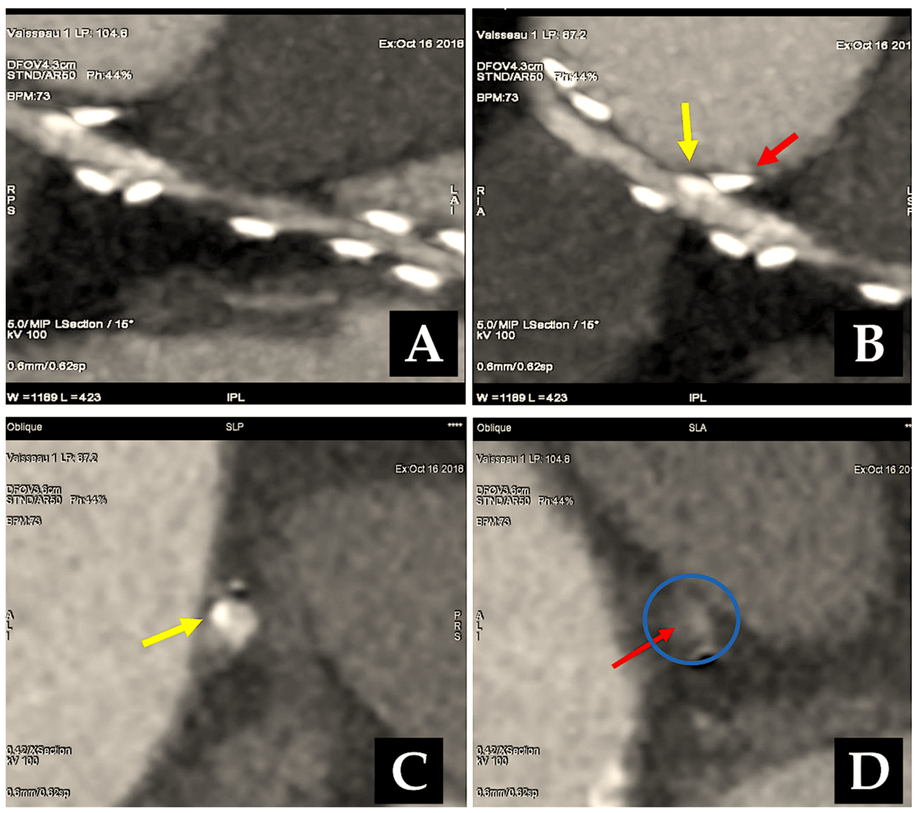
The most common type of angiographic failure observed in radial artery grafts is total obstruction, although a string-like appearance has also been documented. In rare instances, a localised stenosis of the radial artery graft has been identified [151]. Our own experience, spanning two decades, indicates that 6% of RA grafts exhibited stenosis on angiographic controls [151]. In some cases, stenosis was identified at either the proximal or distal anastomosis. In such instances, the potential culprits included a flawed surgical technique or intimal hyperplasia. In more frequent instances, stenosis affected the bodily region of the RA graft, thereby posing challenging pathophysiological concerns. It can often prove problematic to definitively exclude a spasm that is refractory to in situ vasodilators. If identified at an early stage following surgery, the stenosis can be treated with balloon dilation without the need for stenting [151]. If the stenosis is not identified in a timely manner, it is typically of an organic nature. It is possible that some narrowings existed prior to surgery involving the radial artery at the forearm. The development of RA stenosis may be attributable to the presence of an atheromatous plaque that had not been identified at the time of the initial surgical procedure. An additional possibility is that it is associated with fibrosis resulting from arterial trauma caused by inadequate harvesting techniques or prior intravascular procedures. It has been demonstrated that transradial angiography often results in intimal damage and/or medial dissection [152]. In certain instances, the stenosis of the radial artery was identified as a subsequent occurrence, as evidenced by instances where the graft was found to be intact in previous angiograms [151]. It seems plausible to suggest that these vessels may have been the focus of a preceding minor perivascular injury, which subsequently developed into a clinically significant flow-limiting stenosis. It can be reasonably deduced that the occurrence of RA graft stenosis may be prevented by the implementation of a systematic preoperative echo-doppler screening process, coupled with the deliberate preclusion of all calcified RAs and prior interventionally treated conduits. It is possible to safely treat RA graft stenoses by means of PCI and stenting, with the result being durable [151,153]. In regard to percutaneous interventions for venous graft disease, a significant challenge persists due to the high incidence of periprocedural morbidity associated with embolic complications from atherothrombotic detritus (Figure 6) [154].

Determinants of RA Patency

A number of studies have demonstrated that diabetes represents a risk factor for the occlusion of RA grafts [155,156,157]. The majority of angiographic investigations conducted for the purpose of determining the patency of the radial artery are based on the presence or absence of symptoms and thus do not provide an accurate representation of the true state of the patency of the graft. There is a notable increase in the occurrence of graft occlusion when the rationale for angiographic investigation is predicated on the presence of myocardial ischaemia, with an estimated doubling of incidence rates [158,159]. A similar phenomenon is observed in the case of the RA graft, where the occlusion rate at seven years is 12% in asymptomatic patients and 26% in those exhibiting clinical, electrocardiographic, or scintigraphic indications of ischaemia [12].
The identity of the target coronary artery serves as a significant predictor of the graft’s patency, regardless of whether the graft is of a venous [160] or arterial [161] nature. Conduits placed on the left anterior descending coronary artery with a substantial supply from the surrounding perforators demonstrate the most favourable outcome in terms of blood flow, with the diagonal and obtuse marginal branches of the left coronary artery exhibiting a comparatively higher success rate. Given its limited territorial scope, which encompasses primarily the thin right ventricular myocardium, the right coronary artery exhibits the lowest recorded propensity for maintaining patency [160,161]. Similarly, the viability of radial artery conduits is contingent upon the dimensions of the intended coronary target [91,143,145,159,162,163]. The statistical analysis reveals that RA graft longevity for targets of the RCA is inferior to that for targets of the left anterior descending artery, with a borderline correlation observed when compared to the left coronary circumflex perfusion territory. A review of our experience revealed that the number of RA grafts used for anastomosis to the LAD was insufficient for a meaningful statistical analysis. However, for the other targets, the 7-year patency of RA grafts decreased from 92% to 82% to 78% for the diagonal, the obtuse marginal, and the right coronary artery, respectively [10].
It has been established that competitive flow represents a significant contributing factor to the development of failure in radial artery grafts. The anastomotic permeability of targets with moderate stenosis is inferior to that of vessels with high-grade stenosis [10,140,143,145,162]. The definition of critical stenosis remains a topic of debate, with some authors proposing 70% and others 90% as the threshold [164]. The findings of the Maniar and colleagues’ investigation [162] indicated that the mean degree of stenosis for patent anastomoses was 82%, compared to 71% for obstructed anastomoses. This suggests that the threshold for flow competition may lie between these two values. In cases where coronary bypass surgery is combined with valve replacement, the decision to perform coronary bypass often hinges on the identification of coronary stenoses through conventional preoperative angiography rather than on the presence of myocardial ischaemia. The risk of performing coronary bypass grafting on a coronary artery with a low-grade stenosis, which could potentially lead to competitive flow, is higher in such cases. This results in a higher rate of graft occlusion, as evidenced in previous studies [12]. Similarly, other researchers have shown that RA graft occlusion is more prevalent in patients with minimal or no preoperative angina [143]. Another situation at risk for competitive flow is the performance of a coronary bypass for proximal left main stenosis. The obtuse marginal RA graft is particularly susceptible to competitive flow due to the unimpeded retrograde flow from the LITA-to-LAD graft towards the left coronary circumflex artery plexus [11]. Ultimately, an ample compensatory circulation may rival the flow from a graft that has been anastomosed to a persistently obliterated coronary artery [11].
It has been demonstrated that sequential grafting has the effect of enhancing vascular patency, which can be attributed to the augmented distal runoff. In general, sequential RA conduits demonstrate an enhanced patency rate in comparison to grafts with a sole anastomosis, reaching 91% at seven years versus 82% in the event of unifocal anastomosis grafts [12].
In some cases, only one end of the graft may remain patent, while the remainder of the graft is rendered non-functional, resulting in a “string” or “occlusion” effect. This phenomenon has been observed in both experimental and clinical settings [11,126].
The utilisation of composite arterial conduits with the radial artery anastomosed to the LITA has gained considerable recognition within the scientific community. This approach has been extensively documented in numerous studies [163,165,166,167,168,169,170]. The use of shorter segments of the radial artery is a significant advantage of this technique as it allows for the maximisation of all-arterial revascularisation. Those in favour of this method maintain that an increase in shear stress is generated when the radial artery is sutured to the aorta, given that the aorta has a greater calibre than the artery from which the radial artery naturally originates. There is still no clear consensus on whether RA should be proximally anastomosed to the IMA rather than to the aorta, with the current evidence base remaining inconclusive. Comparative studies have yielded conflicting results. Some have reported similar patency rates [117,169], while others have observed the opposite outcome [163,170]. Some have suggested that LITA anastomosed RA grafts are more susceptible to the effects of concurrent flow [163], which may result in higher occlusion rates at both one-year and five-year intervals [170].
The secondary prevention measures employed to ensure the patency of the RA must first be analysed in the context of each individual conduit utilised for the CABG procedure. Given the potential for the RA to be employed in conjunction with SVG, there is an increasing awareness of the factors that contribute to vein graft failure. The vast majority of SVGs that are unsuccessful do so with minimal immediate clinical consequence to the patient [26,32]. A recent scientific statement from the American Heart Association (AHA) on appropriate secondary prevention after coronary artery bypass grafting (CABG) recommends lifelong antiplatelet therapy for patients [1,2,171]. Among the antiplatelet agents, low-dose aspirin (81 mg daily) may be preferable to full-dose aspirin (325 mg daily) due to the lower risk of bleeding [1,2,171]. The extant evidence derived from RCTs and observational studies is inconclusive with regard to the use of aspirin and a P2Y12-receptor inhibitor, such as clopidogrel or ticagrelor, in patients who have undergone CABG. In the event that a patient was undergoing treatment with a P2Y12-receptor inhibitor prior to surgery, it is recommended that this course of medication be continued following surgery for the original indication. The administration of a 12-month course of a P2Y12-receptor inhibitor subsequent to CABG has been evidenced as an effective strategy to enhance vein graft patency, with a utilisation rate exceeding 25% in clinical practice [171,172,173,174,175,176,177,178,179,180].
A patient-level meta-analysis was conducted on four RCTs [177,178,179,180] with the objective of investigating patients undergoing coronary artery bypass graft surgery [181]. The addition of ticagrelor to aspirin was found to be associated with a significantly reduced risk of vein graft failure. However, this was accompanied by a significantly increased risk of clinically important bleeding. The incidence of saphenous vein graft failure was significantly lower in the ticagrelor DAPT group (11.2%) than in the aspirin group (20%). This difference was statistically significant (p < 0.001). Moreover, the incidence of saphenous vein graft failure per patient was significantly lower (13.2% vs. 23.0%, and a p-value of less than 0.001). The incidence of Bleeding Academic Research Consortium type 2, 3, or 5 bleeding events was significantly higher in the ticagrelor DAPT group (22.1%) than in the aspirin group (8.7%). This difference was 13.3% and the OR was 2.98. The incidence of BARC type 2, 3, or 5 bleeding events was significantly higher with ticagrelor DAPT than with aspirin (p < 0.001); however, this was not the case for BARC type 3 or 5 bleeding events (1.8% vs. 1.8%; p = 0.99) [181].
Beta-blockers should be employed in patients who have recently experienced a myocardial infarction, those with left ventricular systolic dysfunction, or those with non-fully revascularised coronary artery disease [75,171]. All patients, irrespective of their lipid values, should be administered lifelong high-intensity statin medication [171,182,183]. Angiotensin-converting enzyme inhibitors are recommended for use in patients with diabetes or left ventricular dysfunction. Aldosterone antagonists may be a suitable option for patients with left ventricular systolic dysfunction [171]. To facilitate long-term compliance with a treatment plan, it is advisable to implement preventive strategies prior to patients being discharged from hospital. Furthermore, patients are encouraged to engage in a brief cardiac rehabilitation regimen, which expedites recuperation and paves the way for constructive lifestyle modifications, including routine aerobic exercise, a diet that is low in saturated fats and carbohydrates, and smoking cessation [1,2,76,171].

7. Radial Artery Versus Other Grafts

In the initial postoperative period, graft deterioration is unlikely to be attributed to intimal hyperplasia; rather, the prevailing mechanism is a sudden graft thrombosis. In comparison to an SVG, a radial artery is likely to be less susceptible to thrombosis due to its enhanced haemodynamic properties. Its diameter is just 20% greater than that of the target vessel, enabling an optimal size match with the coronary arteries [138]. Additionally, the conduit is characterised by a lack of valves and displays a consistent internal diameter along its length, with a slight reduction in diameter observed from the proximal to the distal end. In contrast, the dimensions of the vein are 50% larger than those of the target coronary artery [138]. Consequently, a notable discrepancy often arises. Additionally, the saphenous vein luminal surface is lined with valves, and its diameter is prone to fluctuations at the level of its collateral branches. The SVG diameter exhibits an increment from the proximal to the distal end. These haemodynamic characteristics are less favourable in terms of flow and, consequently, contribute to an elevated thrombosis rate in comparison to that occurring in the RA conduit.
The findings of observational investigations [77,78,79,80] and an RCT (Radial Artery Patency Study, RAPS) [156] indicate that radial artery use is a highly effective method of preventing graft occlusion at 1 year when compared to the vein. With respect to gender, radial artery graft occlusion rates at 1 year are observed to be comparable between men and women, whereas the saphenous vein graft occlusion rate is found to be twice as high in women as in male subjects [156]. A comparison of SVG with TA grafting indicates that the latter results in a higher rate of patency in both diabetic and non-diabetic patients at one year postoperatively. These findings contravene those of the previously mentioned publications. A randomised angiographic study conducted one-year post-total arterial revascularisation failed to detect a difference in graft patency when compared to conventional single LITA with vein grafting (Copenhagen Arterial Revascularisation Randomised Patency and Outcome Trial, CARRPO) [184].
Over time, SVGs are affected by an advancing graft pathology and degeneration, which is responsible for a number of changes including progressive intimal hyperplasia, the calcification of the vessel wall, and the intraluminal clustering of atheromatous debris. At ten years, only 60% of vein grafts remain patent, with 50% of these sites demonstrating the presence of atheroma [116,185]. In contrast, the 10-year efficacy of RA grafts has been demonstrated to exceed 80% in all comprehensive long-term investigations [10,159,186], with the RA exhibiting minimal incidence of graft disease [10,159]. The results of an RCT demonstrated that RA anastomosed to a stenosed branch of the left circumflex coronary artery exhibited a significantly higher five-year patency rate in comparison to SVGs. This was evidenced by the Radial Artery versus Saphenous Vein Patency (RSVP) trial [16]. Similarly, RA graft longevity at five years is more favourable than that of SVGs among individuals who have suffered from in-stent restenosis [187]. In contrast with the findings of previous investigations, the RAPCO trial, which recruited recipients of RA grafting over the age of 70, did not indicate any improvement in patency rates with arterial grafts. At the five-year follow-up, comparable angiographic outcomes were achieved with radial artery or vein grafting to the largest non-left anterior descending artery target [188].
The highest rates of patency have been observed when the ITA is grafted to the left coronary vessels, whether in situ or as a Y- or free graft [159]. Conversely, the lowest rates of efficacy have been reported when the ITA is grafted to the right coronary artery, which is likely due to disparities in size and the advancement of disease at the crux, or to a lesser quantity of viable myocardium. To date, only one published RCT has directly addressed the comparative efficacy of single ITA and BITA grafting. The ART (Arterial Revascularisation Trial) enrolled 3108 patients from seven different nations. The primary outcome is 10-year survival. However, an initial review after one year (a “safety” endpoint) revealed positive results with both strategies. Mortality, myocardial infarction and repeat revascularisation rates were all below 2.5% [13], indicating the efficacy of both approaches. These initial findings were subsequently validated by the 10-year outcomes. The intention-to-treat analysis revealed no significant intergroup disparity in the 10-year mortality rate from any cause amongst patients undergoing CABG and randomly assigned to bilateral or single internal-thoracic-artery grafting. A further examination is necessary to ascertain whether the utilisation of multiple arterial grafts is associated with superior outcomes in comparison to a single internal thoracic artery graft [14].
Two distinct meta-analyses have corroborated this conclusion over the past two years, not only in larger cohorts of patients but also with longer-term follow-up. A single study encompassed 27 observational reports comprising a total of 79,000 patients, comprising approximately one-quarter of those with BITA. The findings indicated a statistically significant reduction in long-term mortality rates with BITA (p < 0.00001) [189]. An investigation conducted by Yi and Associates [190] included nine observational series of over 15,000 patients, representing approximately half of whom had BITA, with follow-up lasting a mean period of nine years. The analysis revealed a notable decline in fatality rates associated with BITA (HR: 0.79; 95% CI: 0.75 to 0.84). It is noteworthy that no study has documented any adverse impact of BITA on the fatality rate (Figure 7).
In the majority of cases, the patency rate of the RA graft is found to be inferior to that of the LITA graft [77,78,79,80]. In addition to the conduit type, it is essential to consider the other factors that can contribute to graft failure when interpreting angiographic data. The size of the target vessel also affects ITA patency, with lower rates for right coronary target grafts and the highest for anastomosis inserted on the LAD [161,191,192]. It should be noted that the ITA graft is also susceptible to competitive flow. In instances where the degree of stenosis is below 60%, there is a fourfold increase in the occlusion rate of the graft compared to the target artery with 80% stenosis [191,192]. In studies where the ITA graft was directed towards similar target vessels, the patency rate was found to be identical to that of the RA grafts at the five-year follow-up [162,188].
There is a paucity of studies comparing the safety and efficacy of GEA with RA [193,194,195,196,197,198,199,200]. SVG is the more extensively studied treatment in CABG [77,78,79,161,191,192,200]. A recent meta-analysis of RA and GEA as alternatives to saphenous vein grafting for CABG surgery has found that both RA and GEA are combined with similar and statistically significant long-term clinical advantages when implanted in the right coronary artery [196]. In the majority of surgical procedures, the choice of conduit is typically limited to one of these two options. It is important to note that sub-occlusive stenosis of the target coronary artery, with a degree of narrowing greater than 90%, is a crucial factor in achieving long-term patency rates. Severe stenosis prevents the onset of spasm and subsequent failure due to chronic competition for coronary flow. The typical objective of RA and GEA utilisation during CABG is the right coronary artery.

8. Comment

There is no consensus on the use of radial artery (RA) as a graft for the second target vessel despite the paucity of contraindications to its use. The analysis of the Society of Thoracic Surgeons database yielded findings indicating that less than 5% of coronary artery bypass graft patients received a radial artery as a second arterial bypass conduit. This trend should be the subject of further investigation and potential intervention. Randomised clinical trials have played a pivotal role in driving this shift in practice [13,14,15,24,28], with numerous cardiac surgeons in Europe [15,16,18], USA [20], Canada [19], and Australia [17,21] participating in these trials. If the attitude of the community towards the use of RA as a second arterial conduit was initially characterised by a high level of reluctance, the evidence presented subsequently has contributed to a progressive softening of this stance.
The results of the ongoing randomised trials investigating arterial revascularisation continue to generate inconsistencies, yet a definitive answer to this long-standing debate has yet to emerge [13,14]. The addition of the radial artery as both a stand-alone and a composite graft (LITA-TA in Y fashion), as demonstrated by Royce et al. and Calafiore et al. [21,37], is gradually gaining more convergence of opinion [15]. We noted that the use of RA as a second arterial graft conferred benefits even in high-risk patients, such as those with reduced ventricular function or unstable angina. The freedom from cardiac death at 15 years was 89% [10,12]. Moreover, the position of the second RA conduit used for sequential anastomosis has no effect on the long-term survival benefit, as evidenced by our series of 910 patients treated since 1989 [9,10] and more recent reports where 52/57 patients exhibited RA conduit integrity for sequential anastomosis at 9.8 years (91.2%, p = 0.08) [11,12].
A dogmatic bias concerning the technical challenges inherent to BITA grafts, coupled with apprehension regarding financial implications related to sternal wound infection, has historically constrained its utilisation. It is evident that this perspective has also influenced the discourse surrounding the use of radial artery grafts in total arterial revascularisation procedures despite the mounting body of evidence in support of their use [15]. With over half a century of clinical experience, we can confidently assert that the RA represents an excellent alternative to other graft types for total arterial revascularisation operations [22].

9. Conclusions

While the surgical technique itself is relatively unchanged since its inception, the use of RA grafts, a technique that originated during the Middle Ages but was subsequently overlooked for many years before making a resurgence in modern times, is gaining traction as a second-line conduit for improving graft harvest and outcomes. This is partly attributed to advancements in graft harvesting techniques as well as the utilisation of calcium antagonists, which have proven effective in reducing vasoreactivity. The efficacy of this technique has been demonstrated in both the short and long term, thereby reinforcing its position as a principal intervention in international guidelines for myocardial revascularisation. A comprehensive long-term assessment of patients from the early 1970’s series has demonstrated that one RA graft by De Oliveira [201] and five RA grafts by Carpentier [7] remained fully patent at the 23-year follow-up and 27-year follow up (Figure 8).
In consideration of the insights gained over the past two decades, it can be posited that the aorto-to-coronary radial bypass graft represents a technique that enables the prevention of pathological changes in grafts, as originally proposed by Carpentier in 1973 in the Annals of Thoracic Surgery (see Table 2) [7].

Author Contributions

Conceptualization, F.N.; methodology, F.N.; software, A.N.; validation, F.N., T.S. and C.A.; formal analysis, F.N.; investigation, F.N.; data curation, F.N. and A.N.; writing—original draft preparation, F.N.; writing—review and editing, F.N.; visualization, F.N. and A.N.; supervision, F.N., T.S. and C.A. All authors have read and agreed to the published version of the manuscript.”

Funding

This research received no external funding.

Institutional Review Board Statement

Ethical approval for this study was obtained from *IRB ADENE Fonds de dotation. APPROVAL NUMBER/ID: IRB _ADENE_20240902. Date 5 September 2024.

Informed Consent Statement

Informed consent was not required for this study because it is regulated by the national CNIL regulation form MR004.

Data Availability Statement

Not applicable.

Conflicts of Interest

The author declares no conflicts of interest.

References

  1. Hillis, L.D.; Smith, P.K.; Anderson, J.L.; Bittl, J.A.; Briges, C.R.; Byrne, J.G.; Cigarroa, J.E.; Disesa, V.J.; Hiratzka, L.F.; Hutter, A.M., Jr.; et al. 2011 ACCF/AHA guideline for coronary artery bypass graft surgery: A report of the American College of Cardiology Foundation/American Heart Association Task Force on Practice Guidelines: Developed in collaboration with the American Association for Thoracic Surgery, Society of Cardiovascular Anesthesiologists, and Society of Thoracic Surgeons. J. Am. Coll. Cardiol. 2011, 58, e123–e210. [Google Scholar] [CrossRef] [PubMed]
  2. Sousa-Uva, M.; Neumann, F.J.; Ahlsson, A.; Alfonso, F.; Banning, A.P.; Benedetto, U.; Byrne, R.A.; Collet, J.P.; Falk, V.; Head, S.J.; et al. 2018 ESC/EACTS Guidelines on myocardial revascularization. Eur. J. Cardiothorac. Surg. 2018, 55, 4–90. [Google Scholar] [CrossRef]
  3. Epstein, A.J.; Polsky, D.; Yang, F.; Yang, L.; Groeneveld, P.W. Coronary revascularization trends in the United States, 2001–2008. JAMA 2011, 305, 1769–1776. [Google Scholar] [CrossRef]
  4. Hannan, E.L.; Racz, M.J.; Gold, J.; Cozzens, K.; Stamato, N.J.; Powell, T.; Hibberd, M.; Walford, G.; American College of Cardiology; American Heart Association. Adherence of catheterization laboratory cardiologists to American College of Cardiology/American Heart Association guidelines for percutaneous coronary interventions and coronary artery bypass graft surgery: What happens in actual practice? Circulation 2010, 121, 267–275. [Google Scholar] [CrossRef]
  5. Head, S.J.; Kaul, S.; Mack, M.J.; Serruys, P.W.; Taggart, D.P.; Holmes, D.R., Jr.; Leon, M.P.; Marco, J.; Bogers, A.J.J.C.; Kappetein, A.P. The rationale for Heart Team decision-making for patients with stable, complex coronary artery disease. Eur. Heart J. 2013, 34, 2510–2518. [Google Scholar] [CrossRef]
  6. Frutkin, A.D.; Lindsey, J.B.; Mehta, S.K.; House, J.A.; Spertus, J.A.; Cohen, D.J.; Rumsfeld, J.S.; Marso, S.P.; NCDR. Drug-eluting stents and the use of percutaneous coronary intervention among patients with class I indications for coronary artery bypass surgery undergoing index revascularization: Analysis from the NCDR (National Cardiovascular Data Registry). JACC Cardiovasc. Interv. 2009, 2, 614–621. [Google Scholar] [CrossRef] [PubMed]
  7. Carpentier, A.; Guermonprez, J.L.; Deloche, A.; Frechette, C.; Dubost, C. The aorta-to-coronary radial bypass graft: A technique avoiding pathological changes in grafts. Ann. Thorac. Surg. 1973, 16, 111–121. [Google Scholar] [CrossRef] [PubMed]
  8. Geha, A.S.; Krone, R.J.; McCormick, J.R.; Baue, A.E. Selection of coronary bypass: Anatomic, physiological and angiographic considerations of vein and mammary artery grafts. J. Thorac. Cardiovasc. Surg. 1975, 70, 414–431. [Google Scholar] [CrossRef]
  9. Acar, C.; Jebara, V.A.; Portoghese, M.; Beyssen, B.; Pagny, J.Y.; Grare, P.; Chachques, J.C.; Fabiani, J.N.; Deloche, A.; Guermonprez, J.L.; et al. Revival of the radial artery for coronary bypass grafting. Ann. Thorac. Surg. 1992, 54, 652–660. [Google Scholar] [CrossRef]
  10. Acar, C.; Ramsheyi, A.; Pagny, J.Y.; Barrier, P.; Fabiani, J.N.; Deloche, A.; Guermonprez, J.L.; Carpentier, A. The radial artery for coronary artery bypass grafting: Clinical and angiographic results at five years. J. Thorac. Cardiovasc. Surg. 1998, 116, 981–989. [Google Scholar] [CrossRef] [PubMed]
  11. Achouh, P.; Boutekadjirt, R.; Toledano, D.; Hammoudi, N.; Pagny, J.Y.; Goube, P.; Ould Isselmou, K.; Lancelin, B.; Fouquet, R.; Acar, C. Long term (5-to-20 year) patency of the radial artery for coronary bypass grafting. J. Thorac. Cardiovasc. Surg. 2010, 140, 73–79. [Google Scholar] [CrossRef] [PubMed]
  12. Achouh, P.; Ould Isselmou, K.; Boutekadjirt, R.; D’Alessandro, C.; Pagny, J.Y.; Fouquet, R.; Fabiani, J.N.; Acar, C. Reappraisal of a twenty-year experience with the radial artery as a conduit for coronary bypass grafting. Eur. J. Cardiothorac. Surg. 2012, 41, 87–92. [Google Scholar] [CrossRef]
  13. Taggart, D.P.; Altman, D.G.; Gray, A.M.; Lees, B.; Gerry, S.; Benedetto, U.; Flather, M.; ART Investigators. Randomized Trial of Bilateral versus Single Internal-Thoracic-Artery Grafts. N. Engl. J. Med. 2016, 375, 2540–2549. [Google Scholar] [CrossRef] [PubMed]
  14. Taggart, D.P.; Benedetto, U.; Gerry, S.; Altman, D.G.; Gray, A.M.; Lees, B.; Gaudino, M.; Zamvar, V.; Bochenek, A.; Buxton, B.; et al. Arterial Revascularization Trial Investigators. Bilateral versus Single Internal-Thoracic-Artery Grafts at 10 Years. N. Engl. J. Med. 2019, 380, 437–446. [Google Scholar] [CrossRef]
  15. Gaudino, M.; Benedetto, U.; Fremes, S.; Biondi-Zoccai, G.; Sedrakyan, A.; Puskas, J.D.; Angelini, G.D.; Buxton, B.; Frati, G.; Hare, D.L.; et al. Radial-Artery or Saphenous-Vein Grafts in Coronary-Artery Bypass Surgery. N. Engl. J. Med. 2018, 378, 2069–2077. [Google Scholar] [CrossRef]
  16. Collins, P.; Webb, C.M.; Chong, C.F.; Moat, N.E.; RSVP Investigators. Radial artery versus saphenous vein patency randomized trial: Five-year angiographic follow-up. Circulation 2008, 117, 2859. [Google Scholar] [CrossRef] [PubMed]
  17. Buxton, B.F.; Raman, J.S.; Ruengsakulrach, P.; Gordon, I.; Rosalion, A.; Bellomo, R.; Horrigan, M.; Hare, D.L. Radial artery patency and clinical outcomes: Five-year interim results of a randomized trial. J. Thorac. Cardiovasc. Surg. 2003, 125, 1363–1371. [Google Scholar] [CrossRef]
  18. Petrovic, I.; Nezic, D.; Peric, M.; Milojevic, P.; Djokic, O.; Kosevic, D.; Tasic, N.; Djukanovic, B.; Otasevic, P. Radial artery vs. saphenous vein graft used asthe second conduit for surgical myocardial revascularization: Long-term clinical follow-up. J. Cardiothorac. Surg. 2015, 10, 127. [Google Scholar] [CrossRef]
  19. Deb, S.; Cohen, E.A.; Singh, S.K.; Une, D.; Laupacis, A.; Fremes, S.E. Radial artery and saphenous vein patency more than 5 years after coronary artery bypass surgery: Results from RAPS (Radial Artery Patency Study). J. Am. Coll. Cardiol. 2012, 60, 28–35. [Google Scholar] [CrossRef] [PubMed]
  20. Goldman, S.; Sethi, G.K.; Holman, W.; Thai, H.; McFalls, E.; Ward, H.B.; Kelly, R.F.; Rhenman, B.; Tobler, G.H.; Bakaeen, F.G.; et al. Radial artery grafts vs. saphenous vein grafts in coronary artery bypass surgery: A randomized trial. JAMA 2011, 305, 167–174. [Google Scholar] [CrossRef]
  21. Royse, A.G.; Brennan, A.P.; Ou-Young, J.; Pawanis, Z.; Canty, D.J.; Royse, C.F. 21 Year Survival of Left Internal Mammary Artery-Radial Artery-Y Graft. J. Am. Coll. Cardiol. 2018, 72, 1332–1340. [Google Scholar] [CrossRef] [PubMed]
  22. Nappi, F.; Sutherland, F.W.; Al-Attar, N.; Spadaccio, C. Incomplete Revascularization in PCI and CABG: When Two Plus Two Does Not Make Four. J. Am. Coll. Cardiol. 2016, 68, 877–878. [Google Scholar] [CrossRef]
  23. Loop, F.D.; Lytle, B.W.; Cosgrove, D.M.; Stewart, R.W.; Goormastic, M.; Williams, G.W.; Golding, L.A.; Gill, C.C.; Taylor, P.C.; Sheldon, W.C.; et al. Influence of the internal-mammary artery graft on 10-year survival and other cardiac events. N. Engl. J. Med. 1986, 314, 1–6. [Google Scholar] [CrossRef]
  24. Lytle, B.W.; Blackstone, E.H.; Loop, F.D.; Houghtaling, P.L.; Arnold, J.H.; Akhrass, R.; McCarthy, P.M.; Cosgrove, D.M. Two internal thoracic artery grafts are better than one. J. Thorac. Cardiovasc. Surg. 1999, 117, 855–872. [Google Scholar] [CrossRef] [PubMed]
  25. Sousa-Uva, M.; Gaudino, M.; Schwann, T.; Acar, C.; Nappi, F.; Benedetto, U.; Ruel, M. Corrigendum to ‘Radial artery as a conduit for coronary artery bypass grafting: A state-of-the-art primer’. Eur. J. Cardiothorac. Surg. 2018, 54, 971–976. [Google Scholar] [CrossRef] [PubMed]
  26. Lopes, R.D.; Mehta, R.H.; Hafley, G.E.; Williams, J.B.; Mack, M.J.; Peterson, E.D.; Allen, K.B.; Harrington, R.A.; Gibson, C.M.; Califf, R.M.; et al. Relationship between vein graft failure and subsequent clinical outcomes after coronary artery bypass surgery. Circulation 2012, 125, 749–756. [Google Scholar] [CrossRef] [PubMed]
  27. Tatoulis, J.; Buxton, B.F.; Fuller, J.A. The right internal thoracic artery: Is it underutilized? Curr. Opin. Cardiol. 2011, 26, 528–535. [Google Scholar] [CrossRef]
  28. Neumann, F.J.; Sousa-Uva, M.; Ahlsson, A.; Alfonso, F.; Banning, A.P.; Benedetto, U.; Byrne, R.A.; Collet, J.P.; Falk, V.; Head, S.J.; et al. 2018 ESC/EACTS Guidelines on myocardial revascularization. Eur. Heart J. 2018, 40, 87–165. [Google Scholar] [CrossRef]
  29. ElBardissi, A.W.; Aranki, S.F.; Sheng, S.; O’Brien, S.M.; Greenberg, C.C.; Gammie, J.S. Trends in isolated coronary artery bypass grafting: An analysis of the Society of Thoracic Surgeons adult cardiac surgery database. J. Thorac. Cardiovasc. Surg. 2012, 143, 273–281. [Google Scholar] [CrossRef]
  30. Hlatky, M.A.; Boothroyd, D.B.; Reitz, B.A.; Shilane, D.A.; Baker, L.C.; Go, A.S. Adoption and effectiveness of internal mammary artery grafting in coronary artery bypass surgery among Medicare beneficiaries. J. Am. Coll. Cardiol. 2014, 63, 33–39. [Google Scholar] [CrossRef]
  31. Tabata, M.; Grab, J.D.; Khalpey, Z.; Edwards, F.H.; O’Brien, S.M.; Cohn, L.H.; Morton Bolman, R., 3rd. Prevalence and variability of internal mammary artery graft use in contemporary multivessel coronary artery bypass graft surgery: Analysis of the Society of Thoracic Surgeons National Cardiac Database. Circulation 2009, 120, 935–940. [Google Scholar] [CrossRef]
  32. Alexander, J.H.; Hafley, G.; Harrington, R.A.; Peterson, E.D.; Ferguson, T.B., Jr.; Lorenz, T.J.; Goyal, A.; Gibson, M.; Mack, M.J.; Gennevois, D.; et al. Efficacy and safety of edifoligide, an E2F transcription factor decoy, for prevention of vein graft failure following coronary artery bypass graft surgery: PREVENT IV: A randomized controlled trial. JAMA 2005, 294, 2446–2454. [Google Scholar] [CrossRef] [PubMed]
  33. Allen, K.; Cheng, D.; Cohn, W.; Connoly, M.; Edgerton, J.; Falk, V.; Martin, J.; Ohtsuka, T.; Vitali, R. Endoscopic vascular harvest in coronary artery bypass grafting surgery: A consensus statement of the International Society of Minimally Invasive Cardiothoracic Surgery (ISMICS) 2005. Innovations 2005, 1, 51–60. [Google Scholar] [CrossRef]
  34. Kelly, R.; Buth, K.J.; Légaré, J.F. Bilateral internal thoracic artery grafting is superior to other forms of multiple arterial grafting in providing survival benefit after coronary bypass surgery. J. Thorac. Cardiovasc. Surg. 2012, 144, 1408–1415. [Google Scholar] [CrossRef]
  35. Suma, H.; Tanabe, H.; Takahashi, A.; Horii, T.; Isomura, T.; Hirose, H.; Amano, A. Twenty years experience with the gastroepiploic artery graft for CABG. Circulation 2007, 116 (Suppl. 11), I188–I191. [Google Scholar] [CrossRef] [PubMed]
  36. Desai, N.D.; Cohen, E.A.; Naylor, C.D.; Fremes, S.E.; Radial Artery Patency Study Investigators. A randomized comparison of radial-artery and saphenous-vein coronary bypass grafts. N. Engl. J. Med. 2004, 351, 2302–2309. [Google Scholar] [CrossRef] [PubMed]
  37. Calafiore, A.M.; Di Giammarco, G.; Luciani, N.; Maddestra, N.; Di Nardo, E.; Angelini, R. Composite arterial conduits for a wider arterial myocardial revascularization. Ann. Thorac. Surg. 1994, 58, 185–190. [Google Scholar] [CrossRef] [PubMed]
  38. Royse, A.; Eccleston, D.; Royse, C.; iGRAFT Collaborators. Bilateral versus Single Internal-Thoracic-Artery Grafts. N. Engl. J. Med. 2017, 376, e37. [Google Scholar] [CrossRef] [PubMed]
  39. National Heart, Lung, and Blood Institute. What to Expect After Coronary Artery Bypass Grafting NIH CABG; National Heart, Lung, and Blood Institute: Bethesda, MD, USA.
  40. McCormack, L.J.; Cauldwell, E.W.; Anson, B.J. Brachial and antebrachial arterial patterns; a study of 750 extremities. Surg. Gyn. Obst. 1953, 96, 43–54. [Google Scholar] [PubMed]
  41. Acar, C.; Jebara, V.; Portoghese, M.; Fontaliran, F.; Dervanian, P.; Chachques, J.C.; Meininger, V.; Carpentier, A. Comparative anatomy and histology of the radial artery and the internal thoracic artery: Implication for coronary artery bypass. Surg. Radiol. Anat. 1991, 13, 283–288. [Google Scholar] [CrossRef] [PubMed]
  42. Cikirikcioglu, M.; Yasa, M.; Kerry, Z.; Posacioglu, H.; Boga, M.; Yagdi, T.; Topçuoglu, N.; Büket, S.; Hamulu, A. The effects of the harmonic scalpel on the vasoreactivity and endothelial integrity of the radial artery: A comparison of two different techniques. J. Thorac. Cardiovasc. Surg. 2001, 122, 624–626. [Google Scholar] [CrossRef]
  43. Galadja, Z.; Peterffy, A. Minimally invasive harvesting of the radial artery as a coronary artery bypass graft. Ann. Thorac. Surg. 2001, 72, 291–293. [Google Scholar] [CrossRef]
  44. Ronan, J.W.; Perry, L.A.; Barner, H.B.; Sundt, T.M., 3rd. Radial artery harvest: Comparison of ultrasonic dissection with standard technique. Ann. Thorac. Surg. 2000, 69, 113–114. [Google Scholar] [CrossRef] [PubMed]
  45. Allen, E.V. Thromboangiitis obliterans: Methods of diagnosis of chronic occlusive arterial lesions distal to the wrist with illustrative cases. Am. J. Med. Sci. 1929, 178, 237. [Google Scholar] [CrossRef]
  46. Kaminski, R.W.; Barnes, R.W. Critique of the Allen test for continuity of the palmar arch assessed by Doppler ultrasound. Surg. Gyn. Obst. 1976, 142, 861–864. [Google Scholar]
  47. Manabe, S.; Tabuchi, N.; Toyama, M.; Yoshizaki, T.; Kato, M.; Wu, H.; Kotani, M.; Sunamori, M. Oxygen pressure measurement during grip exercise reveals exercise intolerance after radial harvest. Ann. Thorac. Surg. 2004, 77, 2066–2070. [Google Scholar] [CrossRef] [PubMed]
  48. Starnes, S.L.; Wolk, S.W.; Lampman, R.M.; Shanley, C.J.; Prager, R.L.; Kong, B.K.; Fowler, J.J.; Page, J.M.; Babcock, S.L.; Lange, L.A.; et al. Non-invasive evaluation of hand circulation before radial artery harvest for coronary artery bypass grafting. J. Thorac. Cardiovasc. Surg. 1999, 117, 261–266. [Google Scholar] [CrossRef]
  49. Pola, P.; Serricchio, M.; Flore, R.; Manasse, E.; Favuzzi, A.; Possati, G.F. Safe removal of the radial artery for myocardial revascularization. J. Thorac. Cardiovasc. Surg. 1996, 112, 737–744. [Google Scholar] [CrossRef]
  50. Jarvis, M.A.; Jarvis, C.L.; Jones, P.R.; Spyt, T.J. Reliability of Allen’s test in selection of patients for radial artery harvest. Ann. Thorac. Surg. 2000, 70, 1362–1365. [Google Scholar] [CrossRef] [PubMed]
  51. Abu-Omar, Y.; Mussa, S.; Anastasiadis, K.; Steel, S.; Hands, L.; Taggart, D.P. Duplex ultrasonography predicts safety of radial artery harvest in the presence of an abnormal Allen test. Ann. Thorac. Surg. 2004, 77, 116–119. [Google Scholar] [CrossRef]
  52. Royse, A.G.; Royse, C.F.; Maleskar, A.; Garg, A. Harvest of the radial artery for coronary artery surgery preserves maximal blood flow of the forearm. Ann. Thorac. Surg. 2004, 78, 539–542. [Google Scholar] [CrossRef] [PubMed]
  53. Rodriguez, E.; Ormont, M.L.; Lambert, E.H.; Needleman, L.; Halpern, E.J.; Diehl, J.T.; Edie, R.N.; Mannion, J.D. The role of preoperative radial artery ultrasound and digital plethysmography prior to coronary artery bypass grafting. Eur. J. Cardiothorac. Surg. 2001, 19, 135–139. [Google Scholar] [CrossRef] [PubMed]
  54. Dumanian, G.A.; Segelman, K.; Mispireta, L.A.; Walsh, J.A.; Hendrickson, M.F.; Wilgis, E.F. Radial artery use in bypass grafting does not change digital blood flow or hand function. Ann. Thorac. Surg. 1998, 65, 1284–1287. [Google Scholar] [CrossRef]
  55. Chong, W.C.; Ong, P.J.; Hayward, C.S.; Collins, P.; Moat, N.E. Effects of radial artery harvesting on forarm function and blood flow. Ann. Thorac. Surg. 2003, 75, 1171–1174. [Google Scholar] [CrossRef] [PubMed]
  56. Lee, H.-S.; Chang, B.-C.; Heo, Y.J. Digital blood flow after radial artery harvest for coronary artery bypass grafting. Ann. Thorac. Surg. 2004, 77, 2071–2074. [Google Scholar] [CrossRef] [PubMed]
  57. Meharwal, Z.S.; Trehan, N. Functional status of the hand after radial artery harvesting: Results in 3977 cases. Ann. Thorac. Surg. 2001, 72, 1557–1561. [Google Scholar] [CrossRef] [PubMed]
  58. Muneretto, C.; Bisleri, G.; Negri, A.; Manfredi, J.; Carone, E.; Morgan, J.A.; Metra, M.; Dei Cas, L. Left internal thoracic artery-radial artery composite grafts as the technique of choice for myocardial revascularization in elderly patients: A prospective randomized evaluation. J. Thorac. Cardiovasc. Surg. 2004, 127, 179–184. [Google Scholar] [CrossRef]
  59. Brodman, R.F.; Hirsh, L.E.; Frame, R. Effect of radial artery harvest on collateral forearm blood flow and digital perfusion. J. Thorac. Cardiovasc. Surg. 2002, 123, 512–516. [Google Scholar] [CrossRef]
  60. Lo, T.S.; Nolan, J.; Fountzopoulos, E.; Behan, M.; Butler, R.; Hetherington, S.L.; Vijayalakshmi, K.; Rajagopal, R.; Fraser, D.; Zaman, A.; et al. Radial artery anomaly and its influence on transradial coronary procedural outcome. Heart 2009, 95, 410–415. [Google Scholar] [CrossRef]
  61. Chamberlain, M.H.; Taggart, D.P. Bifurcating Radial Artery: A Useful Anatomic Variation for Coronary artery bypass grafting. Ann. Thorac. Surg. 2001, 72, 1399. [Google Scholar] [CrossRef] [PubMed]
  62. de Almeida, B.L.; Kambara, A.M.; Rossi, F.H.; Moreira, S.M.; Silva Jordao de Oliveira, E.; de Carvalho Linhares Filho, F.A.; Bastos Metzger, P.; Zampieri Passalacqua, A. Left subclavian artery stenting: An option for the treatment of the coronary-subclavian steal syndrome. Rev. Bras. Cir. Cardiovasc. 2014, 29, 236–240. [Google Scholar] [CrossRef] [PubMed]
  63. Gaudino, M.; Tondi, P.; Serricchio, M.; Spatuzza, P.; Santoliquido, A.; Flora, R.; Girola, F.; Nasso, G.; Pola, P.; Possati, G. Atherosclerotic involvement of the radial artery in patients with coronary artery disease and its relation with midterm radial artery graft patency and endothelial function. J. Thorac. Cardiovasc. Surg. 2003, 126, 1968–1971. [Google Scholar] [CrossRef]
  64. Valsecchi, O.; Vassileva, A.; Musumeci, G.; Rossini, R.; Tespili, M.; Guagliumi, G.; Mihalcsik, L.; Gavazzi, A.; Ferrazzi, P. Failure of transradial approach during coronary interventions: Anatomic considerations. Catheter. Cardiovasc. Interv. 2006, 67, 870–878. [Google Scholar] [CrossRef]
  65. Mounsey, C.A.; Mawhinney, J.A.; Werner, R.S.; Taggart, D.P. Does previous transradial catheterization preclude use of the radial artery as a conduit in coronary artery bypass surgery? Circulation 2016, 134, 681–688. [Google Scholar] [CrossRef]
  66. Staniloae, C.S.; Mody, K.P.; Sanghvi, K.; Mindrescu, C.; Coppola, J.T.; Antonescu, C.R.; Shah, S.; Patel, T. Histopathologic changes of the radial artery wall secondary to transradial catheterization. Vasc. Health Risk Manag. 2009, 5, 527–532. [Google Scholar] [CrossRef]
  67. Rashid, M.; Kwok, C.S.; Pancholy, S.; Chugh, S.; Kedev, S.A.; Bernat, I.; Ratib, K.; Large, A.; Fraser, D.; Nolan, J.; et al. Radial artery occlusion after transradial interventions: A systematic review and meta-analysis. J. Am. Heart Assoc. 2016, 5, 5. [Google Scholar] [CrossRef] [PubMed]
  68. Mittal, S.R. Radial pseudoaneurysm following trans-radial coronary angiography. J. Assoc. Physicians India. 2013, 61, 282–283. [Google Scholar]
  69. Pulikal, G.A.; Cox, I.A.; Talwar, S. Radial arteriovenous fistula after cardiac catheterization. Circulation 2005, 111, e99. [Google Scholar] [CrossRef]
  70. Kamiya, H.; Ushijima, T.; Kanamori, T.; Ikeda, C.; Nakagaki, C.; Ueyama, K.; Watanabe, G. Use of the radial artery graft after transradial catheterization: Is it suitable as a bypass conduit? Ann. Thorac Surg. 2003, 76, 1505–1509. [Google Scholar] [CrossRef]
  71. Edwards, F.H.; Clark, R.E.; Schwartz, M. Coronary artery bypass grafting: The Society of Thoracic Surgeons National Database experience. Ann. Thorac. Surg. 1994, 57, 12–19. [Google Scholar] [CrossRef] [PubMed]
  72. Nashef, S.A.; Roques, F.; Michel, P.; Gauducheau, E.; Lemeshow, S.; Salamon, R. European system for cardiac operative risk evaluation (EuroSCORE). Eur. J. Cardiothorac. Surg. 1999, 16, 9–13. [Google Scholar] [CrossRef]
  73. Shroyer, A.L.; Coombs, L.P.; Peterson, E.D.; Eiken, M.C.; DeLong, E.R.; Chen, A.; Ferguson, T.B., Jr.; Grover, F.L.; Edwards, F.H.; Society of Thoracic Surgeon. The Society of Thoracic Surgeons: 30-day operative mortality and morbidity risk models. Ann. Thorac. Surg. 2003, 75, 1856–1865. [Google Scholar] [CrossRef] [PubMed]
  74. Online STS Adult Cardiac Surgery Risk Calculator (STS Short-Term/Operative Risk Calculator). Available online: https://acsdriskcalc.research.sts.org/ (accessed on 30 October 2024).
  75. Ferguson, T.B., Jr.; Coombs, L.P.; Peterson, E.D. Preoperative beta-blocker use and mortality and morbidity following CABG surgery in North America. JAMA 2002, 287, 2221–2227. [Google Scholar] [CrossRef]
  76. Fihn, S.D.; Blankenship, J.C.; Alexander, K.P.; Bittl, J.A.; Byrne, J.G.; Fletcher, B.J.; Fonarow, G.C.; Lange, R.A.; Levine, G.N.; Maddox, T.M.; et al. 2014 ACC/AHA/AATS/PCNA/SCAI/STS focused update of the guideline for the diagnosis and management of patients with stable ischemic heart disease: A report of the American College of Cardiology/American Heart Association Task Force on Practice Guidelines, and the American Association for Thoracic Surgery, Preventive Cardiovascular Nurses Association, Society for Cardiovascular Angiography and Interventions, and Society of Thoracic Surgeons. J. Am. Coll. Cardiol. 2014, 64, 1929–1949. [Google Scholar] [CrossRef] [PubMed]
  77. da Costa, F.D.; da Costa, I.A.; Poffo, R.; Abuchim, D.; Gaspar, R.; Garcia, L.; Faraco, D.L. Myocardial revascularization with the radial artery: A clinical and angiographic study. Ann. Thorac. Surg. 1996, 62, 475–480. [Google Scholar] [CrossRef] [PubMed]
  78. Dietl, C.A.; Benoit, C.H. Radial artery graft for coronary revascularization; technical considerations. Ann. Thorac. Surg. 1995, 60, 102–110. [Google Scholar] [CrossRef] [PubMed]
  79. Manasse, E.; Sperti, G.; Suma, H.; Canosa, C.; Kol, A.; Martinelli, L.; Schiavello, R.; Crea, F.; Maseri, A.; Possati, G. Use of the radial artery for myocardial revascularization. Ann. Thorac. Surg. 1996, 62, 1076–1083. [Google Scholar] [CrossRef] [PubMed]
  80. Fazel, S.; Mallidi, H.R.; Pelletier, M.P.; Sever, J.Y.; Christakis, G.T.; Goldman, B.S.; Fremes, S.E. Radial artery use is safe in patients with moderate to severe left ventricular dysfunction. Ann. Thorac. Surg. 2003, 75, 1414–1421. [Google Scholar] [CrossRef]
  81. Tatoulis, J.; Buxton, B.F.; Fuller, J.A. The radial artery in coronary reoperations. Eur. J. Cardiothorac. Surg. 2001, 19, 266–273. [Google Scholar] [CrossRef]
  82. Royse, A.G.; Royse, C.F.; Tatoulis, J. Total arterial revascularization and factors influencing in-hospital mortality. Eur. J. Cardiothorac. Surg. 1999, 16, 499–505. [Google Scholar] [CrossRef] [PubMed]
  83. Nappi, F.; Lusini, M.; Spadaccio, C.; Nenna, A.; Covino, E.; Acar, C.; Chello, M. Papillary Muscle Approximation Versus Restrictive Annuloplasty Alone for Severe Ischemic Mitral Regurgitation. J. Am. Coll. Cardiol. 2016, 67, 2334–2346. [Google Scholar] [CrossRef]
  84. Nappi, F.; Spadaccio, C.; Nenna, A.; Lusini, M.; Fraldi, M.; Acar, C.; Chello, M. Is subvalvular repair worthwhile in severe ischemic mitral regurgitation? Subanalysis of the Papillary Muscle Approximation trial. J. Thorac. Cardiovasc. Surg. 2017, 153, 286–295.e2. [Google Scholar] [CrossRef]
  85. Rama, A.; Nappi, F.; Praschker, B.G.; Gandjbakhch, I. Papillary muscle approximation for ischemic mitral valve regurgitation. J. Card. Surg. 2008, 23, 733–735. [Google Scholar] [CrossRef] [PubMed]
  86. Nappi, F.; Nenna, A.; Spadaccio, C.; Lusini, M.; Chello, M.; Fraldi, M.; Acar, C. Predictive factors of long-term results following valve repair in ischemic mitral valve prolapse. Int. J. Cardiol. 2016, 204, 218–228. [Google Scholar] [CrossRef] [PubMed]
  87. Kulkarni, N.M.; Thomas, M.R. Severe spasm of a radial artery coronary bypass graft during coronary intervention. Catheter. Cardiovasc. Interv. 1999, 47, 331–335. [Google Scholar] [CrossRef]
  88. Macina, A.; Torosoff, M.; Millar, R.D. Recurrent spasm of radial artery graft mimicking fixed stenosis. J. Invasive. Cardiol. 2022, 14, 640–641. [Google Scholar]
  89. He, G.W.; Fan, K.Y.; Chiu, S.W.; Chow, W.H. Injection of vasodilators into arterial grafts through cardiac catheter to relieve spasm. Ann. Thorac. Surg. 2000, 69, 625–628. [Google Scholar] [CrossRef] [PubMed]
  90. Gabe, E.D.; Figal, J.C.; Wisner, J.N.; Laguens, R. Radial artery graft vasospasm. Eur. J. Cardiothorac. Surg. 2001, 19, 102–104. [Google Scholar] [CrossRef]
  91. Moran, S.V.; Baeza, R.; Guarda, E.; Zalaquett, R.; Irrazaval, M.J.; Marchant, E.; Deck, C. Predictors of radial artery patency for coronary bypass operations. Ann. Thorac. Surg. 2001, 72, 1552–1556. [Google Scholar] [CrossRef]
  92. Gaudino, M.; Luciani, N.; Nasso, G.; Salica, A.; Canosa, C.; Possati, G. Is postoperative calcium channel blocker therapy needed in patients with radial artery grafts? J. Thorac. Cardiovasc. Surg. 2005, 129, 532–535 . [Google Scholar] [CrossRef]
  93. Patel, A.; Asopa, S.; Dunning, J. Should patients receiving a radial artery conduit have post-operative calcium channel blockers? Interact. Cardiovasc. Thorac. Surg. 2006, 5, 251–257. [Google Scholar] [CrossRef]
  94. Hata, M.; Raman, J.; Matalanis, G.; Rosalion, A.; Storer, M.; Hare, D.; Buxton, B.F. Post harvest wound infection and patient’s perception: Comparative study between radial artery and saphenous vein harvest sites. Ann. Thorac. Cardiovasc. Surg. 2002, 8, 97–101. [Google Scholar] [PubMed]
  95. Saeed, I.; Anyanwu, A.C.; Yacoub, M.H.; Amrani, M. Subjective patient outcome following coronary artery bypass using the radial artery: Results of a cross-sectional survey of harvest site complications and quality of life. Eur. J. Cardiothorac. Surg. 2001, 20, 1142–1146. [Google Scholar] [CrossRef] [PubMed]
  96. Modline, T.; Al-Ruzzeh, S.; Mazrani, W.; Azeem, F.; Bustami, M.; Ilsley, C.; Amrani, M. Use of radial artery graft reduces the morbidity of coronary artery bypass graft surgery in patients aged 65 years and older. Ann. Thorac. Surg. 2002, 74, 1144–1147. [Google Scholar] [CrossRef] [PubMed]
  97. Caputo, M.; Reeves, B.; Marchetto, G.; Mehesh, B.; Lim, K.; Angelini, G.D. Radial versus right internal thoracic artery as a second arterial conduit for coronary surgery: Early and midterm outcomes. J. Thorac. Cardiovasc. Surg. 2003, 126, 39–47. [Google Scholar] [CrossRef]
  98. Lemma, M.; Gelpi, G.; Mangini, A.; Vanelli, P.; Carro, C.; Condemi, A.; Antona, C. Myocardial revascularization with multiple arterial grafts: Comparison between the radial artery and the right internal thoracic artery. Ann. Thorac. Surg. 2001, 71, 1969–1973. [Google Scholar] [CrossRef] [PubMed]
  99. Borger, M.A.; Cohen, G.; Buth, K.J.; Rao, V.; Bozinovski, J.; Liaghati-Nasseri, N.; Mallidi, H.; Feder-Elituy, R.; Sever, J.; Christakis, G.T.; et al. Multiple arterial grafts. Radial versus right internal thoracic arteries. Circulation 1998, 98, II7–II13; discussion II13–II14. [Google Scholar]
  100. Dai, C.; Lu, Z.; Zhu, H.; Xue, S.; Lian, F. Bilateral internal mammary artery grafting and risk of sternal wound infection: Evidence from observational studies. Ann. Thorac. Surg. 2013, 95, 1938–1945. [Google Scholar] [CrossRef]
  101. Puskas, J.D.; Sadiq, A.; Vassiliades, T.A.; Kilgo, P.D.; Lattouf, O.M. Bilateral internal thoracic artery grafting is associated with significantly improved long-term survival, even among diabetic patients. Ann. Thorac. Surg. 2012, 94, 710–715; discussion 715–716. [Google Scholar] [CrossRef] [PubMed]
  102. Deo, S.V.; Shah, I.K.; Dunlay, S.M.; Erwin, P.J.; Locker, C.; Altarabsheh, S.E.; Boislon, B.A.; Park, S.J.; Joyce, L.D. Bilateral internal thoracic artery harvest and deep sternal wound infection in diabetic patients. Ann. Thorac. Surg. 2013, 95, 862–869. [Google Scholar] [CrossRef] [PubMed]
  103. Galadja, Z.; Szentkiralyi, I.; Peterffy, A. Neurologic complications after radial artery harvesting. J. Thorac. Cardiovasc. Surg. 2002, 123, 194–195. [Google Scholar] [CrossRef]
  104. Ikizler, M.; Ozkan, S.; Dernek, S.; Ozdemir, C.; Erdinc, O.O.; Sevin, B.; Ozdemir, G.; KuralWe, T. Does radial artery harvesting for coronary revascularization cause neurological injury in the forearm and hand? Eur. J. Cardiothor. Surg. 2005, 28, 420–424. [Google Scholar] [CrossRef]
  105. Schmid, C.; Tjan, T.D.; Scheld, H.H. Severe complex regional pain syndrome type II after radial artery harvesting. Ann. Thorac. Surg. 2002, 74, 1250–1251. [Google Scholar] [CrossRef]
  106. Serruys, P.W.; Morice, M.-C.; Kappetein, A.P.; Colombo, A.; Holmes, D.R.; Mack, M.J.; Ståhle, E.; Feldman, T.E.; van den Brand, M.; Bass, E.J.; et al. Percutaneous coronary intervention versus coronary-artery bypass grafting for severe coronary artery disease. N. Engl. J. Med. 2009, 360, 961–972. [Google Scholar] [CrossRef] [PubMed]
  107. Newman, M.F.; Kirchner, J.L.; Phillips-Bute, B.; Gaver, V.; Grocott, H.; Jones, R.H.; Mark, D.B.; Rêves, J.G.; Blumenthal, J.A.; Neurological Outcome Research Group and the Cardiothoracic Anesthesiology Research Endeavors Investigators. Longitudinal assessment of neurocognitive function after coronary artery bypass surgery. N. Engl. J. Med. 2001, 344, 395–402. [Google Scholar] [CrossRef] [PubMed]
  108. Roach, G.W.; Kanchuger, M.; Mangano, C.M.; Newman, M.; Nussmeier, N.; Wolman, R.; Aggarwal, A.; Marschall, K.; Graham, S.H.; Ley, C. Adverse cerebral outcomes after coronary bypass surgery. N. Engl. J. Med. 1996, 335, 1857–1863. [Google Scholar] [CrossRef]
  109. Kouchoukos, N.T.; Barzilai, B.; DavilaRomán, V.G. Adverse cerebral outcomes after coronary bypass surgery. N. Engl. J. Med. 1997, 336, 1605–1607. [Google Scholar] [CrossRef]
  110. Shroyer, A.L.; Grover, F.L.; Hattler, B.; Collins, J.F.; McDonald, G.O.; Kozora, E.; Lucke, J.C.; Baltz, J.H.; Novitzky, D.; Veterans Affairs Randomized On/Off Bypass (ROOBY) Study Group. On-pump versus off-pump coronary-artery bypass surgery. N. Engl. J. Med. 2009, 361, 1827–1837. [Google Scholar] [CrossRef]
  111. Hlatky, M.A.; Bacon, C.; Boothroyd, D.; Mahanna, E.; Rêves, J.G.; Newman, M.F.; Johnstone, I.; Winston, C.; Brooks, M.M.; Rosen, A.; et al. Cognitive function 5 years after randomization to coronary angioplasty or coronary artery bypass graft surgery. Circulation 1997, 96 (Suppl. 9), II-11–II-15. [Google Scholar]
  112. Währborg, P.; Booth, J.E.; Clayton, T.; Nugara, F.; Pepper, J.; Weintraub, W.S.; Sigwart, U.; Stables, R.H.; SoS Neuropsychology Substudy Investigators. Neuropsychological outcome after percutaneous coronary intervention or coronary artery bypass grafting: Results from the Stent or Surgery (SoS) Trial. Circulation 2004, 110, 3411–3417. [Google Scholar] [CrossRef]
  113. Selnes, O.A.; Gottesman, R.F.; Grega, M.A.; Baumgartner, W.A.; Zeger, S.L.; McKhann, G.M. Cognitive and neurologic outcomes after coronary-artery bypass surgery. N. Engl. J. Med. 2012, 366, 250–257. [Google Scholar] [CrossRef] [PubMed]
  114. McDonagh, D.L.; Berger, M.; Mathew, J.P.; Graffagnino, C.; Milano, C.A.; Newman, M.F. Neurological complications of cardiac surgery. Lancet Neurol. 2014, 13, 490–502. [Google Scholar] [CrossRef] [PubMed]
  115. Rahimtoola, S.H.; Fessler, C.L.; Grunkemeier, G.L.; Starr, A. Survival 15 to 20 years after coronary bypass surgery for angina. J. Am. Coll. Cardiol. 1993, 21, 151–157. [Google Scholar] [CrossRef] [PubMed]
  116. Fitzgibbon, G.M.; Kafka, H.P.; Leach, A.J.; Keon, W.J.; Hooper, G.D.; Burton, J.R. Coronary bypass graft fate and patient outcome: Angiographic follow-up of 5065 grafts related to survival and reoperation in 1388 patients during 25 years. J. Am. Coll. Cardiol. 1996, 28, 616–626. [Google Scholar] [CrossRef] [PubMed]
  117. Iaco, A.L.; Teodori, G.; Di Giammarco, G.; Di Mauro, M.; Storto, L.; Mazzei, V.; Vitolla, G.; Mostafa, B.; Calafiore, A.M. Radial artery for myocardial revascularization: Long term clinical and angiographic results. Ann. Thorac. Surg. 2001, 72, 464–469. [Google Scholar] [CrossRef]
  118. Tranbaugh, R.F.; Dimitrova, K.R.; Friedmann, P.; Geller, C.M.; Harris, L.J.; Stelzer, P.; Cohen, B.; Hoffman, D.M. Radial artery conduits improve long-term survival after coronary artery bypass grafting. Ann. Thorac. Surg. 2010, 90, 1165–1172. [Google Scholar] [CrossRef]
  119. Zacharias, A.; Schwann, T.A.; Riordan, C.J.; Durham, S.J.; Shah, A.S.; Habib, R.H. Late Results of Conventional Versus All-Arterial Revascularization Based on Internal Thoracic and Radial Artery Grafting. Ann. Thorac. Surg. 2009, 87, 19–26. [Google Scholar] [CrossRef] [PubMed]
  120. BARI Investigators. The final 10-year follow-up results from the BARI randomized trial. J. Am. Coll. Cardiol. 2007, 49, 1600–1606. [Google Scholar] [CrossRef]
  121. Cohen, G.; Tamariz, M.G.; Sever, J.Y.; Liaghati, N.; Guru, V.; Christakis, G.T.; Bhatnagar, G.; Cutrara, C.; Abouzahr, L.; Goldman, B.S.; et al. The radial artery versus the saphenous vein graft in contemporary CABG: A case-matched study. Ann. Thorac. Surg. 2001, 71, 180–186. [Google Scholar] [CrossRef] [PubMed]
  122. Zacharias, A.; Habib, R.H.; Schwann, T.A.; Riordan, C.J.; Durham, S.J.; Shah, A. Improved survival with radial artery versus vein conduits in coronary bypass surgery with left internal thoracic artery to left anterior descending artery grafting. Circulation 2004, 109, 1489–1496. [Google Scholar] [CrossRef]
  123. Nasso, G.; Coppola, R.; Bonifazi, R.; Piancone, F.; Bozzetti, G.; Speziale, G. Arterial revascularization in primary coronary artery bypass grafting: Direct comparison of 4 strategies-results of the Stand-in-Y Mammary Study. J. Thorac. Cardiovasc. Surg. 2009, 137, 1093–10100. [Google Scholar] [CrossRef] [PubMed]
  124. Lawton, J.S.; Barner, H.B.; Bailey, M.S.; Guthrie, T.J.; Moazami, N.; Pasque, M.K.; Moon, M.R.; Damiano, R.J., Jr. Radial artery grafts in women: Utilization and results. Ann. Thorac. Surg. 2005, 80, 559–563. [Google Scholar] [CrossRef] [PubMed]
  125. Schwann, T.A.; Zacharias, A.; Riordan, C.J.; Durham, S.J.; Shah, A.S.; Habib, R.H. Survival and graft patency after coronary artery bypass grafting with coronary endarterectomy: Role of arterial versus vein conduits. Ann. Thorac. Surg. 2007, 84, 25–31. [Google Scholar] [CrossRef] [PubMed]
  126. Schwann, T.A.; Zacharias, A.; Riordan, C.J.; Durham, S.J.; Shah, A.S.; Habib, R.H. Sequential radial artery grafts for multivessel coronary artery bypass graft surgery: 10-year survival and angiography results. Ann. Thorac. Surg. 2009, 88, 31–39. [Google Scholar] [CrossRef]
  127. Zacharias, A.; Schwann, T.A.; Riordan, C.J.; Durham, S.J.; Shah, A.S.; Engoren, M.; Habib, R.H. Late outcomes after radial artery versus saphenous vein grafting during reoperative coronary artery bypass surgery. J. Thorac. Cardiovasc. Surg. 2010, 139, 1511–1518. [Google Scholar] [CrossRef]
  128. Schwann, T.A.; Zacharias, A.; Riordan, C.J.; Durham, S.J.; Shah, A.S.; Habib, R.H. Does radial use as a second arterial conduit for coronary artery bypass grafting improve long-term outcomes in diabetics? Eur. J. Cardiothorac. Surg. 2008, 33, 914–923. [Google Scholar] [CrossRef]
  129. Hayward, P.A.; Hare, D.L.; Gordon, I.; Buxton, B.F. Effect of radial artery or saphenous vein conduit for the second graft on 6-year clinical outcome after coronary artery bypass grafting. Results of a randomised trial. Eur. J. Cardiothorac. Surg. 2008, 34, 113–117. [Google Scholar] [CrossRef] [PubMed]
  130. Hadinata, I.E.; Hayward, P.A.; Hare, D.L.; Matalanis, G.S.; Seevanayagam, S.; Rosalion, A.; Buxton, B.F. Choice of conduit for the right coronary system: 8-year analysis of Radial Artery Patency and Clinical Outcomes trial. Ann. Thorac. Surg. 2009, 88, 1404–1409. [Google Scholar] [CrossRef]
  131. Sergeant, P.; Blackstone, E.; Meyns, B. Is return of angina after coronary artery bypass grafting immutable, can it be delayed, and is it important? J. Thorac. Cardiovasc. Surg. 1998, 116, 440–453. [Google Scholar] [CrossRef] [PubMed]
  132. Lytle, B.W. Radial versus right internal thoracic artery as a second arterial conduit for coronary surgery: Early and midterm outcomes. J. Thorac. Cardiovasc. Surg. 2003, 126, 5–6. [Google Scholar] [CrossRef]
  133. Buxton, B.F.; Bellomo, R.; Gordon, I.; Hare, D.L. Radial versus right internal thoracic artery for myocardial revascularization. J. Thorac. Cardiovasc. Surg. 2004, 127, 893–894. [Google Scholar] [CrossRef]
  134. Di Lazzaro, D.; Ragni, T.; Di Manici, G.; Bardelli, G.; Da Col, U.; Grasselli, F.; Antoniella, A.; Papa, W.; Crusco, F.; Giovagnoni, A. Noninvasive midterm follow-up of radial artery bypass grafts with 16-slice computed tomography. Ann. Thorac. Surg. 2006, 82, 44–50. [Google Scholar] [CrossRef] [PubMed]
  135. Ropers, D.; Pohle, F.K.; Kuettner, A.; Pflederer, T.; Anders, K.; Daniel, W.G.; Bautz, W.; Baum, U.; Achenbach, S. Diagnostic accuracy of noninvasive coronary angiography in patients after bypass surgery using 64-slice spiral computed tomography with 330-ms gantry rotation. Circulation 2006, 114, 2334–2341. [Google Scholar] [CrossRef] [PubMed]
  136. Feuchtner, G.M.; Schachner, T.; Bonatti, J.; Friedrich, G.J.; Soegner, P.; Klauser, A.; zur Nedden, D. Diagnostic performance of 64-slice computed tomography in evaluation of coronary artery bypass grafts. Am. J. Roentgenol. 2007, 189, 574–580. [Google Scholar] [CrossRef] [PubMed]
  137. Achouh, P.; Acar, C. Twenty-year fate of the radial artery graft. Ann Cardiothorac Surg. 2013, 2, 481–484. [Google Scholar]
  138. Chen, A.H.; Nakao, T.; Brodman, R.F.; Greenberg, M.; Charney, R.; Menegus, M.; Johnson, M.; Grose, R.; Frame, R.; Hu, E.C.; et al. Early postoperative angiographic assessment of radial grafts used for coronary artery bypass grafting. J. Thorac. Cardiovasc. Surg. 1996, 111, 1208–1212. [Google Scholar] [CrossRef]
  139. Wendler, O.; Hennen, B.; Demertzis, S.; Markwirth, T.; Tscholl, D.; Lausberg, H.; Huang, Q.; Dübener, L.F.; Langer, F.; Schäfers, H.J. Complete arterial revascularization in multivessel coronary artery disease with 2 conduits (skeletonized grafts and T grafts). Circulation 2000, 102 (Suppl. 3), III79–III83. [Google Scholar] [CrossRef] [PubMed]
  140. Royse, A.G.; Royse, C.F.; Tatoulis, J.; Grigg, L.E.; Shah, P.; Hunt, D.; Better, N.; Marasco, S.F. Postoperative radial artery angiography for coronary artery bypass surgery. Eur. J. Cardiothorac. Surg. 2000, 17, 294–304. [Google Scholar] [CrossRef] [PubMed]
  141. Amano, A.; Hirose, H.; Takahashi, A.; Nagano, N. Coronary artery bypass grafting using the radial artery: Midterm results in a Japanese institute. Ann. Thorac. Surg. 2001, 72, 120–125. [Google Scholar] [CrossRef] [PubMed]
  142. Tatoulis, J.; Royse, A.G.; Buxton, B.F.; Fuller, J.A.; Skillington, P.D.; Goldblatt, J.C.; Brown, R.P.; Rowland, M.A. The radial artery in coronary surgery: A 5 year experience—Clinical and angiographic results. Ann. Thorac. Surg. 2002, 73, 143–148. [Google Scholar] [CrossRef] [PubMed]
  143. Cameron, J.; Trivedi, S.; Stafford, G.; Bett, J.H. Five-year angiographic patency of radial artery bypass grafts. Circulation 2004, 110 (Suppl. 1), II23–II26. [Google Scholar] [CrossRef]
  144. Possati, G.; Gaudino, M.; Prati, F.; Alessandrini, F.; Trani, C.; Glieca, F.; Mazzari, M.A.; Luciani, N.; Schiavoni, G. Long term results of the radial artery used for myocardial revascularization. Circulation 2003, 108, 1350–1354. [Google Scholar] [CrossRef]
  145. Khot, U.N.; Friedman, D.T.; Pettersson, G.; Smedira, N.G.; Li, J.; Ellis, S.G. Radial artery bypass grafts have an increased occurrence of angiographically severe stenosis and occlusion compared with left internal mammary arteries and saphenous vein grafts. Circulation 2004, 109, 2086–2091. [Google Scholar] [CrossRef] [PubMed]
  146. Miwa, S.; Desai, N.; Koyama, T.; Chan, E.; Cohen, E.A.; Fremes, S.E.; Radial Artery Patency Study Investigators. Radial artery angiographic string sign: Clinical consequences and the role of pharmacologic therapy. Ann. Thorac. Surg. 2006, 81, 112–119. [Google Scholar] [CrossRef] [PubMed]
  147. Merlo, M.; Terzi, A.; Tespili, M.; Ferrazzi, P. Reversal of radial artery « string sign » at 6 months follow-up. Eur. J. Cardiothorac. Surg. 2003, 23, 432–434. [Google Scholar] [CrossRef]
  148. Manabe, S.; Fukui, T.; Shimokawa, T.; Tabata, M.; Katayama, Y.; Morita, S.; Takanashi, S. Increased graft occlusion or string sign in composite arterial grafting for mildly stenosed target vessels. Ann. Thorac. Surg. 2010, 89, 683–687. [Google Scholar] [CrossRef]
  149. Gaudino, M.; Prati, F.; Caradonna, E.; Trani, C.; Burzotta, F.; Schiavoni, G.; Glieca, F.; Possati, G. Implantation in the coronary circulation induces morphofunctional transformation of radial grafts from muscular to elastomuscular. Circulation 2005, 112 (Suppl. 9), I208–11. [Google Scholar] [CrossRef] [PubMed]
  150. Myers, M.G.; Fremes, S.E. Prevention of radial artery graft spasm: A survey of Canadian surgical centres. Can. J. Cardiol. 2003, 19, 677–681. [Google Scholar] [PubMed]
  151. Goube, P.; Hammoudi, N.; Pagny, J.Y.; Boutekadjirt, R.; Toledano, D.; Achouh, P.; Acar, C. Radial artery graft stenosis treated by percutaneous intervention. Eur. J. Cardiothorac. Surg. 2010, 37, 697–703. [Google Scholar] [CrossRef] [PubMed]
  152. Yonetsu, T.; Kakuta, T.; Lee, T.; Takayama, K.; Kakita, K.; Iwamoto, T.; Kawaguchi, N.; Takahashi, K.; Yamamoto, G.; Iesaka, Y.; et al. Assessment of acute injuries and chronic intimal thickening of the radial artery after transradial coronary intervention by optical coherence tomography. Eur. Heart J. 2010, 31, 1608–1615. [Google Scholar] [CrossRef] [PubMed]
  153. Sharma, A.K.; Ajani, A.E.; Garg, N.; GebreEyesus, A.; Varghese, J.; Pinnow, E.; Waksman, R.; Pichard, A.D.; Lindsay, J. Percutaneous interventions in radial artery grafts: Clinical and angiographic outcomes. Catheter. Cardiovasc. Interv. 2003, 59, 172–175. [Google Scholar] [CrossRef] [PubMed]
  154. Lefkovits, J.; Holmes, D.R.; Califf, R.M.; Safian, R.D.; Pieper, K.; Keeler, G.; Topol, E.J. Predictors and sequelae of distal embolization during saphenous vein graft intervention from the CAVEAT-II trial: Coronary Angioplasty Versus Excisional Atherectomy Trial. Circulation 1995, 92, 734–740. [Google Scholar] [CrossRef] [PubMed]
  155. Wendler, O.; Hennen, B.; Markwirth, T.; Nikoloudakis, N.; Graeter, T.; Schäfers, H.J. Complete arterial revascularization in the diabetic patient-early postoperative results. Thorac. Cardiovasc. Surg. 2001, 49, 5–9. [Google Scholar] [CrossRef] [PubMed]
  156. Desai, N.D.; Naylor, C.D.; Kiss, A.; Cohen, E.A.; Feder-Elituv, R.; Miwa, S.; Radhakrishnan, S.; Dubbin, J.; Schwartz, L.; Fremes, S.E.; et al. Impact of patient and target-vessel characteristics on arterial and venous bypass graft patency: Insight from a randomized trial. Circulation 2007, 115, 684–691. [Google Scholar] [CrossRef]
  157. Singh, S.K.; Desai, N.D.; Petroff, S.D.; Deb, S.; Cohen, E.A.; Radhakrishnan, S.; Schwartz, L.; Dubbin, J.; Fremes, S.E.; Radial Artery Patency Study Investigators. The impact of diabetic status on coronary artery bypass graft patency: Insights from the radial artery patency study. Circulation 2008, 118 (Suppl. 14), S222–S225. [Google Scholar] [CrossRef] [PubMed]
  158. Buxton, B.F.; Durairaj, M.; Hare, D.L.; Gordon, I.; Moten, S.; Orford, V.; Seevanayagam, S. Do angiographic results from symptom-directed studies reflect true graft patency? Ann. Thorac. Surg. 2005, 80, 896–901. [Google Scholar] [CrossRef]
  159. Tatoulis, J.; Buxton, B.F.; Fuller, J.A.; Meswani, M.; Theodore, S.; Powar, N.; Wynne, R. Long-term patency of 1108 radial arterial-coronary angiograms over 10 years. Ann. Thorac. Surg. 2009, 88, 23–30. [Google Scholar] [CrossRef]
  160. Paz, M.A.; Lupon, J.; Bosch, X.; Pomar, J.L.; Sanz, G. Predictors of early saphenous vein aortocoronary bypass graft occlusion. Ann. Thorac. Surg. 1993, 56, 1101–1106. [Google Scholar] [CrossRef]
  161. Chow, M.S.; Sim, E.; Orszulak, T.A.; Schaff, H.V. Patency of internal thoracic artery grafts: Comparison of right versus left and importance of vessel grafted. Circulation 1994, 90 Pt 2, II129–II132. [Google Scholar] [PubMed]
  162. Maniar, H.S.; Sundt, T.M.; Barner, H.B.; Prasad, S.M.; Peterson, L.; Absi, T.; Moustakidis, P. Effect of target stenosis and location on radial artery graft patency. J. Thorac. Cardiovasc. Surg. 2002, 123, 45–52. [Google Scholar] [CrossRef]
  163. Gaudino, M.; Alessandrini, F.; Pragiola, C.; Cellini, C.; Glieac, F.; Luciani, N.; Giroal, F.; Possati, G. Effect of target artery location and severity of stenosis on long term patency of aorta-anastomosed vs. internal thoracic artery anastomosed radial artery grafts. Eur. J. Cardiothorac. Surg. 2004, 25, 424–428. [Google Scholar] [CrossRef] [PubMed]
  164. Yie, K.; Na, C.Y.; Oh, S.S.; Kim, J.H.; Shinn, S.H.; Seo, H.J. Angiographic results of the radial artery graft patency according to the degree of native coronary stenosis. Eur. J. Cardiothorac. Surg. 2008, 33, 341–348. [Google Scholar] [CrossRef]
  165. Calafiore, A.M.; Di Giammarco, G.; Teodori, G.; D’Annunzio, E.; Vitolla, G.; Fino, C.; Maddestra, N. Radial artery and inferior epigastric artery in composite grafts: Improved midterm angiographic results. Ann Thorac Surg. 1995, 60, 517–523; discussion 523–524. [Google Scholar] [CrossRef] [PubMed]
  166. Weinschelbaum, E.E.; Macchia, A.; Caramutti, V.M.; Machain, H.A.; Raffaelli, H.A.; Favaloro, M.R.; Favaloro, R.R.; Dulbecco, E.A.; Abud, J.A.; De Laurentiis, M.; et al. Myocardial revascularization with radial and mammary arteries: Initial and mid-termresults. Ann. Thorac. Surg. 2000, 70, 1378–1383. [Google Scholar] [CrossRef]
  167. Calafiore, A.M.; Di Mauro, M.; D’Alessandro, S.; Teodori, G.; Vitolla, G.; Contini, M.; Iaco, A.L.; Spira, G. Revascularization of the lateral wall: Long termangiographic and clinical results of radial artery versus right internal thoracic artery grafting. J. Thorac. Cardiovasc. Surg. 2002, 123, 225–231. [Google Scholar] [CrossRef]
  168. Muneretto, C.; Bisleri, G.; Negri, A.; Manfredi, J.; Metra, M.; Nodari, S.; Culot, L.; Dei Cas, L. Total arterial myocardial revascularization with composite grafts improves results of coronary surgery in elderly: A prospective randomized comparison with conventional coronary artery bypass surgery. Circulation 2003, 108 (Suppl 1), II29–II33. [Google Scholar] [CrossRef] [PubMed]
  169. Lemma, M.; Mangini, A.; Gelpi, G.; Innorta, A.; Spina, A.; Antona, C. Is it better to use the radial artery as a composite graft? Clinical and angiographic results of aorto-coronary versus Y-graft. Eur. J. Cardiothorac. Surg. 2004, 26, 110–117. [Google Scholar] [CrossRef] [PubMed]
  170. Jung, S.H.; Song, H.; Choo, S.J.; Je, H.G.; Chung, C.H.; Kang, J.W.; Lee, J.W. Comparison of radial artery patency according to proximal anastomosis site: Direct aorta to radial artery anastomosis is superior to radial artery composite grafting. J. Thorac. Cardiovasc. Surg. 2009, 138, 76–83. [Google Scholar] [CrossRef]
  171. Kulik, A.; Ruel, M.; Jneid, H.; Ferguson, B.; Hiratzka, L.F.; Ikonomidis, J.S.; Lopez-Jimenez, F.; McNallan, S.; Patel, M.; Roger, V.L.; et al. Secondary prevention after coronary artery bypass graft surgery: A scientific statement from the American Heart Association. Circulation 2015, 131, 927–964. [Google Scholar] [CrossRef] [PubMed]
  172. Williams, J.B.; Lopes, R.D.; Hafley, G.E.; Ferguson, T.B.; Mack, M.J.; Gibson, C.M.; Harrington, R.A.; Peterson, E.D.; Smith, P.K.; Mehta, R.H.; et al. Relationship between postoperative clopidogrel use and subsequent angiographic and clinical outcomes following coronary artery bypass grafting. J. Thromb. Thrombolysis 2013, 36, 384–393. [Google Scholar] [CrossRef] [PubMed]
  173. Gao, G.; Zheng, Z.; Pi, Y.; Lu, B.; Lu, J.; Hu, S. Aspirin plus clopidogrel therapy increases early venous graft patency after coronary artery bypass surgery a single-center, randomized, controlled trial. J. Am. Coll. Cardiol. 2010, 56, 1639–1643. [Google Scholar] [CrossRef]
  174. Deo, S.V.; Dunlay, S.M.; Shah, I.K.; Altarabsheh, S.A.; Erwin, P.J.; Boislon, B.A.; Park, S.J.; Joyce, L.D. Dual anti-platelet therapy after coronary artery bypass grafting: Is there any benefit? A systematic review and meta-analysis. J. Card. Surg. 2013, 28, 109–116. [Google Scholar] [CrossRef] [PubMed]
  175. Nocerino, A.G.; Achenbach, S.; Taylor, A.J. Meta-analysis of effect of single versus dual antiplatelet therapy on early patency of bypass conduits after coronary artery bypass grafting. Am. J. Cardiol. 2013, 112, 1576–1579. [Google Scholar] [CrossRef] [PubMed]
  176. Ibrahim, K.; Tjomsland, O.; Halvorsen, D.; Wiseth, R.; Wahba, A.; Karevold, A.; Haaverstad, R. Effect of clopidogrel on midterm graft patency following off-pump coronary revascularization surgery. Heart Surg. Forum 2006, 9, E581–E856. [Google Scholar] [CrossRef] [PubMed]
  177. Saw, J.; Wong, G.C.; Mayo, J.; Bernstein, V.; Mancini, G.B.J.; Ye, J.; Skarsgard, P.; Starovoytov, A.; Cairns, J. Ticagrelor and aspirin for the prevention of cardiovascular events after coronary artery bypass graft surgery. Heart 2016, 102, 763–769. [Google Scholar] [CrossRef] [PubMed]
  178. Zhao, Q.; Zhu, Y.; Xu, Z.; Cheng, Z.; Mei, J.; Chen, X.; Wang, X. Effect of ticagrelor plus aspirin, ticagrelor alone, or aspirin alone on saphenous vein graft patency 1 year after coronary artery bypass grafting: A randomized clinical trial. JAMA 2018, 319, 1677–1686. [Google Scholar] [CrossRef] [PubMed]
  179. Willemsen, L.M.; Janssen, P.W.A.; Peper, J.; Soliman-Hamad, M.A.; van Straten, A.H.M.; Klein, P.; Hackeng, C.M.; Sonker, U.; Bekker, M.W.A.; von Birgelen, C.; et al. Effect of adding ticagrelor to standard aspirin on saphenous vein graft patency in Patients Undergoing Coronary Artery Bypass Grafting POPular CABG): A randomized, double-blind, placebo-controlled trial. Circulation 2020, 142, 1799–1807. [Google Scholar] [CrossRef] [PubMed]
  180. Kulik, A.; Abreu, A.M.; Boronat, V.; Kouchoukos, N.T.; Ruel, M. Ticagrelor versus aspirin and vein graft patency after coronary bypass: A randomized trial. J. Card. Surg. 2022, 37, 563–570. [Google Scholar] [CrossRef]
  181. Sandner, S.; Redfors, B.; Angiolillo, D.J.; Audisio, K.; Fremes, S.E.; Janssen, P.W.A.; Kulik, A.; Mehran, R.; Peper, J.; Ruel, M.; et al. Association of Dual Antiplatelet Therapy with Ticagrelor with Vein Graft Failure After Coronary Artery Bypass Graft Surgery: A Systematic Review and Meta-analysis. JAMA 2022, 328, 554–562. [Google Scholar] [CrossRef]
  182. Stone, N.J.; Robinson, J.G.; Lichtenstein, A.H.; Bairey Merz, C.N.; Blum, C.B.; Eckel, R.H.; Goldberg, A.C.; Gordon, D.; Levy, D.; Lloyd-Jones, D.M.; et al. 2013 ACC/AHA guideline on the treatment of blood cholesterol to reduce atherosclerotic cardiovascular risk in adults: A report of the American College of Cardiology/American Heart Association Task Force on Practice Guidelines. Circulation 2014, 129 (Suppl. 2), S1–S45. [Google Scholar] [CrossRef] [PubMed]
  183. Grundy, S.M.; Stone, N.J. 2018 American Heart Association/American College of Cardiology Multisociety Guideline on the Management of Blood Cholesterol: Primary Prevention. JAMA Cardiol. 2019, 4, 488–489. [Google Scholar] [CrossRef] [PubMed]
  184. Damgaard, S.; Wetterslev, J.; Lund, J.T.; Lilleør, N.B.; Perko, M.J.; Kelbaek, H.; Madsen, J.K.; Steinbrüchel, D.A. One-year results of total arterial revascularization vs. conventional coronary surgery: CARRPO trial. Eur. Heart J. 2009, 30, 1005–1011. [Google Scholar] [CrossRef] [PubMed]
  185. Motwani, J.G.; Topol, E.J. Aortocoronary saphenous vein graft disease: Pathogenesis, predisposition, and prevention. Circulation 1998, 97, 916–931. [Google Scholar] [CrossRef] [PubMed]
  186. Tatoulis, J.; Buxton, B.F.; Fuller, J.A. Patencies of 2127 arterial to coronary conduits over 15 years. Ann. Thorac. Surg. 2004, 77, 93–101. [Google Scholar] [CrossRef] [PubMed]
  187. Gaudino, M.; Cellini, C.; Pragliola, C.; Trani, C.; Burzotta, F.; Schiavoni, G.; Nasso, G.; Possati, G. Arterial versus venous bypass grafts in patients with in-stent restenosis. Circulation 2005, 112, I265–I269. [Google Scholar] [CrossRef]
  188. Hayward, P.A.; Gordon, I.R.; Hare, D.L.; Matalanis, G.; Horrigan, M.L.; Rosalion, A.; Buxton, B.F. Comparable patencies of the radial artery and right internal thoracic artery or saphenous vein beyond 5 years: Results from the Radial Artery Patency and Clinical Outcomes trial. J. Thorac. Cardiovasc. Surg. 2010, 139, 60–67. [Google Scholar] [CrossRef] [PubMed]
  189. Weiss, A.J.; Zhao, S.; Tian, D.H.; Taggart, D.P.; Yan, T.D. A meta-analysis comparing bilateral internal mammary artery with left internal mammary artery for coronary artery bypass grafting. Ann. Cardiothorac. Surg. 2013, 2, 390–400. [Google Scholar] [CrossRef] [PubMed]
  190. Yi, G.; Shine, B.; Rehman, S.M.; Altman, D.G.; Taggart, D.P. Effect of bilateral internal mammary artery on long-term survival: A meta-analysis approach. Circulation 2014, 130, 539–545. [Google Scholar] [CrossRef] [PubMed]
  191. Buxton, B.F.; Ruensakulrach, P.; Fuller, J.; Rosalion, A.; Reid, C.M.; Tatoulis, J. The right internal thoracic artery graft-benefits of grafting the left coronary system and native vessels with a high-grade stenosis. Eur. J. Cardiothorac. Surg. 2000, 18, 255–261. [Google Scholar] [CrossRef] [PubMed]
  192. Shah, P.J.; Durairaj, M.; Gordon, I.; Fuller, J.; Rosalion, A.; Seevanayagam, S.; Tatoulis, J.; Buxton, B.F. Factors affecting patency of internal thoracic artery graft: Clinical and angiographic study in 1434 symptomatic patients operated between 1982 and 2002. Eur. J. Cardiothorac. Surg. 2004, 26, 118–124. [Google Scholar] [CrossRef] [PubMed]
  193. Hirose, H.; Amano, A.; Takahashi, A. Triple arterial coronary revascularization using the radial artery and bilateral internal mammary arteries versus the gastroepiploic artery and bilateral internal mammary arteries. Circ. J. 2002, 66, 544–548. [Google Scholar] [CrossRef] [PubMed]
  194. Glineur, D.; D’hoore, W.; Price, J.; Dormeus, S.; de Kerchove, L.; Dion, R.; Noirhomme, P.; El Khoury, G. Survival benefit of multiple arterial grafting in a 25-year single-institutional experience: The importance of the third arterial graft. Eur. J. Cardiothorac. Surg. 2012, 42, 284–290; discussion 290–291. [Google Scholar] [CrossRef] [PubMed]
  195. Suzuki, T.; Asai, T.; Matsubayashi, K.; Kambara, A.; Kinoshita, T.; Takashima, N.; Hosoba, S. In offpump surgery, skeletonized gastroepiploic artery is superior to saphenous vein in patients with bilateral internal thoracic arterial grafts. Ann. Thorac. Surg. 2011, 91, 1159–1164. [Google Scholar] [CrossRef]
  196. Di Mauro, M.; Lorusso, R.; Di Franco, A.; Foschi, M.; Rahouma, M.; Soletti, G.; Calafiore, A.M.; Gaudino, M. What is the best graft to supplement the bilateral internal thoracic artery to the left coronary system? A meta-analysis. Eur. J. Cardiothorac. Surg. 2019, 56, 21–29. [Google Scholar] [CrossRef] [PubMed]
  197. Pevni, D.; Uretzky, G.; Yosef, P.; Gal Yanay, B.; Shapira, I.; Nesher, N.; Braunshtein, R.; Mohr, R. Revascularization of the right coronary artery in bilateral internal thoracic artery grafting. Ann. Thorac. Surg. 2005, 79, 564–569. [Google Scholar] [CrossRef] [PubMed]
  198. Hwang, H.Y.; Cho, K.R.; Kim, K.B. Equivalency of right internal thoracic artery and right gastroepiploic artery composite grafts: Five-year outcomes. Ann. Thorac. Surg. 2013, 96, 2061–2068. [Google Scholar] [CrossRef] [PubMed]
  199. Glineur, D.; D’hoore, W.; de Kerchove, L.; Noirhomme, P.; Price, J.; Hanet, C.; El Khoury, G. Angiographic predictors of 3-year patency of bypass grafts implanted on the right coronary artery system: A prospective randomized comparison of gastroepiploic artery, saphenous vein, and right internal thoracic artery grafts. J. Thorac. Cardiovasc. Surg. 2011, 142, 980–988. [Google Scholar] [CrossRef] [PubMed]
  200. Di Mauro, M.; Contini, M.; Iacò, A.; Bivona, A.; Gagliardi, M.; Varone, E.; Bosco, P.; Calafiore, A.M. Bilateral internal thoracic artery on the left side: A propensity score- matched study of impact of the third conduit on the right side. J. Thorac. Cardiovasc. Surg. 2009, 137, 869–874. [Google Scholar] [CrossRef] [PubMed]
  201. De Oliveira, S.A. Radial artery for coronary artery bypass grafting: 23-year patency. Ann. Thorac. Surg. 1999, 68, 2390–2391. [Google Scholar] [CrossRef]
  202. Buxton, B.F.; Shi, W.Y.; Tatoulis, J.; Fuller, J.A.; Rosalion, A.; Hayward, P.A. Total arterial revascularization with internal thoracic and radial artery grafts in triple-vessel coronary artery disease is associated with improved survival. J. Thorac. Cardiovasc. Surg. 2014, 148, 1238–1243; discussion 1243–1244. [Google Scholar] [CrossRef]
  203. Raja, S.G.; Benedetto, U.; Jothidasan, A.; Jujjavarapu, R.K.; Ukwu, U.F.; De Robertis, F.; Bahrami, T.; Gaer, J.A.; Amrani, M. For the Harefield Cardiac Outcomes Research Group. Right internal mammary artery versus radial artery as second arterial conduit in coronary artery bypass grafting: A case-control study of 1526 patients. Int. J. Surg. 2015, 16, 183–189. [Google Scholar] [CrossRef]
  204. Ruttmann, E.; Fischler, N.; Sakic, A.; Chevtchik, O.; Alber, H.; Schistek, R.; Ulmer, H.; Grimm, M. Second internal thoracic artery versus radial artery in coronary artery bypass grafting: A long-term, propensity score-matched follow-up study. Circulation 2011, 124, 1321–1329. [Google Scholar] [CrossRef] [PubMed]
Figure 1. The illustration depicts a coronary artery bypass graft (CABG) procedure utilising radial artery (RA), saphenous vein grafts (SVGs), and left internal thoracic artery (LITA) (A,B). (A): The SVGs have been anastomosed to the posterior descending artery branch of the right coronary artery (1) and to the obtuse branch of the left coronary circumflex artery. The proximal anastomosis is secured on the ascending aorta (yellow arrow). The LITA has been anastomosed to the left anterior descending artery (LAD). (B): The RA (2) is anastomosed distally on the right coronary artery and proximally on the ascending aorta (yellow arrow), while the LITA is anastomosed on the LAD. Note that the venous graft size is greater than the arterial graft size.
Surgeries 06 00006 g001
Figure 2. The figure illustrates the post-processing of CT angiography of CABG using volume rendering (A) and two-dimensional curved imaging with automatic tracking. (B): CT angiographic control of the radial artery at 27 years. The radial artery (yellow arrow) was utilised as the second target conduit on the second obtuse branch of the left coronary circumflex artery and the posterior descending artery. Abbreviations: CABG, coronary artery bypass grafting; CT, computed tomography.
Surgeries 06 00006 g002
Figure 3. The CABG procedure was conducted using arterial conduit (AC) and including BITA in a Y-shaped configuration (A), the gastroepiploic artery (B), and pedicled LITA-RITA and RA (C). (A): 1. Left internal thoracic artery (LITA); 2. LITA on diagonal branch; 3. LITA on left anterior descending artery (LAD). (B): Gastroepiploic artery on right coronary artery (RCA). (C): 1. Right internal thoracic artery (RITA) on RCA; 2. LITA on LAD; 3. radial artery (RA) on first obtuse branch.
Surgeries 06 00006 g003
Figure 4. The illustration depicts a CABG procedure utilising bilateral internal thoracic arteries anastomosed with Y-graft technique (AC). (A): Conventional angiogram demonstrates the patency of the RA. The radial artery is employed as a Y-graft technique (LITA-RA-Y) for sequential anastomoses. (B,C): The Y-graft technique (LITA-RITA-Y) is utilised on the left anterior descending artery (LAD) and on the second marginal branch. 1. LITA-LAD; 2. LITA-RITA-Y; 3. RITA-second marginal branch. Abbreviations: LAD, left anterior descending; LITA, left internal thoracic artery; RA, radial artery; RITA, right internal thoracic artery.
Surgeries 06 00006 g004
Figure 5. This shows how the radial artery can be used for CABG, either in a Y-shape for sequential grafting or for isolated grafting (AF). (A): The RA is still open after five years. RA is used as a Y-graft technique for sequential anastomoses. (B,C): Y-graft technique on LAD and LCC2 at 6 years. (D,E): The blood flow is competitive, which causes the distal segment of the RA to shrink. The RA graft was anastomosed to the first and second obtuse branch of LCC at 8 years. (D): The circumflex coronary artery is free from significant narrowing, allowing unrestricted flow to the second obtuse branch. The first obtuse marginal is hardly visible. (E): The RA supplies the first obtuse branch (LCC1) with a string-like involution of its distal segment and no opacification of the second obtuse branch. (F): RA grafted on LAD and controlled at 18 years. Yellow arrow: 1. LITA; 2. Y-graft; 3. RA (LCC2). Abbreviations: CABG, coronary artery bypass grafting; LAD, left anterior descending; LCC, left coronary circumflex; LCC1, first obtuse branch; LCC2, second obtuse branch; RA, radial artery; RCA, right coronary artery.
Surgeries 06 00006 g005
Figure 6. The illustration depicts the 20-year angiographic control of the radial artery as a secondary conduit on the right coronary artery (RCA), demonstrating the presence of a stenosis at the distal anastomosis. Panels (AC) illustrate the balloon dilatation of the radial artery (RA) without the use of a stent.
Surgeries 06 00006 g006
Figure 7. CABG has been performed using best isolated technically appropriate anastomosis, either as an isolated termino-lateral graft with pediculate ITA or in a Y-shaped configuration with sequential BITA grafts. Controls have been achieved through the utilisation of computed tomography angiography. (AF): The post-processing of computed tomography angiography of CABG was conducted using volume rendering and two-dimensional curved imaging with automatic tracking. (AC): Illustrations of the utilisation of two-dimensional curved imaging in conjunction with automatic tracking in the context of CABG. (A): LITA has been anastomosed to the LAD. (B): LITA sequential grafting on LAD and the first diagonal branch. (C): RITA anastomosed on the first obtuse branch. (DF): Volume rendering imaging of CABG. (D): 1. LITA, 2. LITA on the diagonal branch, and 3. LITA-LAD. (E,F): Illustrations of the grafting of the RITA on the CCA. The white arrow (1) indicates the course of the RITA between the aorta and the left atrium. (2) This image depicts distal grafting on the first obtuse branch. Abbreviations: CABG, coronary artery bypass grafting; CCA, circumflex coronary artery; LITA, left internal thoracic artery; LAA, left atrial appendance; LAD, left anterior descending artery; RCA, right coronary artery; RITA, right internal artery.
Surgeries 06 00006 g007
Figure 8. Depicts a check with angio CT scan angiography 2D curved imaging (AD). (A): This depicts stenosis of the body of the RA graft. (B): The red arrow indicates a metallic clip, while the yellow arrow highlights a fibrous plaque that is contiguous. (C): The yellow arrow shows a calcific plaque of RA conduit. (D): The blue circle highlights a stenosis of RA due to a fibrous plaque; the red arrow shows a remodelling plaque.
Surgeries 06 00006 g008
Table 1. Historical note.
1971Carpentier was the first to utilise the radial artery as a conduit for bypassing the coronary arteries. A series of 30 patients underwent surgical procedures utilising the radial artery [7].
1975The annual meeting of the American Association for Thoracic Surgery was held in New York. Carpentier reported that occlusions occurred in approximately one-third of patients. The occlusion of the arterial conduit was attributed to spasm of the denervated vessel. The use of the radial artery as a graft was discontinued until the physiological issue was resolved [8].
1989The methodology employed in the harvesting and preparation of the radial artery underwent a modification. The artery was dissected in a pedicled fashion with its satellite veins, a procedure known as “en bloc” dissection. The dilatation of the artery was achieved by infusing it with blood and the vasodilator papaverine at low pressure, with the administration of the antispasmodic drug diltiazem.
1992A total of 104 patients who underwent radial artery surgery in the early 1970s were successfully managed [9].
Table 2. Randomised controlled trial and observational studies comparing the RA with other conduits.
First Author
Year of Publication.
(Ref. Φ)
Years of EnrolmentNumber of PatientsMean Age
(yrs)
Male Sex
%
Second ConduitSecond ConduitArterial Grafts to CCA (%)Clinical Follow-Up Span
(yrs)
Main Findings
Death
%
≠ Patency %Death, Myocardial Infarction, or Repeat Revascularisation %
## Gaudino et al.
Radial Investigator #
[15]
1996–2004103666.8 ± 9.5570.1RA
534
SVG
502
RA
77.7
5No difference
RA 7.5 vs. SVG 8.4
(p = 0.68)
Better patency
RA 8.1% vs. SVG 19.9% (p < 0.001)
Better clinical outcome
RA 12.5 vs. SVG 18.7
(p = 0.01)
Petrovic et al.
[18]
2001–200320056.4 ± 6.172.5RA
100
SVG
100
RA
83
8No difference
RA 12 vs. SVG 12
(p = 0.979)
Better patency RA 8% vs. SVG 14% (p = 0.67)No difference in clinical outcome
RA 28 vs. SVG 36
(p = 0.509)
Deb S et al. (RAPS) #
[19]
1996–200156160.4 ± 8.084.8RA
269
SVG
269
RA
49.8
8.4No difference
RA and SVG
Overall mortality † 11.5
Better patency
RA 18% vs. SVG 28%
(p = 0.02)
¥ MACE worse in patients with study graft stenosis MACE was lower for the RA
(p < 0.0001)
Collins et al. (RSVP) #
[16]
1998–200014258.5 ± 6.796.5RA
82
SVG
60
RA
100
5.5No difference
RA and SVG
Overall mortality † 5.63
Better patency
RA 1.7% vs. SVG 13.6%
(p = 0.04)
No difference between RA and SVG group
Buxton et al. (RAPCO) #
[17]
1997–200461972.8 ± 4.780.9RA
198
RITA
196
RA
113
SVG
112
RA
100
6No difference
RA 10.6 vs. RITA 11.4
(p = 0.06)
RA 7.8 vs. SVG 15.2
(p = 0.54)
No difference
RA 10.6 vs. RITA 11.4
(p = 0.06)
RA 7.8 vs. SVG 15.2.
(p = 0.54)
Better clinical outcome
RA 10 vs. RITA 18 (p = 0.8)
No difference RA 40 vs. SVG 47
(p = 0.53) Lower reitervention RA vs. SVG
No difference RA vs. RITA vs. SVG
Goldman et al.
[20]
2003–200875762 ± 899RA
366
SVG
367
RA
26.8
1No difference
RA 2 vs. SVG 2
(p = 0.61)
No difference
RA 11 vs. SVG 11
(p = 0.82)
No difference in clinical outcome
RA 45 vs. SVG 47
(p = 0.31)
Observational Study
Buxton et al.
[202]
2001–2013† 115661.799.8RA
779
RITA
747
RA
37
8Better survival
RITA 4.50 vs. RA 12.1
[HR] 1.9; 95% (CI) 1.2–3.1 (p = 0.008)
Better patency
RITA 7.64 vs. RA 16.8
HR 1.5; 95% CI 1.0–2.2.
(p = 0.044)
Increased risk of death and repeat revascularisation in diabetic and obese patients with RA
Tranbaugh et al.
[118]
1995–2009† 134461.6 ± 9.576.8RA
528
RITA
528
RA
100
9Better survival
RA 17 vs. RITA 22,
(p = 0.025)
Better patency
RA 16.1 vs. RITA 12.6
(p = 0.155)
Fewer event
RA 7.6% vs. RITA 14.0%. (p = 0.001)
[OR] 0.48; 95% CI, 0.30–0.77; p = 0.002)
Raja et al.
[203]
1995–2010605968 ± 9.178RA
4325
RITA
1089
SVG
786
RA
45
10Better survival
Ra vs. SVG
HR 0.79; 95% CL 0.70–0.90
(p < 0.001)
Better patency
RA vs. SVG
No difference
RA vs. RITA
Higher incidence of sternal wound infection
RITA vs. RA
Royce et al.
[21]
1996–2003† 661067.7 ± 9.877.2RA
236
SVG
236
RA
332
LITA
332
RA
100
11.9Better survival
RA vs. SVG
HR 1.3; 95% CL 1.0–1.6
(p 0.038)
Not evaluatedNot evaluated
Achouh et al.
[12]
1989–200381971.2 ± 10.278.5RA
632
RITA
58
SVG
180
RA
60
9.8Similar survival between RA, RITA, and SVGNo difference
RA 17.2 vs. SVG 18,1
(p = 0.704)
RA 17,2 vs. RITA 12,1
(p= 0.32)
No difference between RA, RITA, and SVG
Ruttmann et al.
[204]
2001–2010† 100157.2 ± 9.389.9RA
277
RITA
277
RA
96.4
57 monthsBetter survival
RITA 1.1 vs. RA 7
HR 0.23; 95% CL 0.066–0.81
(p = 0.022)
Better patency
RA 37.9 vs. RITA 10.2
(p = 0.001)
Better event free
RITA 4.1 vs. RA 17.8 (HR) 0.18; 95% CL 0.08–0.42; (p < 0.001)
Achouh et al.
[11]
1989–200171169 ± 979* RA
202
RITA
30
SVG
70
RA
60
9.3Similar survival between RA, RITA, and SVGNo difference
RA 17 vs. SVG 19
(p = 0.50)
RA 17 vs. RITA 13
(p = 0.66)
No difference between RA, RITA, and SVG
Di Mauro et al.
[200]
1991–2002† 149662.5 ± 7.786.9RA
87
RGEA
208
RA
36
8Similar survival
RA 8.1 vs. RGE 8.3
(p = 0.129)
No difference
BITA plus RA and BITA plus RGEA
Similar events
RGEA 1.3 vs. RA 3.3
(p = 0.350)
Caputo et al.
[97]
1996–200166156.6 ± 7.975.7RA
325
RITA
336
RA
58
18 monthsBetter survival
RA vs. RITA
HR. RA 0.25; 95% CI, 0.06–1.10
(p = 0.07)
Not reportedBetter event-free RA vs. RITA
HR; RA, 0.37; 95% CI, 0.16–0.84.
(p = 0.02)
Hirose et al.
[193]
1995 -200121962.1 ± 8.965RA
96
RGEA
123
RA
54
2.3Similar survival
RA 9.7 vs. RGEA 17
No difference
RA 10.1 vs. RGEA 7.1
Similar event-free RA vs. RGEA
Acar et al.
[10]
1989–199791067 ± 980.7* RA
122
SVG
23
RA
50
791.6% ± 3.11%10 RA (10.78%)Similar event-free RA vs. LITA
* Acar et al.
[9]
1989–199110462.2 ± 880.7* RA
122
SVG
24
RA
48.3
9.2 monthsSimilar survival RA vs. SVG
No death in RA group
2 RA graft occluded (6.5%)
1 diagonal branch; 1 LAD
All RA were alive and free of symptoms
Sternal wound infection in BITA (2.88 %)
** Carpentier 1973
[7]
1971–197530 168 months Occlusions in about 1/3 of patientsOcclusion of RA was due to spasm of the denervated vessel. In 1989, angiography showed RA patency anastomosed to the LAD at 14 years
# Acronym of study: radial investigator. ## Patient level meta-analysis: RAPCO = Radial Artery Patency and Clinical Outcomes; RAPS = Radial Artery Patency Study; RSVP = Radial Artery Versus Saphenous Vein Patency; BITA = Bilateral Thoracic Artery; CI = confidence interval; HR = hazard ratio; OR = odds ratio; RA = radial artery; GEA = right gastroepiploic artery; RITA = right internal thoracic artery; SVG = saphenous vein graft. ¥ Results from last RAPS study [19]. † Propensity Matched Study. N patients matched: Raja n = 1020 [19]; Tranbaugh n = 1056 [118]; Royce n = 443 [21]; Ruttmann n = 554 [204]; Di Mauro [200] n = 885. * Series of Acar [9,10] and Achou [11]; included 202 angiographic control. ** Historical series of the Broussais Hospital. Acar 122 RA, which included sequential anastomoses. * Historical series of the Broussais Hospital. Carpentier used RA as conduit on LAD, OM, and RCA.
Disclaimer/Publisher’s Note: The statements, opinions and data contained in all publications are solely those of the individual author(s) and contributor(s) and not of MDPI and/or the editor(s). MDPI and/or the editor(s) disclaim responsibility for any injury to people or property resulting from any ideas, methods, instructions or products referred to in the content.