Systematic Review

Nail–Plate Constructs for Treating Distal Femur Fractures: A Systematic Review of Biomechanical Studies

1
Perelman School of Medicine, University of Pennsylvania, Philadelphia, PA 19104, USA
2
College of Medicine, Northeast Ohio Medical University, Rootstown, OH 44272, USA
3
Department of Orthopedic Surgery, Duke University, Durham, NC 27705, USA
*
Author to whom correspondence should be addressed.
Surgeries 2024, 5(3), 799-816; https://doi.org/10.3390/surgeries5030064
Submission received: 9 August 2024 / Revised: 2 September 2024 / Accepted: 5 September 2024 / Published: 6 September 2024

Abstract

:
The biomechanical efficacy of nail–plate constructs (NPCs) used in the treatment of traumatic distal femur fractures (DFFs) remains understudied compared to traditional approaches. This systematic review examines the biomechanical efficacy of NPCs compared to alternative approaches for the surgical fixation of DFFs to guide surgical decision-making and improve patient outcomes. This systematic review searched the PubMed, CINAHL, MEDLINE, Web of Science, and SPORT Discus databases from inception until 24 January 2024. Inclusion criteria were biomechanical studies that involved nail–plate combination constructs for DFFs. Six observational studies were included. Of the included studies, five studies utilized synthetic bone models in testing, and one study used both synthetic and cadaveric bone models. All studies found NPCs to have significantly higher axial and torsional stiffness and resistance to loading than distal lateral femoral locking plate (DLFLP) constructs. The 11 mm NPCs were significantly stiffer than the 9 mm NPCs under torsional and axial loading. Only one of two studies found NPCs to have greater axial stiffness than dual-plate (DP) constructs. NPCs and DP constructs had greater torsional and axial stiffness than the plate-only or DP with medial distal tibial plate constructs. NPCs had less displacement and torque than the plate- or nail-only constructs under axial and torsional loads. NPCs demonstrate superior axial and torsional stiffness and resistance to mechanical loads compared to DLFLP. The varying performance between 11 mm and 9 mm NPCs suggests that construct diameter plays a role in mechanical stability. NPCs and DP constructs performed better than plate-only constructs. Future research should explore the impact of varying nail diameters and plate configurations on stability, as well as the clinical efficacy of NPCs across different patient populations, particularly those with varying bone densities, to better understand their performance in real-world scenarios.

1. Introduction

Distal femur fractures (DFFs) represent a common and significant challenge in orthopedic trauma, characterized by an incidence rate of approximately 8.7 per 100,000 annually [1,2,3]. Management of these fractures has evolved over the last 50 years, shifting from non-operative methods in the 1960s to ORIF with non-locked lateral plates in the 1980s [4,5,6,7,8]. The latest shift in treatment involves using both a retrograde intramedullary nail (RIMN) and a single lateral locking plate (LLP) to create a “nail–plate construct” (NPC) for fixation [9,10,11]. DFFs often present as comminuted and intra-articular in nature, with a proclivity to occur through osteoporotic bone, factors which ultimately further complicate the process of fixation [3,12,13]. DFFs are of great clinical relevance due to their complex nature and the high risk of complications, such as nonunions and the need for revision surgery [3,14]. Furthermore, DFFs often lead to prolonged immobility, delayed weight-bearing, and long-term functional impairments, particularly in osteoporotic patients, resulting in reduced mobility and increased morbidity [3,15].
Currently, DFF are treated with a variety of surgical approaches, including dual-plate (DP), single-plate (SP), RIMN, or NPC [16,17,18]. The core constructs used in distal femur fracture fixation is the LLP, which can be used in conjunction with the RIMN to create the NPC. The LLP is positioned along the lateral aspect of the femur, providing fixed-angle stability by securing the bone from the side, which is particularly effective in cases of osteoporotic bone. The RIMN is inserted through the knee and extends centrally within the femoral canal, offering strong axial and rotational support from within the bone. NPCs combine these approaches, with the plate affixed laterally and the nail providing internal support, delivering enhanced stability for complex or comminuted fractures. These constructs are foundational, allowing for variations like DP, parallel plating (PP), and orthogonal plating (OP), which adjust the positioning and combination of components to optimize fracture healing based on specific fracture patterns.
Though multiple technique types exist, DFF treatment remains challenging, with high rates of complications such as nonunion, necessitating revision surgery in 4–19% of cases [4,14,19,20,21]. DFFs often occur in young patients involved in high-energy trauma and in geriatric female populations with a high prevalence of osteoporosis. In the latter scenario, general post-operative one-year mortality rates range between 18–30% [3,22,23,24]. These high complication and mortality rates have prompted interest in understanding which construct offers greater stability to prevent postoperative failure and enable early weight-bearing [25,26,27]. Bone density plays a critical role in the treatment of DFF, particularly influencing the mechanical stability and fixation success of different constructs [28,29,30,31]. Patients with lower bone density, such as those with osteoporosis, often face higher complication rates due to reduced bone quality, which affects the anchoring and overall performance of fixation devices [15,32,33,34,35,36].
This review evaluates the biomechanical effectiveness of NPCs versus other surgical techniques for DFF fixation. Previous systematic reviews have largely focused on clinical outcomes and complications associated with various fixation methods [4,37]. However, there is a notable gap in the literature regarding comprehensive biomechanical analyses of NP constructs versus other methods such as DP and SP and variable sub-approaches. This review aims to offer evidence-based guidance for DFF surgical management, hypothesizing that nail–plate constructs provide superior biomechanical stability compared to traditional methods.

2. Materials and Methods

2.1. Search Creation

This study is a systematic review using the PubMed, CINAHL, MEDLINE, Web of Science, and SPORTDiscus platforms for initial search from database inception until 24 January 2024. This study was completed in accordance with the most recent preferred reporting items for systematic reviews and meta-analyses (PRISMA) guidelines in 2020 [38]. The search algorithm used in each of the five databases was (“distal femur fracture” OR “distal femoral fracture” OR “distal femur fractures”) AND (“nail and plate” OR “nail–plate” OR “plate-nail” OR “nail plate” OR “nail plate constructs” OR supplementation OR combination) AND (biomechanical OR mechanical OR stiffness OR stability). This systematic review was retrospectively registered in the Open Science Framework Registries and can be found at https://doi.org/10.17605/OSF.IO/9YJMD (accessed on 25 April 2024).

2.2. Inclusion and Exclusion Criteria

Inclusion criteria were comparative biomechanical studies that involve NPCs for DFFs, as well as any other surgical construct for treating distal femur fractures. Exclusion criteria were non-biomechanical studies and studies that reported clinical outcomes such as randomized controlled trials (RCTs) and cohort studies.

2.3. Study Definitions

For the purpose of this study, NPCs include constructs which include both an LLP and an RIMN. DP constructs are composed of two LLPs together. Other construct types will be defined as needed.

2.4. Article Sorting Process

Article sorting was completed by two authors. After the search algorithm was used in the five databases, the retrieved articles were uploaded into Rayyan for ease of article sorting [39]. One individual screener performed a manual de-duplication of articles. Two reviewers performed article screening based on title and abstract, followed by full-text screening based on inclusion and exclusion criteria. Any conflicts during the article selection process were resolved through discussion between the two reviewers. If consensus could not be reached, the first author was consulted for the final decision. The quality of the included studies was carefully considered based on the relevance and rigor of their biomechanical methodologies. Emphasis was placed on the consistency of testing conditions, the use of appropriate models (e.g., synthetic vs. cadaveric), and the clarity of reported outcomes to ensure that only robust studies were included in the review. Lastly, the references of each included article were manually searched for articles not initially captured. Any conflicts during the article screening process were resolved by the first author.

2.5. Data Extraction

Data extraction was completed by a single author. Data recorded included first author, year of publication, construct type, model type (Ex: cadaveric, synthetic), construct brand, number of constructs in each model, method of biomechanical analysis, and values pertaining to torsional stiffness, axial stiffness, torsional loading, and axial loading. The included studies used both synthetic and cadaveric bone models, each with unique impacts on biomechanical results. Synthetic models provide consistency but may not fully replicate human bone properties, potentially leading to differences in construct performance. Cadaveric models offer more realistic anatomy and variability, making them more representative of clinical scenarios, but this may cause greater variability in results. These differences were accounted for when interpreting the biomechanical outcomes of each study.

2.6. Statistical Analysis

The statistical analysis in this review primarily involved narrative synthesis of the biomechanical data reported by the individual studies. We reported descriptive statistics as presented in the studies, such as means and standard deviations, to summarize the biomechanical parameters. This study utilized the Statistical Package for the Social Sciences (SPSS) version 29.0 (Armonk, NY, USA: IBM Corp) for reporting basic statistics such as ranges to summarize study data where possible.

3. Results

3.1. Search Results

From the 109 articles initially retrieved from the five databases, a total of six biomechanical studies met the inclusion criteria for this systematic review and were included in this study in accordance with the PRISMA system (Figure 1) [40,41,42,43,44,45]. Studies were generally excluded due to inadequate reporting of results, non-biomechanical studies, or biomechanical studies not of the femur. A full reference search of the included articles yielded no additional articles for inclusion.
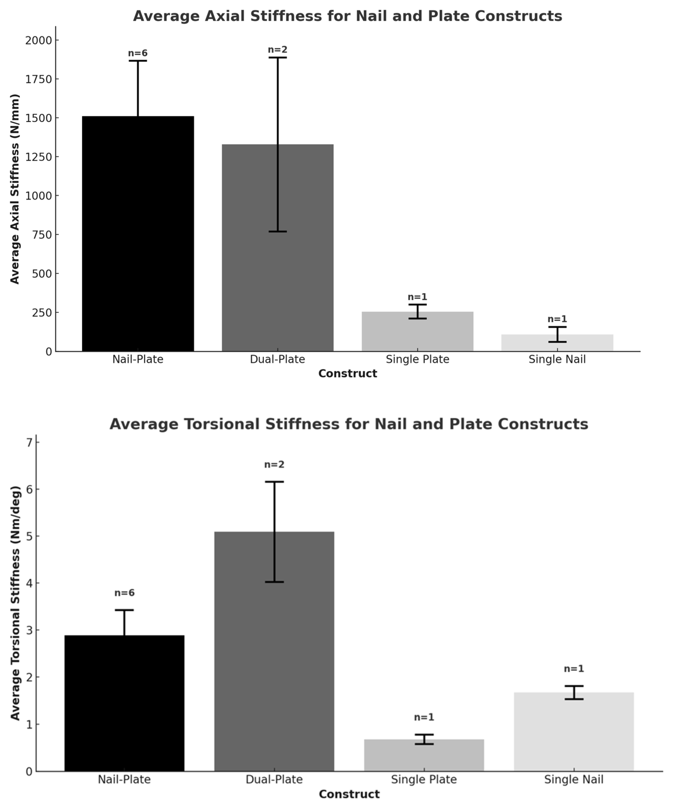
All six studies retrieved were biomechanical in nature. Five studies utilized synthetic bone models in testing, and one study used both synthetic and cadaveric bone models. Each study used, on average, seven bone models for testing (Range: 5–8). All studies utilized axial load testing as a method of quantification for biomechanical analysis and five studies used torsional stiffness testing. Further information is presented in Table 1. Mean axial stiffness and torsional stiffness for various constructs are summarized in Figure 2.
Four studies found a significant difference between the axial and torsional parameters of NPCs compared to other constructs [40,42,43,44]. The complete focused interpretation of the biomechanical data can be found in Table 2.

3.2. NPCs vs. DP Constructs

Two studies (33.33%) investigated the biomechanical properties of NPCs in comparison to DP constructs. One study found NPCs to have greater axial stiffness than DP constructs, and one study found NPCs in synthetic and cadaveric models to have less axial and torsional stiffness than DP constructs. Fontenot et al. (2019) found that NPCs had a significantly higher baseline axial stiffness and (366.0 ± 31.8 N/mm vs. (258.5 ± 40.4 N/mm) and max load to failure when compared with the DP construct utilizing a lateral locked plate + medial distal tibial plate construct (p < 0.001; p < 0.01 respectively) [42]. Fontenot et al. (2019) found no difference between cycles to failure between NPCs or DP constructs and no observable differences between NPCs or DP construct with a distal femoral plate + medial reconstruction plate [42]. Wright et al. (2020) found that in synthetic osteoporotic models, the NPC had less torsional stiffness than the DP construct and less axial stiffness than the DP construct (p = 0.00; p < 0.05; respectively) [43]. Wright et al. (2020) further saw that DP constructs in cadaveric models were nearly twice as stiff as the NPC when torsional load was applied (8.41 ± 0.58 Nm/deg vs. 4.24 ± 0.41 Nm/deg, p < 0.001) and over one-and-a-half times stiffer in axial compression (2148.1 ± 820.4 vs. 1387.7 ± 467.9 N/mm, p = 0.02) [43].

3.3. NPCs vs. DLFLP

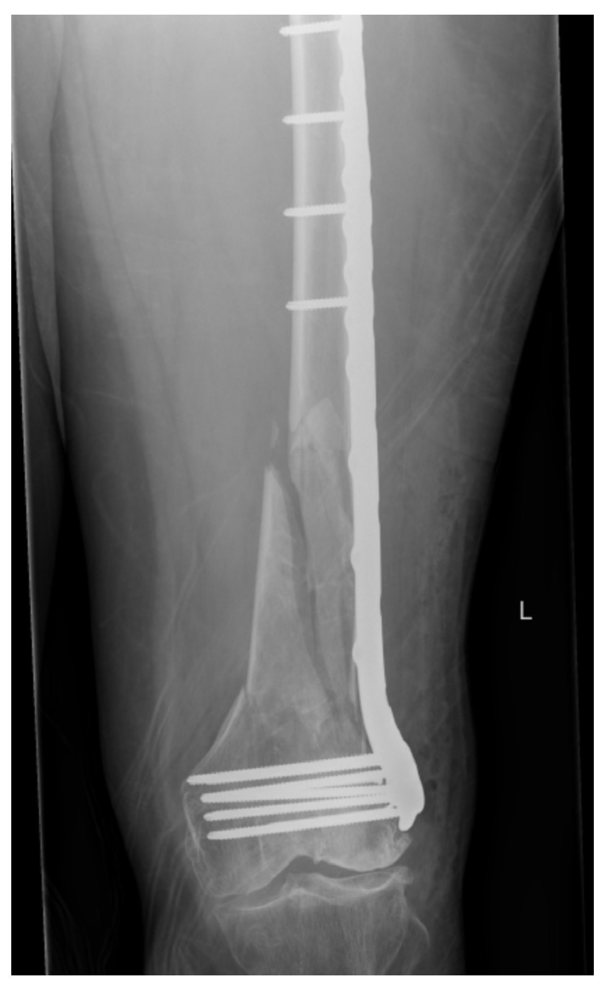
Three studies (50%) investigated NPCs compared to DLFLP constructs alone (Figure 3 and Figure 4). All studies found NPCs to have significantly higher axial and torsional stiffness and resistance to loading than DLFLP constructs. Fontenot et al. (2019) found synthetic composite NPCs to have a significantly higher baseline axial stiffness when compared to DLFLP constructs (366.0 ± 31.8 N/mm vs. 213.9 ± 18.3; p < 0.001) [42]. Additionally, no specimens with the DLFLP survived maximum loading tests. Wright et al. (2020) found that the NPC had a significantly higher mean torsional and axial stiffness compared to the DLFLP construct in synthetic osteoporotic models (1.44 ± 0.17 N/mm, 371.4 ± 41.9 N/mm vs. 0.68 ± 0.10 N/mm, 255.0 ± 45.3 N/mm; p < 0.001, p < 0.05 respectively) [43]. Basci et al. (2015) found that in synthetic composite femurs, NPCs were significantly more resistant than the DLFLP constructs in axial (p = 0.001) and torsional (p = 0.012) load tests and load-to-failure tests (p = 0.008) [40].

3.4. NPCs vs. Parallel Plating and Orthogonal Plating

One study (16.66%) investigated NPCs vs. PP and OP. Cheung et al. (2023) found no significant difference in torsional stiffness (p = 0.51) or axial stiffness (p = 0.53) between the NPC, PP, and OP construct [41].

3.5. NPCs vs. Retrograde Intramedullary Nail

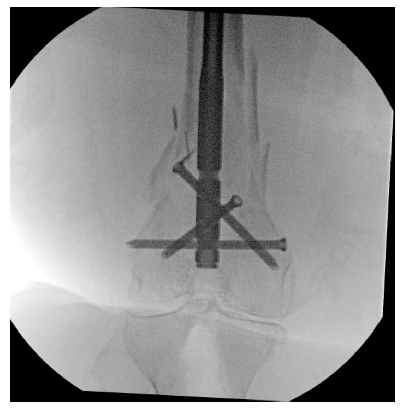
Two studies (33%) investigated NPCs vs. RIMN (Figure 5). Both found NPCs to have greater axial and stiffness and resistance than RIMN constructs. Wright et al. (2020) found that NPCs had less torsional stiffness than RIMN (1.44 ± 0.17 N/mm vs. 1.67 ± 0.14 N/mm) but greater axial stiffness (371.4 ± 41.9 N/mm vs. 109.2 ± 47.6) in synthetic bone models (p = 0.001, p < 0.05; respectively) [43]. Basci et al. (2015) found NPCs to be significantly more resistant than the RIMN constructs in the axial load test and torsional load test (0.96 N/mm, 0.86 N/mm vs. 1.72 N/mm, 1.62 N/mm, respectively) but significantly less resistance than the RIMN in the load-to-failure test (p = 0.059) [40].

3.6. NPCs Using Variable Nail Size

One study (16.66%) investigated NPCs with variable retrograde intramedullary nail size. Wright et al. (2022) found that the 11 mm NPC was significantly stiffer than the 9 mm NPC averaged across all torsional and axial loads (p < 0.005; p < 0.005, respectively) [44]. Torsional stiffness and axial stiffness were found to be 1.6 and 1.3 times greater, respectively, in the 11 mm nail group compared to the 9 mm nail group (2.39 ± 0.41 N/mm and 506.84 ± 44.50 N/mm vs. 1.44 ± 0.17 N/mm and 376.77 ± 37.65 N/mm, respectively) [44].

3.7. Linked NPCs vs. Non-Linked NPCs

One study (16.66%) investigated the biomechanics between linked NPCs (LNPC) vs. non-linked NPCs (NLNPC). Lin et al. (2023) found no significant difference in average torsional stiffness (p = 0.675) or axial stiffness (p = 0.308) between the linked and unlinked NPC [45].

4. Discussion

This study focuses on the biomechanical implications of NPCs versus various other surgical methods for DFF stabilization. While previous systematic reviews have reported outcomes related to the biomechanical performance of different constructs for treating DFFs, with a significant focus on dual plate constructs, new studies have since been published. This analysis aims to compare NPC fixation to other constructs from a biomechanical standpoint. We evaluated the biomechanical parameters of nail–plate constructs (NPCs) for treating distal femur fractures (DFFs). The assessed parameters included axial stiffness, torsional stiffness, and resistance to loading. Axial stiffness measures compressive force resistance, while torsional stiffness assesses twisting force resistance. Resistance to loading evaluates overall durability and stability. Our findings highlight that while the NPC demonstrates superior axial stiffness in some models, it exhibits variable torsional stiffness across different comparative studies. Specifically, NPCs exhibited greater axial stiffness than DP constructs, with NPCs showing higher baseline axial stiffness and maximum load to failure. Still, these constructs offered less axial and torsional stiffness than DP constructs in synthetic and cadaveric models. This suggests that NPC might offer differential benefits in specific clinical scenarios.
A prior systematic review by Wadhwa et al. (2023) highlights the biomechanical benefits of supplemental fixation techniques in DFF, including DP constructs and NPCs [37]. These authors demonstrate increased stability, load-to-failure, and construct stiffness when supplemental techniques are compared to LLP fixation alone [37]. In comparison, the present study similarly emphasizes the superiority of NPCs over DP constructs in axial stiffness, with Fontenot et al. (2019) showing NP constructs exhibiting significantly higher baseline axial stiffness and maximum load to failure [42]. In contrast to these findings, Wright et al. (2020) presented results demonstrating that DP constructs in cadaveric models significantly outperformed NPCs regarding torsional stiffness [43]. Wadhwa et al. describe the lack of clinical studies to date investigating NPCs against DP or other constructs, which limits our ability to interpret these findings [37]. This juxtaposition of biomechanical outcomes underlines the complexity of selecting the most effective stabilization strategy for DFFs, suggesting a tailored approach based on the specific biomechanical properties required for optimal patient outcomes.
In a previous systematic review of biomechanical and anatomic considerations in DFFs, DeKeyser et al. (2022) found DP constructs to demonstrate a greater axial and rotational stiffness [4]. Moreover, these authors report that DP constructs lead to less displacement and fewer incidences of failure than all other constructs, such as less-invasive stabilization system (LISS) plating versus LISS plates with other constructs and dual plating [4]. This finding supports the results of the present study, where both Fontenot et al. (2019) and Wright et al. (2020) report DP constructs to offer greater torsional and axial stiffness than other constructs, such as single-plate or only RIMN constructs [42,43]. However, there was a split in findings between these studies regarding NPC capabilities compared to DP constructs. What can be ascertained from other studies, however, is that supplemental constructs such as DP constructs and NPCs both tend to offer faster fracture healing, a higher union rate, and possibly lower revision surgery rates than single constructs [46,47,48]. When comparing construct styles such as parallel or orthogonal DP constructs, Cheung et al. (2023) found no significant differences between torsional and axial stiffness in either of the construct types [41]. Unfortunately, there is a lack of comparative biomechanical studies focusing on variations in construct types, with the majority reporting on the biomechanical outcomes of these plating constructs in humeral fractures [41,49,50,51]. Within femur fractures, Cheung hypothesizes that in NPC systems, the RIMN serves as a load-sharing device and potentially could withstand greater resistance to axial loading than a medially based implant [41]. Wright et al. propose that the lower axial stiffness observed in the NPC compared to DP in general may be due to limited distal fixation such as the inability to place all distal interlocking screws in the intramedullary nail or all of the distal screws in the lateral plate [43].
The current biomechanical results present in the literature and the findings from our systematic review are important to contextualize and adequately explain clinical results across the literature. Given the need to increase overall system stability in the NPC, Liporace et al. (2019) have recommended linking the plate and nail distally to increase the overall stability in osteoporotic patients [52]. Liporace et al. (2019) suggest that the NPC is ideal for the geriatric, osteoporotic patients suffering from DFFs who are ambulatory at baseline, where immediate weight bearing is desired [52]. The NPC technique used by Liporace et al. (2019) and recreated by Liporace et al. (2022) increases points of fixation, creating a fixed angle construct allowing for a medial shift in the weight-bearing axis along the femur to offer increased stability [52,53]. Given that the NPC is touted for its ability to offer rapid weight-bearing with reliable outcomes, this may be a better fit for geriatric and osteoporotic patients as RIMN and single-plate constructs often require protected weight bearing during recovery [52,54,55,56]. In addition to the conversation surrounding biomechanical stability, postoperative outcomes are another important paradigm.
Espey et al. (2023) found that NPCs had the lowest reoperation rate (2.5%) compared to that of single traditional construct (STC) fixation using either an LLP or RIMN (12.9%); however, the number of studies used in the [55]. However, Passias et al. (2021) reported that the NPCs were about twice as expensive as STCs, given the additional hardware involved in the fixation process, indicating that further research is necessary in order to determine whether decreased reoperation rates are justified in selecting a more expensive construct over STCs [17].
The biocompatibility of the materials used in fixation constructs is a crucial factor influencing patient outcomes and overall treatment success. Titanium alloys, such as Ti6Al4V, are commonly used in NPCs, DP, and DLFLP constructs due to their excellent mechanical properties and biocompatibility. Titanium alloys are favored for their high strength, corrosion resistance, and minimal adverse tissue reactions, which contribute to lower infection rates and better integration with bone tissue [57,58]. However, titanium can release trace amounts of ions, which may have cytotoxic effects and can distort imaging modalities like CT and MRI, potentially complicating postoperative assessments [57]. Stainless steel is another material used in some orthopedic constructs. It offers good mechanical properties but has a higher risk of corrosion and metal ion release, which can lead to local tissue reactions and systemic effects [59,60]. Both titanium and stainless steel have been shown to integrate well with bone, promoting effective healing while minimizing inflammatory responses [58,60]. Recent advancements in coating technologies, such as hydroxyapatite and zeolite coatings on titanium implants, have shown promise in enhancing biocompatibility and osseointegration, providing better support for bone regeneration and reducing the risk of implant-related complications [59,60]. These coatings can help mitigate some of the cytotoxic effects associated with metal ion release and improve the overall performance of the implants [58]. The choice of material for fixation constructs must consider both mechanical performance and biocompatibility to ensure optimal patient outcomes. Further research into advanced materials and coatings is essential to enhance the biocompatibility and clinical effectiveness of these constructs.
This study presents a thorough biomechanical comparison of NPCs versus other fixation constructs for DFFs. However, limitations include a relatively small number of studies meeting the inclusion criteria, potentially limiting the generalizability of findings. The narrow set of biomechanical criteria may not capture all relevant clinical factors affecting DFF mechanics and their relevance to full body biomechanics and clinical recovery. While the biomechanical data suggest that NPCs may offer greater stability compared to other techniques, it is crucial to evaluate the limitations of the models used critically. Synthetic bone models, despite their consistency, lack the heterogeneity of human bone, particularly in osteoporotic patients where bone density and quality vary significantly. While standardized, they do not account for the variability in bone density and quality among patients, especially those with osteoporosis which may not fully replicate the complexity of human bone characteristics, particularly in varied patient populations with differing bone qualities such as osteoporosis. The impact and influence of osteoporosis on bone health and its mechanism of triggering deterioration in trabecular bone are a major health problem and concern. This is of great concern in orthopedic patients who are at greater risk of suffering traumatic fractures due to age or occupation [61]. On the other hand, cadaveric models, while providing more realistic anatomy, may still present challenges, such as variability in bone preservation and quality, which can influence biomechanical outcomes. These limitations underscore the need for cautious interpretation when applying these findings to clinical practice. In everyday clinical practice, the potential biomechanical advantages of NPCs should be carefully balanced with individual patient factors, such as bone quality and fracture type, as well as the surgeon’s experience, to ensure that these findings translate effectively into improved patient outcomes. Overall, as synthetic models were the majority of constructs available to be reviewed in this study, this may show greater confidence in the results than in real human femurs. Future research should incorporate patient-specific computational models and cadaveric studies that better simulate the variability in bone quality and fracture patterns seen in clinical practice. This would help validate the biomechanical advantages of NPCs and improve their applicability in real-world scenarios.

5. Conclusions

This study indicates that the NPC offers greater biomechanical stability, such as increased axial stiffness and maximum load to failure, compared to traditional methods like RIMN and DLFLP, though it is not consistently superior to DP constructs. For complex DFFs, especially where bone quality or comminution complicates surgery, the NPC may provide a more robust solution. However, caution is needed in generalizing these results to clinical practice. The biomechanical advantages observed should be validated with clinical studies to ensure that they translate effectively into improved patient outcomes. This study also emphasizes the importance of considering individual patient factors, surgical expertise, and potential variability in outcomes, underscoring the need for further research to validate these findings and assess their clinical impact. Future research should focus on exploring specific biomechanical parameters, such as the impact of varying nail diameters and plate configurations on stability. Additionally, studies should examine the clinical efficacy of NPCs across different patient populations, particularly in those with varying bone densities, to better understand how these constructs perform in real-world scenarios. Investigating long-term outcomes, including complication rates and functional recovery, will also be essential to assessing the clinical benefits of NPCs fully.

Author Contributions

A.N.B.; conceptualization, O.S.A. and S.P.; data collection, O.S.A.; writing—original draft preparation, A.T.A., A.N.B., C.P. and M.R.D.; writing—review and editing. All authors have read and agreed to the published version of the manuscript.

Funding

This research received no external funding.

Institutional Review Board Statement

Not applicable.

Informed Consent Statement

Not applicable.

Conflicts of Interest

The authors declare no conflicts of interest.

References

  1. Elsoe, R.; Ceccotti, A.A.; Larsen, P. Population-based epidemiology and incidence of distal femur fractures. Int. Orthop. 2018, 42, 191–196. [Google Scholar] [CrossRef] [PubMed]
  2. Gwathmey, F.W., Jr.; Jones-Quaidoo, S.M.; Kahler, D.; Hurwitz, S.; Cui, Q. Distal femoral fractures: Current concepts. J. Am. Acad. Orthop. Surg. 2010, 18, 597–607. [Google Scholar] [CrossRef] [PubMed]
  3. Coon, M.S.; Best, B.J. Distal Femur Fractures. In StatPearls; StatPearls Publishing: Treasure Island, FL, USA, 2024. [Google Scholar]
  4. DeKeyser, G.J.; Hakim, A.J.; O’Neill, D.C.; Schlickewei, C.W.; Marchand, L.S.; Haller, J.M. Biomechanical and anatomical considerations for dual plating of distal femur fractures: A systematic literature review. Arch. Orthop. Trauma. Surg. 2022, 142, 2597–2609. [Google Scholar] [CrossRef] [PubMed]
  5. Merchan, E.C.; Maestu, P.R.; Blanco, R.P. Blade-plating of closed displaced supracondylar fractures of the distal femur with the AO system. J. Trauma. 1992, 32, 174–178. [Google Scholar] [CrossRef]
  6. Siliski, J.M.; Mahring, M.; Hofer, H.P. Supracondylar-intercondylar fractures of the femur. Treatment by internal fixation. J. Bone Jt. Surg. Am. 1989, 71, 95–104. [Google Scholar] [CrossRef]
  7. Giles, J.B.; DeLee, J.C.; Heckman, J.D.; Keever, J.E. Supracondylar-intercondylar fractures of the femur treated with a supracondylar plate and lag screw. J. Bone Jt. Surg. Am. 1982, 64, 864–870. [Google Scholar] [CrossRef]
  8. Neer, C.S.; Grantham, S.A.; Shelton, M.L. Supracondylar fracture of the adult femur. A study of one hundred and ten cases. J. Bone Jt. Surg. Am. 1967, 49, 591–613. [Google Scholar] [CrossRef]
  9. Hoskins, W.; Sheehy, R.; Edwards, E.R.; Hau, R.C.; Bucknill, A.; Parsons, N.; Griffin, X.L. Nails or plates for fracture of the distal femur? data from the Victoria Orthopaedic Trauma Outcomes Registry. Bone Jt. J. 2016, 98, 846–850. [Google Scholar] [CrossRef]
  10. Markmiller, M.; Konrad, G.; Sudkamp, N. Femur-LISS and distal femoral nail for fixation of distal femoral fractures: Are there differences in outcome and complications? Clin. Orthop. Relat. Res. 2004, 426, 252–257. [Google Scholar] [CrossRef]
  11. Schutz, M.; Muller, M.; Krettek, C.; Hontzsch, D.; Regazzoni, P.; Ganz, R.; Haas, N. Minimally invasive fracture stabilization of distal femoral fractures with the LISS: A prospective multicenter study. Results of a clinical study with special emphasis on difficult cases. Injury 2001, 32 (Suppl. S3), 48–54. [Google Scholar] [CrossRef]
  12. Rollo, G.; Pichierri, P.; Grubor, P.; Marsilio, A.; Bisaccia, M.; Grubor, M.; Pace, V.; Lanzetti, R.M.; Giaracuni, M.; Filipponi, M.; et al. The challenge of nonunion and malunion in distal femur surgical revision. Med. Glas. 2019, 16. [Google Scholar] [CrossRef]
  13. Saxena, V.; Akshay, V.; Panwar, A.; Kumar, S. Management of Non-union Distal Femur Fractures With Augmentation Nail Plate Construct. Cureus 2023, 15, e37173. [Google Scholar] [CrossRef]
  14. Ricci, W.M.; Streubel, P.N.; Morshed, S.; Collinge, C.A.; Nork, S.E.; Gardner, M.J. Risk factors for failure of locked plate fixation of distal femur fractures: An analysis of 335 cases. J. Orthop. Trauma. 2014, 28, 83–89. [Google Scholar] [CrossRef] [PubMed]
  15. Canton, G.; Giraldi, G.; Dussi, M.; Ratti, C.; Murena, L. Osteoporotic distal femur fractures in the elderly: Peculiarities and treatment strategies. Acta Biomed. 2019, 90, 25–32. [Google Scholar] [CrossRef] [PubMed]
  16. Garala, K.; Ramoutar, D.; Li, J.; Syed, F.; Arastu, M.; Ward, J.; Patil, S. Distal femoral fractures: A comparison between single lateral plate fixation and a combined femoral nail and plate fixation. Injury 2022, 53, 634–639. [Google Scholar] [CrossRef] [PubMed]
  17. Passias, B.J.; Emmer, T.C.; Sullivan, B.D.; Gupta, A.; Myers, D.; Skura, B.W.; Taylor, B.C. Treatment of Distal Femur Fractures with a Combined Nail-Plate Construct: Techniques and Outcomes. J. Long. Term. Eff. Med. Implant. 2021, 31, 15–26. [Google Scholar] [CrossRef]
  18. Medda, S.; Kessler, R.B.; Halvorson, J.J.; Pilson, H.T.; Babcock, S.; Carroll, E.A. Technical Trick: Dual Plate Fixation of Periprosthetic Distal Femur Fractures. J. Orthop. Trauma. 2021, 35, e148–e152. [Google Scholar] [CrossRef]
  19. Henderson, C.E.; Kuhl, L.L.; Fitzpatrick, D.C.; Marsh, J.L. Locking plates for distal femur fractures: Is there a problem with fracture healing? J. Orthop. Trauma. 2011, 25 (Suppl. S1), S8–S14. [Google Scholar] [CrossRef]
  20. Tank, J.C.; Schneider, P.S.; Davis, E.; Galpin, M.; Prasarn, M.L.; Choo, A.M.; Munz, J.W.; Achor, T.S.; Kellam, J.F.; Gary, J.L. Early Mechanical Failures of the Synthes Variable Angle Locking Distal Femur Plate. J. Orthop. Trauma. 2016, 30, e7–e11. [Google Scholar] [CrossRef]
  21. Yoon, B.H.; Park, I.K.; Kim, Y.; Oh, H.K.; Choo, S.K.; Sung, Y.B. Incidence of nonunion after surgery of distal femoral fractures using contemporary fixation device: A meta-analysis. Arch. Orthop. Trauma. Surg. 2021, 141, 225–233. [Google Scholar] [CrossRef]
  22. Martinet, O.; Cordey, J.; Harder, Y.; Maier, A.; Buhler, M.; Barraud, G.E. The epidemiology of fractures of the distal femur. Injury 2000, 31 (Suppl. S3), 62–94. [Google Scholar] [CrossRef] [PubMed]
  23. Myers, P.; Laboe, P.; Johnson, K.J.; Fredericks, P.D.; Crichlow, R.J.; Maar, D.C.; Weber, T.G. Patient Mortality in Geriatric Distal Femur Fractures. J. Orthop. Trauma. 2018, 32, 111–115. [Google Scholar] [CrossRef]
  24. Streubel, P.N.; Ricci, W.M.; Wong, A.; Gardner, M.J. Mortality after distal femur fractures in elderly patients. Clin. Orthop. Relat. Res. 2011, 469, 1188–1196. [Google Scholar] [CrossRef] [PubMed]
  25. Consigliere, P.; Iliopoulos, E.; Ads, T.; Trompeter, A. Early versus delayed weight bearing after surgical fixation of distal femur fractures: A non-randomized comparative study. Eur. J. Orthop. Surg. Traumatol. 2019, 29, 1789–1794. [Google Scholar] [CrossRef] [PubMed]
  26. Lieder, C.M.; Gaski, G.E.; Virkus, W.W.; Kempton, L.B. Is Immediate Weight-Bearing Safe After Single Implant Fixation of Elderly Distal Femur Fractures? J. Orthop. Trauma. 2021, 35, 49–55. [Google Scholar] [CrossRef]
  27. Schandelmaier, P.; Partenheimer, A.; Koenemann, B.; Grun, O.A.; Krettek, C. Distal femoral fractures and LISS stabilization. Injury 2001, 32 (Suppl. S3), 55–63. [Google Scholar] [CrossRef]
  28. Black, D.M.; Bauer, D.C.; Vittinghoff, E.; Lui, L.Y.; Grauer, A.; Marin, F.; Khosla, S.; de Papp, A.; Mitlak, B.; Cauley, J.A.; et al. Treatment-related changes in bone mineral density as a surrogate biomarker for fracture risk reduction: Meta-regression analyses of individual patient data from multiple randomised controlled trials. Lancet Diabetes Endocrinol. 2020, 8, 672–682. [Google Scholar] [CrossRef]
  29. Ellingsen Husebye, E.; Lyberg, T.; Madsen, J.E.; Nordsletten, L.; Roise, O. The early effects of intramedullary reaming of the femur on bone mineral density; an experimental study in pigs. Scand. J. Surg. 2009, 98, 189–194. [Google Scholar] [CrossRef]
  30. Johnson, K.D.; Hicken, G. Distal femoral fractures. Orthop. Clin. N. Am. 1987, 18, 115–132. [Google Scholar] [CrossRef]
  31. Leslie, W.D.; Martineau, P.; Bryanton, M.; Lix, L.M. Which is the preferred site for bone mineral density monitoring as an indicator of treatment-related anti-fracture effect in routine clinical practice? A registry-based cohort study. Osteoporos. Int. 2019, 30, 1445–1453. [Google Scholar] [CrossRef]
  32. Cirnigliaro, C.M.; Myslinski, M.J.; La Fountaine, M.F.; Kirshblum, S.C.; Forrest, G.F.; Bauman, W.A. Bone loss at the distal femur and proximal tibia in persons with spinal cord injury: Imaging approaches, risk of fracture, and potential treatment options. Osteoporos. Int. 2017, 28, 747–765. [Google Scholar] [CrossRef] [PubMed]
  33. Inacio, J.V.; Malige, A.; Schroeder, J.T.; Nwachuku, C.O.; Dailey, H.L. Mechanical characterization of bone quality in distal femur fractures using pre-operative computed tomography scans. Clin. Biomech. 2019, 67, 20–26. [Google Scholar] [CrossRef] [PubMed]
  34. Kannus, P.; Jarvinen, M.; Sievanen, H.; Jarvinen, T.A.; Oja, P.; Vuori, I. Reduced bone mineral density in men with a previous femur fracture. J. Bone Miner. Res. 1994, 9, 1729–1736. [Google Scholar] [CrossRef]
  35. Leppala, J.; Kannus, P.; Niemi, S.; Sievanen, H.; Vuori, I.; Jarvinen, M. An early-life femoral shaft fracture and bone mineral density at adulthood. Osteoporos. Int. 1999, 10, 337–342. [Google Scholar] [CrossRef]
  36. Schmidt, C.; Riedel, C.; Sturznickel, J.; Mushumba, H.; Delsmann, M.M.; Ries, C.; Kleiss, S.; Bannas, P.; Beil, F.T.; Amling, M.; et al. Investigation of distal femur microarchitecture and factors influencing its deterioration: An ex vivo high-resolution peripheral quantitative computed tomography study. J. Orthop. Res. 2022, 40, 2057–2064. [Google Scholar] [CrossRef] [PubMed]
  37. Wadhwa, H.; Goodnough, H.L.; Sharma, J.; Maschhoff, C.W.; Van Rysselberghe, N.L.; Bishop, J.A.; Gardner, M.J. Supplemental fixation of distal femur fractures: A review of biomechanical and clinical evidence. Curr. Orthop. Pract. 2023, 34, 201–207. [Google Scholar] [CrossRef]
  38. Page, M.J.; McKenzie, J.E.; Bossuyt, P.M.; Boutron, I.; Hoffmann, T.C.; Mulrow, C.D.; Shamseer, L.; Tetzlaff, J.M.; Akl, E.A.; Brennan, S.E.; et al. The PRISMA 2020 statement: An updated guideline for reporting systematic reviews. BMJ 2021, 372, n71. [Google Scholar] [CrossRef] [PubMed]
  39. Ouzzani, M.; Hammady, H.; Fedorowicz, Z.; Elmagarmid, A. Rayyan-a web and mobile app for systematic reviews. Syst. Rev. 2016, 5, 210. [Google Scholar] [CrossRef]
  40. Basci, O.; Karakasli, A.; Kumtepe, E.; Guran, O.; Havitcioglu, H. Combination of anatomical locking plate and retrograde intramedullary nail in distal femoral fractures: Comparison of mechanical stability. Eklem Hastalik. Cerrahisi 2015, 26, 21–26. [Google Scholar] [CrossRef]
  41. Cheung, Z.B.; Nasser, P.; Iatridis, J.C.; Forsh, D.A. Orthogonal plating of distal femur fractures: A biomechanical comparison with plate-nail and parallel plating constructs. J. Orthop. 2023, 37, 34–40. [Google Scholar] [CrossRef]
  42. Fontenot, P.B.; Diaz, M.; Stoops, K.; Barrick, B.; Santoni, B.; Mir, H. Supplementation of Lateral Locked Plating for Distal Femur Fractures: A Biomechanical Study. J. Orthop. Trauma. 2019, 33, 642–648. [Google Scholar] [CrossRef] [PubMed]
  43. Wright, D.J.; DeSanto, D.J.; McGarry, M.H.; Lee, T.Q.; Scolaro, J.A. Supplemental Fixation of Supracondylar Distal Femur Fractures: A Biomechanical Comparison of Dual-Plate and Plate-Nail Constructs. J. Orthop. Trauma. 2020, 34, 434–440. [Google Scholar] [CrossRef] [PubMed]
  44. Wright, D.J.; DeSanto, D.J.; McGarry, M.H.; Lee, T.Q.; Scolaro, J.A. Nail diameter significantly impacts stability in combined plate-nail constructs used for fixation of supracondylar distal femur fractures. OTA Int. 2022, 5, e174. [Google Scholar] [CrossRef]
  45. Lin, C.C.; Parody, N.; Anil, U.; Egol, K.A. Effect of Implant Linkage on Axial and Rotational Stiffness of Nail-Plate Constructs for Comminuted Distal Femoral Fractures. J. Orthop. Trauma. 2023, 37, 351–355. [Google Scholar] [CrossRef] [PubMed]
  46. Andring, N.A.; Kaupp, S.M.; Henry, K.A.; Helmig, K.C.; Babcock, S.; Halvorson, J.J.; Pilson, H.T.; Carroll, E.A. Dual Plate Fixation of Periprosthetic Distal Femur Fractures. J. Orthop. Trauma. 2024, 38, 36–41. [Google Scholar] [CrossRef] [PubMed]
  47. Bologna, M.G.; Claudio, M.G.; Shields, K.J.; Katz, C.; Salopek, T.; Westrick, E.R. Dual plate fixation results in improved union rates in comminuted distal femur fractures compared to single plate fixation. J. Orthop. 2020, 18, 76–79. [Google Scholar] [CrossRef]
  48. Tripathy, S.K.; Mishra, N.P.; Varghese, P.; Panigrahi, S.; Purudappa, P.P.; Goel, A.; Sen, R.K. Dual-Plating in Distal Femur Fracture: A Systematic Review and Limited Meta-analysis. Indian. J. Orthop. 2022, 56, 183–207. [Google Scholar] [CrossRef]
  49. Lee, S.K.; Kim, K.J.; Park, K.H.; Choy, W.S. A comparison between orthogonal and parallel plating methods for distal humerus fractures: A prospective randomized trial. Eur. J. Orthop. Surg. Traumatol. 2014, 24, 1123–1131. [Google Scholar] [CrossRef]
  50. Taylor, P.A.; Owen, J.R.; Benfield, C.P.; Wayne, J.S.; Boardman, N.D., 3rd. Parallel Plating of Simulated Distal Humerus Fractures Demonstrates Increased Stiffness Relative to Orthogonal Plating With a Distal Humerus Locking Plate System. J. Orthop. Trauma. 2016, 30, e118–e122. [Google Scholar] [CrossRef]
  51. Yu, X.; Xie, L.; Wang, J.; Chen, C.; Zhang, C.; Zheng, W. Orthogonal plating method versus parallel plating method in the treatment of distal humerus fracture: A systematic review and meta-analysis. Int. J. Surg. 2019, 69, 49–60. [Google Scholar] [CrossRef]
  52. Liporace, F.A.; Yoon, R.S. Nail Plate Combination Technique for Native and Periprosthetic Distal Femur Fractures. J. Orthop. Trauma. 2019, 33, e64–e68. [Google Scholar] [CrossRef] [PubMed]
  53. Liporace, F.A.; Tang, A.; Jankowski, J.M.; Yoon, R.S. Distal femur: Nail plate combination and the linked construct. OTA Int. 2022, 5, e172. [Google Scholar] [CrossRef]
  54. Attum, B.; Douleh, D.; Whiting, P.S.; White-Dzuro, G.A.; Dodd, A.C.; Shen, M.S.; Mir, H.R.; Obremskey, W.T.; Sethi, M.K. Outcomes of Distal Femur Nonunions Treated With a Combined Nail/Plate Construct and Autogenous Bone Grafting. J. Orthop. Trauma. 2017, 31, e301–e304. [Google Scholar] [CrossRef]
  55. Espey, R.; Stevenson, L.; Tucker, A. Combined nail-plate constructs in the management of osteoporotic native distal femoral fractures: A systematic review of the available evidence. Eur. J. Orthop. Surg. Traumatol. 2023, 33, 3215–3223. [Google Scholar] [CrossRef] [PubMed]
  56. Adesina, S.A.; Amole, I.O.; Adefokun, I.G.; Adegoke, A.O.; Akinwumi, A.I.; Odekhiran, E.O.; Durodola, A.O.; Ojo, S.A.; Eyesan, S.U. Retrograde intramedullary nailing with supplemental plate and lag screws allows early weight bearing following distal end-segment femur fractures (AO/OTA 33) in a low-resource setting. Eur. J. Orthop. Surg. Traumatol. 2024, 34, 1519–1527. [Google Scholar] [CrossRef]
  57. Liu, J.; Wang, K.; Li, X.; Zhang, X.; Gong, X.; Zhu, Y.; Ren, Z.; Zhang, B.; Cheng, J. Biocompatibility and osseointegration properties of a novel high strength and low modulus beta- Ti10Mo6Zr4Sn3Nb alloy. Front. Bioeng. Biotechnol. 2023, 11, 1127929. [Google Scholar] [CrossRef]
  58. Sommer, U.; Laurich, S.; de Azevedo, L.; Viehoff, K.; Wenisch, S.; Thormann, U.; Alt, V.; Heiss, C.; Schnettler, R. In Vitro and In Vivo Biocompatibility Studies of a Cast and Coated Titanium Alloy. Molecules 2020, 25, 3399. [Google Scholar] [CrossRef]
  59. Liu, C.; Zhang, J.; Zhao, X.; Xu, M.; Liu, H.; Zhou, H. Stability, biomechanics and biocompatibility analysis following different preparation strategies of hierarchical zeolite coatings on titanium alloy surfaces. Front. Bioeng. Biotechnol. 2023, 11, 1337709. [Google Scholar] [CrossRef]
  60. He, S.; Zhu, J.; Jing, Y.; Long, S.; Tang, L.; Cheng, L.; Shi, Z. Effect of 3D-Printed Porous Titanium Alloy Pore Structure on Bone Regeneration: A Review. Coatings 2024, 14, 253. [Google Scholar] [CrossRef]
  61. Al-Mukhtar, A.M.; Könke, C. Fracture Mechanics and Micro Crack Detection in Bone: A Short Communication. In Proceedings of the MPMD, Minneapolis, MN, USA, 8–10 August 2011. [Google Scholar]
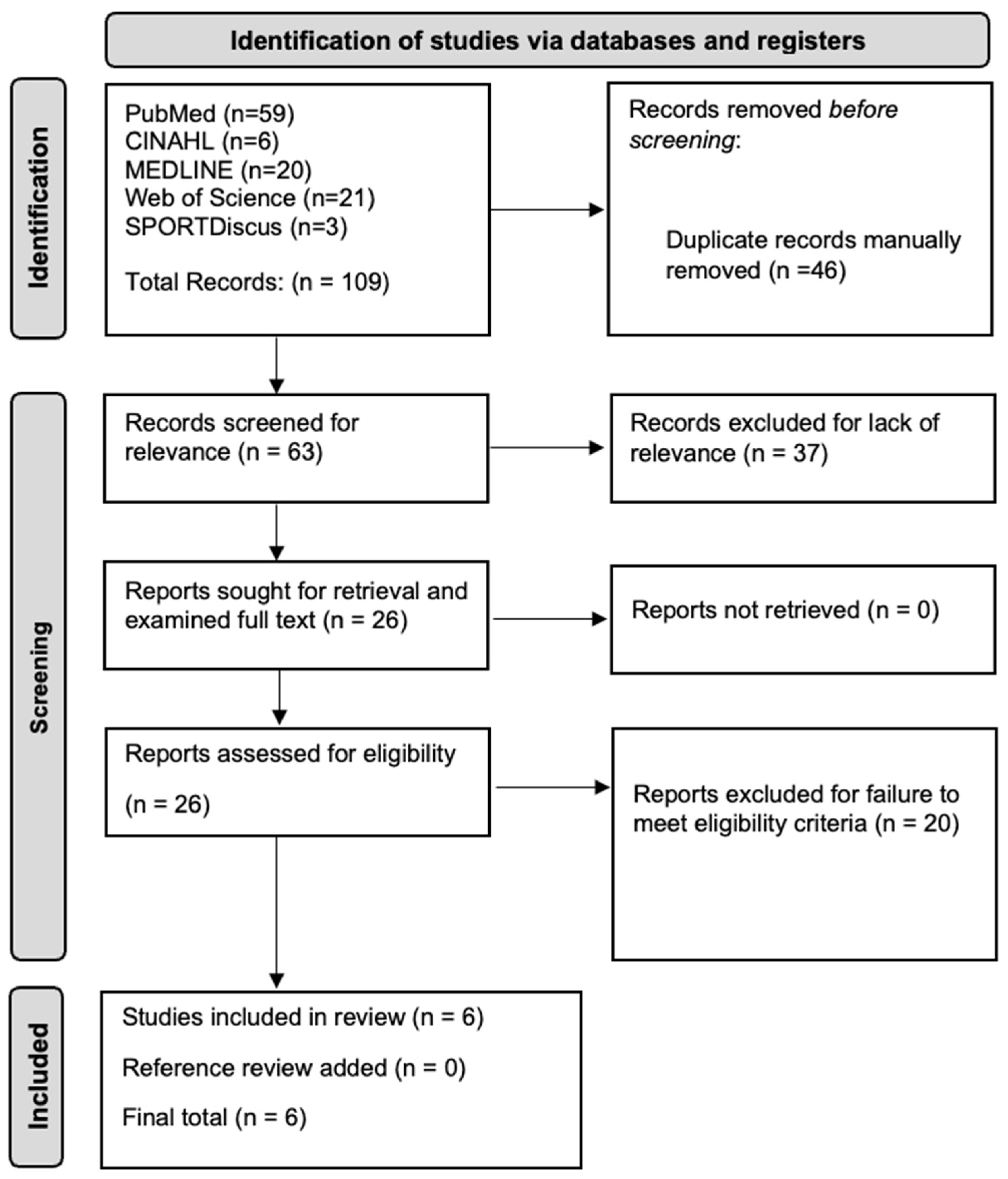
Figure 1. The preferred reporting items for systematic reviews and meta-analyses (PRISMA) diagram for this study, depicting the search process.
Surgeries 05 00064 g001
Figure 2. Mean axial and torsional stiffness for nail–plate, dual-plate, single-plate, and single-nail constructs.
Surgeries 05 00064 g002
Figure 3. Radiographic image of a nail–plate construct composed of intramedullary nail and lateral locking plate. The intramedullary nail provides internal stabilization through the femoral canal, while the lateral locking plate is secured externally with locking screws to enhance the stability of the fractured end of the distal femur.
Surgeries 05 00064 g003
Figure 4. Radiographic image of a distal lateral femoral locking plate-only construct. The plate is secured externally along the lateral aspect of the entire length of the femur with multiple locking screws that penetrate the bone, providing stable fixation for the distal fracture site.
Surgeries 05 00064 g004
Figure 5. The radiographic image displays distal femur fixation using a retrograde intramedullary nail-only construct. The nail is inserted through the knee joint and extends proximally through the femoral canal, providing internal stabilization. Several transverse screws are placed through the nail and the bone to secure the construct and maintain alignment.
Surgeries 05 00064 g005
Table 1. Study characteristics from the included articles in this systematic review. Data include type of study, model type, subgroup comparison, number of constructs, method of biomechanical analysis, biomechanical analysis data, and key findings.
Author (Year)Model Type UsedConstructs BrandSubgroup Comparison Type# of Models Tested Biomechanical TestBiomechanical Analysis DataKey Findings from Biomechanical Analysis
Torsional Stiffness (Nm/Degree)Axial Stiffness (N/mm)
Basci (2015) [40]Synthetic Composite FemursSawbonesNail–platen = 7Torsional Load Testing, Axial Load TestingN/AN/AThe NPC was significantly more resistant than the femoral plate and RIMN constructs in the axial load test and torsional load test (p ≤ 0.012). The NPC had a significantly higher resistance than the femoral plate (p = 0.008) but not RIMN (p = 0.059) in the load to failure test.
Lateral locked distal femur platen = 7
Retrograde intramedullary nailn = 7
Cheung (2023) [41]Synthetic Osteoporotic FemursSawbones, SKU 3503Nail–platen = 5Torsional Load Testing,74.5 ± 20.7 (baseline)3.57 × 10⁵ ± 1.63 × 10⁵ (baseline)No difference found in torsional stiffness or axial stiffness ((p = 0.51), p = 0.53)) between the NPC, parallel plating construct, and orthogonal plating construct.
Parallel platingn = 5Axial Load Testing81.2 ± 23.7 (baseline)4.31 × 10⁵ ± 3.62 × 10⁵ (baseline)
Orthogonal platingn = 5 66.2 ± 31.1 (baseline)2.44 × 10⁵ ± 1.32 × 10⁵ (baseline)
Fontenot (2019) [42]Synthetic Composite FemursSawbonesNail–platen = 7Axial Load TestingN/A366.0 ± 31.8 (baseline)The lateral locked NPC and LLP + medial reconstruction plate construct have significantly higher baseline axial stiffness (p < 0.001) compared with LLP only or the LLP + medial distal tibia plate construct.
Lateral locked distal femoral plate alonen = 7213.9 ± 18.3 (baseline)
Lateral locked distal femoral plate + medial distal tibial platen = 7258.5 ± 40.4 (baseline)
Lateral locked distal femoral plate + medial reconstruction platen = 7364.0 ± 32.5 (baseline)
Lin (2023) [45]Synthetic Osteoporotic FemursSawbones, SKU 3503Linked nail–plate constructn = 8Torsional Load Testing, Axial Load Testing3.95 ± 0.45 (average)3067.1 ± 332.0 (average)No significant difference in mean torsional or axial stiffness (p = 0.675, p = 0.308) between the linked and unlinked NPC.
Non-linked nail–plate constructn = 83.85 ± 0.53 (average)3345.2 ± 665.3 (average)
Wright (2022) [44]Synthetic Osteoporotic FemursSawbones, SKU 1130–130Lateral plate + 9 mm retrograde intramedullary nailn = 6Torsional Load Testing, Axial Load Testing1.44 ± 0.17 (average)376.77 ± 37.65 (average)The 11 mm NPC was significantly stiffer than the 9 mm NPC across all torsional (1.6 times more, p < 0.005) and axial loads (1.3 times more, p < 0.005).
Lateral plate + 11 mm retrograde intramedullary nailn = 62.39 ± 0.41 (average)506.84 ± 44.50 (average)
Wright (2020) [43]Synthetic Osteoporotic FemursSawbones, SKU 1130–130Nail–platen = 6Torsional Load Testing, Axial Load Testing1.44 ± 0.17 (average)371.4 ± 41.9 (average)The average torsional (p = 0.001) and axial (p < 0.05) stiffness were significantly different between the NPC, DP, DLFLP, and rIMN constructs. The NPC had less torsional stiffness than both the DP and RIMN constructs and less axial stiffness than the DP construct.
Dual-platen = 61.76 ± 0.33 (average)507.9 ± 83.1 (average)
Distal lateral femoral locking llate (DLFLP)n = 60.68 ± 0.10 (average)255.0 ± 45.3 (average)
Retrograde intramedullary nail (rIMN)n = 61.67 ± 0.14 (average)109.2 ± 47.6 (average)
Fresh-frozen cadaveric femursN/ANail–platen = 7Torsional Load Testing, Axial Load Testing4.24 ± 1.09 (average)1387.7 ± 467.9 (average)DP construct was almost twice as stiff as the NPC under torsional load (p < 0.001) and over one-and-a-half times stiffer under axial compression (p = 0.02).
Dual-platen = 78.41 ± 1.55 (average)2148.1 ± 820.4 (average)
Table 2. Focused analysis of study findings. Data includes first author, groups compared, main parameters measured, significant differences, and conclusions.
AuthorGroups Being ComparedParameters MeasuredSignificant Difference in Torsinal Parameters?Significant Difference in Axial Parameters?Conclusions
Cheung (2023) [41]Nail–plate combination vs. parallel plates vs. orthogonal platesBaseline stiffnessNoNoN/A
Fontenot (2019) [42]Nail–plate combination vs. lateral-plate-only vs. lateral plate + medial distal tibial plate vs. lateral plate + medial reconstruction plateBaseline stiffness-YesThe NPC and DP (lateral plate + medial reconstruction plate) had more torsional and axial stiffness than the plate-only and DP with medial distal tibial plate constructs.
Wright (2022) [44]11 mm nail–plate construct vs. 9 mm nail–plate constructAverage stiffness across multiple loadsYesYesThe 11 mm NPC had more torsional and axial stiffness than the 9 mm NPC.
Lin (2023) [45]Linked nail–plate construct vs. non-linked nail–plate constructAverage stiffness across multiple loadsNoNoN/A
Wright (2020) [43]Nail–plate vs. dual-plate vs. plate-only vs. nail-only (dynthetic femurs)Average stiffness across multiple loadsYesYesThe DP and nail-only constructs had more torsional stiffness than the NPC. The DP construct had more axial stiffness than the NPC.
Nail–plate vs. dual-plate (cadaveric femurs)Average stiffness across multiple loadsYesYesThe DP construct had more torsional and axial stiffness than the NPC.
Basci (2015) [40]Nail–plate vs. plate-only vs. nail-onlyAverage torque/displacement across multiple loadsYesYesThe NPC had less displacement and torque than the plate-only and nail-only constructs under axial and torsional loads.
Disclaimer/Publisher’s Note: The statements, opinions and data contained in all publications are solely those of the individual author(s) and contributor(s) and not of MDPI and/or the editor(s). MDPI and/or the editor(s) disclaim responsibility for any injury to people or property resulting from any ideas, methods, instructions or products referred to in the content.