Abstract
Smooth muscle tumors of unknown malignant potential (STUMP) represent a group of heterogeneous uterine neoplasms showing worrisome histological features, raising concerns for a malignant behavior, but do not satisfy the diagnostic criteria for leiomyosarcoma. The existing literature remains scarce due to its rarity; therefore, there is no consensus regarding diagnostic criteria, malignant potential, treatment of choice, and follow-up. The authors report their case series to analyze the heterogeneous features of this poorly understood neoplasm. A retrospective, monocentric cohort study of all patients who underwent surgery diagnosed with STUMP at the Garibaldi-Nesima Hospital in Catania from February 2018 to August 2021 was conducted. Each patient’s age at diagnosis, symptoms, surgical treatment, tumor features, postoperative management, and follow-up were recorded. Nine STUMP cases were included. All patients reported menometrorrhagia and pelvic pain. In all women, the tumor lesion was single and intramural, and the mean diameter was 7.5 cm. Four women underwent laparotomic conservative surgery. The mean postoperative follow-up time was 36 months, and recurrences were observed in two patients. In conclusion, STUMP cases should be systematically discussed by tumor boards, pathologists with good expertise in gynecological diseases should examine the histological samples, and close surveillance is mandatory because of the possibility of recurrence or metastasis.
1. Introduction
Uterine smooth muscle tumors (SMTs) represent a group of neoplasms that originate from the smooth muscle cells in the wall of the uterus, known as the myometrium [1]. The precise pathogenesis of these tumors is still not fully understood, but a combination of genetic, hormonal, and local environmental factors is considered to contribute. Specific gene mutations and chromosomal abnormalities have been associated with the development of SMTs. For instance, leiomyomas often carry mutations in the MED12 gene, and chromosomal aberrations are common [2]. Leiomyosarcomas can also carry complex karyotypic abnormalities, and altered phosphorylated proteins are involved in tumorigenic signaling pathways, anti-apoptotic processes, and cell survival [3]. Moreover, the expression of the p53 and Ki-67 receptors seems promising in the immunodifferentiation of SMTs with malignant potential [4]. Estrogen and progesterone are believed to play crucial roles in the growth of SMTs because they stimulate the proliferation of uterine smooth muscle cells, and both benign and malignant uterine tumors often have a higher number of estrogen and progesterone receptors compared to the normal uterine smooth muscle [5]. Local growth factors and cytokines, proteins that regulate cell growth and immune responses, are also involved in the development of SMTs. These include transforming growth factor-beta (TGF-β) [6], vascular endothelial growth factor (VEGF) [7], and various interleukins [8]. Epigenetic modifications have also been implicated. These include DNA methylation, histone modification, and changes in non-coding RNA molecules [9]. Some researchers proposed that a small population of stem cells in the myometrium may transform into tumor-initiating cells, leading to the development of SMTs [10]. Despite these insights, more research is needed to fully understand the complex interplay of factors involved in the pathogenesis of these tumors. SMTs have traditionally been classified into benign leiomyomas and malignant leiomyosarcomas. Leiomyomas, more commonly known as uterine fibroids, are the most-common benign pelvic tumors in women, affecting an estimated 70–80% of women by the age of 50 years [11]. Their prevalence is higher in African American women and tends to occur at a younger age compared to other ethnic groups [11]. They are often asymptomatic, but can cause heavy menstrual bleeding, pelvic pain, and reproductive issues in some cases [12]. They are often discovered during routine pelvic examinations, and their treatment can range from watchful waiting to medication or various surgical interventions [12]. Leiomyosarcomas are extremely rare, representing about 1–2% of all uterine malignancies [13] and aggressive malignant tumors [14]. Symptoms may include abnormal vaginal bleeding, pelvic pain, and a rapidly enlarging uterus [13]. They are often diagnosed at an advanced stage, making their prognosis generally poor [15]. Treatment usually involves surgery and may also include radiation and chemotherapy [15]. The histological differentiation between these two categories is based on certain distinctive features such as cytological atypia, the mitotic index, and the presence of coagulative tumor cell necrosis (CTCN) [1]. Cytological atypia refers to the variation in the size, shape, and organization of cells, while the mitotic index is an estimate of the proportion of cells undergoing cell division or mitosis. CTCN, conversely, signifies the presence of localized areas of cell death, a common characteristic of rapidly proliferating tumors. The Stanford criteria, developed by Bell et al. [16], provide a standard for the histological diagnosis of leiomyosarcomas. These criteria stipulate that at least two of the following characteristics must be present: diffuse moderate to severe atypia, a mitotic count of at least 10 mitotic figures (MFs) per 10 high-power fields (HPFs), and CTCN. Additional criteria that are considered, but given lesser weight in the diagnosis include the cellularity of the tumor, the demarcation of the tumor boundaries, and the relationship of the tumor to the surrounding myometrium. Bell and his team also described four histological categories of problematic SMTs in their work [16]. The first, termed “atypical leiomyoma with low risk of recurrence” (AL-LRR), is characterized by diffuse moderate-to-severe atypia, less than 10 MFs/10 HPFs, and absence of CTCN. The second category, “atypical leiomyoma with limited experience” (AL-LE), shows focal or multifocal severe atypia, less than 10 MFs/10 HPFs, and no CTCN. The third category, “smooth muscle tumor with low malignant potential” (SMT-LMP), is characterized by absent or minimal atypia, less than 10 MFs/10 HPFs, and the presence of CTCN. Finally, the fourth category, “mitotically active leiomyoma with limited experience” (MAL-LE), displays more than or equal to 20 MFs/10 HPFs, but without evidence of atypia and tumor cell necrosis. Subsequently, the World Health Organization (WHO) classification [17] introduced a new category of SMTs termed “smooth muscle tumor of unknown malignant potential” (STUMP). This term was introduced to categorize tumors that present worrisome histological features indicative of possible malignant behavior, but do not meet all the Stanford diagnostic criteria for leiomyosarcoma. The introduction of this term highlights the challenges in diagnosing and classifying uterine SMTs, especially when they possess histological features that do not neatly fall into the traditional categories of benign or malignant. Imaging techniques such as ultrasound scan [18] and magnetic resonance imaging [19] play a crucial role in the diagnosis of SMTs. These techniques provide valuable information about the size, location, and extent of the tumors and are indispensable for treatment planning [20]. Nevertheless, despite the advancement in imaging techniques, the diagnosis of STUMP can still be challenging due to its imaging features, which blur the line between benign and malignant tumors. The existing literature on STUMP remains scarce, primarily due to the rarity of these tumors. This lack of information has made it difficult to reach a consensus regarding the optimal management strategies. Research on this topic is important because understanding the biological behavior of STUMP may have significant implications for patient management.
In this paper, the authors report their case series to analyze the heterogeneous features of this poorly understood neoplasm.
2. Materials and Methods
This manuscript describes a retrospective, single-center cohort study conducted at the Garibaldi-Nesima Hospital in Catania. The study encompasses the time frame from February 2018 to August 2021 and focuses on patients who underwent hysterectomy or myomectomy, resulting in a STUMP diagnosis. The methodology involved a thorough evaluation of all medical records of women who underwent surgical procedures based on an ultrasonographic presumed diagnosis of leiomyoma. The primary objective of this evaluation was to identify the prevalence rate of STUMP among these patients. Following the strict anonymization of personal data, key patient details such as age at diagnosis, symptoms, type of surgical treatment (laparoscopy versus laparotomy and myomectomy versus hysterectomy), tumor features (including imaging and histological characteristics), postoperative management, and follow-up outcomes were documented for each patient with STUMP. The type of surgical treatment was influenced by various factors such as the size and location of the tumor, the patient’s overall health status, the prior surgical history, as well as the surgeon’s expertise and preferences, to ensure the most-effective and -personalized treatment approach. Follow-up for each participant continued until August 2021. The study adhered to the ethical guidelines outlined in the Declaration of Helsinki and received approval from the Institutional Review Board (or Ethics Committee) of Azienda di Rilievo Nazionale e di Alta Specializzazione (ARNAS) Garibaldi, Catania (No. Prot. 263/C.E approved on 11 May 2020) (Report No. 68/2020/CECT2).
3. Results
In total, 1010 medical records of patients who underwent gynecological surgery for a presumed diagnosis of leiomyoma were examined. In most cases (1001), the imaging diagnosis of leiomyoma was corroborated by postoperative histology. However, in nine (0.9%) cases, a postoperative histological diagnosis of STUMP was made (Figure 1).
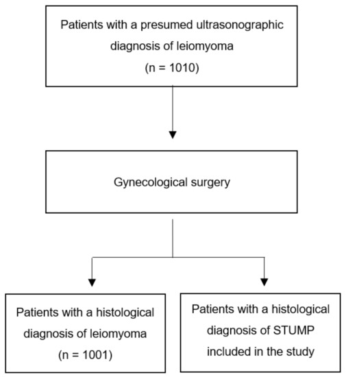
Figure 1.
Flowchart showing all the cases examined and those that were finally selected for inclusion in the study.
All patients reported menometrorrhagia and pelvic pain, among others, suggesting a symptomatic presentation. A detailed examination of the tumor characteristics revealed that, in all cases, the lesion was solitary and intramural, with a mean diameter of 7.5 cm (range 2 to 13 cm). Four patients opted for laparotomic conservative surgery to preserve their fertility. The mean postoperative follow-up time was 36 months and recurrences were observed in two patients (Table 1).

Table 1.
Features of the 9 cases with postoperative histological diagnosis of STUMP.
4. Discussion
In accordance with the literature [11,17], the present analysis confirmed that STUMPs constitute a rare category within the broad array of uterine neoplasms, having been diagnosed in only 9% of the examined patients, and it highlighted the potential for diagnostic ambiguity in the initial imaging results. It also reaffirmed that it predominantly appears before the age of 50, as reported in previous studies [21].
One of the challenges of STUMPs is the pressing need to establish a set of universally agreed-upon diagnostic criteria [17,22,23]. Due to their singular histological attributes, which sit on the borderline between benign and malignant tumors [17], these entities often plunge the diagnostic process into a sea of uncertainty. Nevertheless, accurate diagnosis is a fundamental prerequisite for delineating the most-beneficial treatment pathway, providing a realistic prognosis, and managing the patient’s clinical course in the most-effective way possible.
Further compounding the complexity of the issue, STUMPs present a set of characteristics that make preoperative diagnosis an exceedingly arduous task [20]. These elusive tumors often masquerade as other uterine conditions, thus concealing their nature until revealed by postoperative histopathological assessments.
These detections often occur inadvertently during surgical procedures, such as myomectomies or hysterectomies [22,24,25], which are typically performed on patients who present with symptoms suggestive of leiomyomas, thereby rendering the detection of STUMPs even more challenging [21,22,25]. In accordance with this, all patients included in the present study reported menometrorrhagia and pelvic pain, among other symptoms, that overlapped with those of more-commonplace gynecological conditions.
Tumor diameters ranged from 2 cm to 13 cm, as reported in previous studies [26,27], and the ultrasound characteristics were not different from those typically present in leiomyomas [20].
Adding to the convoluted puzzle of STUMPs is the ongoing debate about their potential for malignancy [14]. Because of their rarity, the task of gathering comprehensive data to accurately assess the risk of malignant transformation and consequent metastasis is a significant challenge [14]. This inherent difficulty fuels the ambiguous nature of STUMPs, further complicating the mission of formulating precise risk stratification models and implementing effective treatment regimens. The process of developing and standardizing treatment strategies for STUMPs calls for an in-depth exploration of available research data and a thorough consensus-building process within the medical community. The infrequent occurrence of these tumors poses a substantial obstacle to the execution of extensive clinical studies. This limitation hampers the development of evidence-based, globally accepted treatment protocols. Therefore, the collective knowledge and understanding of STUMPs remain in a constant state of flux, with each new case enriching our understanding and providing unique insights into this form of tumor.
Among the enrolled patients, it is possible to note the different features with two examples. A 28-year-old nulliparous woman underwent a laparotomic myomectomy for 16 myometrial lesions. One of these lesions received a histological diagnosis of STUMP. Postoperatively, she had a negative follow-up, and remarkably, she became pregnant 17 months after the surgery and delivered via full-term cesarean section. On the other hand, there was a case of a 57-year-old postmenopausal woman who underwent a laparotomic hysterectomy due to a substantial myometrial formation. Upon histological examination, the formation was diagnosed as STUMP. She had a negative postoperative follow-up for 36 months. She experienced a recurrence in the left pararectal fossa and the right thigh. A subsequent definitive diagnosis identified a well-differentiated leiomyosarcoma, and she is currently scheduled to undergo chemotherapy.
The management of STUMP remains a controversial topic. The optimal treatment strategy is still under debate due to the lack of high-quality, evidence-based guidelines. The decision-making process for treatment should consider several factors, including the patient’s age, fertility desires, the tumor’s characteristics, and the patient’s overall health status. The primary treatment for STUMP is generally surgical, with the preferred approach being total hysterectomy with or without bilateral adnexectomy [28]. However, in women who desire to preserve their fertility, a conservative surgical approach may be considered, as opted for by four of our patients who underwent myomectomy, although this may be associated with a higher risk of recurrence [28]. Morcellation, where large masses are divided into smaller pieces for removal, is generally not recommended in the treatment of STUMP due to the risk of spreading peritoneal implants, thereby increasing the risk of recurrence [29,30].
Emerging research is focused on deepening our understanding of the molecular pathogenesis of STUMP. Researchers utilize advanced molecular biology techniques to identify the primary molecular pathways that drive STUMP oncogenesis. This could potentially pave the way for developing innovative molecularly targeted therapies [30,31]. STUMP tumors are known to relapse and metastasize as either STUMP or leiomyosarcoma [31,32]. Interestingly, recent findings have highlighted the role of the inflammatory myofibroblastic tumor gene (IMT) rearranged with ALK in the precise characterization of relapse [30,33]. Based on these findings, it is recommended that all pathological samples be examined by pathologists with considerable expertise in gynecological diseases. These professionals play a critical role in distinguishing STUMPs from benign leiomyomas and malignant leiomyosarcomas, a task that requires a high level of expertise due to the overlapping histological features among these entities [17].
Recurrent STUMP represents a clinical, diagnostic, and therapeutic challenge, and a unique consensus about its management has not yet been reached considering the scarcity of the currently available literature and the rarity of this tumor [30,34]. In the present study, recurrence was observed in only two cases; both presented with an immunohistological diagnosis of AL-LE and positive staining for p16 and p53. We suggest the potential role of the immunohistochemistry of p16 and p53 in identifying the most-aggressive forms of STUMP. Atkins et al. [35] reported three cases out of eight patients of metastatic disease following an initial interpretation of STUMP: the first patient developed peritoneal and lymph node metastases and was treated with progesterone after tumor debulking, remaining free from disease for three years; the other patients developed liver metastases, which were treated surgically. In agreement with Ip et al. [36], the immunohistochemical analysis showed a strong p16 positivity with a diffuse and focal distribution in two and one of the relapses, respectively. Progesterone [22], gonadotropin-releasing hormone agonists [37], and chemotherapy (such as doxorubicin and cisplatin) [22] could have a potential role as adjuvant therapy, but none have proven effective in the prevention of recurrence.
Furthermore, there is no consensus on the postoperative follow-up. Ip et al. [36] recommend an intensive follow-up protocol with an evaluation performed every 6 months for the first 5 years followed by annual surveillance for the next 5 years.
Given the many uncertainties and complexities associated with STUMP, it is highly recommended that a multidisciplinary tumor board systematically review and discuss each case. This collaborative approach ensures comprehensive decision-making that encompasses all relevant clinical, pathological, and radiological aspects, ultimately leading to the best-possible patient outcomes.
The strengths of this study include the analysis of many patients undergoing gynecological surgery and the consequent inclusion of many patients with STUMP, considering the rarity of these neoplasms. Furthermore, all diagnoses and management were performed at the same center. Nevertheless, further studies with a longer follow-up should be conducted to better understand the varied characteristics of these tumors.
5. Conclusions
Uterine STUMP is an uncommon neoplasm usually detected at the pathologic examination of gynecological surgery specimens in patients with presumed leiomyomas. To date, there is no consensus regarding diagnosis, malignant potential, treatment of choice, and follow-up. STUMP cases should be systematically discussed by tumor boards; pathologists with good expertise in gynecological diseases should examine the histological samples; close surveillance is mandatory because of the possibility of recurrence or metastasis.
Author Contributions
Conceptualization, C.E.; methodology, E.R.; investigation, E.R.; data curation, E.R.; writing—original draft preparation, G.G.I. and F.A.G.; writing—review and editing, F.C.; supervision, G.E. All authors have read and agreed to the published version of the manuscript.
Funding
This research received no external funding.
Institutional Review Board Statement
The study was conducted in accordance with the Declaration of Helsinki and approved by the Institutional Review Board (or Ethics Committee) of Azienda di Rilievo Nazionale e di Alta Specializzazione (ARNAS) Garibaldi, Catania (Protocol Code 263/C.E approved on 11 May 2020) (Report No. 68/2020/CECT2).
Informed Consent Statement
Informed consent was obtained from all subjects involved in the study. Written informed consent was obtained from the patients to publish this paper.
Data Availability Statement
Not applicable.
Conflicts of Interest
The authors declare no conflict of interest.
References
- Dall’Asta, A.; Gizzo, S.; Musarò, A.; Quaranta, M.; Noventa, M.; Migliavacca, C.; Sozzi, G.; Monica, M.; Mautone, D.; Berretta, R. Uterine smooth muscle tumors of uncertain malignant potential (STUMP): Pathology, follow-up, and recurrence. Int. J. Clin. Exp. Pathol. 2014, 7, 8136–8142. [Google Scholar] [PubMed]
- Croce, S.; Chibon, F. MED12 and uterine smooth muscle oncogenesis: State of the art and perspectives. Eur. J. Cancer 2015, 51, 1603–1610. [Google Scholar] [CrossRef] [PubMed]
- Ura, B.; Monasta, L.; Arrigoni, G.; Battisti, I.; Licastro, D.; Di Lorenzo, G.; Romano, F.; Aloisio, M.; Peterlunger, I.; Stabile, G.; et al. Phosphoproteins Involved in the Inhibition of Apoptosis and in Cell Survival in the Leiomyoma. J. Clin. Med. 2019, 8, 691. [Google Scholar] [CrossRef] [PubMed]
- Gökaslan, H.; Türkeri, L.; Kavak, Z.N.; Eren, F.; Şişmanoğlu, A.; İlvan, Ş.; Durmuşoğlu, F. Differential diagnosis of smooth muscle tumors utilizing p53, pTEN and Ki-67 expression with estrogen and progesterone receptors. Gynecol. Obstet. Investig. 2005, 59, 36–40. [Google Scholar] [CrossRef]
- Bodner, K.; Bodner-Adler, B.; Kimberger, O.; Czerwenka, K.; Mayerhofer, K. Estrogen and progesterone receptor expression in patients with uterine smooth muscle tumors. Fertil. Steril. 2004, 81, 1062–1066. [Google Scholar] [CrossRef]
- Ciebiera, M.; Włodarczyk, M.; Wrzosek, M.; Męczekalski, B.; Nowicka, G.; Łukaszuk, K.; Ciebiera, M.; Słabuszewska-Jóźwiak, A.; Jakiel, G. Role of Transforming Growth Factor β in Uterine Fibroid Biology. Int. J. Mol. Sci. 2017, 18, 2435. [Google Scholar] [CrossRef]
- Uluer, E.T.; Inan, S.; Ozbilgin, K.; Karaca, F.; Dicle, N.; Sancı, M. The role of hypoxia related angiogenesis in uterine smooth muscle tumors. Biotech. Histochem. 2015, 90, 102–110. [Google Scholar] [CrossRef]
- Plewka, A.; Madej, P.; Plewka, D.; Kowalczyk, A.; Miskiewicz, A.; Wittek, P.; Leks, T.; Bilski, R. Immunohistochemical localization of selected pro-inflammatory factors in uterine myomas and myometrium in women of various ages. Folia Histochem. Cytobiol. 2013, 51, 73–83. [Google Scholar] [CrossRef]
- Conconi, D.; Redaelli, S.; Lissoni, A.A.; Cilibrasi, C.; Perego, P.; Gautiero, E.; Sala, E.; Paderno, M.; Dalprà, L.; Landoni, F.; et al. Genomic and Epigenomic Profile of Uterine Smooth Muscle Tumors of Uncertain Malignant Potential (STUMPs) Revealed Similarities and Differences with Leiomyomas and Leiomyosarcomas. Int. J. Mol. Sci. 2021, 22, 1580. [Google Scholar] [CrossRef]
- Machado-Lopez, A.; Simón, C.; Mas, A. Molecular and Cellular Insights into the Development of Uterine Fibroids. Int. J. Mol. Sci. 2021, 22, 8483. [Google Scholar] [CrossRef]
- Styer, A.K.; Rueda, B.R. The Epidemiology and Genetics of Uterine Leiomyoma. Best Pract. Res. Clin. Obstet. Gynaecol. 2016, 34, 3–12. [Google Scholar] [CrossRef] [PubMed]
- De La Cruz, M.S.; Buchanan, E.M. Uterine Fibroids: Diagnosis and Treatment. Am. Fam. Physician 2017, 95, 100–107. [Google Scholar] [PubMed]
- Giuntoli, R.L., II; Metzinger, D.S.; DiMarco, C.S.; Cha, S.S.; Sloan, J.A.; Keeney, G.L.; Gostout, B.S. Retrospective review of 208 patients with leiomyosarcoma of the uterus: Prognostic indicators, surgical management, and adjuvant therapy. Gynecol. Oncol. 2003, 89, 460–469. [Google Scholar] [CrossRef]
- Abeler, V.M.; Røyne, O.; Thoresen, S.; Danielsen, H.E.; Nesland, J.M.; Kristensen, G.B. Uterine sarcomas in Norway. A histopathological and prognostic survey of a total population from 1970 to 2000 including 419 patients. Histopathology 2009, 54, 355–364. [Google Scholar] [CrossRef]
- Devaud, N.; Vornicova, O.; Razak, A.R.A.; Khalili, K.; Demicco, E.G.; Mitric, C.; Bernardini, M.Q.; Gladdy, R.A. Leiomyosarcoma: Current Clinical Management and Future Horizons. Surg. Oncol. Clin. N. Am. 2022, 31, 527–546. [Google Scholar] [CrossRef]
- Bell, S.W.; Kempson, R.L.; Hendrickson, M.R. Problematic uterine smooth muscle neoplasms. A clinicopathologic study of 213 cases. Am. J. Surg. Pathol. 1994, 18, 535–558. [Google Scholar] [CrossRef]
- Kurman, R.J.; Carcangiu, M.L.; Herrington, C.S.; Young, R.H. WHO Classification of Tumours of Female Reproductive Organs; International Agency for Research on Cancer: Lyon, France, 2014. [Google Scholar]
- Bonneau, C.; Thomassin-Naggara, I.; Dechoux, S.; Cortez, A.; Darai, E.; Rouzier, R. Value of ultrasonography and magnetic resonance imaging for the characterization of uterine mesenchymal tumors. Acta Obstet. Gynecol. Scand. 2014, 93, 261–268. [Google Scholar] [CrossRef]
- Lin, G.; Yang, L.-Y.; Huang, Y.-T.; Ng, K.-K.; Ng, S.-H.; Ueng, S.-H.; Chao, A.; Yen, T.-C.; Chang, T.-C.; Lai, C.-H. Comparison of the diagnostic accuracy of contrast-enhanced MRI and diffusion-weighted MRI in the differentiation between uterine leiomyosarcoma/smooth muscle tumor with uncertain malignant potential and benign leiomyoma. J. Magn. Reson. Imaging 2016, 43, 333–342. [Google Scholar] [CrossRef] [PubMed]
- Testa, A.C.; Di Legge, A.; Bonatti, M.; Manfredi, R.; Scambia, G. Imaging techniques for evaluation of uterine myomas. Best Pract. Res. Clin. Obstet. Gynaecol. 2016, 34, 37–53. [Google Scholar] [CrossRef] [PubMed]
- Ng, J.S.; Han, A.; Chew, S.H.; Low, J. A clinicopathologic study of uterine smooth muscle tumours of uncertain malignant potential (STUMP). Ann. Acad. Med. Singap. 2010, 39, 625–628. [Google Scholar] [CrossRef]
- Guntupalli, S.R.; Ramirez, P.T.; Anderson, M.L.; Milam, M.R.; Bodurka, D.C.; Malpica, A. Uterine smooth muscle tumor of uncertain malignant potential: A retrospective analysis. Gynecol. Oncol. 2009, 113, 324–326. [Google Scholar] [CrossRef] [PubMed]
- D’Angelo, E.; Prat, J. Uterine sarcomas: A review. Gynecol. Oncol. 2010, 116, 131–139. [Google Scholar] [CrossRef] [PubMed]
- Incognito, G.G.; D’Urso, G.; Incognito, D.; Lello, C.; Miceli, A.; Palumbo, M. Management of a giant uterine smooth muscle tumor of uncertain malignant potential in a 32-year-old woman: Case report and review of the literature. Minerva Obstet. Gynecol. 2022, 74, 466–470. [Google Scholar] [PubMed]
- Ip, P.P.; Cheung, A.N.; Clement, P.B. Uterine smooth muscle tumors of uncertain malignant potential (STUMP): A clinicopathologic analysis of 16 cases. Am. J. Surg. Pathol. 2009, 33, 992–1005. [Google Scholar] [PubMed]
- White, M.P.; Rahimi, S.; Garely, A.; Buhl, A.; Dean, R.M. Uterine smooth Muscle tumors of Uncertain Malignant Potential (STUMP): Review of Pathophysiology, Classification, diagnosis, treatment, and surveillance. J. Healthc. Commun. 2017, 2, 40. [Google Scholar]
- Hughes, L.; Roex, A.; Parange, A. STUMP, a surprise finding in a large fibroid uterus in a 20-year-old woman. Int. J. Women’s Health 2018, 10, 211–214. [Google Scholar] [CrossRef] [PubMed]
- Vilos, G.A.; Marks, J.; Ettler, H.C.; Vilos, A.G.; Prefontaine, M.; Abu-Rafea, B. Uterine smooth muscle tumors of uncertain malignant potential: Diagnostic challenges and therapeutic dilemmas. Report of 2 cases and review of the literature. J. Minim. Invasive Gynecol. 2012, 19, 288–295. [Google Scholar]
- Macciò, A.; Chiappe, G.; Kotsonis, P.; Lavra, F.; Serra, M.; Demontis, R.; Madeddu, C. Abdominal leiomyosarcomatosis after surgery with external morcellation for occult smooth muscle tumors of uncertain malignant potential: A case report. Int. J. Surg. Case Rep. 2017, 38, 107–110. [Google Scholar]
- Rizzo, A.; Ricci, A.D.; Saponara, M.; De Leo, A.; Perrone, A.M.; De Iaco, P.; Nannini, M.A.P.A.M. Recurrent Uterine Smooth-Muscle Tumors of Uncertain Malignant Potential (STUMP): State of The Art. Anticancer Res. 2020, 40, 1229–1238. [Google Scholar]
- Canzonieri, V.; D’Amore, E.S.; Bartoloni, G.; Piazza, M.; Blandamura, S.; Carbone, A. Leiomyomatosis with vascular invasion. A unified pathogenesis regarding leiomyoma with vascular microinvasion, benign metastasizing leiomyoma and intravenous leiomyomatosis. Virchows Arch. 1994, 425, 541–545. [Google Scholar] [CrossRef]
- Han, A.K.W.; Hong, K.; Kim, M.; Kim, M.K.; Kim, M.L.; Jung, Y.W.; Yun, B.S.; Seong, S.J. Unexpected uterine smooth muscle tumor of uncertain malignant potential and sarcoma: A single center cohort study in South Korea. Taiwan. J. Obstet. Gynecol. 2020, 59, 275–281. [Google Scholar] [PubMed]
- Miettinen, M. Smooth muscle tumors of soft tissue and non-uterine viscera: Biology and prognosis. Mod. Pathol. 2014, 27 (Suppl. 1), S17–S29. [Google Scholar] [CrossRef] [PubMed]
- Gadducci, A.; Zannoni, G.F. Uterine smooth muscle tumors of unknown malignant potential: A challenging question. Gynecol. Oncol. 2019, 154, 631–637. [Google Scholar] [PubMed]
- Atkins, K.A.; Arronte, N.; Darus, C.J.; Rice, L.W. The Use of p16 in enhancing the histologic classification of uterine smooth muscle tumors. Am. J. Surg. Pathol. 2008, 32, 98–102. [Google Scholar] [CrossRef]
- Ip, P.P.; Tse, K.Y.; Tam, K.F. Uterine smooth muscle tumors other than the ordinary leiomyomas and leiomyosarcomas: A review of selected variants with emphasis on recent advances and unusual morphology that may cause concern for malignancy. Adv. Anat. Pathol. 2010, 17, 91–112. [Google Scholar]
- Berretta, R.; Rolla, M.; Merisio, C.; Giordano, G.; Nardelli, G.B. Uterine smooth muscle tumor of uncertain malignant potential: A three-case report. Int. J. Gynecol. Cancer 2008, 18, 1121–1126. [Google Scholar] [CrossRef]

Disclaimer/Publisher’s Note: The statements, opinions and data contained in all publications are solely those of the individual author(s) and contributor(s) and not of MDPI and/or the editor(s). MDPI and/or the editor(s) disclaim responsibility for any injury to people or property resulting from any ideas, methods, instructions or products referred to in the content. |
© 2023 by the authors. Licensee MDPI, Basel, Switzerland. This article is an open access article distributed under the terms and conditions of the Creative Commons Attribution (CC BY) license (https://creativecommons.org/licenses/by/4.0/).