Article

Transforaminal Fusion Using Physiologically Integrated Titanium Cages with a Novel Design in Patients with Degenerative Spinal Disorders: A Pilot Study

by
Renat Nurmukhametov
1,
Medet Dosanov
1,
Manuel De Jesus Encarnacion
2,
Rossi Barrientos
2,
Yasser Matos
2,
Alexander Ivanovich Alyokhin
1,
Ismael Peralta Baez
3,
Ibrahim Efecan Efe
4,
Manuela Restrepo
5,
Vishal Chavda
6,
Bipin Chaurasia
7 and
Nicola Montemurro
8,*
1
Central Clinical Hospital of the Russian Academy of Sciences, 121359 Moscow, Russia
2
Department of Neurosurgery, University of Friendship of Peoples, 117198 Moscow, Russia
3
Hospital Dr. Alejandro Cabral, San Juan de la Maguana 72000, Dominican Republic
4
Charité—Universitätsmedizin Berlin, Corporate Member of Freie Universität Berlin, Berlin Institute of Health, and Humboldt-Universität zu Berlin, 10117 Berlin, Germany
5
Ibero-American University, México City 01030, Mexico
6
Department of Pathology, Stanford School of Medicine, Stanford University Medical Center, San Francisco, CA 94305, USA
7
Bhawani Hospital and Research Centre, Birgunj 44300, Nepal
8
Department of Neurosurgery, Azienda Ospedaliera Universitaria Pisana (AOUP), University of Pisa, 56100 Pisa, Italy
*
Author to whom correspondence should be addressed.
Surgeries 2022, 3(3), 175-184; https://doi.org/10.3390/surgeries3030019
Submission received: 23 May 2022 / Revised: 27 June 2022 / Accepted: 28 June 2022 / Published: 30 June 2022

Abstract

:
More contemporary options have been presented in the last few years as surgical methods and materials have improved in patients with degenerative spine illnesses. The use of biologically integrated titanium cages of a unique design based on computer 3D modeling for the surgical treatment of patients with degenerative illnesses of the spine’s intervertebral discs has been proposed and experimentally tested. The goal of this study is to compare the radiographic and clinical outcomes of lumbar posterior interbody fusion with a 3D porous titanium alloy cage versus a titanium-coated polyetheretherketone (PEEK) cage, including fusion quality, time to fusion, preoperative and postoperative patient assessments, and the presence, severity, and other side effect characteristics. (1) Methods: According to the preceding technique, patients who were operated on with physiologically integrated titanium cages of a unique design based on 3D computer modeling were included in the study group. This post-surveillance study was conducted as a randomized, prospective, interventional, single-blind, center study to look at the difference in infusion rates and the difference compared to PEEK cages. The patients were evaluated using CT scans, Oswestry questionnaires (every 3, 6, and 12 months), and VAS scales. (2) Results: Six months following surgery, the symptoms of fusion and the degree of cage deflation in the group utilizing the porous titanium 3D cage were considerably lower than in the group using the PEEK cage (spinal fusion sign, p = 0.044; cage subsidence, p = 0.043). The control group had one case of cage migration into the spinal canal with screw instability, one case of screw instability without migration but with pseudoarthrosis formation and two surrounding segment syndromes with surgical revisions compared with the 3D porous titanium alloy cage group. (3) Conclusions: The technique for treating patients with degenerative disorders or lumbar spine instability with aspects of neural compression utilizing biologically integrated titanium cages of a unique design based on computer 3D printing from CT scans has been proven. This allows a new approach of spinal fusion to be used in practice, restoring the local sagittal equilibrium of the spinal motion segment and lowering the risk of pseudarthrosis and revision surgery.

1. Introduction

Currently, posterior lumbar interbody fusion (PLIF) or transforaminal lumbar interbody fusion (TLIF) techniques have been introduced to treat degenerative pathological disease of the spine using cages made of various materials, including titanium alloys [1,2,3] and other materials that have a shape memory effect [4,5]. The major task is to support the load on the spine and distribute axial stresses that fall on the operated section. A structural-holistic graft is frequently used for interbody fusion. The use of specific implants known as cages, which are inserted from the posterior to anterior surgical accesses to the operating segment of the spine, can provide a solution to such difficulties. Some current approaches to the lumbar spine are TLIF, extreme lateral interbody fusion (XLIF), oblique lateral interbody fusion (OLIF), and anterior lumbar interbody fusion (ALIF). The use of cage-based fusion technology yields better outcomes in repairing and maintaining the standard height of the intervertebral disc [6,7,8], contributing to the restoration of normal neurological function and pain relief for patients. At its best, the implant should provide mechanical support and grow into the surrounding tissues, displaying strong integrative properties. Such qualities are achieved by using special cage materials, such as porous and plastic compounds that can provide an intermediary layer of tissue between the bone and cartilage components, as well as the implant itself. Cages can cause complications in practice, such as non-fusion problems with body tissues, biomechanical conflict between the cage and the adjacent vertebra, or implant migration, which occurs in 1 to 5% of cases in clinical spine surgery [9]. To reduce cage movement, it may be worthwhile to establish a good fixation element geometry and improve the cage’s design, which is functionally focused on aiding spinal fusion and sustaining axial load.
As a result, the work of surgical treatment of degenerative spinal illnesses is localized in terms of surgical indication, cage material selection, and cage design. Based on a synthesis of advanced domestic and international experiences, it appears appropriate to broaden the scope of research to the possibilities of using biologically integrated titanium cages of an individual design for the stated purposes, with the latter being developed based on the results of 3D modeling as well as competing technologies such as PEEK (polyetheretherketone). Because of its outstanding strength, fatigue resistance, and biocompatibility, titanium alloy (Ti) is currently the ideal material for pedicle-based screw fixation systems [10,11].
The diversity of degenerative spinal lesions and the polymorphism of clinical and pathomorphological manifestations necessitate the development and testing of promising methods and tools based on the principles of reliable fusion and complex clinical and morphological compliance to ensure early and long-term results. The findings of an empirical investigation using physiologically integrated titanium cages of a unique design based on 3D computer modeling for TLIF in patients with degenerative disorders of the spine are presented in this publication. 3D printing technology has increasing applications in spine surgery due to its many realized and potential benefits. A recent review showed the current and future potential of 3D printing technology in various aspects of spine surgery [12]. The ability of 3D printing technology to geometrically mimic the spinal anatomy enables the manufacturing of realistic anatomical models that can aid in patient education and surgical training. In addition, the atypical spinal condition of some patients required a custom implant for surgical management, as the restoration of anatomy could not be achieved using standard implants [12].
The goal of this study is to compare the radiographic and clinical outcomes of lumbar posterior interbody fusion with a 3D porous titanium alloy cage versus a titanium-coated PEEK cage, including fusion quality, time to fusion, preoperative and postoperative patient assessments, and the presence, severity, and other side effect characteristics. A randomized, prospective, interventional, double, single-blind, post-marketing study was done to examine the difference in the rate of fusion formation and the difference in comparison to PEEK cages. Stryker develops PEEK cages, which are used as a comparison item (herein after referred to as PEEK cages).

2. Materials and Methods

2.1. Materials and Design

The authors improved on the previously patented edition Implant (a frame made of metal wire connected by diffusion welding and formed in the shape of a cylindrical sleeve formed by a spirally wound wire with subsequent laying of turns along the axis of the sleeve, having a rigidity of 1000 to 5000 N/mm with an average size of open pores of 150 to 600 m and a porosity coefficient of 50–60 percent). Based on 3D computer modeling and the following technological specifications, biologically integrable titanium cages of a unique design were designed.
The cages are intended to repair disc and cartilage fragments in the spine, restore the height of the interbody gap, and decrease the instability of the spinal column’s anterior support. They are self-made titanium implants based on a unique 3D model of cage placement in the interbody space created from a CT scan (Figure 1).
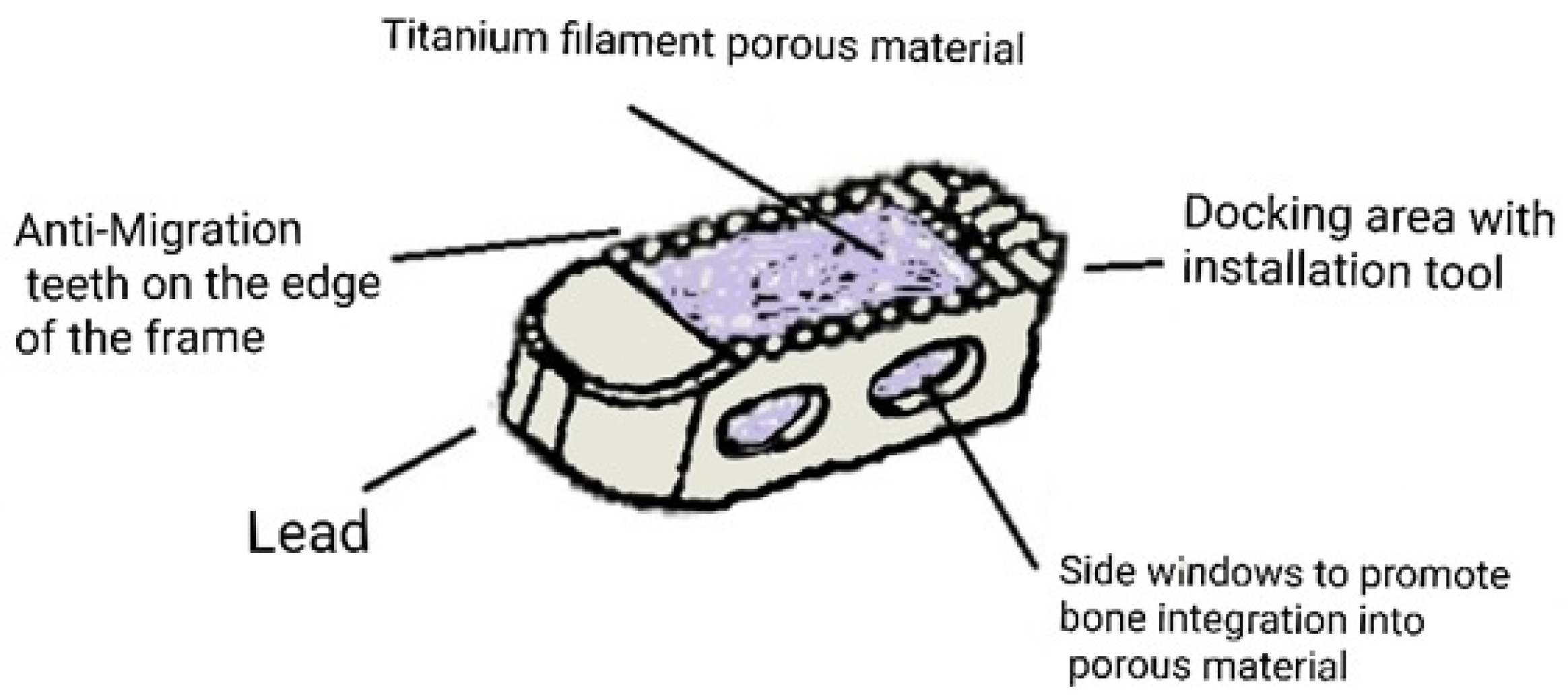
A monolithic titanium frame and an internal porous base (pressed titanium thread) are used to create implants (Figure 2). The cage’s stiff section is a titanium frame with a bullet-like geometric form. The technological holes for fixing the installation tool are grooves or threaded holes on the back of the cage. The height of the anti-migration teeth on the rigid frame’s body is 1.2–1.5 mm, and the rigid frame’s sidewalls are 1.2 mm thick. Implantation method: PLIF/TLIF. Product name: TLIF-cage “Biodynamics”.

2.2. Participants

All patients included in this study were treated at the Central Clinical Hospital of the Russian Academy of Sciences in Moscow, Russia. Informed consent was obtained by all patients in written form. According to the preceding technique, patients who were operated on with physiologically integrated titanium cages of a unique design based on 3D computer modeling were included in the study group. Participants had to have a mature skeleton, be between the ages of 21 and 75, be free of issues such as obesity (BMI > 40) and sensitivity to titanium materials, be free of obvious contraindications to surgery and/or anomalies impacting the normal bone regeneration process. Patients in the control group underwent surgery using a standard non-individual medical plastic (PEEK) cage. Follow-up was 3–6 months following the operation.

2.3. Interventions

Patients were investigated with a preoperative and a postoperative computed tomography (CT), with an Oswestry questionnaire (every 3, 6 months), and with a visual analog scales (VAS) evaluation, when needed. A clinical and neurological examination survey and functional radiography, multislice computed tomography (MSCT), and magnetic resonance imaging were among the preoperative radiation diagnostics (MRI). To calculate the essential parameters and create an individual cage, all patients in the Study Group underwent multislice computed tomography (MSCT).

2.4. Outcome

The bone block was evaluated using recommendations [13]. If there was at least one continuous bone bridge between the vertebral bodies, both through the interbody implant and around it, a full-fledged bone block was recognized; otherwise, the existence of a failure of the bone block development was recognized. In the meantime, the state of the fixation system was evaluated; in the presence of a bilateral fracture of the longitudinal rods at the same level, a fracture of both screws in at least one vertebra, and/or osteolysis around both screws in at least one vertebra, the presence of fixation system instability was identified.
During the postoperative follow-up, CT interpretation was used to estimate the duration of fusion formation and to identify long-term side effects and migrations. Patients’ functional activity and quality of life were assessed using the Oswestry questionnaire. The Russian-adapted version of questionnaire 2.1a (Russian Version of the Oswestry Disability Index) was utilized [14]. The results are interpreted as follows: a disability of 0–20 percent is considered minimal, a disability of 20–40 percent is moderate, a disability of 40–60 percent is pronounced, a disability of 60–80 percent is individualizing, and a disability of 80–100 percent is extremely pronounced or exaggerated. The pain intensity was measured using the VAS scale. If the score was less than a certain threshold (2 points), the indicator was considered important. In the presence of chronic back pain syndrome (VAS > 4 points and/or ODI > 30% for at least the last three months), syndrome of intermittent neurogenic claudication, radicular pain syndrome and sensitivity disorders (any options) in the absence of the effect of conservative therapy, violation of the motor sphere, a clinical and neurological examination was also performed (with muscle strength of 4 or less points).

2.5. Statistical Methods

The statistical evaluation of this study was done with a Student’s paired T-test using GraphPad Prism 5.0.2, and the level of significance was considered as (p < 0.05).

3. Results

A total of 80 patients were enrolled in the study (Table 1), with 40 in the study group (3D porous titanium alloy cage) and 40 in the control group (PEEK cages). Both groups included patients with degenerative and dystrophic lumbar spine problems who had surgery using the posterior transforaminal fusion method with transpedicular fixation at the operating levels. Except for the elderly, men predominate in able-bodied age groups, which can be explained by larger physical and static pressures on the spinal column. The majority of patients with degenerative-dystrophic illnesses of the spine who require surgical intervention are from the able-bodied population, highlighting the importance of study in this field.
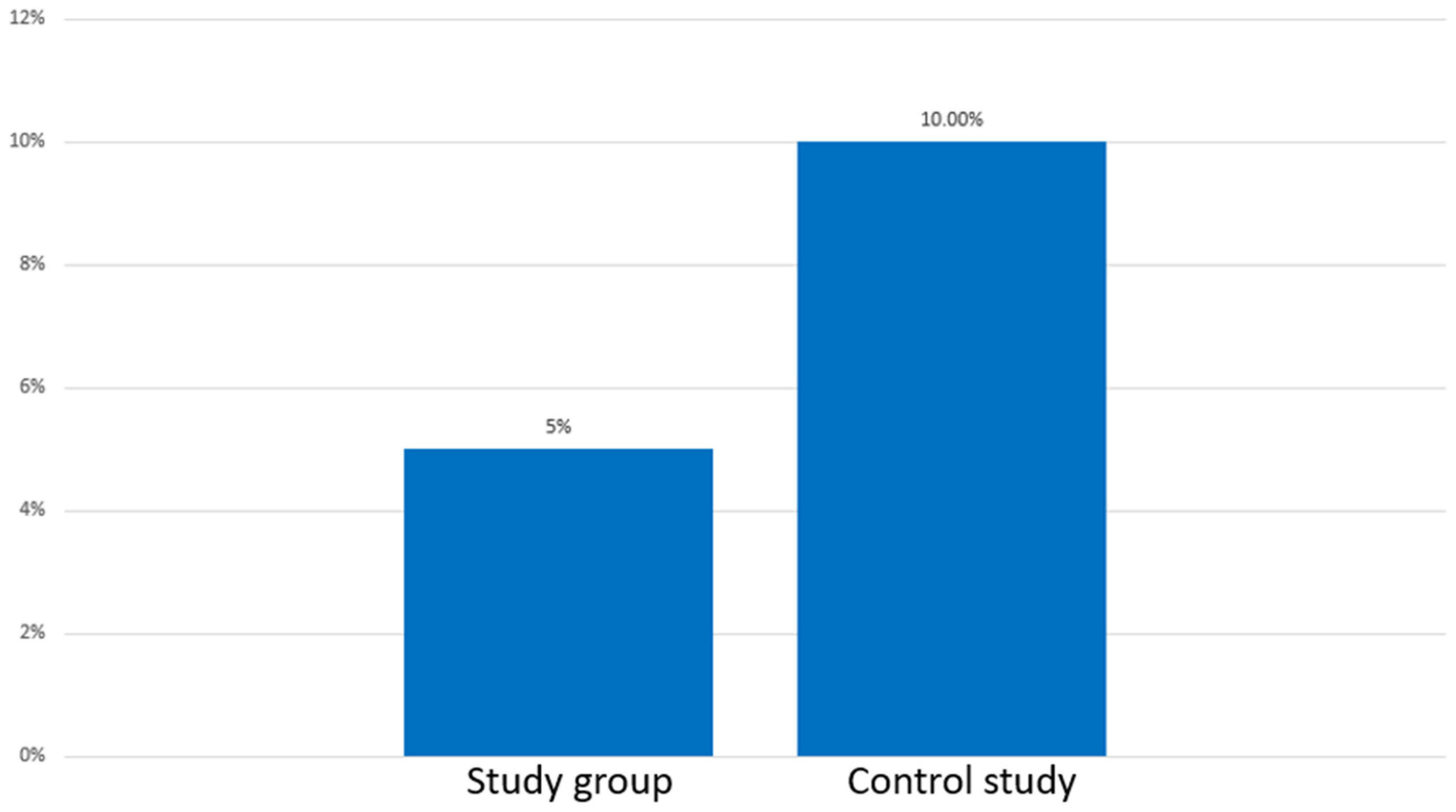
Six months following surgery, the symptoms of fusion and the degree of cage deflation in the group utilizing the porous titanium 3D cage were considerably lower than in the group using the PEEK cage (spinal fusion sign, p = 0.044; cage subsidence, p = 0.043). However, the efficiency of the operation did not differ between the two groups six months and one year following the operation. The clinical outcomes discussed with patients one year after surgeries were similar in the porous 3D titanium alloy cage and titanium-coated PEEK cage groups. However, postoperative endplate fusion is more common and extensive. Modeling was a remarkable radiological finding in patients with custom-designed biointegrable titanium cages based on the 3D computer. During the follow-up period (from 3–6 months after surgery), the Study Group had no cases of screw migration or instability, whereas the control group had one case of cage migration into the spinal canal with screw instability and one case of screw instability without migration but with the formation of pseudarthrosis (Figure 3).
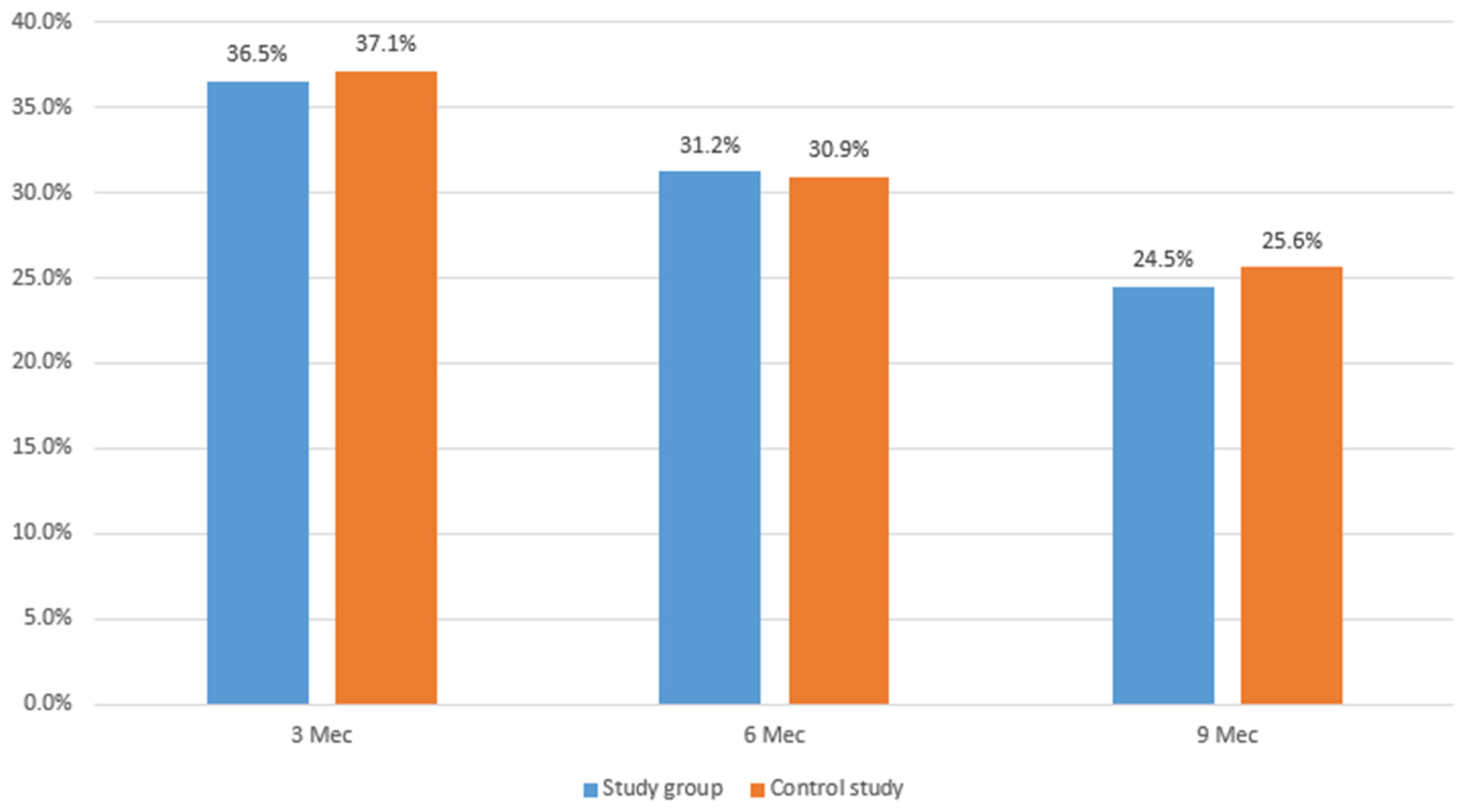
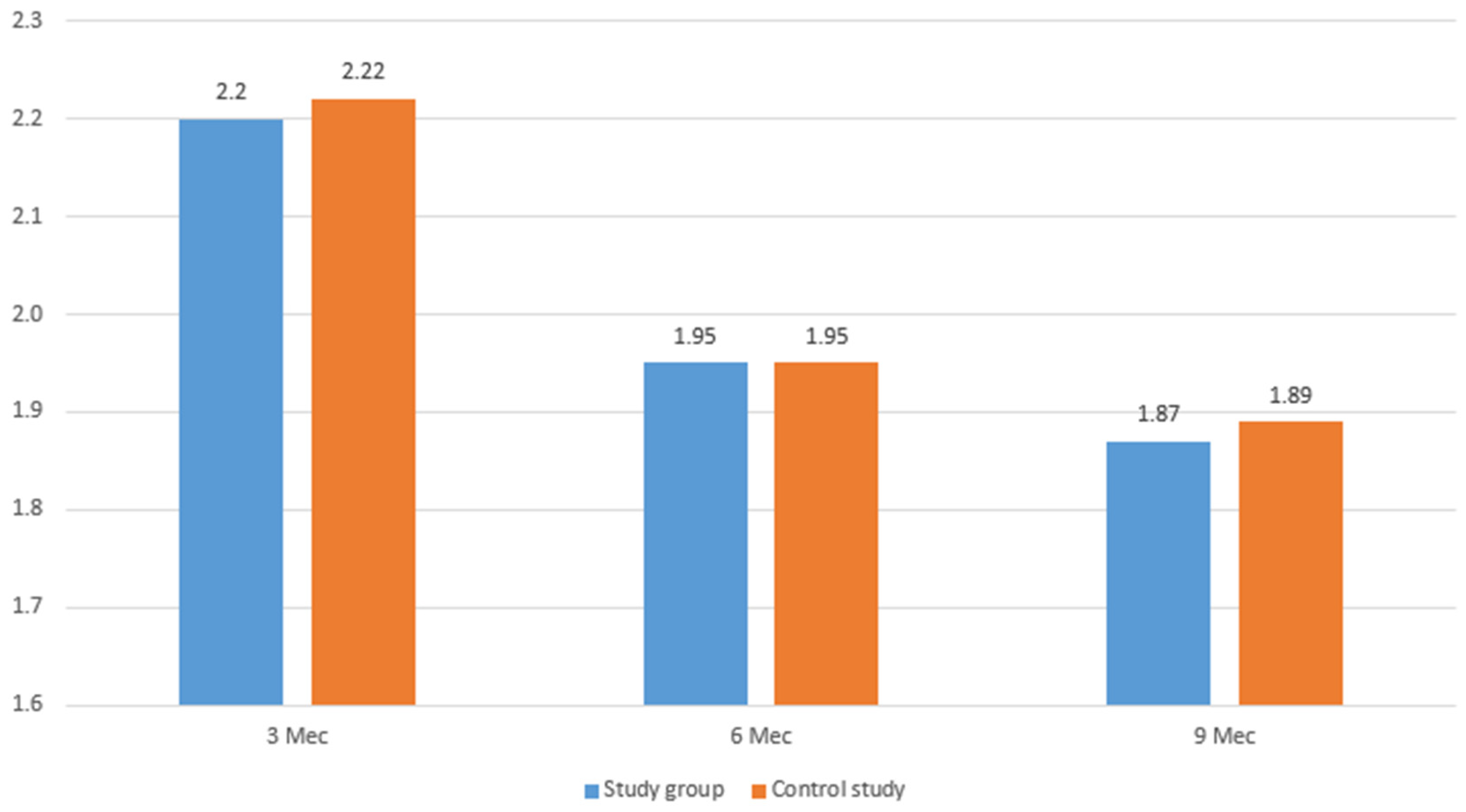
There were two units of reoperations in the study group for the syndrome of the adjacent segment. Back pain was recognized at a minimum level in all patients in the study group (100 percent), whereas back pain was identified at a minimum level in 36 patients in the control group (90 percent), with the exception of four patients who required a second revision operation (Figure 4). Screws were stable and cage incisors were cut into the endplates in both groups, according to CT scan data taken 3–6 months after surgery. Patients in the study group performed slightly better on the Oswestry questionnaire (Figure 5) and on the VAS scale (Figure 6), but there were no statistically significant differences between the two groups.

4. Discussion

Our findings revealed a slight improvement in the controlled parameters in study group patients compared to control group patients, confirming that the method of removing pathological conditions of the spine using biologically integrated titanium cages of a unique design based on computer 3D modeling is not inferior and generally outperforms the standardized non-custom design medical-grade plastic (PEEK) cage technique. It also showed good biocompatibility and osseointegration, which provides clinical value for applying bone implants in biomedical applications. Interbody fusion with a titanium cage combined with transpedicular fixation is acknowledged in modern medical practice as an efficient option for removing spinal instability [15,16,17]. Cages are special closable devices made out of a titanium frame with an upper and lower aperture and an internal hollow (to be filled with bone chips or a replacement material). The usual procedure for the treatment of spinal injuries, as well as for stabilization during operations for vertebral body fractures, periarticular fracture subsidence, or degenerative-dystrophic diseases, is the replacement of bone and cartilage structures, which is usually done using implants (cages) made of various materials [18]. The selection of material for the cage’s construction is a critical scientific effort; the material must encourage successful germination, avoid biomechanical conflicts with human tissues, and meet load and stress absorption requirements. Metal, ceramic, and polymer materials are among the materials available for the fabrication of cages for interbody fusion of the spine [19]. Cages composed of composite materials and special alloys, particularly those with a shape memory effect, such as titanium and titanium alloys as well as titanium-coated PEEK, are the most prevalent in relation to challenging clinical duties and the development of medical materials technologies [10].
These technologies and materials are more in line with the stated requirements for interbody fusion, although they may have drawbacks as well. PEEK cages, in particular, may not always provide complete protection against migration, and the use of PEEK cages does not always result in good indications of tissue integration [20]. Porous titanium materials may have a greater biointegration performance than PEEK [21,22]. The osteointegration properties of the implant are closely related to surface roughness, biocompatibility, proper pore size, and porosity. Morphological observations of the novel 3D printed cage show that it is an ideal carrier for the adhesion of bone cells [23]. The characteristics of internal porosity and intercommunication and the diameter of the metal trabecular diameter are all conducive to the growth of bone tissue, and it can obtain a better fusion effect [23,24].
The need for comparative investigations of the two technologies is due to the much higher cost of PEEK-titanium cages manufactured and imported from other countries, which limits their widespread use in Russian medicine. The titanium cage design alone does not allow for tissue integration; the material used to fill the apertures is frequently isolated and does not create adhesions, resulting in pseudarthrosis [25]. Biomechanical conflicts may emerge because of the difference in elastic moduli between the cage (the elastic modulus of titanium is 120 GPa) and the vertebral body (0.75–10 GPa) [26].
The biological integration of the implantable titanium cage and bone, which is ensured by utilizing a porous material consisting of titanium fibers, can successfully tackle the combination of these difficulties. Recent studies have shown that the surface characteristics of titanium make it superior to other materials in terms of osteoinductivity and osseointegration, as osteoblasts produced significantly higher levels of vascular endothelial growth factor-A (VEGF-A) and fibroblast growth factor-2 (FGF-2) on the surface of titanium alloys than on PEEK materials [24,27]. When the structure is forced against the bone, this technology gives consistent results in terms of the penetration of the cage into the bone and the production of shock absorption. It appears that present technologies must be improved in order to improve the geometry of the structures employed, as well as their elasticity and stress absorption capability. The individualization of implant design is possible because of modern information technologies and related industrial solutions, which eliminate current flaws. 3D modeling and 3D printing technologies are examples of such solutions [28,29,30,31,32,33]. As a result, it can be said that the employment of biologically integrated titanium cages with a unique design for transforaminal fusion can produce better outcomes in the eradication of disease-ridden maladies of the spine, albeit with further empirical verification being required.
Based on the results obtained, the active use of a promising technology for eliminating pathological conditions of the spine using biologically integrated titanium cages of a unique design based on computer 3D modeling is recommended, taking into account the high cost and associated limited use of PEEK technologies, as well as the objective need for import substitutions in Russian medicine. The technology allows one to manage the modulus of elasticity and implant integration while maintaining biological inertness at the highest possible degree. Furthermore, you can utilize this technique to imitate the qualities of a missing intervertebral disc or vertebral body by growing your own freshly created tissue over the implant.

Limitations of the Study

The main limitations of this study are the small sample of patients and the heterogeneity of these patients. These limitations make it impossible for us to perform a deep statistical analysis or a proper association. This is a pilot study, and further randomized multicenter studies are needed.

5. Conclusions

It appears that the surgical treatment of patients with degenerative diseases of the intervertebral discs of the spine using biologically integrated titanium cages of a unique design based on 3D computer modeling will produce scientific and technical results that will serve as the foundation for the innovative development of the domestic market for products and services, as well as the transition to personalized medicine and life-saving devices. The ideal implant should be both patient-specific and condition-specific to maximize the compatibility of the implant with the body and the effectiveness of the instrumentation and adjuvant treatments. As the state of 3D printing technology advances, it would result in a better resolution of printing and the availability of more 3D printable implant materials. This new advancement would allow for more optimal patient-specific and condition-specific implants and emerging applications such as biodegradable implants and localized drug delivery systems. Based on our findings, the effectiveness of the technology for treating patients with degenerative diseases of the spine or an instability of spinal motion segments with elements of neural compression using biologically integrated titanium cages of a unique design based on computer 3D is confirmed, allowing the practical use of a new spinal fusion technique that allows for the restoring of local sagittal balance and the reduction of the incidence of pseudarthrosis.

Author Contributions

Conceptualization, R.N., M.D., M.D.J.E. and N.M.; methodology, M.D.J.E., V.C. and R.B.; validation, Y.M., A.I.A., I.P.B., B.C. and N.M.; investigation, I.E.E. and M.R.; resources, M.D.J.E. and M.R.; data curation, R.N., M.D., B.C. and N.M.; writing—original draft preparation, R.N., M.D., R.B. and N.M.; writing—review and editing, V.C., M.R., B.C. and N.M.; visualization, V.C., M.D., Y.M. and A.I.A.; supervision, R.N., B.C., V.C. and N.M. All authors have read and agreed to the published version of the manuscript.

Funding

This research received no external funding.

Institutional Review Board Statement

The study was conducted in accordance with the Declaration of Helsinki and approved by the Ethics Committee of RAS for studies involving humans.

Informed Consent Statement

Informed consent was obtained from all subjects involved in the study.

Data Availability Statement

Not applicable.

Conflicts of Interest

The authors declare no conflict of interest.

References

  1. Nemoto, O.; Asazuma, T.; Yato, Y.; Imabayashi, H.; Yasuoka, H.; Fujikawa, A. Comparison of fusion rates following transforaminal lumbar interbody fusion using polyetheretherketone cages or titanium cages with transpedicular instrumentation. Eur. Spine J. 2014, 23, 2150–2155. [Google Scholar] [CrossRef] [PubMed]
  2. Wang, B.; Hua, W.; Ke, W.; Lu, S.; Li, X.; Zeng, X.; Yang, C. Biomechanical Evaluation of Transforaminal Lumbar Interbody Fusion and Oblique Lumbar Interbody Fusion on the Adjacent Segment: A Finite Element Analysis. World Neurosurg. 2019, 126, e819–e824. [Google Scholar] [CrossRef] [PubMed]
  3. Ponnappan, R.K.; Serhan, H.; Zarda, B.; Patel, R.; Albert, T.; Vaccaro, A.R. Biomechanical evaluation and comparison of polyetheretherketone rod system to traditional titanium rod fixation. Spine J. 2009, 9, 263–267. [Google Scholar] [CrossRef] [PubMed]
  4. Wang, X.; Xu, J.; Zhu, Y.; Li, J.; Zhou, S.; Tian, S.; Xiang, Y.; Liu, X.; Zheng, Y.; Pan, T. Biomechanical analysis of a newly developed shape memory alloy hook in a transforaminal lumbar interbody fusion (TLIF) in vitro model. PLoS ONE 2014, 9, e114326. [Google Scholar] [CrossRef]
  5. Zhou, C.; Sethi, K.; Willing, R. Shape optimisation of a transforaminal lumbar interbody fusion cage featuring an I-beam cross-section. Orthop. Proc. 2017, 99, 125–126. [Google Scholar]
  6. Cole, C.D.; McCall, T.D.; Schmidt, M.H.; Dailey, A.T. Comparison of low back fusion techniques: Transforaminal lumbar interbody fusion (TLIF) or posterior lumbar interbody fusion (PLIF) approaches. Curr. Rev. Musculoskelet. Med. 2009, 2, 118–126. [Google Scholar] [CrossRef] [Green Version]
  7. Ahsan, K.; Khan, S.I.; Zaman, N.; Ahmed, N.; Montemurro, N.; Chaurasia, B. Fusion versus nonfusion treatment for recurrent lumbar disc herniation. J. Craniovertebr. Junction Spine 2021, 12, 44–53. [Google Scholar] [CrossRef]
  8. Ahsan, M.K.; Hossain, M.R.; Khan, M.S.I.; Zaman, N.; Ahmed, N.; Montemurro, N.; Chaurasia, B. Lumbar revision microdiscectomy in patients with recurrent lumbar disc herniation: A single-center prospective series. Surg. Neurol. Int. 2020, 11, 404. [Google Scholar] [CrossRef]
  9. Faizan, A.; Kiapour, A.; Kiapour, A.M.; Goel, V.K. Biomechanical analysis of various footprints of transforaminal lumbar interbody fusion devices. Clin. Spine Surg. 2014, 27, E118–E127. [Google Scholar] [CrossRef]
  10. Kersten, R.F.; van Gaalen, S.M.; de Gast, A.; Öner, F.C. Polyetheretherketone (PEEK) cages in cervical applications: A systematic review. Spine J. 2015, 15, 1446–1460. [Google Scholar] [CrossRef]
  11. Montemurro, N.; Condino, S.; Cattari, N.; D’Amato, R.; Ferrari, V.; Cutolo, F. Augmented Reality-Assisted Craniotomy for Parasagittal and Convexity En Plaque Meningiomas and Custom-Made Cranio-Plasty: A Preliminary Laboratory Report. Int. J. Environ. Res. Public Health 2021, 18, 9955. [Google Scholar] [CrossRef] [PubMed]
  12. Kumara, N.; Ramakrishnana, A.S.; Lopez, K.G.; Chin, B.Z.; Devyapriya, S.; Kumar, L.; Baskar, S.; Vellayappan, B.A.; His Fuh, J.Y.; Kumar, S.; et al. Current trends and future scope in 3D printing for surgical management of spine pathologies. Bioprinting 2022, 26, 197. [Google Scholar] [CrossRef]
  13. Watters, W.C., III; Resnick, D.K.; Eck, J.C.; Ghogawala, Z.; Mummaneni, P.V.; Dailey, A.T.; Choudhri, T.F.; Sharan, A.; Groff, M.W.; Wang, J.C.; et al. Guideline update for the performance of fusion procedures for degenerative disease of the lumbar spine. Part 13: Injection therapies, low-back pain, and lumbar fusion. J. Neurosurg. Spine 2014, 21, 79–90. [Google Scholar] [CrossRef] [PubMed] [Green Version]
  14. Cherepanov, E.A. Russian version of the Oswestry Disability Index: Cross-cultural adaptation and validity. Hir. Pozvonočnika 2009, 3, 93–98. [Google Scholar] [CrossRef] [Green Version]
  15. Byvaltsev, V.A.; Kalinin, A.A.; Okoneshnikova, A.K.; Kerimbaev, T.T.; Belykh, E.G. Facet Fixation Combined with Lumbar Interbody Fusion: Comparative Analysis of Clinical Experience and A New Method of Surgical Treatment of Patients with Lumbar Degenerative Diseases. Vestn. Ross. Akad. Med. Nauk 2016, 71, 375–384. [Google Scholar] [CrossRef] [PubMed] [Green Version]
  16. Chen, H.H.; Cheung, H.H.; Wang, W.K.; Li, A.; Li, K.C. Biomechanical analysis of unilateral fixation with interbody cages. Spine 2005, 30, E92–E96. [Google Scholar] [CrossRef]
  17. Zhao, J.; Zhang, F.; Chen, X.; Yao, Y. Posterior interbody fusion using a diagonal cage with unilateral transpedicular screw fixation for lumbar stenosis. J. Clin. Neurosci. 2011, 18, 324–328. [Google Scholar] [CrossRef]
  18. Alvarez, K.; Nakajima, H. Metallic Scaffolds for Bone Regeneration. Materials 2009, 2, 790–832. [Google Scholar] [CrossRef]
  19. Jain, S.; Eltorai, A.E.; Ruttiman, R.; Daniels, A.H. Advances in Spinal Interbody Cages. Orthop. Surg. 2016, 8, 278–284. [Google Scholar] [CrossRef]
  20. Basgul, C.; Yu, T.; MacDonald, D.W.; Siskey, R.; Marcolongo, M.; Kurtz, S.M. Does annealing improve the interlayer adhesion and structural integrity of FFF 3D printed PEEK lumbar spinal cages? J. Mech. Behav. Biomed. Mater. 2020, 102, 103455. [Google Scholar] [CrossRef]
  21. Kurtz, S.M. PEEK Biomaterials Handbook, 2nd ed.; William Andrew: Norwich, NY, USA, 2019; pp. 101–102. [Google Scholar]
  22. Schroeder, G.D.; Canseco, J.A.; Patel, P.D.; Divi, S.N.; Karamian, B.A.; Kandziora, F.; Vialle, E.N.; Oner, F.C.; Schnake, K.J.; Dvorak, M.F.; et al. Establishing the Injury Severity of Subaxial Cervical Spine Trauma: Validating the Hierarchical Nature of the AO Spine Subaxial Cervical Spine Injury Classification System. Spine 2021, 46, 649–657. [Google Scholar] [CrossRef] [PubMed]
  23. Li, Y.; Qi, Y.; Gao, Q.; Niu, Q.; Shen, M.; Fu, Q.; Hu, K.; Kong, L. Effects of a micro/nano rough strontium-loaded surface on osseointegration. Int. J. Nanomed. 2015, 10, 4549–4563. [Google Scholar]
  24. Li, P.; Jiang, W.; Yan, J.; Hu, K.; Han, Z.; Wang, B.; Zhao, Y.; Cui, G.; Wang, Z.; Mao, K.; et al. A novel 3D printed cage with microporous structure and in vivo fusion function. J. Biomed. Mater. Res. A 2019, 107, 1386–1392. [Google Scholar] [CrossRef]
  25. Lin, W.; Ha, A.; Boddapati, V.; Yuan, W.; Riew, K.D. Diagnosing Pseudoarthrosis After Anterior Cervical Discectomy and Fusion. Neurospine 2018, 15, 194–205. [Google Scholar] [CrossRef] [PubMed] [Green Version]
  26. Kandziora, F.; Pflugmacher, R.; Schaefer, J.; Scholz, M.; Ludwig, K.; Schleicher, P.; Haas, N.P. Biomechanical comparison of expandable cages for vertebral body replacement in the cervical spine. J. Neurosurg. 2003, 99, 91–97. [Google Scholar] [CrossRef] [PubMed] [Green Version]
  27. Olivares-Navarrete, R.; Hyzy, S.L.; Pan, Q.; Dunn, G.; Williams, J.K.; Schwartz, Z.; Boyan, B.D. Osteoblast maturation on microtextured titanium involves paracrine regulation of bone morphogenetic protein signaling. J. Biomed. Mater. Res. A 2015, 103, 1721–1731. [Google Scholar] [CrossRef]
  28. Provaggi, E.; Leong, J.J.H.; Kalaskar, D.M. Applications of 3D printing in the management of severe spinal conditions. J. Eng. Med. 2017, 231, 471–486. [Google Scholar] [CrossRef]
  29. Mishra, R.; Narayanan, M.D.K.; Umana, G.E.; Montemurro, N.; Chaurasia, B.; Deora, H. Virtual Reality in Neurosurgery: Beyond Neurosurgical Planning. Int. J. Environ. Res. Public Health 2022, 19, 1719. [Google Scholar] [CrossRef]
  30. Provaggi, E.; Capelli, C.; Rahmani, B.; Burriesci, G.; Kalaskar, D. 3D printing assisted finite element analysis for optimising the manufacturing parameters of a lumbar fusion cage. Mater. Des. 2019, 163, 107540. [Google Scholar] [CrossRef]
  31. Condino, S.; Montemurro, N.; Cattari, N.; D’Amato, R.; Thomale, U.; Ferrari, V.; Cutolo, F. Evaluation of a Wearable AR Platform for Guiding Complex Craniotomies in Neurosurgery. Ann. Biomed. Eng. 2021, 49, 2590–2605. [Google Scholar] [CrossRef]
  32. Montemurro, N.; Scerrati, A.; Ricciardi, L.; Trevisi, G. The Exoscope in Neurosurgery: An Overview of the Current Literature of Intraoperative Use in Brain and Spine Surgery. J. Clin. Med. 2021, 11, 223. [Google Scholar] [CrossRef] [PubMed]
  33. Sheha, E.D.; Gandhi, S.D.; Colman, M.W. 3D printing in spine surgery. Ann. Transl. Med. 2019, 7, S164. [Google Scholar] [CrossRef] [PubMed]
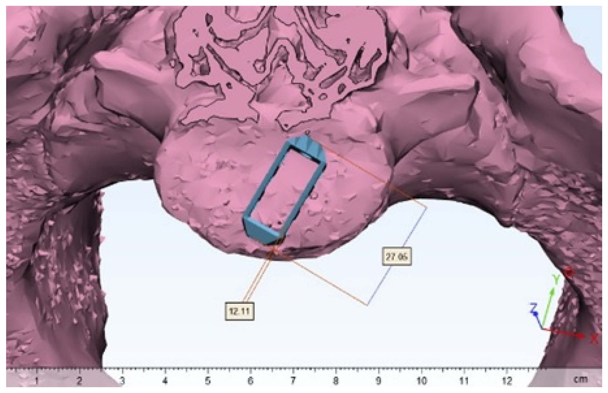
Figure 1. 3D modeling of the implant based on CT scan reconstruction.
Surgeries 03 00019 g001
Figure 2. Schematic illustration of the simulated cage.
Surgeries 03 00019 g002
Figure 3. Proportion of patients with screw instability and cage migration (%).
Surgeries 03 00019 g003
Figure 4. The proportion of patients who underwent reoperations, % of the total number of group participants.
Surgeries 03 00019 g004
Figure 5. Assessment according to the Oswestry questionnaire (average ODI by group), %.
Surgeries 03 00019 g005
Figure 6. Average group values of the VAS indicator (points).
Surgeries 03 00019 g006
Table 1. Characteristics of study participants.
Age (Years)Number of Patients (%)Sex
Female (%)Male (%)
<306 (8%)2 (3%)4 (18%)
30–396 (8%)4 (7%)2 (9%)
40–4912 (15%)5 (9%)7 (32%)
50–5919 (24%)16 (28%)3 (14%)
60–7037 (46%)31 (53%)6 (27%)
Total80 (100%)58 (100%)22 (100%)
Publisher’s Note: MDPI stays neutral with regard to jurisdictional claims in published maps and institutional affiliations.