Case Report

A Multilayered Dural Repair Technique Using Duragen for Early Cranioplasty Following Decompressive Craniotomy

Department of Neurosurgery, Kawasaki Municipal Hospital, Shinkawadori, Kawasaki-ku, Kanagawa 210-0013, Japan
*
Author to whom correspondence should be addressed.
Surgeries 2021, 2(4), 371-377; https://doi.org/10.3390/surgeries2040036
Submission received: 4 October 2021 / Revised: 26 October 2021 / Accepted: 27 October 2021 / Published: 28 October 2021

Abstract

:
Decompressive craniotomy is a neurosurgical emergency procedure in which a large skull bone is removed and the dura matter is extensively opened. Duraplasty is required to avoid cerebrospinal fluid (CSF) leakage during the decompressive craniotomy. DuraGen® is a safe and effective type I collagen matrix graft, which is frequently used in decompressive craniotomy procedures. Since DuraGen® does not require labor-intensive suturing, the operative time is shortened by DuraGen® closure with sufficient tightness preventing CSF leakage. Recently, early cranioplasty is preferred to achieve efficient rehabilitation after decompressive craniotomy. Although evidence of efficacy and safety of DuraGen® has been increasing in the management of duraplasty, no reports have previously discussed the condition of DuraGen® during the second surgery (cranioplasty) at this early timing. DuraGen®-derived neodura develops a mature dura 1 year post its placement, and the neodura remain fragile at this early time point. A deconstructed fragile neodura may result in postoperative CSF leakage. Here, we illustrated a multilayered dural repair technique with DuraGen® to avoid disruption of the fragile neodura during early cranioplasty.

1. Introduction

Decompressive craniotomy is a neurosurgical emergency procedure in which a large skull bone is removed and the dura matter is extensively opened [1]. Indications for decompressive craniotomy are elevated intracranial pressure due to severe traumatic brain injury and stroke [2]. Subsequent cranioplasty and rehabilitation will be required once the damaged brain recovers from its initial pathology [2].
Duraplasty is required to avoid cerebrospinal fluid (CSF) leakage during decompressive craniotomy. Current surgical methods of dural repair consist of the use of muscular fascia, periosteum, and artificial dural substitutes such as Gore-Tex DM® (W.L. Gore & Associates Inc., Phoenix, AZ, USA) and SEAMDURA® (Gunze, Kyoto, Japan) [3]. Although autografts do not induce an immunological reaction, these tissues are not always available in the required size. Gore-Tex DM® is a non-absorbable membrane and requires suturing for dural closure. SEAMDURA®, a biocompatible dural substitute made of a copolymer of L-lactide, ε-caprolacton and polyglycolic acid, lacks pliability and is difficult to manipulate [3].
DuraGen® (Integra Life Sciences Corp., Princeton, NJ, USA) is a safe and effective type I collagen matrix graft, manufactured from bovine Achilles tendon [4]. Since DuraGen® does not require labor-intensive suturing, the operative time is shortened by DuraGen® closure with sufficient tightness preventing CSF leakage. Recently, DuraGen® has been frequently used in decompressive craniotomy procedures [5,6].
Previous studies have demonstrated the potential for early cranioplasty to be a safe and viable option compared to delayed cranioplasty [7,8]. A possible advantage is the abbreviation of excessive bed-ridden periods due to the skull defect which would result in physical vulnerability, and allowing more aggressive rehabilitation [8]. Cranioplasty is generally performed 30–60 days post the initial surgery [9]. Although evidence of efficacy and safety of DuraGen® has been increasing in the management of duraplasty [10], no reports have previously discussed the condition of DuraGen® during a second surgery (cranioplasty) at this early timing.
DuraGen®-derived neodura develops a mature dura 1 year post its placement, and the DuraGen®-derived neodura will remain fragile at this early time point [4,11,12]. It is difficult to create a surgical plane between the myocutaneous flap and the DuraGen®- derived neodura covering the brain surface. A deconstructed fragile neodura may result in postoperative CSF leakage.
Here, we illustrate a multilayered dural repair technique with DuraGen®, which can be potentially used for preventing disrupture of the fragile neodura.

2. Case Report

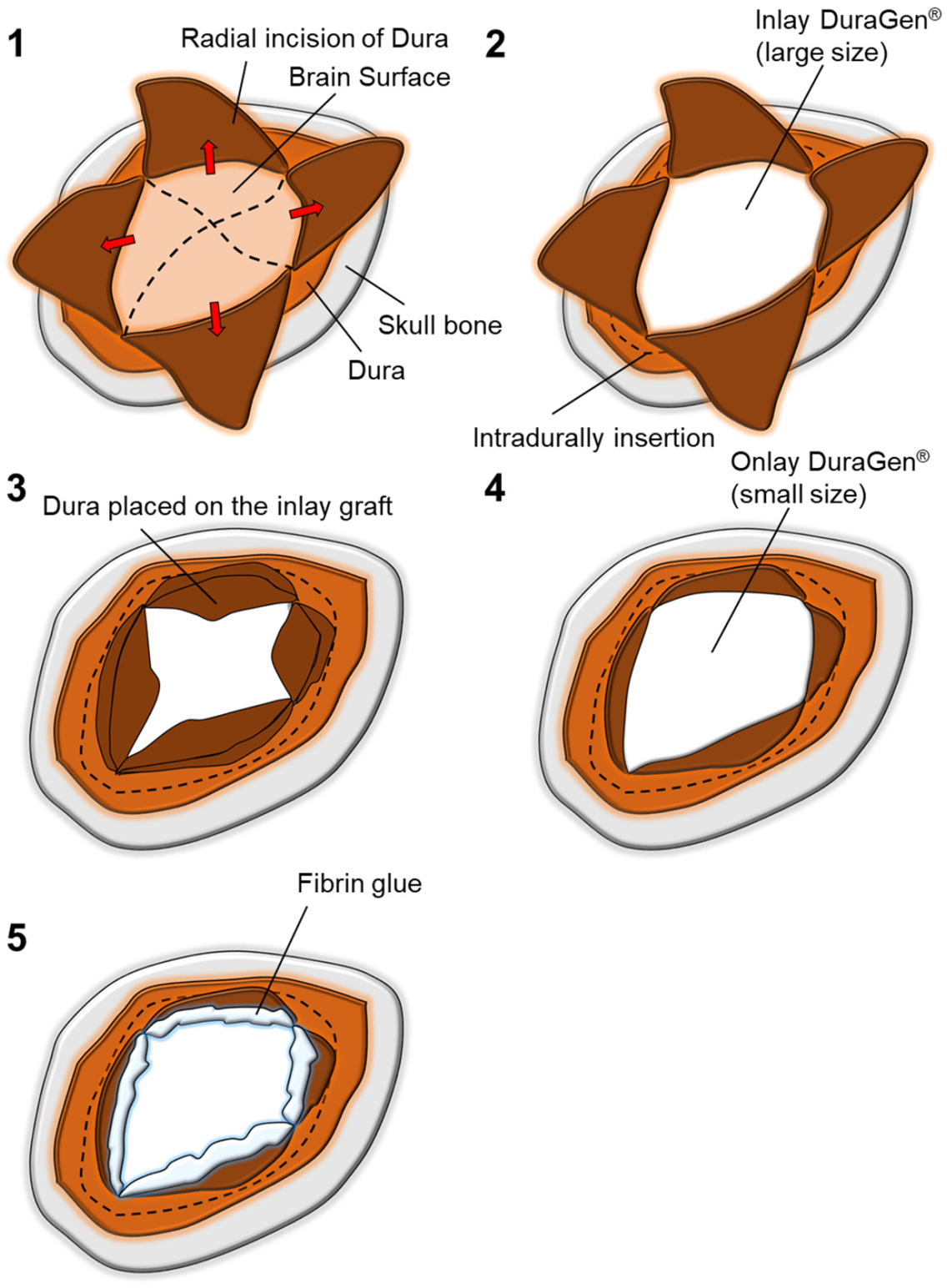
A 51-year-old woman presented with sudden-onset headaches, vomiting and abnormal consciousness level. Her CT scan in the emergency department revealed Fisher grade 3 subarachnoid hemorrhage due to rupture of a left middle cerebral artery aneurysm. Aneurysmal clipping and decompressive frontotemporal craniotomy were performed (Figure 1A). The dura was cut radially. A 10 × 12.5 cm DuraGen®, cut into 2 pieces, was placed in a multilayered fashion in order to achieve duraplasty. A larger graft (1.5-times larger in diameter than the exposed brain surface area) was inserted intradurally (inlay placement) to obtain sufficient contact with the inner surface of the dura mater. Then, the radially incised native dura was placed over the inlay graft. An adequate size of onlay graft was placed extradurally on top of the exposed inlay graft and native dura. The smaller portion of the graft was sufficient in order to cover the incised native dura. The repaired dura was fixed with fibrin glue to prevent CSF leakage and migration of the grafts (Figure 1B and Figure 2). Absence of CSF leakage was confirmed with the Valsalva maneuver after completion of the dural reconstruction. Postoperative care was as per the standard protocols.
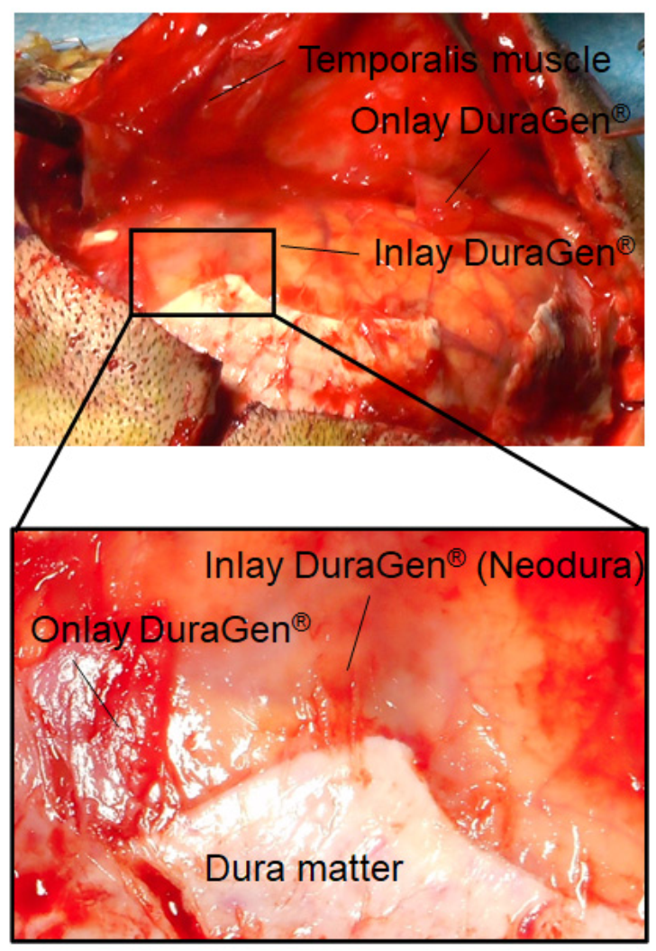
There was no clinical evidence of postoperative CSF leakage. Cranioplasty was performed 27 days after decompressive craniotomy. The musculocutaneous flap was re-opened without irritating the radically incised native dura and inlay graft. A slight adhesion between the onlay graft and overlying scalp and temporalis muscle was noted. The dissection plane could be easily identified by the native dura between inlay and onlay grafts. Although the onlay graft was partly fractured, the inlayed graft remained completely intact. Furthermore, the inlayed graft appeared to have already been mostly resorbed, creating a secondary dura between the musculocutaneous flap and brain: a neodura. No CSF leakage was observed post the cranioplasty procedure. This technique made the musculocutaneous flap dissection faster and potentially safer. The patient achieved efficient rehabilitation after early cranioplasty. She was discharged three weeks after the cranioplasty without any severe neurological sequelae.

3. Discussion

DuraGen® is extensively used in neurosurgery as a dural substitute. Several reports have demonstrated low complication rates associated with CSF leakage [4,5,6,11,13]. There are some studies showing the multilayered dural closure techniques with DuraGen® to prevent CSF leakage [14,15]. However, no reports have demonstrated the issues of its condition during a second surgery (cranioplasty) at an early timing.
Fibroblasts begin to migrate into the matrix 2 to 3 days after implantation of DuraGen® which initializes its process of laying down new collagen. Within 2 weeks, a neodural membrane forms between the dural margins to permanently close the dural defect. After 6–8 weeks, the implant is resorbed and replaced by dural tissue. After 1 year, the neodura is replaced completely by a mature dura [4,5,6,11,12].
In our case, a fragile neodura was observed on the brain surface 27 days after decompressive craniotomy. The myocutaneous flap should be opened meticulously in order to avoid excessive, and/or additional damage to both the flap and underlying brain [16]. A previous study reported a multilayered onlay repair utilizing both DuraGen and a gelatin film barrier (Gelfilm®; Pfizer, New York, NY, USA) to prevent scarring and tissue adhesion, which facilitates a safe and rapid cranioplasty following decompressive craniotomy [16]. However, one disadvantage of using a gelatin film is that patients may be at increased risk of infection and adverse biological reaction [17]. An increase of tissue adhesion between graft and brain surface were observed with DuraGen® compared with periosteum [13]. Notably, the inlayed DuraGen® must be carefully preserved during the procedure to avoid CSF leakage and related possible complications. The patients, who have persistent subcutaneous CSF collection or a pseudo meningocele, may require a second intervention [12].
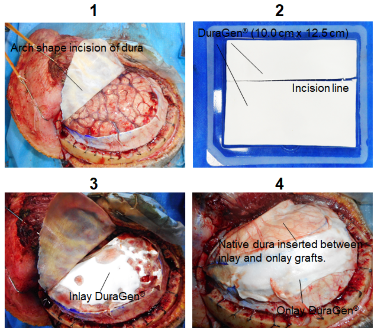
The present multilayered dural repair technique was used to achieve a successful cranioplasty preventing irritation of the graft adjacent to the brain during early cranioplasty, resulting in no CSF leakage. In our case, a 10 cm × 12.5 cm DuraGen® was cut into 2 pieces. Inlay–onlay sandwich graft technique can be achieved using only one sheet of DuraGen® by insertion of native dura between the inlay and onlay grafts, sparing medical resources (Figure 3). Sufficient overlap of the onlay graft and native dura is facilitated by radially incised native dura. Arch shape incision is not necessarily recommended (Figure A1). Inlay–onlay sandwich graft technique for arch shape incision tends to need two sheets of DuraGen® for sufficient overlap of the onlay graft and native dura.
DuraGen® can be used for a variety of conditions and procedures including traumatic brain injury and stroke. The necessity for earlier cranioplasty will also continue to increase in order to achieve prompt and efficient rehabilitation. This technique can make cranioplasty dissection faster and potentially safer, and subsequently improve clinical outcomes. Further experiences are needed to validate the advantages and disadvantages of the procedure in large sample sizes.

4. Conclusions

Early cranioplasty is preferred to achieve efficient rehabilitation after decompressive craniotomy. Neodura derived from DuraGen®, which is an absorbable engineered collagen-based artificial graft remains fragile at this early timing. We illustrated a multilayered dural repair technique with DuraGen® to avoid disruption of the fragile neodura during early cranioplasty.

Author Contributions

R.T. conceptualized, designed and performed the study and wrote the manuscript. Y.K. and M.M. assisted in the acquisition of data. M.K. assisted with discussion and review of the manuscript. All authors have read and agreed to the published version of the manuscript.

Funding

This research received no external funding.

Institutional Review Board Statement

The study was conducted according to the guidelines of the Declaration of Helsinki, and approved by the Institutional Review Board (Kawasaki Municipal Hospital).

Informed Consent Statement

Informed consent was obtained from all individual participants included in the study.

Conflicts of Interest

The authors have no conflict of interest directly relevant to the content of this article.

Appendix A

Figure A1. Arch shape incision of dura is performed (1). A 10 cm ×12.5 cm Duragen® is cut into 2 pieces (2). A larger graft is inserted intradurally (3). Incised native dura is placed over the inlay graft. An adequate size of onlay graft is placed extradurally on top of the exposed inlay graft and native dura (4).
Surgeries 02 00036 g0a1

References

  1. Timofeev, I.; Santarius, T.; Kolias, A.G.; Hutchinson, P.J. Decompressive craniectomy-operative technique and perioperative care. In Advances and Technical Standards in Neurosurgery; Springer: Vienna, Austria, 2012; Volume 38, pp. 115–136. [Google Scholar]
  2. De Bonis, P.; Frassanito, P.; Mangiola, A.; Nucci, C.G.; Anile, C.; Pompucci, A. Cranial repair: How complicated is filling a “hole”? J. Neurotrauma 2012, 29, 1071–1076. [Google Scholar] [CrossRef] [PubMed]
  3. Matsumoto, H.; Minami, H.; Yamaura, I.; Yoshida, Y. Postoperative subdural hematoma with blood flow from an epidural hematoma through a tear at the suture point of an artificial dura substitute. Acta Neurochir. 2019, 161, 755–760. [Google Scholar] [CrossRef] [PubMed]
  4. Narotam, P.K.; van Dellen, J.R.; Bhoola, K.D. A clinicopathological study of collagen sponge as a dural graft in neurosurgery. J. Neurosurg. 1995, 82, 406–412. [Google Scholar] [CrossRef] [PubMed] [Green Version]
  5. Danish, S.F.; Samdani, A.; Hanna, A.; Storm, P.; Sutton, L. Experience with acellular human dura and bovine collagen matrix for duraplasty after posterior fossa decompression for Chiari malformations. J. Neurosurg. 2006, 104, 16–20. [Google Scholar] [CrossRef] [PubMed]
  6. Narotam, P.K.; Reddy, K.; Fewer, D.; Qiao, F.; Nathoo, N. Collagen matrix duraplasty for cranial and spinal surgery: A clinical and imaging study. J. Neurosurg. 2007, 106, 45–51. [Google Scholar] [CrossRef] [PubMed] [Green Version]
  7. Bjornson, A.; Tajsic, T.; Kolias, A.G.; Wells, A.; Naushahi, M.J.; Anwar, F.; Helmy, A.; Timofeev, I.; Hutchinson, P.J. A case series of early and late cranioplasty-comparison of surgical outcomes. Acta Neurochir. 2019, 161, 467–472. [Google Scholar] [CrossRef] [PubMed] [Green Version]
  8. Kim, J.H.; Hwang, S.Y.; Kwon, T.H.; Chong, K.; Yoon, W.K.; Kim, J.H. Defining “early” cranioplasty to achieve lower complication rates of bone flap failure: Resorption and infection. Acta Neurochir. 2019, 161, 25–31. [Google Scholar] [CrossRef] [PubMed]
  9. Kim, Y.M.; Park, T.; Lee, S.P.; Baek, J.W.; Ryou, K.S.; Kim, S.H. Optimal timing and complications of cranioplasty: A single-center retrospective review of 109 cases. J. Neurointensive Care 2020, 3, 48–57. [Google Scholar] [CrossRef]
  10. Stendel, R.; Danne, M.; Fiss, I.; Klein, I.; Schilling, A.; Hammersen, S.; Pietilae, T.; Jänisch, W.; Hopfenmüller, W. Efficacy and safety of a collagen matrix for cranial and spinal dural reconstruction using different fixation techniques. J. Neurosurg. 2008, 109, 215–221. [Google Scholar] [CrossRef] [PubMed]
  11. Narotam, P.K.; José, S.; Nathoo, N.; Taylon, C.; Vora, Y. Collagen matrix (DuraGen) in dural repair: Analysis of a new modified technique. Spine 2004, 29, 2861–2867. [Google Scholar] [CrossRef] [PubMed]
  12. Sade, B.; Oya, S.; Lee, J.H. Non-watertight dural reconstruction in meningioma surgery: Results in 439 consecutive patients and a review of the literature. Clinical article. J. Neurosurg. 2011, 114, 714–718. [Google Scholar] [CrossRef] [PubMed]
  13. Neulen, A.; Gutenberg, A.; Takács, I.; Wéber, G.; Wegmann, J.; Schulz-Schaeffer, W.; Giese, A. Evaluation of efficacy and biocompatibility of a novel semisynthetic collagen matrix as a dural onlay graft in a large animal model. Acta Neurochir. 2011, 153, 2241–2250. [Google Scholar] [CrossRef] [PubMed] [Green Version]
  14. Chen, P.F.; Yip, C.M.; Lin, Y.H. Endoscopic transpterygoid approach to repair lateral sphenoid recess cerebrospinal fluid leakage by multilayered reconstruction. Eur. Ann. Otorhinolaryngol. Head Neck Dis. 2021. [Google Scholar] [CrossRef] [PubMed]
  15. Inoue, T.; Shitara, S.; Shima, A.; Goto, Y.; Fukushima, T. Double collagen matrix grafting for dural closure in microvascular decompression: An alternative use of autologous fascial grafting. Acta Neurochir. 2021, 163, 2395–2401. [Google Scholar] [CrossRef] [PubMed]
  16. Oladunjoye, A.O.; Schrot, R.J.; Zwienenberg-Lee, M.; Muizelaar, J.P.; Shahlaie, K. Decompressive craniectomy using gelatin film and future bone flap replacement. J. Neurosurg. 2013, 118, 776–782. [Google Scholar] [CrossRef] [PubMed] [Green Version]
  17. Przybylowski, C.J.; So, V.; DeTranaltes, K.; Walker, C.; Baranoski, J.F.; Chapple, K.; Sanai, N. Sterile Gelatin Film Reduces Cortical Injury Associated with Brain Tumor Re-Resection. Oper. Neurosurg. 2021, 20, 383–388. [Google Scholar] [CrossRef] [PubMed]
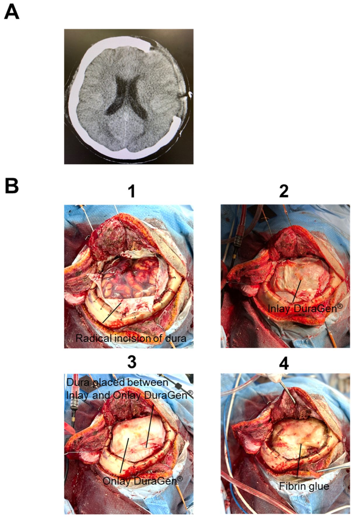
Figure 1. A multilayered dural repair technique using Duragen®. (A) Postoperative CT scan is shown. Decompressive frontotemporal craniotomy is performed. (B) Intraoperative images of a multilayered dural repair technique are shown. The dura is cut radially (1). DuraGen® is cut into 2 pieces. A larger graft is inserted intradurally (2). The radially incised native dura is placed over the inlay graft. An adequate size of onlay graft is placed extradurally on top of the exposed inlay graft and native dura (3). The repaired dura is fixed with fibrin glue to prevent CSF leak and migration of the grafts (4).
Surgeries 02 00036 g001
Figure 2. Intraoperative image of inlay and onlay Duragen® during early craniotomy. A slight adhesion between the onlay graft and overlying scalp and temporalis muscle is noted. Although the onlay graft is partly fractured, the inlayed graft remains completely intact. No CSF leakage is observed postoperatively. Furthermore, the inlayed graft appears to be mostly resorbed, creating a secondary dura mater between the skin graft and brain: a neodura.
Surgeries 02 00036 g002
Figure 3. Scheme of a multilayered dural repair technique using Duragen (related to Figure 1B).
Surgeries 02 00036 g003
Publisher’s Note: MDPI stays neutral with regard to jurisdictional claims in published maps and institutional affiliations.