
Article

Oxytocin Variants Induce Cellular Signaling and Neurite Outgrowth in Human-Derived Neuron-like SH-SY5Y Cell Line

1
Chicago College of Osteopathic Medicine, Midwestern University, 555 31st St., Downers Grove, IL 60515, USA
2
Department of Biomedical Sciences, College of Graduate Studies, Midwestern University, 555 31st St., Downers Grove, IL 60515, USA
3
Department of Pharmacology, College of Graduate Studies, Midwestern University, 555 31st St., Downers Grove, IL 60515, USA
*
Author to whom correspondence should be addressed.
NeuroSci 2026, 7(5), 100; https://doi.org/10.3390/neurosci7050100 (registering DOI)
Submission received: 5 August 2026 / Revised: 27 August 2026 / Accepted: 29 August 2026 / Published: 3 September 2026

Abstract

In the central nervous system, the neuropeptide oxytocin stimulates neural networks that regulate social behaviors, including social attachment, aggression, and complex social cognition. Perturbations in oxytocin and/or oxytocin receptor expression results in social behavioral deficits and are associated with a number of psychopathologies including autism spectrum disorder, schizophrenia, anxiety, and depression. In rodent models of autism spectrum disorder, oxytocin is effective at improving social behavior. However, limited translatability between animal models and human physiology has hindered successful translation of these findings into human therapeutics. Oxytocin variant-induced cellular signaling pathways and G-protein coupling have largely been investigated in HEK and CHO heterologous expression systems; however, cellular context is crucial, and these profiles likely differ from intact neurons. This project assessed oxytocin variants in in vitro human-derived neuron-like SH-SY5Y cells that endogenously express the oxytocin receptor. Results demonstrated that the naturally occurring oxytocin variants Leu8-OT, Pro8-OT and Val3-Pro8-OT activated intracellular signaling pathways and promoted neuronal differentiation-associated responses, including calcium mobilization, membrane hyperpolarization, and neurite outgrowth in a concentration-dependent manner. Knowledge of how oxytocin variants alter cellular structure and function has the potential to both identify mechanisms that produce social dysfunction and to inform the development of therapeutic agents.

1. Introduction

Neuropeptides oxytocin (OT) and arginine vasopressin (AVP) play a critical role in shaping social phenotypes [1,2]. In the central nervous system, they are expressed in the Social Behavioral Network (SBN) which includes midbrain nuclei and connections to the mesolimbic system, and affects parental bonding, peer relationships, social perception, cognition and decision making [3,4,5,6,7,8,9,10]. Remarkable evolutionary conservation is observed in OT structure and function [11,12,13,14,15,16,17,18]. Abnormalities in social behavior are a central element of multiple psychopathologies, and dysfunction with the OT system has been identified as an underlying cause of some psychiatric symptoms [19,20,21]. Animal models are valuable tools to assess the role of the OT system [22,23,24,25,26,27,28], and provide critical pre-clinical insight into expected functions in humans. A fundamental problem for translation of animal studies, particularly with rodents, to human therapeutics is insufficient translatability due to drastic differences in cortical structures and neural pathways [29,30].
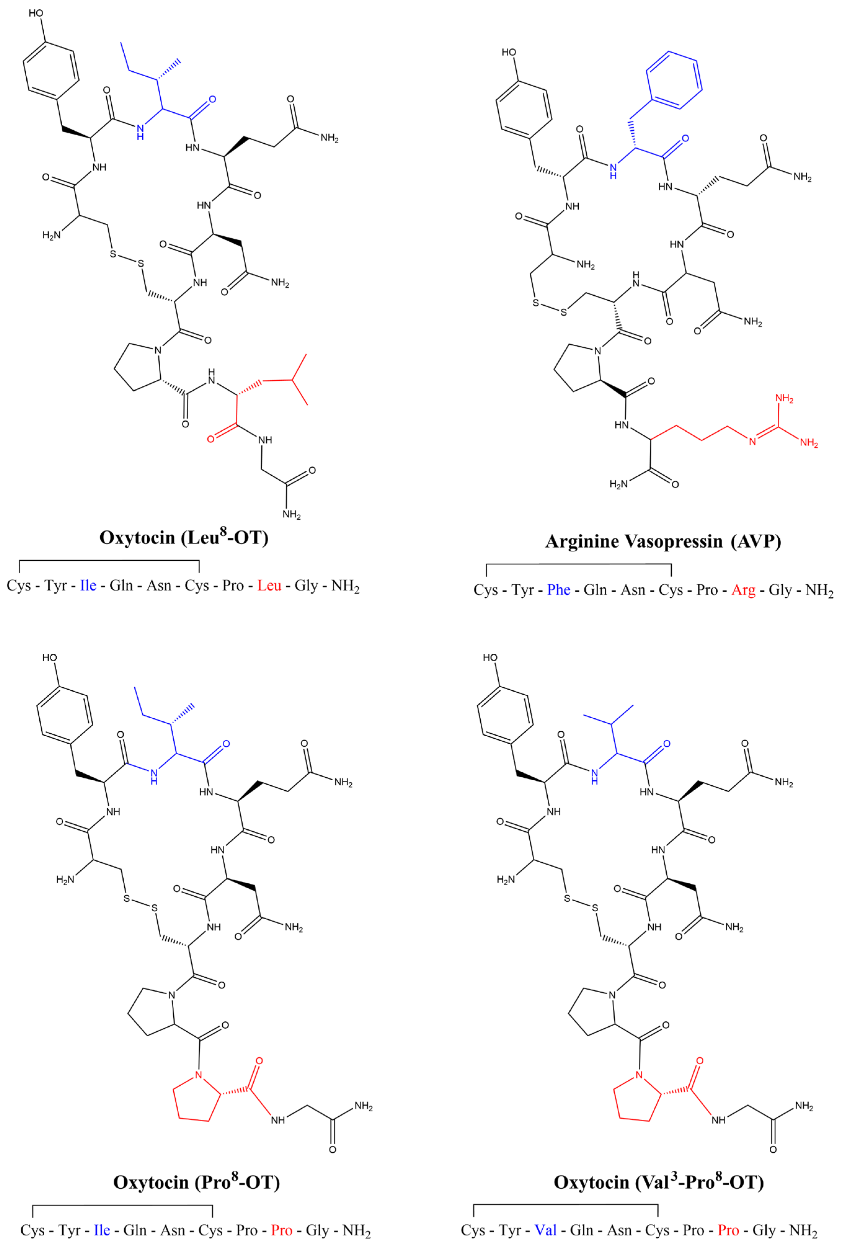
OT is a highly evolutionarily conserved signaling molecule that binds primarily to the OT receptor, and to a lesser extent the vasopressin receptors, which are all members of the G-protein-coupled receptor (GPCR) superfamily [31,32]. Invertebrates generally have only a single ancestral oxytocin/vasopressin-like homolog; however, OT and vasopressin are believed to have arisen from a gene duplication event prior to vertebrate divergence [15,33]. The consensus sequences for mammalian nonapeptides OT and AVP differ at amino acids 3 and 8 [11]. The consensus mammalian OT is a nine amino acid polypeptide comprising a hexacyclic ring with a disulfide bridge (residues 1–6) and a linear tripeptide tail (residues 7–9) with leucine in the 8th position, Leu8-OT (Cys-Tyr-Ile-Gln-Asn-Cys-Pro-Leu-Gly) [15,18,34,35,36]. In New World Monkeys, at least six distinct OT ligand variants have been identified, including two that have a proline in the 8th position, which forms a polyproline helix and significantly alters the ligand structure and stability, Pro8-OT (Cys-Tyr-Ile-Gln-Asn-Cys-Pro-Pro-Gly) and Val3-Pro8-OT (Cys-Tyr-Val-Gln-Asn-Cys-Pro-Pro-Gly) (Figure 1) [13,14,18,36,37].
OT and vasopressin activity in the social brain network generally displays opposing actions, with OT primarily functioning as an anxiolytic neuropeptide and vasopressin demonstrating anxiogenic properties [20,38,39]. Notably, in heterologous systems using HEK or CHO cells expressing human vasopressin receptor 1a (V1a receptor), Leu8-OT, Pro8-OT and the nonpeptide OT agonist LIT-001 all function as partial antagonists [40,41], which was not seen at V1a receptors from other nonhuman primates [40]. These data suggest the ability of OT variants to inhibit anxiogenic signaling pathways in the social behavioral network [20,42]. The OT receptor promiscuously couples to and activates various G proteins to produce diverse effects on cellular function, including activation of phospholipase C (Gq), stimulation of potassium channel currents (Gi), and inhibition of adenylyl cyclase (Gi/o) that vary across cellular context [43,44,45,46,47,48,49].
OT is synthesized in the magnocellular neurons of the supraoptic and paraventricular nuclei of the hypothalamus, and OT neurons primarily project to the posterior pituitary where it is released into the bloodstream [50]. In the central nervous system, OT neurons also project to multiple regions within the SBN [16]. In humans, OT receptors are expressed in areas important for behavior, emotion, impulse control, decision-making, speech, and memory including the prefrontal cortex, anterior cingulate, the bed nucleus of the stria terminalis, medial preoptic area, amygdala, hippocampus, and hypoglossal and solitary nuclei [51,52]. Additionally, in nonhuman primates OT receptor neurons have been shown to project across the primary visual path to the superior colliculus and visual cortex [53,54,55]. OT receptor expressing neurons include glutamatergic pyramidal cells [56], GABAergic interneurons [57], and other neuroendocrine cells [50]. Heterologous expression systems expressing human OT receptor have been used to assess G-protein cellular signaling [46,47,49], but lack the neuronal context necessary for understanding cellular function in the SBN. Since practical and ethical considerations preclude the use of human primary neuronal cultures that express the OT receptor, neuronal-like cell lines offer an alternative model to assess the endogenous human OT receptor. Several neuroblastoma cell lines maintain their potential for neurite outgrowth and differentiation [58,59], and express OT receptor [60,61]. The human neuroblastoma cell line SH-SY5Y is a subclone of SK-N-SH cell line isolated from bone marrow of a 4-year-old female [62,63]. Studies in SH-SY5Y cell line demonstrated that 1 μM Leu8-OT stimulated calcium mobilization and resulted in a significant increase in neurite length, with this single dose being analyzed in combination with various inhibitors [64,65].
In this study, we investigated the cellular signaling and neurotrophic effects of three naturally occurring OT variants (Leu8-OT, Pro8-OT, and Val3-Pro8-OT) using human-derived neuron-like SH-SY5Y cells. We first assessed OT- and AVP-mediated calcium mobilization in SH-SY5Y cells and confirmed the integrity of our peptides in human-OT receptor (hOTR) expressing Chinese hamster ovary (CHO) cells. The selective OT receptor antagonist L-371,257 confirmed that calcium mobilization was mediated through the OT receptor. We then evaluated calcium signaling induced by the three OT variants and demonstrated through thapsigargin (Tg) pretreatment that these dose-dependent responses were primarily driven by intracellular calcium [Ca2+]i stores. In addition, all three OT variants produced membrane hyperpolarization and promoted neurite outgrowth in a concentration-dependent manner. Although differences among the variants were not generally statistically significant, they followed the same trend (Leu8-OT > Pro8-OT ≈ Val3-Pro8-OT). This project combined assessment of structural (neurite outgrowth) and functional (cellular signaling assays) in human-derived in vitro neuronal-like SH-SY5Y cells to provide insights into therapeutic potential of naturally occurring OT variants.

2. Materials and Methods

SH-SY5Y cell cultures. SH-SY5Y cells were purchased (Cat. No. CRL-2266™, ATCC®, Manassas, VA, USA) and cultured in equal parts Ham’s F12 (Cat. No. SH30231.01, Hyclone, Logan, UT, USA) and Eagle’s Minimal Essential Medium (EMEM; Cat. No. 30-2003, ATCC®. Manassas, VA, USA), with 10% fetal bovine serum (FBS; Cat. No. S11550H, Atlanta Biologicals, Oakwood, GA, USA), 1% Penicillin-Streptomycin (Cat. No. 15140122, Gibco, Grand Island, NY, USA), 1% non-essential amino acids (NEAA; Cat. No. 11140050, Gibco, Grand Island, NY, USA), 2 mM l-glutamine (Cat. No. 25030149, Gibco, Grand Island, NY, USA). 96-well microplates (Cat. No. 07-000-061, Fisher Scientific, Waltham, MA, USA) are pretreated with poly-l-lysine (Cat. No. P1274, Millipore-Sigma, Burlington, MA, USA) for one hour, the solution is aspirated, the plates are allowed to dry, and 1 million cells/mL are then plated and cultured overnight in culture media at 37 °C in 5% CO2 and 95% humidity.
hOTR-CHO cell cultures. Stably transfected human OT receptor (hOTR)-expressing Chinese hamster ovary (CHO) cells were purchased from Genscript (Cat. No. M00195, Genscript, Piscataway, NJ, USA). hOTR-CHO cells were cultured in Ham’s F12 (Cat. No. SH30026.01, Hyclone, Logan, UT, USA), 10% FBS (Cat. No. S11550H, Atlanta Biologicals, Oakwood, GA, USA), 1.5% HEPES 1 M Solution (Cat. No. SH30231.01, Hyclone, Logan, UT, USA), 1% Penicillin-Streptomycin (Cat. No. 15140122, Gibco, Grand Island, NY, USA) and 500 mg/mL G418 (Cat. No. G64000-5.0, RPI Corp., Mount Prospect, IL, USA). Cells were plated at 0.3 million cells/mL and cultured at 37 °C in 5% CO2 and 95% humidity as previously described [46,47].
Drugs. AVP (Cat. No. V-9798, Millipore-Sigma, Burlington, MA, USA), Leu8-OT (Cat. No. 06379, Millipore-Sigma, Burlington, MA, USA), Pro8-OT and Val3-Pro8-OT (Cat. No. 58863, Anaspec, Fremont, CA, USA), L-371,257 (Cat. No. SC-204038, Santa Cruz Biotchnology, Dallas, TX, USA), SR49059 (Cat. No.S5701, Millipore-Sigma, Burlington, MA, USA), and thapsigargin (Cat. No. T9033, Millipore-Sigma, Burlington, MA, USA) were reconstituted in dimethyl sulfoxide (DMSO; Cat. No. 472301, Millipore-Sigma, Burlington, MA, USA).
Calcium mobilization assay. Briefly, 100 mL of dye loading medium was added per well containing 4 μM Fluo8-AM (Cat. No. 21080, AAT Bioquest, Pleasanton, CA, USA), 0.04% pluronic acid (Cat. No. P3000MP, Molecular Probes, Eugene, OR, USA) in Locke’s buffer with glycine, 0.5 mM probenecid (Cat. No.P8761, Millipore-Sigma, Burlington, MA, USA), and incubated for one hour as previously described [46,47]. Immediately before running the assay, the dye loading solution was replaced with Locke’s buffer. After recording the baseline fluorescence for 60 s, logarithmic concentrations of OT variants were added to cells with the FLIPR pipettor at a rate of 2 μL/s, yielding a final volume of 120 μL/well. The cells were excited at 488 nm and Ca2+-bound Fluo8 emission was recorded at 538 nm at 2 s intervals. The fluorescence was then monitored for an additional 200 s. The effects of OT variant addition on Ca2+ mobilization or membrane potential were monitored with a FLIPR2 plate reader (Molecular Devices, San Jose, CA, USA). The FLIPR2 system measures intracellular fluorescence by illuminating the underside of a 96-well microplate with an air-cooled laser and simultaneously detecting fluorescence signals from cell-permeant dyes in all wells. An integrated automated 96-channel pipettor delivered precise volumes of test compounds from a source plate to each well simultaneously. To assess OT and V1a receptor involvement, cells were pretreated for 10 min with 1 μM OT receptor inhibitor L-371,257 or 100 nM V1a receptor inhibitor SR49059 prior to running the assay. To assess the role of intracellular calcium [Ca2+]i in OT-mediated mobilization of Ca2+, cells were pretreated for five minutes with the sarco/endoplasmic reticulum calcium ATPase (SERCA) inhibitor thapsigargin to deplete [Ca2+]i stores.
Membrane potential assay. The FLIPR membrane potential assay (FMP blue; Cat. No. F1241, Molecular Devices, San Jose, CA, USA) was used to assess the changes in membrane potential as previously described [46,47]. The FMP blue dye is a lipophilic, negatively charged dye that distributes across the cell membrane as a function of membrane potential [61]. Cell culture medium was aspirated and 100 μL of diluted FMP solution was added. After a 45 min equilibration, fluorescence measurements were performed with cells excited at 530 nm, and emission recorded at 565 nm, at 2 s intervals. After recording the baseline for 60 s, logarithmic concentrations of OT variants were added to a final volume of 120 μL at a rate of 2 μL/s, and fluorescence monitored for an additional 200 s.
Neurite outgrowth assays. Cultures were performed as described above with the exception that they were plated on poly-L-lysine treated coverslips in a 24-well plate at 25,000 cells/mL, and cells were allowed to settle for three hours prior to addition of vehicle or OT variant doses. Cells were allowed to incubate for 24 h before they were fixed. Each experiment had two replicate wells per plate, and the experiment was replicated three times. Cells were stained using the F-actin visualization kit (Cytoskeleton BK005) according to the manufacturer’s protocol, with 0.1 μg/mL DAPI (Cat. No. D9542, Millipore-Sigma, Burlington, MA, USA). A Nikon A1R confocal microscope (Nikon, Melleville, NY, USA) was used to collect 60× images of F-actin- and DAPI-stained cells. Images were quantified using ImageJ package FIJI with the NeuronJ plugin as previously described [66] for total neurite length in micrometers, number of processes coming directly off the soma, and total number of branches. Cells within each independent experiment were evaluated separately and the resulting values were averaged to generate a single mean value for each biological replicate. The biological replicate means were entered into GraphPad Prism for statistical analyses.
Statistical Analyses. For cellular signaling experiments, sigmoidal dose-response curves for OT variants were generated using three-parameter nonlinear regression analysis in GraphPad Prism 8.3 software (GraphPad, Boston, MA, USA). The half-maximal effective (EC50) and half-maximal inhibitory (IC50) values, maximal responses (EMAX), and goodness of fit (R2) for OT variants were obtained from the average of each individual experiment following nonlinear regression analyses. These concentration–response profiles were performed using all three OT variants. For neurite outgrowth assays, dose-response graphs for OT variants were generated in GraphPad Prism 8.3 software. A one-way ANOVA was performed with Šidák’s multiple comparisons to determine statistical significance.

3. Results

3.1. AVP and OT-Induced Ca2+ Mobilization via the Human OT Receptor

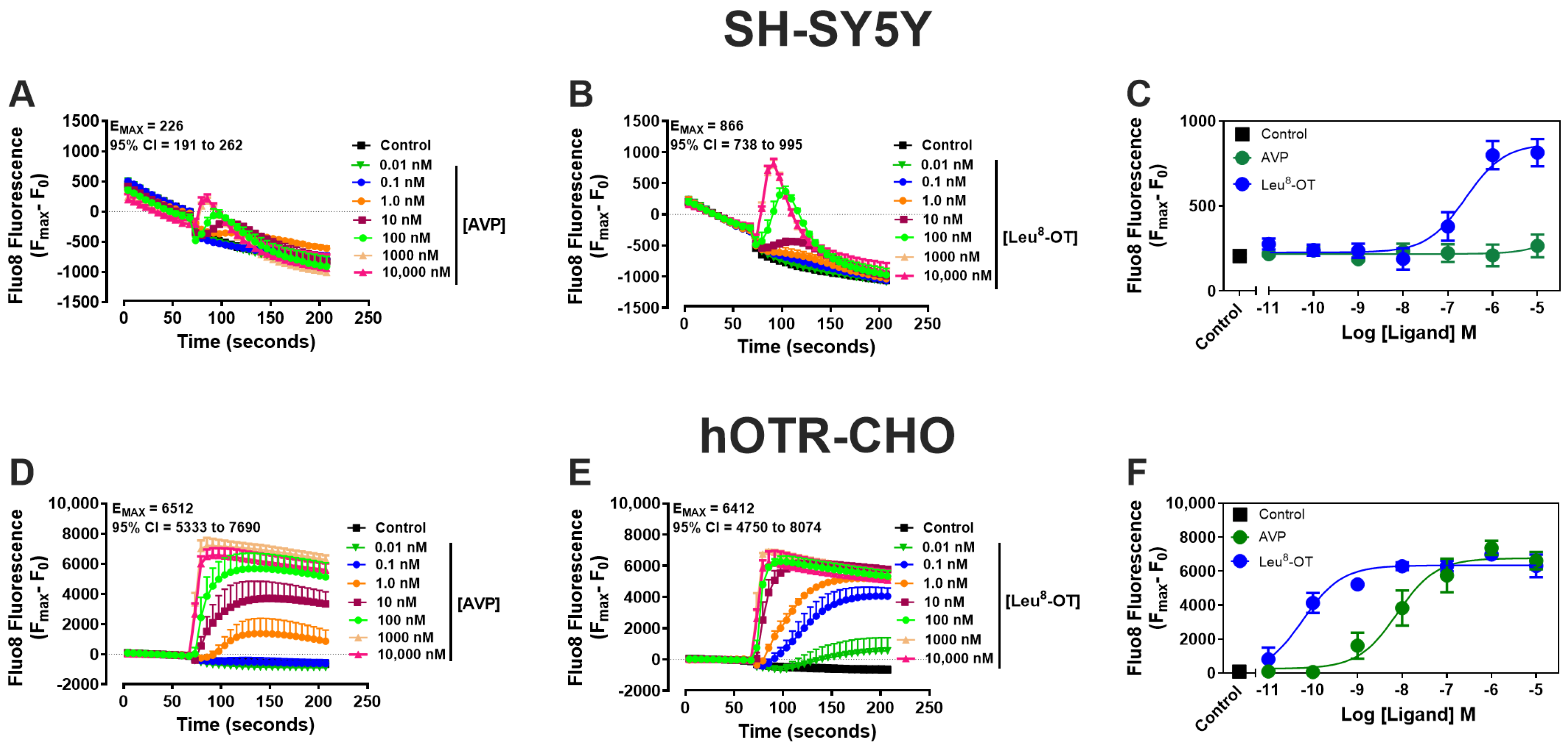
Previous studies have shown in SH-SY5Y cells that 1 μM Leu8-OT stimulated calcium mobilization and resulted in a significant increase in neurite length, although only a single dose was analyzed in combination with various inhibitors [64,65]. To directly compare the activation of the human OT receptor by AVP and Leu8-OT in SH-SY5Y cells, we performed concentration–response assays spanning 10 picomolar (pM) to 10 micromolar (μM) using the Ca2+-sensitive dye Fluo8-AM. In SH-SY5Y cells, Leu8-OT displayed a concentration-dependent increase in Ca2+ signaling (Figure 2B), as well as being both more potent (half-maximal effective concentration, EC50; Table 1) and efficacious (EMAX; Figure 2A,B) than AVP (Figure 2A). In addition, the concentration response curve for Leu8-OT exhibited a strong goodness of fit (R2 > 0.70; Table 1), whereas the R2 value could not reliably be calculated for AVP, indicating poor correlation of the data and a weak response in SH-SY5Y cells. To confirm peptide integrity and activity, the assays were repeated in heterologous CHO cells expressing the human OT receptor (hOTR-CHO). In these cells, both AVP and Leu8-OT elicited a concentration-dependent increase in Ca2+ signaling (Figure 2D–F). Consistent with our previous report [47], Leu8-OT remained more potent than AVP, while both peptides generated concentration–response curves with comparable goodness of fit (Figure 2D–F, Table 1). Collectively, these findings demonstrate that AVP is less potent and less efficacious than Leu8-OT at stimulating Ca2+ signaling in both SH-SY5Y cells and hOTR-CHO cells.
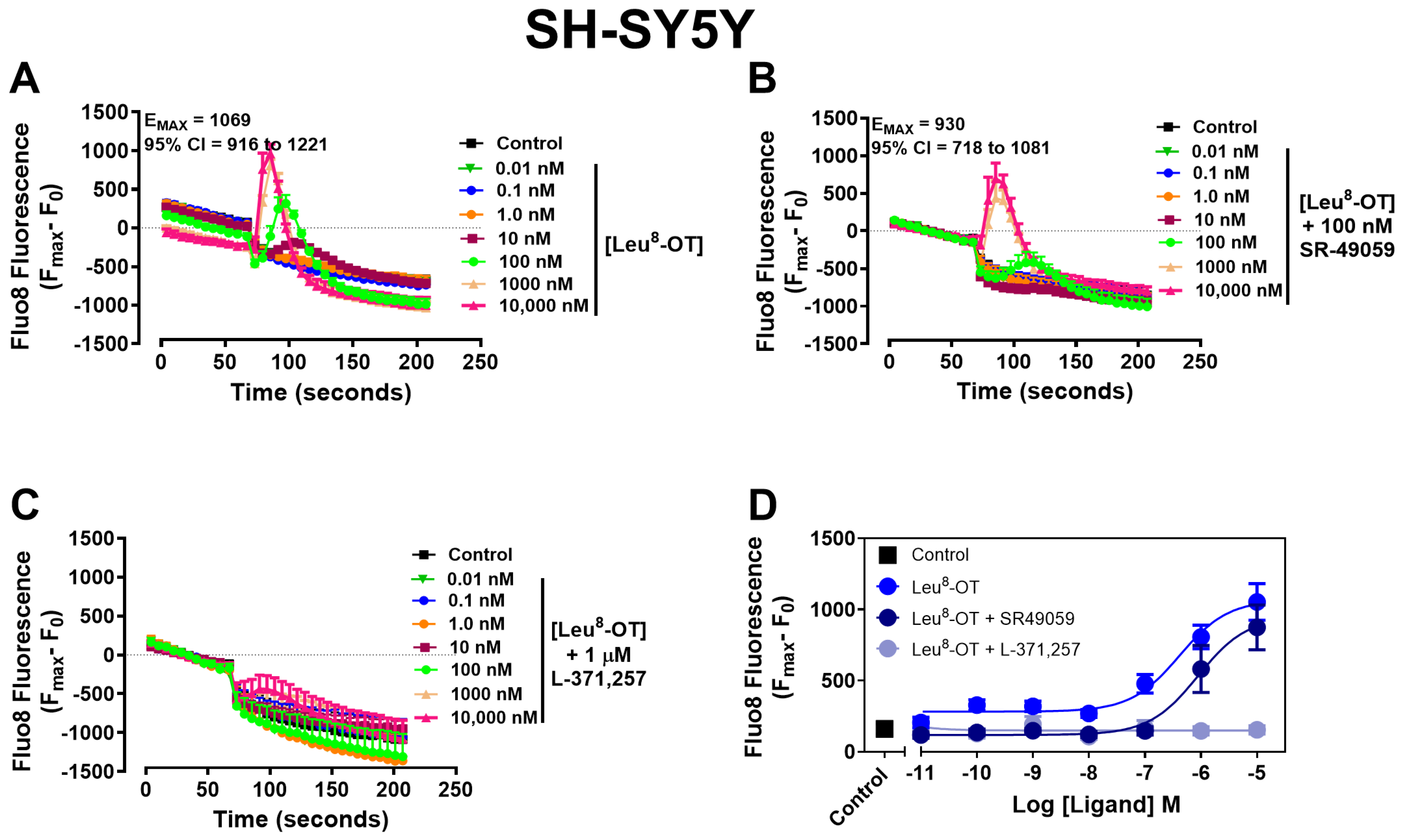
Because both AVP and OT can activate OT and vasopressin receptors [47,67], receptor-selective antagonists were used to determine which receptor is mediating Leu8-OT-induced calcium mobilization. Concentration–response studies with Leu8-OT alone demonstrated a dose-dependent increase in [Ca2+]i signaling (Figure 3A, Table 2), consistent with Figure 2. Pretreatment with the selective V1a receptor antagonist SR-49059 did not significantly alter the concentration response curve for Leu8-OT (Figure 3B, Table 2), indicating that V1a receptor activation contributes minimally, if at all, to Leu8-OT-mediated calcium signaling. In contrast, pretreatment with the selective OT receptor antagonist L-371,257 markedly attenuated Leu8-OT-mediated calcium mobilization (Figure 3C,D, Table 2). These findings demonstrate that Leu8-OT-induced calcium signaling in SH-SY5Y cells is mediated predominantly through activation of the OT receptor rather than the V1a receptor, which is consistent with Yin et al.’s findings that SH-SY5Y cells do not express V1a or V1b receptors [60].

3.2. Leu8-OT-, Pro8-OT-, and Val3-Pro8-OT-Induced Intracellular Ca2+ Mobilization

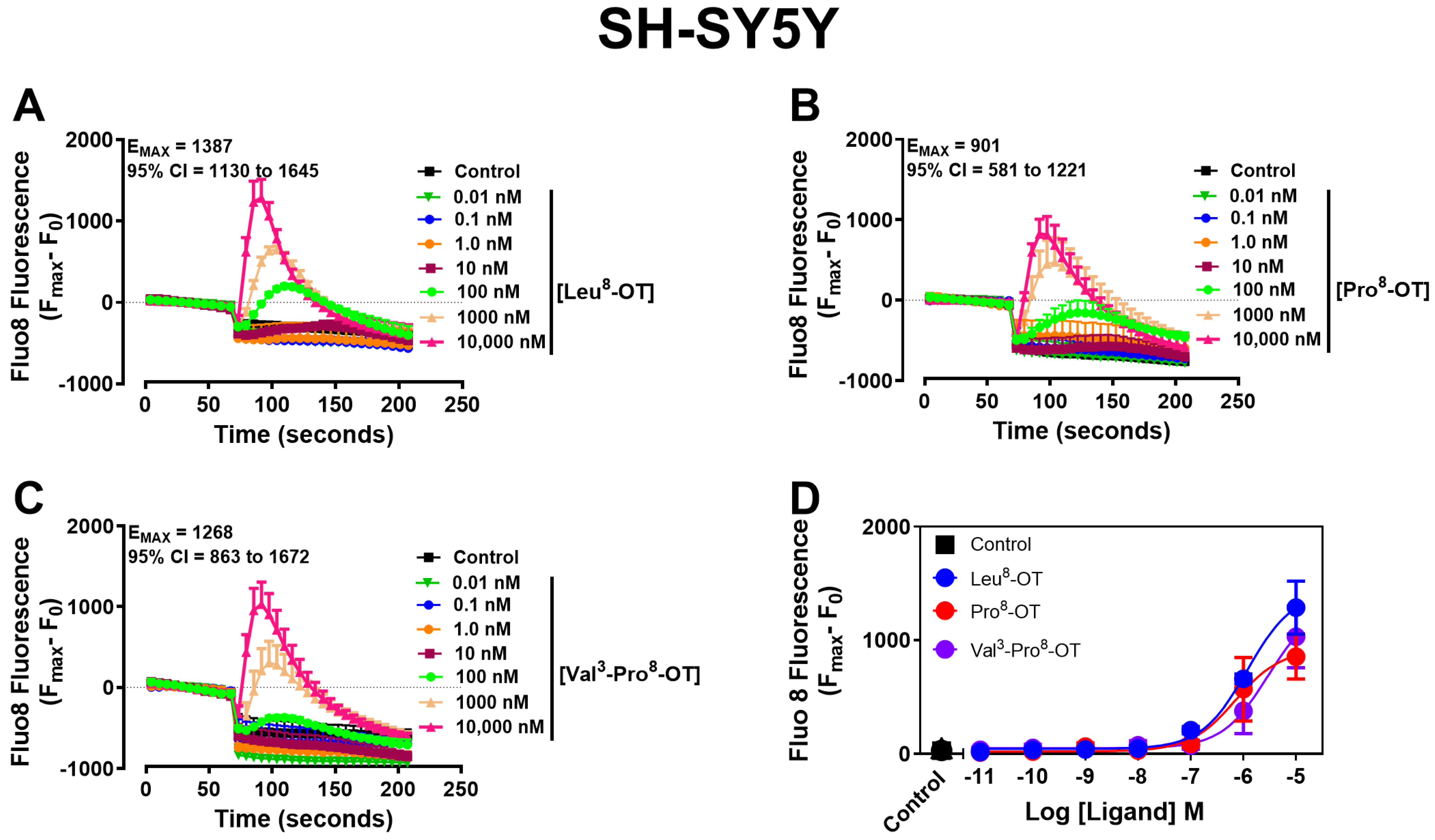
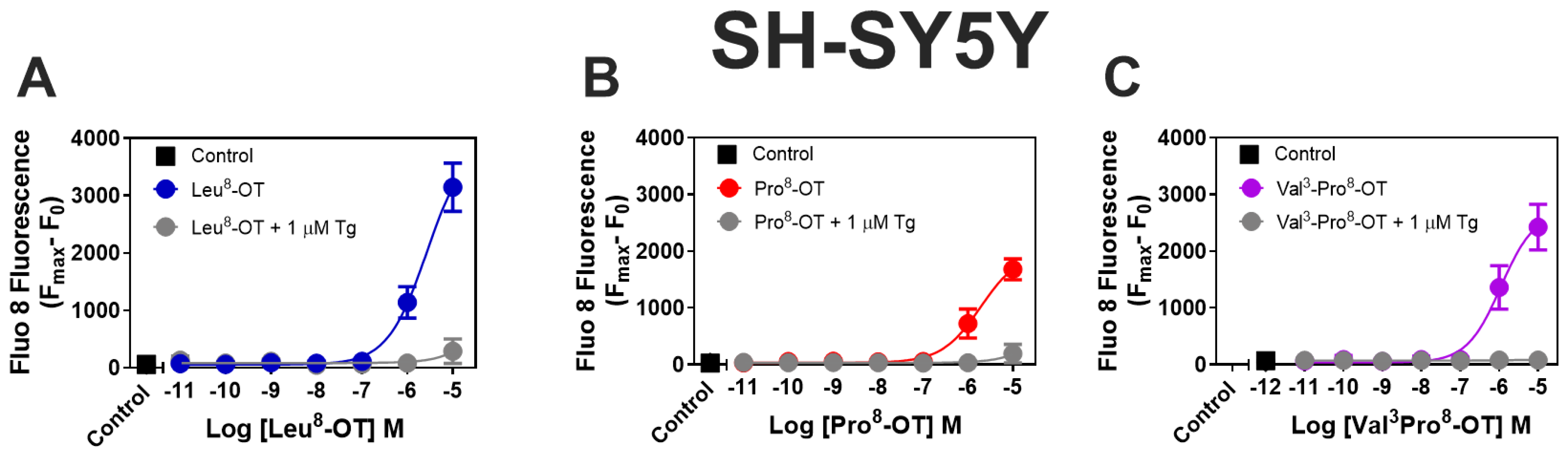
New World Monkeys exhibit a hotspot of genetic variation in both OT peptides and receptors that correlate with differences in social behavior [13,14]. Among these are OT variants containing a proline residue at position 8, which alters structural and functional properties, including stability compared with the highly conserved consensus mammalian Leu8-OT [13,14,68]. To determine whether Pro8-OT and Val3-Pro8-OT exhibit signaling properties comparable to endogenous Leu8-OT, we performed a concentration–response assays measuring [Ca2+]i in SH-SY5Y cells. All three peptides produced robust, concentration-dependent increases in [Ca2+]i signaling and well-fit concentration–response curves (Figure 4, Table 3). Although no statistically significant differences were observed in potency or efficacy, the variants exhibited a trend toward reduced activity relative to Leu8-OT (Leu8-OT > Pro8-OT ≈ Val3-Pro8-OT). Collectively, these findings suggest that substitution of proline at position 8 largely retains functional activity at the human OT receptor in SH-SY5Y cells.
The sarco/endoplasmic reticulum Ca2+-ATPase (SERCA) maintains [Ca2+]i homeostasis by transporting Ca2+ from the cytosol into the endoplasmic reticulum [69,70]. Consequently, pretreatment with thapsigargin, a potent SERCA inhibitor, depletes [Ca2+]i stores. Depletion of [Ca2+]i stores with Tg pretreatment attenuated OT-variant-induced Ca2+ mobilization, demonstrating that the observed response is dependent on intracellular calcium stores (Figure 5).

3.3. Leu8-OT-, Pro8-OT-, and Val3-Pro8-OT-Induced Changes in Membrane Potential

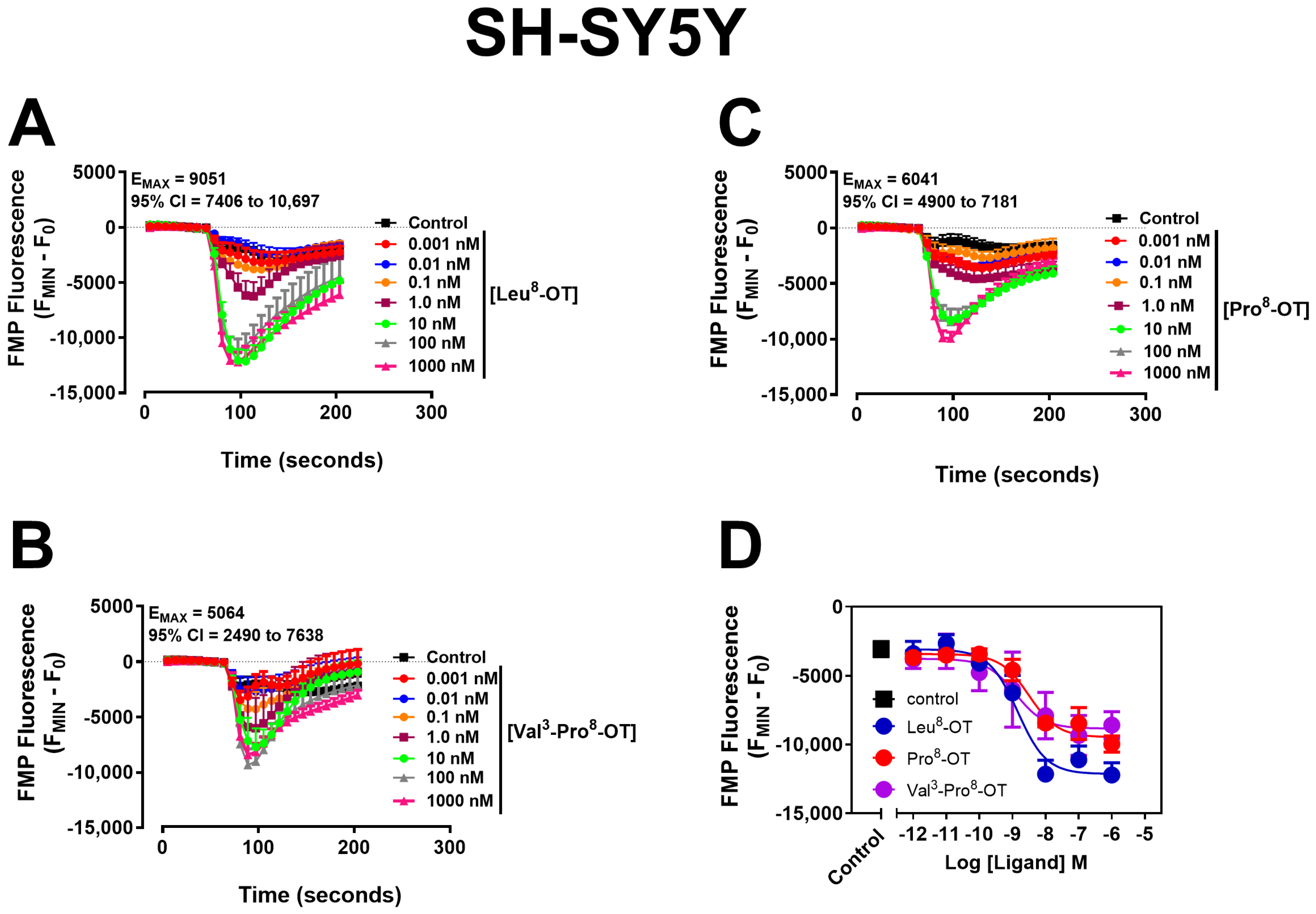
OT variants in hOTR-CHO cells have been shown to induce membrane hyperpolarization [46,47]; however, cellular context can play a key role in cellular signaling. Membrane potential assays were performed using FMP blue, a voltage-sensitive dye whose fluorescence increases with depolarization and decreases with hyperpolarization [71,72], to assess whether OT variants alter membrane excitability in SH-SY5Y cells. All three OT variants produced concentration-dependent decreases in FMP blue fluorescence, consistent with a hyperpolarizing response (Figure 6). While the variants did not differ significantly in potency (half-maximal inhibitory concentration; IC50) or efficacy (EMAX), they exhibited a trend toward lower activity compared to Leu8-OT (Leu8-OT > Pro8-OT ≈ Val3-Pro8-OT), mirroring a pattern observed with [Ca2+]i mobilization (Figure 6, Table 4). The goodness of fit for Val3-Pro8-OT was moderate, with an R2 of 0.49, whereas it was strong for Leu8-OT and Pro8-OT.

3.4. Leu8-OT-, Pro8-OT-, and Val3-Pro8-OT-Induced Neurite Outgrowth Is Dose-Dependent

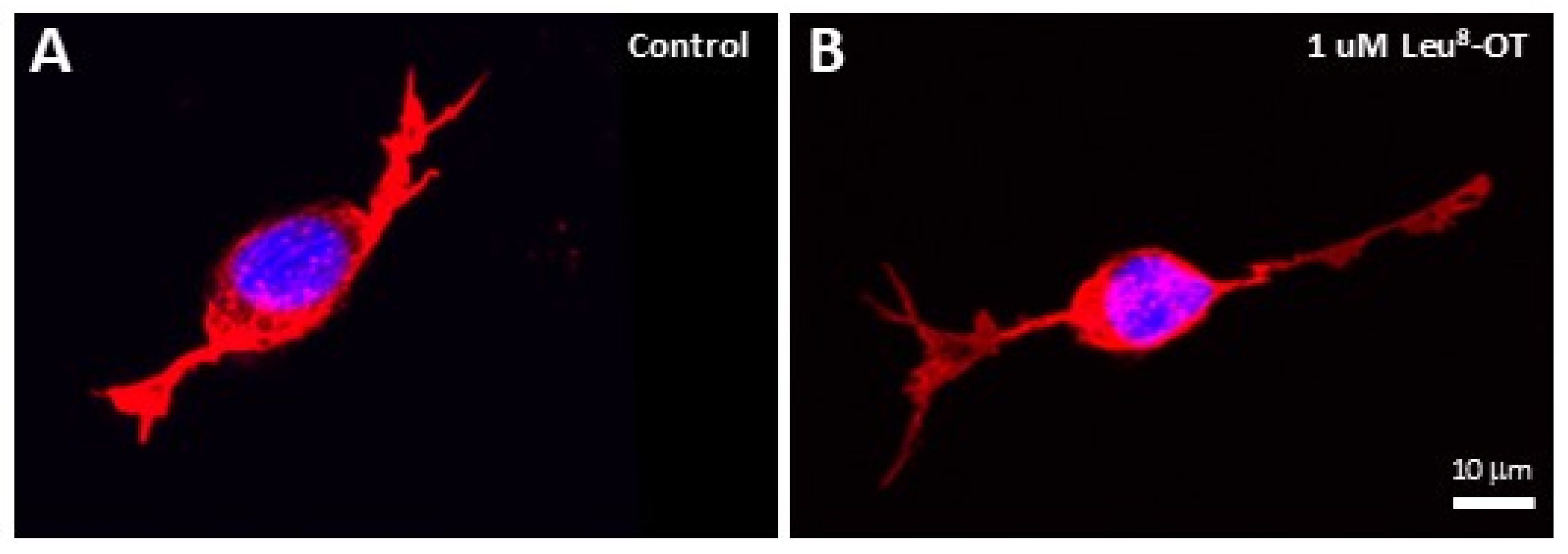
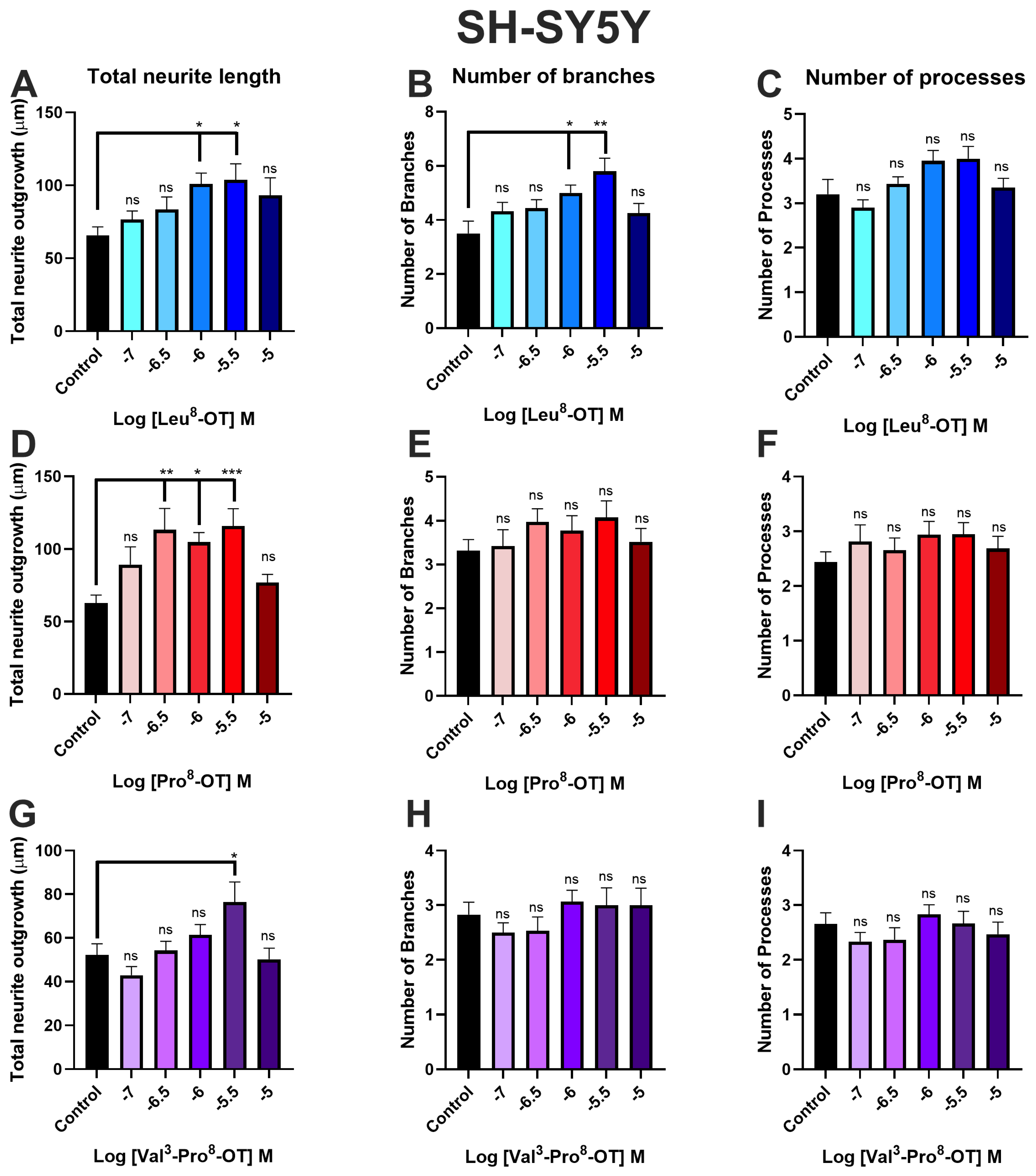
Two studies previously demonstrated that 1 μM Leu8-OT resulted in a significant increase in neurite length [64,65]. To determine whether Pro8-OT and Val3-Pro8-OT exert similar neurotrophic effects, cells were exposed to half-log doses ranging from 100 nM to 10 μM, corresponding to the concentration range that produced the greatest effects on Ca2+ mobilization. A representative image is shown for the phalloidin and DAPI staining of SH-SY5Y cells that were then processed using ImageJ package FIJI with NeuronJ plugin (Figure 7). All OT variants produced concentration-dependent increases in neurite outgrowth (Figure 8A,D,G), although their efficacy differed slightly. Leu8-OT significantly increased neurite length and branching at 1 and 3 μM, but showed no significant difference in the number of processes (Figure 8A–C). Pro8-OT significantly increased neurite length at 0.3, 1, and 3 μM doses with no significant increase in number of branches or processes (Figure 8D–F), whereas Val3-Pro8-OT produced significant increases in neurite outgrowth only at 3 μM (Figure 8G–I), indicating a lower efficacy. Collectively, these findings demonstrate that all three OT variants promote neurite outgrowth, with a rank order of activity broadly consistent with that observed for Ca2+ mobilization and membrane hyperpolarization (Leu8-OT > Pro8-OT ≈ Val3-Pro8-OT).

4. Discussion

OT is a highly conserved neuropeptide that regulates social behavior and neurodevelopment via activation of the OT receptor [13,14,26,35]. Previous studies largely relied on stably transfected heterologous expression systems such as CHO and human embryonic kidney (HEK) cells [40,46,47,49,67,73], where OT variants exhibit differences in potency and efficacy. Although OT receptor pharmacology has been extensively characterized in heterologous expression systems, much less is known about how naturally occurring OT variants influence signaling and neuronal structure in human-derived neuronal cells [48,64,74,75,76]. In the present study, we examined the effects of the consensus mammalian OT (Leu8-OT) and two naturally occurring New World Monkey OT variants, Pro8-OT and Val3-Pro8-OT in SH-SY5Y cells that endogenously express hOTR [60,64].
As a preliminary investigation, this study was designed to establish whether naturally occurring OT variants retain biological activity in a human neuronal-like context. This study demonstrates that OT variants derived from New World Monkeys with a proline in the 8th position activate endogenous OT receptor signaling pathways in human-derived SH-SY5Y cells. Specifically, Pro8-OT and Val3-Pro8-OT stimulated intracellular calcium mobilization, membrane hyperpolarization, and neurite outgrowth similar to Leu8-OT via the OT receptor. The calcium mobilization experiments are consistent with the established role of the OT receptor as a Gq-coupled GPCR that stimulates phospholipase C activation and intracellular calcium release [46,47,48,67,76]. Furthermore, attenuation of signaling following Tg pretreatment indicates that OT-mediated calcium responses are dependent on intracellular calcium stores [70], supporting activation of IP3-sensitive pathways. Collectively, these findings extend previous pharmacological studies demonstrating that naturally occurring OT variants retain biologically relevant signaling activity in a neuronal context.
The ability of the OT receptor to regulate both intracellular calcium dynamics and membrane potential highlights the signaling versatility of the OT receptor. All three OT variants also exhibited membrane hyperpolarization, which can be related to either Gi/o-dependent signaling or Gq-mediated Ca2+-activated K+ channels [43,44,45,46,47,48,49,77]. These findings support that cellular context influences receptor coupling and downstream physiological responses, emphasizing the importance of studying OT pharmacology in neuronal models as well as heterologous expression systems. Although these results provide evidence that Pro8-OT and Val3-Pro8-OT activate signaling pathways, a more comprehensive assessment of downstream signaling mechanisms and cellular outcomes is required to fully elucidate their pharmacological profiles.
Likewise, all three OT variants promoted neurite outgrowth in a dose-dependent manner. While previous work demonstrated that a single concentration of Leu8-OT increased neurite length in SH-SY5Y cells [64,65], our study expands upon these observations by characterizing concentration–response relationships and comparing two other naturally occurring OT variants from New World Monkeys. These findings highlight the importance of cellular context, as Leu8-OT-induced neurite retraction was observed across all concentrations tested (10 nM, 100 nM, and 1 μM) in the rat hypothalamic neuronal cell line H32 regulated by the transcription factor myocyte enhancer factor 2A (MEF2A) [78]. Meyer et al. noted that this is inconsistent with the previous studies showing neurite outgrowth [64,65], and analysis of the SH-SY5Y transcriptome revealed no detectable MEF2A expression [78,79]. They next examined the mouse hypothalamic cell line mHypnoE-N11, which lacks endogenous MEF2A expression and the rat hypothalamic line H32, which expresses MEF2A. Exogenous MEF2A expression was induced in mHypnoE-N11 cells via plasmid transfection, while CRISPR-Cas9 was used to generate MEF2A H32 knockout cells and H32 cells with an inactive MEF2A mutant. These studies demonstrated that cells expressing transcriptionally active MEF2A exhibited neurite retraction, whereas cells lacking MEF2A or expressing the inactive mutant displayed neurite outgrowth, indicating that MEF2A is a key regulator of OT-induced neurite outgrowth [79]. Increased neurite length and complexity are interpreted as indicators of enhanced neuronal differentiation and connectivity. Consequently, these findings suggest that OT signaling may contribute to structural plasticity in human neurons and provide a potential cellular mechanism underlying the established role of the OT system in social cognition, learning, and neurodevelopment [2,4,15,27,80,81].
The findings that Pro8-OT and Val3-Pro8-OT remain functionally active at the human OT receptor have important evolutionary and translational implications. New World Monkeys exhibit exceptional diversity within the OT system, where variation in OT and OT receptor structure has been linked to species-specific social behaviors [13,14]. Despite these sequence differences, the present findings demonstrate substantial conservation of receptor activation and downstream signaling. The evolutionary conservation suggests that structural modifications to the OT peptide can be tolerated while maintaining biological function, indicating that naturally occurring OT variants may help inform the development of novel therapeutic agents with enhanced stability compared to Leu8-OT. Despite considerable interest in OT as a treatment for disorders characterized by social dysfunction, therapeutic development has been hindered by the peptide’s short half-life, poor blood–brain barrier penetration and dosing difficulties, variable clinical efficacy, and the complex pharmacology of OT receptor signaling [82,83,84,85,86,87]. Consequently, there remains a need to identify novel OT variants with improved pharmacological properties and a better understanding of the cellular mechanisms underlying OT-mediated neural plasticity.
Several limitations are important when interpreting these findings. Although SH-SY5Y cells provide a useful human-derived neuronal model, neuroblastoma cells do not fully recapitulate the complexity of mature human neurons or neural circuits [63,88]. Additionally, several important questions remain unresolved. Due to the exploratory nature and available resources of the current study, mechanistic analyses beyond proximal GPCR signaling events for calcium mobilization, membrane potential, and neurite morphology were not performed. In CHO cells expressing oxytocin receptors from nonhuman primates including marmoset, macaque, titi monkey, or hOTR, Pro8-OT showed a higher binding affinity than Leu8-OT at oxytocin receptors of all four species [89]. In HEK-293T cells expressing hOTR, Pro8-OT and Val3-Pro8-OT exhibited reduced β-arrestin recruitment compared with Leu8-OT, suggesting that the clinical significance of these variants is likely in reduced receptor desensitization, which has been indicated as a limitation in Leu8-OT treatment effectiveness [26]. The present study did not assess binding kinetics, signaling bias, receptor trafficking, downstream transcriptional responses, or long-term effects on neuronal differentiation, which will be important for further defining the signaling profiles of these OT variants. Consequently, the present study should be viewed as a foundational characterization of OT variant activity in the human neuronal context, while providing a framework for future studies examining downstream signaling pathways, receptor regulation, and neuronal network formation. Future studies employing more physiologically relevant neuronal models and broader mechanistic analyses will be necessary to fully define the therapeutic potential of these peptides, including studies of OT variant activity in human-induced pluripotent stem-cell-derived neurons that express the OT receptor [90] and the molecular pathways linking receptor activation to structural plasticity. Determining whether naturally occurring OT variants display biased signaling profiles, altered receptor desensitization, or improved pharmacokinetic properties in neurons also may facilitate the development of next-generation OT-based therapeutics for neuropsychiatric disorders characterized by social dysfunction.

5. Conclusions

This study demonstrates that the naturally occurring OT variants Leu8-OT, Pro8-OT, and Val3-Pro8-OT remain functionally active in human-derived SH-SY5Y cells, where they stimulate intracellular calcium mobilization, membrane hyperpolarization, and neurite outgrowth. Although subtle differences in potency and efficacy were observed, all three peptides produced signaling and neurotrophic responses consistent with a role in neuronal plasticity. As an initial characterization of evolutionarily distinct variants in a human neuronal-like model, these findings suggest that naturally occurring OT variants may serve as useful templates for the development of OT-based therapeutics for disorders involving social and cognitive dysfunction.

Author Contributions

Conceptualization, M.L.P. and N.V.; methodology, M.L.P. and J.A. (Jihad Aburas); validation, N.V., A.L., A.A., J.A. (Janki Amin) and M.B.; formal analysis, N.V., A.L., A.A., J.A. (Janki Amin), M.B. and J.A. (Jihad Aburas); investigation, N.V., A.L., A.A., J.A. (Janki Amin) and M.B.; resources, M.L.P.; data curation, N.V., A.L., A.A., J.A. (Janki Amin), M.B. and J.A. (Jihad Aburas); writing—original draft preparation, M.L.P., N.V., J.A. (Janki Amin) and M.S.; writing—review and editing, M.L.P. and M.S.; visualization, M.L.P.; supervision, M.L.P. and J.A. (Jihad Aburas); project administration, M.L.P.; funding acquisition, M.L.P. All authors have read and agreed to the published version of the manuscript.

Funding

This research was funded by Midwestern University Startup Funds to M.L.P. and intramural funding to M.L.P.

Institutional Review Board Statement

Not applicable. This research did not involve human or animal subjects; therefore, informed consent was not required.

Informed Consent Statement

Informed consent was not required for this study because no human subjects were involved. All experiments were conducted using the SH-SY5Y human neuroblastoma cell line or hOTR-expressing CHO cells.

Data Availability Statement

The original contributions presented in this study are included in the article. Further inquiries can be directed to the corresponding author.

Acknowledgments

The authors acknowledge the valuable contributions of former students in the laboratory, whose early work, insights, and dedication laid important groundwork for this study, including Ashleigh Gore, Marissa Gehr, Asma Basith, Rachel Han, Supriya Bhuvanagiri, Bisini Panicker, and former Research Technician Madeline Scoles.

Conflicts of Interest

The authors declare no conflicts of interest.

Abbreviations

The following abbreviations are used in this manuscript:
ANOVAAnalysis of variance
AVPArginine vasopressin
Ca2+Calcium
[Ca2+]iIntracellular calcium
CHOChinese hamster ovary
DAPI4′,6-diamidino-2-phenylindole
DMSODimethyl sulfoxide
EC50Half-maximal effective concentration
EMAXEfficacy; maximal effect
FBSFetal bovine serum
FLIPRFluorescence imaging plate reader
FMPFLIPR membrane potential
GPCRG-protein-coupled receptor
HEKHuman embryonic kidney
hOTRHuman oxytocin receptor
hOTR-CHOHuman oxytocin receptor-expressing Chinese hamster ovary cells
IC50Half-maximal inhibitory concentration
Leu8-OTConsensus mammalian sequence, leucine-8 oxytocin
MEF2AMyocyte enhancer factor 2A
OTOxytocin
Pro8-OTProline-8 oxytocin
R2Goodness of fit
SBNSocial behavioral network
SERCASarco/endoplasmic reticulum Ca2+-ATPase
TgThapsigargin
V1aVasopressin 1a
V1bVasopressin 1b
Val3-Pro8-OTValine-3, proline-8 oxytocin

References

  1. Vanderwolf, C.H.; Cain, D.P. The behavioral neurobiology of learning and memory: A conceptual reorientation. Brain Res. Rev. 1994, 19, 264–297. [Google Scholar] [CrossRef] [Scilit] [PubMed]
  2. Neumann, I.D. Brain oxytocin: A key regulator of emotional and social behaviours in both females and males. J. Neuroendocrinol. 2008, 20, 858–865. [Google Scholar] [CrossRef] [Scilit] [PubMed]
  3. Ferguson, J.N.; Young, L.J.; Hearn, E.F.; Matzuk, M.M.; Insel, T.R.; Winslow, J.T. Social amnesia in mice lacking the oxytocin gene. Nat. Genet. 2000, 25, 284–288. [Google Scholar] [CrossRef] [Scilit] [PubMed]
  4. Winslow, J.T.; Insel, T.R. The social deficits of the oxytocin knockout mouse. Neuropeptides 2002, 36, 221–229. [Google Scholar] [CrossRef] [Scilit] [PubMed]
  5. Pobbe, R.L.; Pearson, B.L.; Defensor, E.B.; Bolivar, V.J.; Young, W.S., 3rd; Lee, H.J.; Blanchard, D.C.; Blanchard, R.J. Oxytocin receptor knockout mice display deficits in the expression of autism-related behaviors. Horm. Behav. 2012, 61, 436–444. [Google Scholar] [CrossRef] [Scilit] [PubMed]
  6. Lee, H.J.; Caldwell, H.K.; Macbeth, A.H.; Tolu, S.G.; Young, W.S., 3rd. A conditional knockout mouse line of the oxytocin receptor. Endocrinology 2008, 149, 3256–3263. [Google Scholar] [CrossRef] [Scilit] [PubMed]
  7. Takayanagi, Y.; Yoshida, M.; Bielsky, I.F.; Ross, H.E.; Kawamata, M.; Onaka, T.; Yanagisawa, T.; Kimura, T.; Matzuk, M.M.; Young, L.J.; et al. Pervasive social deficits, but normal parturition, in oxytocin receptor-deficient mice. Proc. Natl. Acad. Sci. USA 2005, 102, 16096–16101. [Google Scholar] [CrossRef] [Scilit] [PubMed]
  8. McCall, C.; Singer, T. The animal and human neuroendocrinology of social cognition, motivation and behavior. Nat. Neurosci. 2012, 15, 681–688. [Google Scholar] [CrossRef] [Scilit] [PubMed]
  9. Gao, P.T.; Zhang, Y.; Bai, Y.T.; Zhang, L.Z.; Xie, K.R.; Lin, Y.R.; Li, W.; Wei, Z.Y.; Tai, F.D.; He, Z.X. Oxytocin receptor neurons in the adBNST modulate maternal-like behavior in mice. Zool. Res. 2026, 47, 1209–1219. [Google Scholar] [CrossRef] [Scilit] [PubMed]
  10. Zhao, W.; Zhang, X.; Kendrick, K.M. From fundamental research to clinical translation: The neural modulation of social behavior by oxytocin and vasopressin. Neurosci. Biobehav. Rev. 2026, 185, 106624. [Google Scholar] [CrossRef] [Scilit] [PubMed]
  11. Wallis, M. Molecular evolution of the neurohypophysial hormone precursors in mammals: Comparative genomics reveals novel mammalian oxytocin and vasopressin analogues. Gen. Comp. Endocrinol. 2012, 179, 313–318. [Google Scholar] [CrossRef] [Scilit] [PubMed]
  12. Grinevich, V.; Knobloch-Bollmann, H.S.; Eliava, M.; Busnelli, M.; Chini, B. Assembling the Puzzle: Pathways of Oxytocin Signaling in the Brain. Biol. Psychiatry 2016, 79, 155–164. [Google Scholar] [CrossRef] [Scilit] [PubMed]
  13. French, J.A.; Taylor, J.H.; Mustoe, A.C.; Cavanaugh, J. Neuropeptide diversity and the regulation of social behavior in New World primates. Front. Neuroendocrinol. 2016, 42, 18–39. [Google Scholar] [CrossRef] [Scilit] [PubMed]
  14. Mustoe, A.; Taylor, J.H.; French, J.A. Oxytocin Structure and Function in New World Monkeys: From Pharmacology to Behavior. Integr. Zool. 2018, 13, 634–654. [Google Scholar] [CrossRef] [Scilit] [PubMed]
  15. Donaldson, Z.R.; Young, L.J. Oxytocin, vasopressin, and the neurogenetics of sociality. Science 2008, 322, 900–904. [Google Scholar] [CrossRef] [Scilit] [PubMed]
  16. Stoop, R. Neuromodulation by oxytocin and vasopressin in the central nervous system as a basis for their rapid behavioral effects. Curr. Opin. Neurobiol. 2014, 29, 187–193. [Google Scholar] [CrossRef] [Scilit] [PubMed]
  17. Venter, J.C.; di Porzio, U.; Robinson, D.A.; Shreeve, S.M.; Lai, J.; Kerlavage, A.R.; Fracek, S.P., Jr.; Lentes, K.U.; Fraser, C.M. Evolution of neurotransmitter receptor systems. Prog. Neurobiol. 1988, 30, 105–169. [Google Scholar] [CrossRef] [Scilit] [PubMed]
  18. Ren, D.; Lu, G.; Moriyama, H.; Mustoe, A.C.; Harrison, E.B.; French, J.A. Genetic diversity in oxytocin ligands and receptors in New World monkeys. PLoS ONE 2015, 10, e0125775. [Google Scholar] [CrossRef] [Scilit] [PubMed]
  19. Quintana, D.S.; Dieset, I.; Elvsashagen, T.; Westlye, L.T.; Andreassen, O.A. Oxytocin system dysfunction as a common mechanism underlying metabolic syndrome and psychiatric symptoms in schizophrenia and bipolar disorders. Front. Neuroendocrinol. 2017, 45, 1–10. [Google Scholar] [CrossRef] [Scilit] [PubMed]
  20. Neumann, I.D.; Landgraf, R. Balance of brain oxytocin and vasopressin: Implications for anxiety, depression, and social behaviors. Trends Neurosci. 2012, 35, 649–659. [Google Scholar] [CrossRef] [Scilit] [PubMed]
  21. Boulton, K.A.; Guastella, A.J. Oxytocin and autism—A precision medicine framework to unpack mechanisms and evidence. Neurosci. Biobehav. Rev. 2026, 187, 106746. [Google Scholar] [CrossRef] [Scilit] [PubMed]
  22. Sharma, K.; LeBlanc, R.; Haque, M.; Nishimori, K.; Reid, M.M.; Teruyama, R. Sexually dimorphic oxytocin receptor-expressing neurons in the preoptic area of the mouse brain. PLoS ONE 2019, 14, e0219784. [Google Scholar] [CrossRef] [Scilit] [PubMed]
  23. Cavanaugh, J.; Carp, S.B.; Rock, C.M.; French, J.A. Oxytocin modulates behavioral and physiological responses to a stressor in marmoset monkeys. Psychoneuroendocrinology 2016, 66, 22–30. [Google Scholar] [CrossRef] [Scilit] [PubMed]
  24. Taylor, J.H.; French, J.A. Oxytocin and vasopressin enhance responsiveness to infant stimuli in adult marmosets. Horm. Behav. 2015, 75, 154–159. [Google Scholar] [CrossRef] [Scilit] [PubMed]
  25. Mustoe, A.C.; Cavanaugh, J.; Harnisch, A.M.; Thompson, B.E.; French, J.A. Do marmosets care to share? Oxytocin treatment reduces prosocial behavior toward strangers. Horm. Behav. 2015, 71, 83–90. [Google Scholar] [CrossRef] [Scilit] [PubMed]
  26. Parreiras, E.S.L.T.; Vargas-Pinilla, P.; Duarte, D.A.; Longo, D.; Espinoza Pardo, G.V.; Dulor Finkler, A.; Paixao-Cortes, V.R.; Pare, P.; Rovaris, D.L.; Oliveira, E.B.; et al. Functional New World monkey oxytocin forms elicit an altered signaling profile and promotes parental care in rats. Proc. Natl. Acad. Sci. USA 2017, 114, 9044–9049. [Google Scholar] [CrossRef] [Scilit] [PubMed]
  27. Harony-Nicolas, H.; Kay, M.; du Hoffmann, J.; Klein, M.E.; Bozdagi-Gunal, O.; Riad, M.; Daskalakis, N.P.; Sonar, S.; Castillo, P.E.; Hof, P.R.; et al. Oxytocin improves behavioral and electrophysiological deficits in a novel Shank3-deficient rat. eLife 2017, 6, e18904. [Google Scholar] [CrossRef] [Scilit] [PubMed]
  28. Dai, Y.C.; Zhang, H.F.; Schon, M.; Bockers, T.M.; Han, S.P.; Han, J.S.; Zhang, R. Neonatal Oxytocin Treatment Ameliorates Autistic-Like Behaviors and Oxytocin Deficiency in Valproic Acid-Induced Rat Model of Autism. Front. Cell. Neurosci. 2018, 12, 355. [Google Scholar] [CrossRef] [Scilit] [PubMed]
  29. Freeman, S.M.; Young, L.J. Comparative Perspectives on Oxytocin and Vasopressin Receptor Research in Rodents and Primates: Translational Implications. J. Neuroendocrinol. 2016, 28, 1–12. [Google Scholar] [CrossRef] [Scilit] [PubMed]
  30. Althammer, F.; Jirikowski, G.; Grinevich, V. The oxytocin system of mice and men-Similarities and discrepancies of oxytocinergic modulation in rodents and primates. Peptides 2018, 109, 1–8. [Google Scholar] [CrossRef] [Scilit] [PubMed]
  31. Gimpl, G.; Fahrenholz, F. The oxytocin receptor system: Structure, function, and regulation. Physiol. Rev. 2001, 81, 629–683. [Google Scholar] [CrossRef] [Scilit] [PubMed]
  32. Manning, M.; Stoev, S.; Chini, B.; Durroux, T.; Mouillac, B.; Guillon, G. Peptide and non-peptide agonists and antagonists for the vasopressin and oxytocin V1a, V1b, V2 and OT receptors: Research tools and potential therapeutic agents. Prog. Brain Res. 2008, 170, 473–512. [Google Scholar] [CrossRef] [Scilit] [PubMed]
  33. Stoop, R. Neuromodulation by oxytocin and vasopressin. Neuron 2012, 76, 142–159. [Google Scholar] [CrossRef] [Scilit] [PubMed]
  34. Ren, D.; Chin, K.R.; French, J.A. Molecular variation in AVP and AVPR1a in New World monkeys (Primates, Platyrrhini): Evolution and implications for social monogamy. PLoS ONE 2014, 9, e111638. [Google Scholar] [CrossRef] [Scilit] [PubMed]
  35. Jurek, B.; Neumann, I.D. The Oxytocin Receptor: From Intracellular Signaling to Behavior. Physiol. Rev. 2018, 98, 1805–1908. [Google Scholar] [CrossRef] [Scilit] [PubMed]
  36. Lee, A.G.; Cool, D.R.; Grunwald, W.C., Jr.; Neal, D.E.; Buckmaster, C.L.; Cheng, M.Y.; Hyde, S.A.; Lyons, D.M.; Parker, K.J. A novel form of oxytocin in New World monkeys. Biol. Lett. 2011, 7, 584–587. [Google Scholar] [CrossRef] [Scilit] [PubMed]
  37. Vargas-Pinilla, P.; Paixao-Cortes, V.R.; Pare, P.; Tovo-Rodrigues, L.; Vieira, C.M.; Xavier, A.; Comas, D.; Pissinatti, A.; Sinigaglia, M.; Rigo, M.M.; et al. Evolutionary pattern in the OXT-OXTR system in primates: Coevolution and positive selection footprints. Proc. Natl. Acad. Sci. USA 2015, 112, 88–93. [Google Scholar] [CrossRef] [Scilit] [PubMed]
  38. Bosch, O.J.; Neumann, I.D. Both oxytocin and vasopressin are mediators of maternal care and aggression in rodents: From central release to sites of action. Horm. Behav. 2012, 61, 293–303. [Google Scholar] [CrossRef] [Scilit] [PubMed]
  39. Viviani, D.; Stoop, R. Opposite effects of oxytocin and vasopressin on the emotional expression of the fear response. Prog. Brain Res. 2008, 170, 207–218. [Google Scholar] [CrossRef] [Scilit] [PubMed]
  40. Mustoe, A.; Schulte, N.A.; Taylor, J.H.; French, J.A.; Toews, M.L. Leu(8) and Pro(8) oxytocin agonism differs across human, macaque, and marmoset vasopressin 1a receptors. Sci. Rep. 2019, 9, 15480. [Google Scholar] [CrossRef] [Scilit] [PubMed]
  41. Frantz, M.C.; Pellissier, L.P.; Pflimlin, E.; Loison, S.; Gandia, J.; Marsol, C.; Durroux, T.; Mouillac, B.; Becker, J.A.J.; Le Merrer, J.; et al. LIT-001, the First Nonpeptide Oxytocin Receptor Agonist that Improves Social Interaction in a Mouse Model of Autism. J. Med. Chem. 2018, 61, 8670–8692. [Google Scholar] [CrossRef] [Scilit] [PubMed]
  42. Heinrichs, M.; Domes, G. Neuropeptides and social behaviour: Effects of oxytocin and vasopressin in humans. Prog. Brain Res. 2008, 170, 337–350. [Google Scholar] [CrossRef] [Scilit] [PubMed]
  43. Reversi, A.; Cassoni, P.; Chini, B. Oxytocin receptor signaling in myoepithelial and cancer cells. J. Mammary Gland Biol. Neoplasia 2005, 10, 221–229. [Google Scholar] [CrossRef] [Scilit] [PubMed]
  44. Wang, Y.F.; Hatton, G.I. Interaction of extracellular signal-regulated protein kinase 1/2 with actin cytoskeleton in supraoptic oxytocin neurons and astrocytes: Role in burst firing. J. Neurosci. 2007, 27, 13822–13834. [Google Scholar] [CrossRef] [Scilit] [PubMed]
  45. Phaneuf, S.; Europe-Finner, G.N.; Varney, M.; MacKenzie, I.Z.; Watson, S.P.; Lopez Bernal, A. Oxytocin-stimulated phosphoinositide hydrolysis in human myometrial cells: Involvement of pertussis toxin-sensitive and -insensitive G-proteins. J. Endocrinol. 1993, 136, 497–509. [Google Scholar] [CrossRef] [Scilit] [PubMed]
  46. Pierce, M.L.; Mehrotra, S.; Mustoe, A.C.; French, J.A.; Murray, T.F. A Comparison of the Ability of Leu(8)- and Pro(8)-Oxytocin to Regulate Intracellular Ca(2+) and Ca(2+)-Activated K(+) Channels at Human and Marmoset Oxytocin Receptors. Mol. Pharmacol. 2019, 95, 376–385. [Google Scholar] [CrossRef] [Scilit] [PubMed]
  47. Pierce, M.L.; French, J.A.; Murray, T.F. Comparison of the pharmacologic profiles of arginine vasopressin and oxytocin analogs at marmoset, titi monkey, macaque, and human oxytocin receptors. Biomed. Pharmacother. 2020, 125, 109832. [Google Scholar] [CrossRef] [Scilit] [PubMed]
  48. Gravati, M.; Busnelli, M.; Bulgheroni, E.; Reversi, A.; Spaiardi, P.; Parenti, M.; Toselli, M.; Chini, B. Dual modulation of inward rectifier potassium currents in olfactory neuronal cells by promiscuous G protein coupling of the oxytocin receptor. J. Neurochem. 2010, 114, 1424–1435. [Google Scholar] [CrossRef] [Scilit] [PubMed]
  49. Busnelli, M.; Sauliere, A.; Manning, M.; Bouvier, M.; Gales, C.; Chini, B. Functional selective oxytocin-derived agonists discriminate between individual G protein family subtypes. J. Biol. Chem. 2012, 287, 3617–3629. [Google Scholar] [CrossRef] [Scilit] [PubMed]
  50. Ludwig, M.; Leng, G. Dendritic peptide release and peptide-dependent behaviours. Nat. Rev. Neurosci. 2006, 7, 126–136. [Google Scholar] [CrossRef] [Scilit] [PubMed]
  51. Meyer-Lindenberg, A.; Domes, G.; Kirsch, P.; Heinrichs, M. Oxytocin and vasopressin in the human brain: Social neuropeptides for translational medicine. Nat. Rev. Neurosci. 2011, 12, 524–538. [Google Scholar] [CrossRef] [Scilit] [PubMed]
  52. Boccia, M.L.; Petrusz, P.; Suzuki, K.; Marson, L.; Pedersen, C.A. Immunohistochemical localization of oxytocin receptors in human brain. Neuroscience 2013, 253, 155–164. [Google Scholar] [CrossRef] [Scilit] [PubMed]
  53. Freeman, S.M.; Walum, H.; Inoue, K.; Smith, A.L.; Goodman, M.M.; Bales, K.L.; Young, L.J. Neuroanatomical distribution of oxytocin and vasopressin 1a receptors in the socially monogamous coppery titi monkey (Callicebus cupreus). Neuroscience 2014, 273, 12–23. [Google Scholar] [CrossRef] [Scilit] [PubMed]
  54. Freeman, S.M.; Inoue, K.; Smith, A.L.; Goodman, M.M.; Young, L.J. The neuroanatomical distribution of oxytocin receptor binding and mRNA in the male rhesus macaque (Macaca mulatta). Psychoneuroendocrinology 2014, 45, 128–141. [Google Scholar] [CrossRef] [Scilit] [PubMed]
  55. Schorscher-Petcu, A.; Dupre, A.; Tribollet, E. Distribution of vasopressin and oxytocin binding sites in the brain and upper spinal cord of the common marmoset. Neurosci. Lett. 2009, 461, 217–222. [Google Scholar] [CrossRef] [Scilit] [PubMed]
  56. Lin, Y.T.; Chen, C.C.; Huang, C.C.; Nishimori, K.; Hsu, K.S. Oxytocin stimulates hippocampal neurogenesis via oxytocin receptor expressed in CA3 pyramidal neurons. Nat. Commun. 2017, 8, 537. [Google Scholar] [CrossRef] [Scilit] [PubMed]
  57. Huber, D.; Veinante, P.; Stoop, R. Vasopressin and oxytocin excite distinct neuronal populations in the central amygdala. Science 2005, 308, 245–248. [Google Scholar] [CrossRef] [Scilit] [PubMed]
  58. Teppola, H.; Sarkanen, J.R.; Jalonen, T.O.; Linne, M.L. Morphological Differentiation Towards Neuronal Phenotype of SH-SY5Y Neuroblastoma Cells by Estradiol, Retinoic Acid and Cholesterol. Neurochem. Res. 2016, 41, 731–747. [Google Scholar] [CrossRef] [Scilit] [PubMed]
  59. Barnes, E.N.; Biedler, J.L.; Spengler, B.A.; Lyser, K.M. The fine structure of continuous human neuroblastoma lines SK-N-SH, SK-N-BE(2), and SK-N-MC. In Vitro 1981, 17, 619–631. [Google Scholar] [CrossRef] [Scilit] [PubMed]
  60. Yin, K.; Baillie, G.J.; Vetter, I. Neuronal cell lines as model dorsal root ganglion neurons: A transcriptomic comparison. Mol. Pain 2016, 12, 1–17. [Google Scholar] [CrossRef] [Scilit] [PubMed]
  61. Cassoni, P.; Sapino, A.; Stella, A.; Fortunati, N.; Bussolati, G. Presence and significance of oxytocin receptors in human neuroblastomas and glial tumors. Int. J. Cancer 1998, 77, 695–700. [Google Scholar] [CrossRef]
  62. Biedler, J.L.; Helson, L.; Spengler, B.A. Morphology and growth, tumorigenicity, and cytogenetics of human neuroblastoma cells in continuous culture. Cancer Res. 1973, 33, 2643–2652. [Google Scholar] [PubMed]
  63. Kovalevich, J.; Langford, D. Considerations for the use of SH-SY5Y neuroblastoma cells in neurobiology. Methods Mol. Biol. 2013, 1078, 9–21. [Google Scholar] [CrossRef] [Scilit] [PubMed]
  64. Zatkova, M.; Reichova, A.; Bacova, Z.; Strbak, V.; Kiss, A.; Bakos, J. Neurite Outgrowth Stimulated by Oxytocin Is Modulated by Inhibition of the Calcium Voltage-Gated Channels. Cell. Mol. Neurobiol. 2018, 38, 371–378. [Google Scholar] [CrossRef] [Scilit] [PubMed]
  65. Lestanova, Z.; Bacova, Z.; Kiss, A.; Havranek, T.; Strbak, V.; Bakos, J. Oxytocin Increases Neurite Length and Expression of Cytoskeletal Proteins Associated with Neuronal Growth. J. Mol. Neurosci. 2016, 59, 184–192. [Google Scholar] [CrossRef] [Scilit] [PubMed]
  66. Pemberton, K.; Mersman, B.; Xu, F. Using ImageJ to Assess Neurite Outgrowth in Mammalian Cell Cultures: Research Data Quantification Exercises in Undergraduate Neuroscience Lab. J. Undergrad. Neurosci. Educ. 2018, 16, A186–A194. [Google Scholar] [PubMed]
  67. Pierce, M.L.; French, J.A.; Murray, T.F. Comparison of the pharmacological profiles of arginine vasopressin and oxytocin analogs at marmoset, macaque, and human vasopressin 1a receptor. Biomed. Pharmacother. 2020, 126, 110060. [Google Scholar] [CrossRef] [Scilit] [PubMed]
  68. Veber, B.; Fuscaldo, R.D.S.; Vargas-Pinilla, P.; Missaggia, B.O.; Bortolini, M.C.; Netz, P.A. Computational modeling of oxytocin-receptors interactions with the common marmoset Callithrix jacchus Pro8OT variant. Genet. Mol. Biol. 2025, 48, e20250058. [Google Scholar] [CrossRef] [Scilit] [PubMed]
  69. Dravid, S.M.; Murray, T.F. Spontaneous synchronized calcium oscillations in neocortical neurons in the presence of physiological [Mg(2+)]: Involvement of AMPA/kainate and metabotropic glutamate receptors. Brain Res. 2004, 1006, 8–17. [Google Scholar] [CrossRef] [Scilit] [PubMed]
  70. Quynh Doan, N.T.; Christensen, S.B. Thapsigargin, Origin, Chemistry, Structure-Activity Relationships and Prodrug Development. Curr. Pharm. Des. 2015, 21, 5501–5517. [Google Scholar] [CrossRef] [Scilit] [PubMed]
  71. Baxter, D.F.; Kirk, M.; Garcia, A.F.; Raimondi, A.; Holmqvist, M.H.; Flint, K.K.; Bojanic, D.; Distefano, P.S.; Curtis, R.; Xie, Y. A novel membrane potential-sensitive fluorescent dye improves cell-based assays for ion channels. J. Biomol. Screen. 2002, 7, 79–85. [Google Scholar] [CrossRef] [Scilit] [PubMed]
  72. Whiteaker, K.L.; Gopalakrishnan, S.M.; Groebe, D.; Shieh, C.C.; Warrior, U.; Burns, D.J.; Coghlan, M.J.; Scott, V.E.; Gopalakrishnan, M. Validation of FLIPR membrane potential dye for high throughput screening of potassium channel modulators. J. Biomol. Screen. 2001, 6, 305–312. [Google Scholar] [CrossRef] [Scilit] [PubMed]
  73. Kii, I.; Hirahara-Owada, S.; Yamaguchi, M.; Niwa, T.; Koike, Y.; Sonamoto, R.; Ito, H.; Takahashi, K.; Yokoyama, C.; Hayashi, T.; et al. Quantification of receptor activation by oxytocin and vasopressin in endocytosis-coupled bioluminescence reduction assay using nanoKAZ. Anal. Biochem. 2018, 549, 174–183. [Google Scholar] [CrossRef] [Scilit] [PubMed]
  74. Bakos, J.; Srancikova, A.; Havranek, T.; Bacova, Z. Molecular Mechanisms of Oxytocin Signaling at the Synaptic Connection. Neural Plast. 2018, 2018, 4864107. [Google Scholar] [CrossRef] [Scilit] [PubMed]
  75. Bakos, J.; Strbak, V.; Ratulovska, N.; Bacova, Z. Effect of oxytocin on neuroblastoma cell viability and growth. Cell. Mol. Neurobiol. 2012, 32, 891–896. [Google Scholar] [CrossRef] [Scilit] [PubMed]
  76. van den Burg, E.H.; Stindl, J.; Grund, T.; Neumann, I.D.; Strauss, O. Oxytocin Stimulates Extracellular Ca2+ Influx Through TRPV2 Channels in Hypothalamic Neurons to Exert Its Anxiolytic Effects. Neuropsychopharmacology 2015, 40, 2938–2947. [Google Scholar] [CrossRef] [Scilit] [PubMed]
  77. Zhou, X.B.; Lutz, S.; Steffens, F.; Korth, M.; Wieland, T. Oxytocin receptors differentially signal via Gq and Gi proteins in pregnant and nonpregnant rat uterine myocytes: Implications for myometrial contractility. Mol. Endocrinol. 2007, 21, 740–752. [Google Scholar] [CrossRef] [Scilit] [PubMed]
  78. Meyer, M.; Berger, I.; Winter, J.; Jurek, B. Oxytocin alters the morphology of hypothalamic neurons via the transcription factor myocyte enhancer factor 2A (MEF-2A). Mol. Cell. Endocrinol. 2018, 477, 156–162. [Google Scholar] [CrossRef] [Scilit] [PubMed]
  79. Meyer, M.; Kuffner, K.; Winter, J.; Neumann, I.D.; Wetzel, C.H.; Jurek, B. Myocyte Enhancer Factor 2A (MEF2A) Defines Oxytocin-Induced Morphological Effects and Regulates Mitochondrial Function in Neurons. Int. J. Mol. Sci. 2020, 21, 2200. [Google Scholar] [CrossRef] [Scilit] [PubMed]
  80. Kirsch, P.; Esslinger, C.; Chen, Q.; Mier, D.; Lis, S.; Siddhanti, S.; Gruppe, H.; Mattay, V.S.; Gallhofer, B.; Meyer-Lindenberg, A. Oxytocin modulates neural circuitry for social cognition and fear in humans. J. Neurosci. 2005, 25, 11489–11493. [Google Scholar] [CrossRef] [Scilit] [PubMed]
  81. Zhou, M.; Zhu, S.; Xu, T.; Wang, J.; Zhuang, Q.; Zhang, Y.; Becker, B.; Kendrick, K.M.; Yao, S. Neural and behavioral evidence for oxytocin’s facilitatory effects on learning in volatile and stable environments. Commun. Biol. 2024, 7, 109. [Google Scholar] [CrossRef] [Scilit] [PubMed]
  82. Gulliver, D.; Werry, E.; Reekie, T.A.; Katte, T.A.; Jorgensen, W.; Kassiou, M. Targeting the Oxytocin System: New Pharmacotherapeutic Approaches. Trends Pharmacol. Sci. 2019, 40, 22–37. [Google Scholar] [CrossRef] [Scilit] [PubMed]
  83. Iovino, M.; Messana, T.; Tortora, A.; Giusti, C.; Lisco, G.; Giagulli, V.A.; Guastamacchia, E.; De Pergola, G.; Triggiani, V. Oxytocin Signaling Pathway: From Cell Biology to Clinical Implications. Endocr. Metab. Immune Disord. Drug Targets 2021, 21, 91–110. [Google Scholar] [CrossRef] [Scilit] [PubMed]
  84. Huang, Y.; Huang, X.; Ebstein, R.P.; Yu, R. Intranasal oxytocin in the treatment of autism spectrum disorders: A multilevel meta-analysis. Neurosci. Biobehav. Rev. 2021, 122, 18–27. [Google Scholar] [CrossRef] [Scilit] [PubMed]
  85. Tanaka, A.; Furubayashi, T.; Arai, M.; Inoue, D.; Kimura, S.; Kiriyama, A.; Kusamori, K.; Katsumi, H.; Yutani, R.; Sakane, T.; et al. Delivery of Oxytocin to the Brain for the Treatment of Autism Spectrum Disorder by Nasal Application. Mol. Pharm. 2018, 15, 1105–1111. [Google Scholar] [CrossRef] [Scilit] [PubMed]
  86. Bowen, M.T.; Neumann, I.D. The Multidimensional Therapeutic Potential of Targeting the Brain Oxytocin System for the Treatment of Substance Use Disorders. Curr. Top. Behav. Neurosci. 2018, 35, 269–287. [Google Scholar] [CrossRef] [Scilit] [PubMed]
  87. Benner, S.; Yamasue, H. Clinical potential of oxytocin in autism spectrum disorder: Current issues and future perspectives. Behav. Pharmacol. 2018, 29, 1–12. [Google Scholar] [CrossRef] [Scilit] [PubMed]
  88. LePage, K.T.; Dickey, R.W.; Gerwick, W.H.; Jester, E.L.; Murray, T.F. On the use of neuro-2a neuroblastoma cells versus intact neurons in primary culture for neurotoxicity studies. Crit. Rev. Neurobiol. 2005, 17, 27–50. [Google Scholar] [CrossRef] [Scilit] [PubMed]
  89. Taylor, J.H.; Schulte, N.A.; French, J.A.; Toews, M.L. Binding Characteristics of Two Oxytocin Variants and Vasopressin at Oxytocin Receptors from Four Primate Species with Different Social Behavior Patterns. J. Pharmacol. Exp. Ther. 2018, 367, 101–107. [Google Scholar] [CrossRef] [Scilit] [PubMed]
  90. Asaba, T.; Hamano, S.; Nanmo, A.; Seo, J.; Kageyama, T.; Fukuda, J. Human iPSC-derived cerebral organoids reveal oxytocin-mediated protection against amyloid-beta pathology. Regen. Ther. 2025, 30, 259–267. [Google Scholar] [CrossRef] [Scilit] [PubMed]
Figure 1. Structural diagrams showing the consensus OT (Leu8-OT), as well as Pro8-OT, Val3-Pro8-OT, and AVP. This figure was generated with ChemDraw 19. Amino acid residue 3 is colored in blue and amino acid residue 8 is colored in red to facilitate the comparison of similarities and differences at these sites.
Neurosci 07 00100 g001
Figure 2. AVP and Leu8-OT-variant-induced calcium mobilization in SH-SY5Y cells and hOTR-expressing CHO cells. Time-response curves for AVP (A,D) and Leu8-OT (B,E) and dose-response (C,F) curves. N = 3 experiments at five replicates/dose/experiment.
Neurosci 07 00100 g002
Figure 3. OT receptor inhibitor L-371,257 blocks Leu8-OT-induced calcium mobilization in SH-SY5Y cells. Leu8-OT time response (A), with pretreatment with V1a receptor inhibitor SR-49059 (B), with pretreatment with OT receptor inhibitor L-371,257 (C), and concentration–response relationships (D) in SH-SY5Y cells. N = 3 experiments at five replicates/dose/experiment.
Neurosci 07 00100 g003
Figure 4. OT variant-induced calcium mobilization. Time-response curves for Leu8-OT (A), Pro8-OT (B), Val3-Pro8-OT (C), and dose-response curves (D). N = 3 experiments at five replicates/dose/experiment.
Neurosci 07 00100 g004
Figure 5. The sarco/endoplasmic reticulum Ca2+ ATPase (SERCA) inhibitor thapsigargin (Tg) depletes intracellular calcium stores. Pretreatment with Tg abrogated the response for Leu8-OT (A) and Pro8-OT (B), and Val3-Pro8-OT (C). N = 3 experiments at five replicates/dose/experiment.
Neurosci 07 00100 g005
Figure 6. OT-variant-induced membrane hyperpolarization. Time-response for Leu8-OT (A) and Pro8-OT variants (B), and Val3-Pro8-OT (C). Dose-response curves (D). N = 3 experiments at three replicates/dose/experiment.
Neurosci 07 00100 g006
Figure 7. Representative images of control and 1 μM Leu8-OT-stimulated neurite outgrowth. Phalloidin (red) and DAPI (blue) staining of SH-SY5Y cell control (A) or cells pretreated with 1 μM Leu8-OT, fixed and stained 24 h after the drug treatment (B).
Neurosci 07 00100 g007
Figure 8. OT variants induce neurite outgrowth in SH-SY5Y cells in a dose-dependent manner. Dose-response for Leu8-OT neurite outgrowth (A), branching (B), and processes (C), Pro8-OT neurite outgrowth (D), branching (E), and processes (F), and Val3-Pro8-OT neurite outgrowth (G), branching (H), and processes (I). N = 3 experiments × 2 replicates/experiment × 5 neurons per replicate. * p < 0.05, ** p < 0.01, *** p < 0.001. ns = not significant.
Neurosci 07 00100 g008
Table 1. AVP and Leu8-OT-induced calcium mobilization in SH-SY5Y cells and hOTR-CHO cells.
ConditionParameterStatistics
AVPEC50N/A
SH-SY5Y95% CIN/A
R2N/A
Leu8-OTEC50248.5 nM
SH-SY5Y95% CI85.87 to 719.30
R20.86
AVPEC507.71 nM
hOTR-CHO95% CI3.00 to 19.82
R20.88
Leu8-OTEC500.07 nM
hOTR-CHO95% CI0.02 to 0.15 nM
R20.87
Potency, efficacy, and goodness of fit for AVP and Leu8-OT-induced calcium mobilization in SH-SY5Y cells and hOTR-CHO cells. N = 3 experiments at five replicates/dose/experiment. Time response and sigmoidal curves (Figure 2).
Table 2. OT receptor inhibitor L-371,257 blocks Leu8-OT-induced calcium mobilization in SH-SY5Y cells.
ConditionParameterStatistics
Leu8-OTEC50455.50 nM
95% CI176.00 to 1179.00
R20.71
Leu8-OTEC50885.40 nM
+100 nM95% CI285.60 to 2744.00
SR49059R20.66
Leu8-OTEC50N/A
+1 μM95% CIN/A
L-371,257R2N/A
Potency, efficacy, and goodness of fit for Leu8-OT-induced calcium mobilization in control, pretreated with V1a receptor inhibitor SR49059, and OT receptor inhibitor L-371,257. N = 3 experiments at five replicates/dose/experiment. Time response and sigmoidal curves (Figure 3).
Table 3. Leu8-OT-, Pro8-OT-, and Val3-Pro8-OT-induced Ca2+ mobilization in SH-SY5Y cells.
ConditionParameterStatistics
Leu8-OTEC501.18 μM
95% CI0.58 to 2.40
R20.92
Pro8-OTEC507.10 μM
95% CI3.1 to 16.2
R20.72
Val3-Pro8-OTEC502.90 μM
95% CI0.59 to 14.4
R20.77
Potency, efficacy, and goodness of fit for Leu8-OT-, Pro8-OT-, and Val3-Pro8-OT-induced calcium mobilization in SH-SY5Y cells. N = 3 experiments at five replicates/dose/experiment. Time response and sigmoidal curves (Figure 4).
Table 4. OT variant-induced membrane hyperpolarization in SH-SY5Y cells.
ConditionParameterStatistics
Leu8-OTIC501.43 nM
95% CI0.50 to 3.70
R20.83
Pro8-OTIC503.18 nM
95% CI1.12 to 9.03
R20.87
Val3-Pro8-OTIC501.23 nM
95% CI0.19 to 17.26
R20.49
Potency, efficacy and goodness of fit for Leu8-OT, Pro8-OT, and Val3-Pro8-OT FLIPR membrane potential assay in SH-SY5Y cells. N = 3 experiments (three replicates per dose per experiment). Time response and sigmoidal curves (Figure 6).
Disclaimer/Publisher’s Note: The statements, opinions and data contained in all publications are solely those of the individual author(s) and contributor(s) and not of MDPI and/or the editor(s). MDPI and/or the editor(s) disclaim responsibility for any injury to people or property resulting from any ideas, methods, instructions or products referred to in the content.