
Repetitive Transcranial Magnetic Stimulation for Major Depressive Disorder Comorbid with Huntington’s Disease: A Case Report

 , ,
, , 
Abstract
:1. Introduction
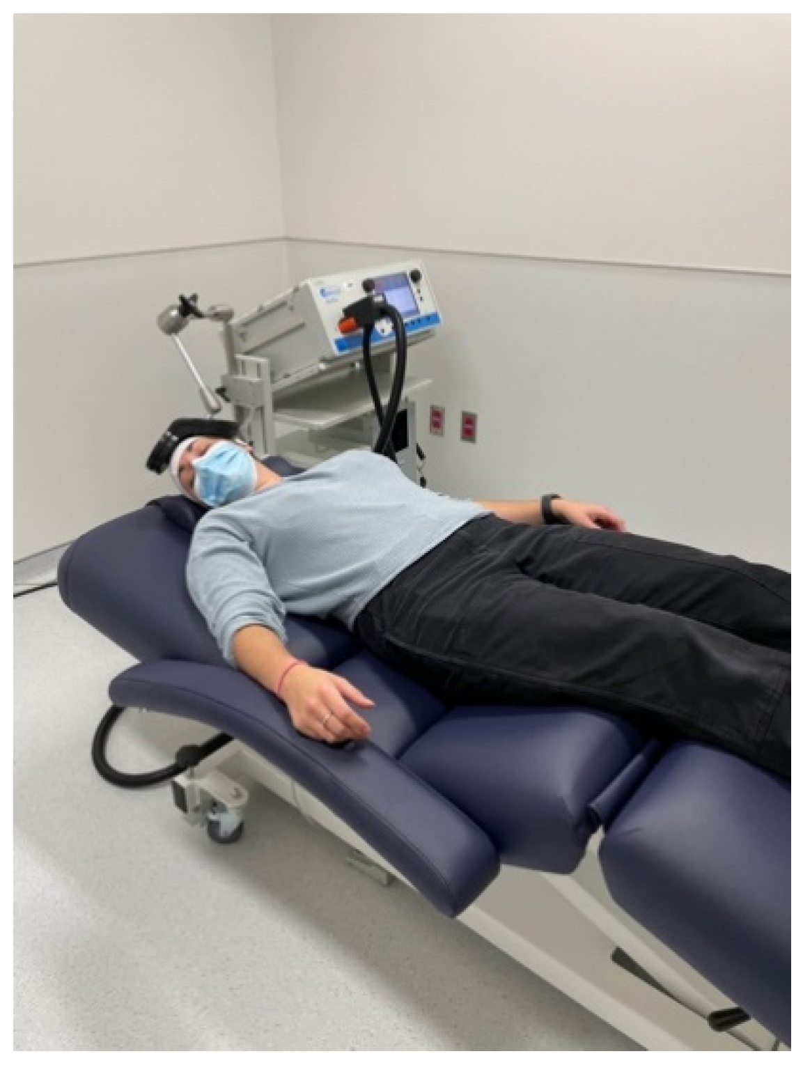
2. Case Report
3. Discussion
4. Conclusions
Author Contributions
Funding
Institutional Review Board Statement
Informed Consent Statement
Acknowledgments
Conflicts of Interest
References
- Ross, C.A.; Tabrizi, S. Huntington’s disease: From molecular pathogenesis to clinical treatment. Lancet Neurol. 2011, 10, 83–98. [Google Scholar] [CrossRef]
- Jamwal, S.; Kumar, P. Insight Into the Emerging Role of Striatal Neurotransmitters in the Pathophysiology of Parkinson’s Disease and Huntington’s Disease: A Review. Curr. Neuropharmacol. 2019, 17, 165–175. [Google Scholar] [CrossRef] [PubMed]
- Galts, C.P.; Bettio, L.; Jewett, D.C.; Yang, C.C.; Brocardo, P.S.; Rodrigues, A.L.S.; Thacker, J.S.; Gil-Mohapel, J. Depression in neurodegenerative diseases: Common mechanisms and current treatment options. Neurosci. Biobehav. Rev. 2019, 102, 56–84. [Google Scholar] [CrossRef]
- Kirkwood, S.C.; Su, J.L.; Conneally, P.M.; Foroud, T. Progression of Symptoms in the Early and Middle Stages of Huntington Disease. Arch. Neurol. 2001, 58, 273–278. [Google Scholar] [CrossRef] [PubMed]
- Bindler, L.; Travers, D.; Millet, B. [Suicide in Huntington’s disease: A review]. Rev. Med. Suisse 2009, 5, 646–648. [Google Scholar]
- Hawton, K.; Comabella, C.C.I.; Haw, C.; Saunders, K. Risk factors for suicide in individuals with depression: A systematic review. J. Affect. Disord. 2013, 147, 17–28. [Google Scholar] [CrossRef]
- Milev, R.V.; Giacobbe, P.; Kennedy, S.; Blumberger, D.M.; Daskalakis, Z.J.; Downar, J.; Modirrousta, M.; Patry, S.; Vila-Rodriguez, F.; Lam, R.W.; et al. Canadian Network for Mood and Anxiety Treatments (CANMAT) 2016 Clinical Guidelines for the Management of Adults with Major Depressive Disorder. Can. J. Psychiatry 2016, 61, 561–575. [Google Scholar] [CrossRef]
- Akil, H.; Gordon, J.; Hen, R.; Javitch, J.; Mayberg, H.; McEwen, B.; Meaney, M.J.; Nestler, E.J. Treatment resistant depression: A multi-scale, systems biology approach. Neurosci. Biobehav. Rev. 2018, 84, 272–288. [Google Scholar] [CrossRef]
- Rossi, S.; Hallett, M.; Rossini, P.M.; Pascual-Leone, A. Safety, ethical considerations, and application guidelines for the use of transcranial magnetic stimulation in clinical practice and research. Clin. Neurophysiol. 2009, 120, 2008–2039. [Google Scholar] [CrossRef] [Green Version]
- Lesenskyj, A.M.; Samples, M.P.; Farmer, J.M.; Maxwell, C.R. Treating refractory depression in Parkinson’s disease: A meta-analysis of transcranial magnetic stimulation. Transl. Neurodegener. 2018, 7, 8. [Google Scholar] [CrossRef]
- Hordacre, B.; Comacchio, K.; Williams, L.; Hillier, S. Repetitive transcranial magnetic stimulation for post-stroke depression: A randomised trial with neurophysiological insight. J. Neurol. 2021, 268, 1474–1484. [Google Scholar] [CrossRef] [PubMed]
- Beam, W.; Borckardt, J.J.; Reeves, S.T.; George, M.S. An efficient and accurate new method for locating the F3 position for prefrontal TMS applications. Brain Stimul. 2009, 2, 50–54. [Google Scholar] [CrossRef] [PubMed] [Green Version]
- Jodoin, V.D.; Miron, J.-P.; Lespérance, P. Safety and Efficacy of Accelerated Repetitive Transcranial Magnetic Stimulation Protocol in Elderly Depressed Unipolar and Bipolar Patients. Am. J. Geriatr. Psychiatry 2019, 27, 548–558. [Google Scholar] [CrossRef] [PubMed]
- Ahmed, M.A.; Darwish, E.S.; Khedr, E.M.; Elserogy, Y.; Ali, A.M. Effects of low versus high frequencies of repetitive transcranial magnetic stimulation on cognitive function and cortical excitability in Alzheimer’s dementia. J. Neurol. 2012, 259, 83–92. [Google Scholar] [CrossRef] [PubMed]
- Guse, B.; Falkai, P.; Wobrock, T. Cognitive effects of high-frequency repetitive transcranial magnetic stimulation: A systematic review. J. Neural Transm. 2009, 117, 105–122. [Google Scholar] [CrossRef] [Green Version]
- Paoli, R.A.; Botturi, A.; Ciammola, A.; Silani, V.; Prunas, C.; Lucchiari, C.; Zugno, E.; Caletti, E. Neuropsychiatric Burden in Huntington’s Disease. Brain Sci. 2017, 7, 67. [Google Scholar] [CrossRef]
- Slaughter, J.R.; Martens, M.P.; Slaughter, K.A. Depression and Huntington’s Disease: Prevalence, Clinical Manifestations, Etiology, and Treatment. CNS Spectrums 2001, 6, 306–326. [Google Scholar] [CrossRef]
- Cepeda, C.; Murphy, K.P.S.; Parent, M.; Levine, M.S. The role of dopamine in huntington’s disease. In Progress in Brain Research; Elsevier: Amsterdam, The Netherlands, 2014; Volume 211, pp. 235–254. [Google Scholar]
- Yukimasa, T.; Yoshimura, R.; Tamagawa, A.; Uozumi, T.; Shinkai, K.; Ueda, N.; Tsuji, S.; Nakamura, J. High-Frequency Repetitive Transcranial Magnetic Stimulation Improves Refractory Depression by Influencing Catecholamine and Brain-Derived Neurotrophic Factors. Pharmacopsychiatry 2006, 39, 52–59. [Google Scholar] [CrossRef] [Green Version]
- Zhao, X.; Li, Y.; Tian, Q.; Zhu, B.; Zhao, Z. Repetitive transcranial magnetic stimulation increases serum brain-derived neurotrophic factor and decreases interleukin-1β and tumor necrosis factor-α in elderly patients with refractory depression. J. Int. Med. Res. 2019, 47, 1848–1855. [Google Scholar] [CrossRef]
- Cho, S.S.; Strafella, A.P. rTMS of the Left Dorsolateral Prefrontal Cortex Modulates Dopamine Release in the Ipsilateral Anterior Cingulate Cortex and Orbitofrontal Cortex. PLoS ONE 2009, 4, e6725. [Google Scholar] [CrossRef] [Green Version]
- Pogarell, O.; Koch, W.; Pöpperl, G.; Tatsch, K.; Jakob, F.; Zwanzger, P.; Mulert, C.; Rupprecht, R.; Möller, H.-J.; Hegerl, U. Striatal dopamine release after prefrontal repetitive transcranial magnetic stimulation in major depression: Preliminary results of a dynamic [123I] IBZM SPECT study. J. Psychiatr. Res. 2006, 40, 307–314. [Google Scholar] [CrossRef]
- Pogarell, O.; Koch, W.; Pöpperl, G.; Tatsch, K.; Jakob, F.; Mulert, C.; Grossheinrich, N.; Rupprecht, R.; Möller, H.-J.; Hegerl, U.; et al. Acute prefrontal rTMS increases striatal dopamine to a similar degree as d-amphetamine. Psychiatry Res. Neuroimaging 2007, 156, 251–255. [Google Scholar] [CrossRef] [PubMed]
- Latorre, A.; Rocchi, L.; Berardelli, A.; Bhatia, K.P.; Rothwell, J.C. The use of transcranial magnetic stimulation as a treatment for movement disorders: A critical review. Mov. Disord. 2019, 34, 769–782. [Google Scholar] [CrossRef]
- Brusa, L.; Versace, V.; Koch, G.; Bernardi, G.; Iani, C.; Stanzione, P.; Centonze, D. Improvement of choreic movements by 1 Hz repetitive transcranial magnetic stimulation in Huntington’s disease patients. Ann. Neurol. 2005, 58, 655–656. [Google Scholar] [CrossRef] [PubMed]
- Shukla, A.; Jayarajan, R.N.; Muralidharan, K.; Jain, S. Repetitive Transcranial Magnetic Stimulation Not Beneficial in Severe Choreiform Movements of Huntington Disease. J. ECT 2013, 29, e16–e17. [Google Scholar] [CrossRef]
- Keck, M.; Welt, T.; Müller, M.; Erhardt, A.; Ohl, F.; Toschi, N.; Holsboer, F.; Sillaber, I. Repetitive transcranial magnetic stimulation increases the release of dopamine in the mesolimbic and mesostriatal system. Neuropharmacology 2002, 43, 101–109. [Google Scholar] [CrossRef]
- Unified Huntington’s disease rating scale: Reliability and consistency. Mov. Disord. 1996, 11, 136–142. [CrossRef]
- Rutherford, G.; Lithgow, B.; Moussavi, Z. Short and Long-term Effects of rTMS Treatment on Alzheimer’s Disease at Different Stages: A Pilot Study. J. Exp. Neurosci. 2015, 9, S24004. [Google Scholar] [CrossRef] [Green Version]
- Xie, Y.; Li, Y.; Nie, L.; Zhang, W.; Ke, Z.; Ku, Y. Cognitive Enhancement of Repetitive Transcranial Magnetic Stimulation in Patients with Mild Cognitive Impairment and Early Alzheimer’s Disease: A Systematic Review and Meta-Analysis. Front. Cell Dev. Biol. 2021, 9, 734046. [Google Scholar] [CrossRef]

Publisher’s Note: MDPI stays neutral with regard to jurisdictional claims in published maps and institutional affiliations. |
© 2021 by the authors. Licensee MDPI, Basel, Switzerland. This article is an open access article distributed under the terms and conditions of the Creative Commons Attribution (CC BY) license (https://creativecommons.org/licenses/by/4.0/).
Share and Cite
Noiseux, C.; Miron, J.-P.; Desbeaumes Jodoin, V.; Chu, T.R.; Chouinard, S.; Lespérance, P. Repetitive Transcranial Magnetic Stimulation for Major Depressive Disorder Comorbid with Huntington’s Disease: A Case Report. NeuroSci 2021, 2, 400-404. https://doi.org/10.3390/neurosci2040029
Noiseux C, Miron J-P, Desbeaumes Jodoin V, Chu TR, Chouinard S, Lespérance P. Repetitive Transcranial Magnetic Stimulation for Major Depressive Disorder Comorbid with Huntington’s Disease: A Case Report. NeuroSci. 2021; 2(4):400-404. https://doi.org/10.3390/neurosci2040029
Chicago/Turabian StyleNoiseux, Clémence, Jean-Philippe Miron, Véronique Desbeaumes Jodoin, Tian Ren Chu, Sylvain Chouinard, and Paul Lespérance. 2021. "Repetitive Transcranial Magnetic Stimulation for Major Depressive Disorder Comorbid with Huntington’s Disease: A Case Report" NeuroSci 2, no. 4: 400-404. https://doi.org/10.3390/neurosci2040029
APA StyleNoiseux, C., Miron, J.-P., Desbeaumes Jodoin, V., Chu, T. R., Chouinard, S., & Lespérance, P. (2021). Repetitive Transcranial Magnetic Stimulation for Major Depressive Disorder Comorbid with Huntington’s Disease: A Case Report. NeuroSci, 2(4), 400-404. https://doi.org/10.3390/neurosci2040029