

Antiviral Activity of Rambutan Peel Polyphenols Obtained Using Green Extraction Technology and Solvents

 , , , ,
  and
and
Abstract
1. Introduction
2. Materials and Methods
2.1. Extraction Procedures
2.1.1. Preparation of Materials and Extraction Conditions
2.1.2. Hybrid Extraction
2.1.3. Column Fractionation
2.1.4. HPLC/ESI/MS Analysis
2.1.5. Polyphenol Quantification
2.2. Antiproliferative Assays
2.2.1. Cell Culture
2.2.2. RPE Treatment and Cytotoxic Assay
2.3. Antiviral Activity
2.3.1. RNA Extraction
2.3.2. Reverse Transcription-Quantitative Polymerase Chain Reaction (RT-qPCR)
2.3.3. Quantification of HCV-RNA
2.3.4. Protein Extraction
2.3.5. Western Blot Analysis
3. Results
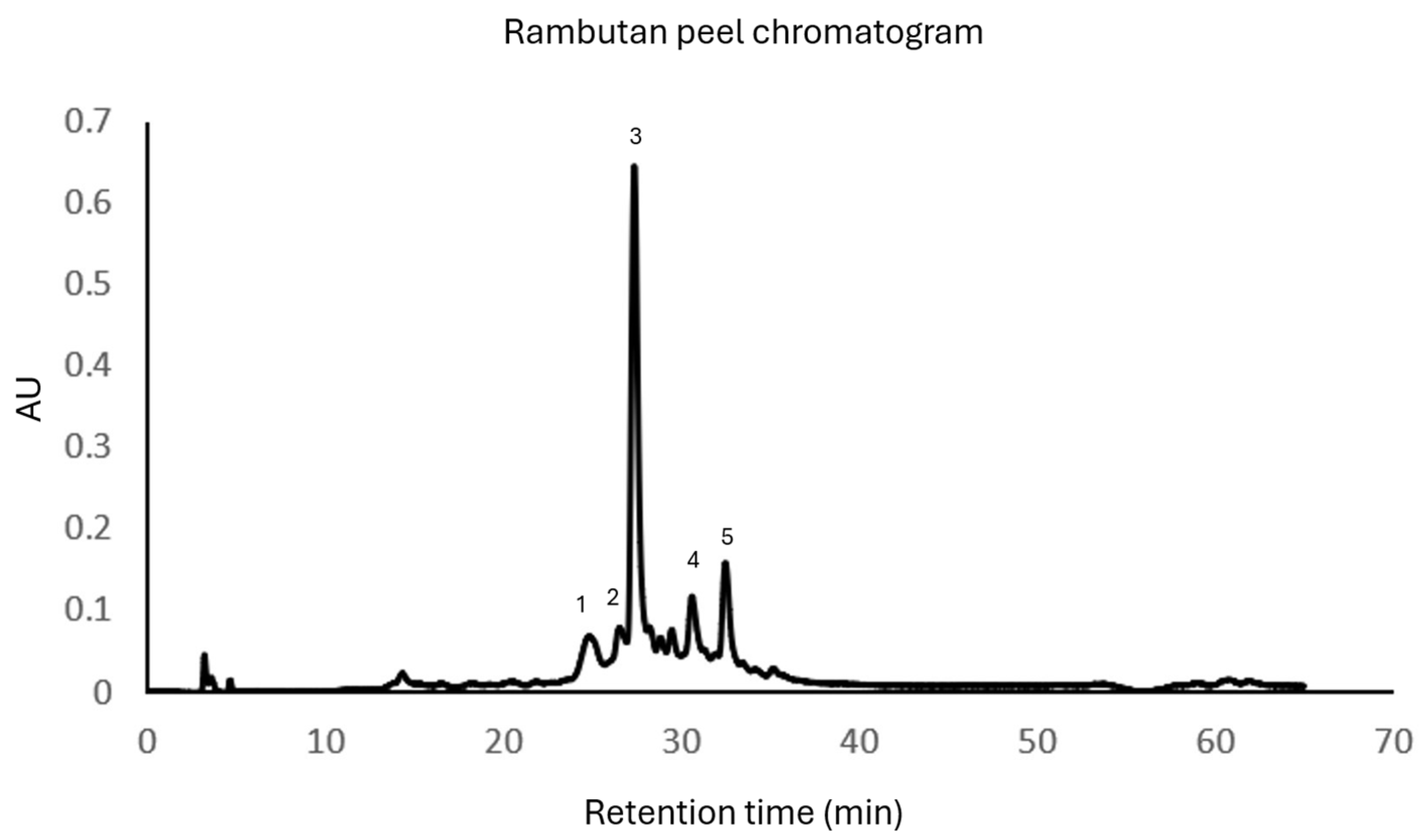
3.1. Mexican Rambutan Peel Extract
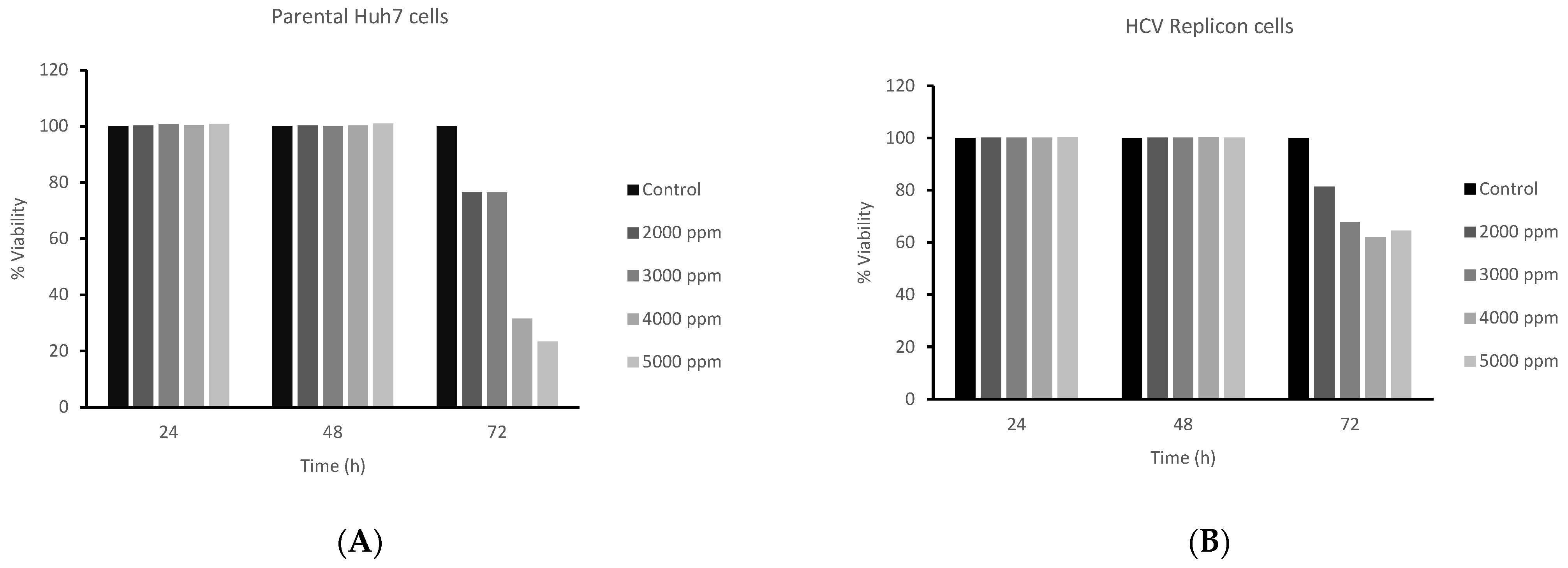
3.2. Antiproliferative Activity MTT Assay with Rambutan Peel Extracts
3.3. Antiviral Effect of Rambutan Peel Extract
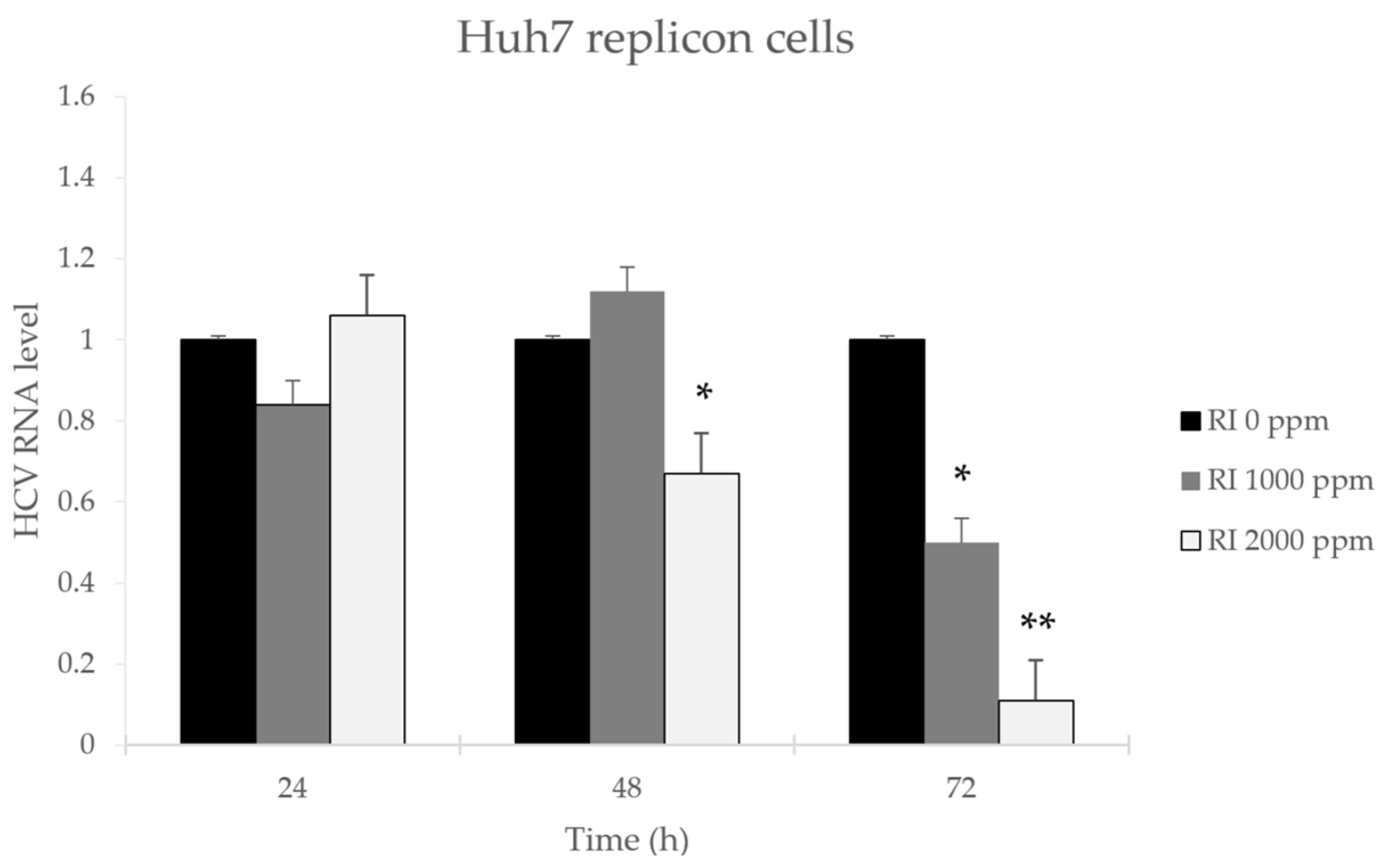
3.3.1. Effect of Rambutan Peel Extract on HCV Replication
3.3.2. HCV-NS3 Protein Levels Are Downregulated by Polyphenols from Rambutan Peel Extract in Huh7 HCV Replicon Cells
4. Discussion
5. Conclusions
Author Contributions
Funding
Institutional Review Board Statement
Informed Consent Statement
Data Availability Statement
Acknowledgments
Conflicts of Interest
References
- Maroun, R.G.; Rajha, H.N.; El Darra, N.; El Kantar, S.; Chacar, S.; Debs, E.; Vorobiev, E.; Louka, N. Emerging technologies for the extraction of polyphenols from natural sources. In Polyphenols: Properties, Recovery, and Applications; Elsevier Inc.: Amsterdam, The Netherlands, 2018; pp. 265–293. [Google Scholar]
- Wen, L.; Zhang, Z.; Sun, D.-W.; Sivagnanam, S.P.; Tiwari, B.K. Combination of emerging technologies for the extraction of bioactive compounds. Crit. Rev. Food Sci. Nutr. 2020, 60, 1826–1841. [Google Scholar] [CrossRef] [PubMed]
- Kumar, K.; Srivastav, S.; Sharanagat, V.S. Ultrasound assisted extraction (UAE) of bioactive compounds from fruit and vegetable processing by-products: A review. Ultrason. Sonochem. 2021, 70, 105325. [Google Scholar] [CrossRef]
- Chemat, F.; Rombaut, N.; Sicaire, A.-G.; Meullemiestre, A.; Fabiano-Tixier, A.-S.; Abert-Vian, M. Ultrasound assisted extraction of food and natural products. Mechanisms, techniques, combinations, protocols and applications. A review. Ultrason. Sonochem. 2017, 34, 540–560. [Google Scholar] [CrossRef] [PubMed]
- Zhang, H.-F.; Yang, X.-H.; Wang, Y. Microwave assisted extraction of secondary metabolites from plants: Current status and future directions. Trends Food Sci. Technol. 2011, 22, 672–688. [Google Scholar] [CrossRef]
- Woodhouse, I.H. Introduction to Microwave Remote Sensing; CRC Press: Boca Raton, FL, USA, 2017. [Google Scholar]
- López-Salazar, H.; Camacho-Díaz, B.H.; Ocampo, M.L.A.; Jiménez-Aparicio, A.R. Microwave-assisted extraction of functional compounds from plants: A Review. BioResources 2023, 18, 6614. [Google Scholar] [CrossRef]
- Picot-Allain, C.; Mahomoodally, M.F.; Ak, G.; Zengin, G. Conventional versus green extraction techniques—A comparative perspective. Curr. Opin. Food Sci. 2021, 40, 144–156. [Google Scholar] [CrossRef]
- SIAP. Produccion Agricola. Anuario Estadístico de la Producción Agrícola. 2023. Available online: https://nube.siap.gob.mx/cierreagricola/ (accessed on 2 August 2024).
- INIFAP. ¿Qué es el Rambután? 2022. Available online: https://www.gob.mx/inifap/articulos/que-es-el-rambutan?idiom=es (accessed on 2 August 2024).
- Hernández-Hernández, C.; Aguilar, C.; Rodríguez-Herrera, R.; Flores-Gallegos, A.; Morlett-Chávez, J.; Govea-Salas, M.; Ascacio-Valdés, J. Rambutan (Nephelium lappaceum L.): Nutritional and functional properties. Trends Food Sci. Technol. 2019, 85, 201–210. [Google Scholar] [CrossRef]
- Mahmood, K.; Fazilah, A.; Yang, T.A.; Sulaiman, S.; Kamilah, H. Valorization of rambutan (Nephelium lappaceum) by-products: Food and non-food perspectives. Int. Food Res. J. 2018, 25, 890–902. [Google Scholar]
- Estrada-Gil, L.; Contreras-Esquivel, J.C.; Flores-Gallegos, C.; Zugasti-Cruz, A.; Govea-Salas, M.; Mata-Gómez, M.A.; Rodríguez-Herrera, R.; Ascacio-Valdés, J.A. Recovery of Bioactive Ellagitannins by Ultrasound/Microwave-Assisted Extraction from Mexican Rambutan Peel (Nephelium lappaceum L.). Molecules 2022, 27, 1592. [Google Scholar] [CrossRef]
- Cerda-Cejudo, N.D.; Buenrostro-Figueroa, J.J.; Sepúlveda-Torre, L.; Torres-León, C.; Chávez-González, M.L.; Ascacio-Valdés, J.A.; Aguilar, C.N. Solid-State Fermentation for the Recovery of Phenolic Compounds from Agro-Wastes. Resources 2023, 12, 36. [Google Scholar] [CrossRef]
- De La Rosa-Esteban, K.; Sepúlveda, L.; Chávez-González, M.L.; Torres-León, C.; Estrada-Gil, L.E.; Aguilar, C.N.; Ascacio-Valdés, J.A. Valorization of Mexican Rambutan Peel through the Recovery of Ellagic Acid via Solid-State Fermentation Using a Yeast. Fermentation 2023, 9, 723. [Google Scholar] [CrossRef]
- Cheng, H.S.; Ton, S.H.; Kadir, K.A. Ellagitannin geraniin: A review of the natural sources, biosynthesis, pharmacokinetics and biological effects. Phytochem. Rev. 2017, 16, 159–193. [Google Scholar] [CrossRef]
- Reddy, B.U.; Mullick, R.; Kumar, A.; Sharma, G.; Bag, P.; Roy, C.L.; Sudha, G.; Tandon, H.; Dave, P.; Shukla, A.; et al. A natural small molecule inhibitor corilagin blocks HCV replication and modulates oxidative stress to reduce liver damage. Antivir. Res. 2018, 150, 47–59. [Google Scholar] [CrossRef] [PubMed]
- Wang, D.; Dong, X.; Wang, B.; Liu, Y.; Li, S. Geraniin Attenuates Lipopolysaccharide-Induced Cognitive Impairment in Mice by Inhibiting Toll-Like Receptor 4 Activation. J. Agric. Food Chem. 2019, 67, 10079–10088. [Google Scholar] [CrossRef] [PubMed]
- Yoganathan, S.; Alagaratnam, A.; Acharekar, N.; Kong, J. Ellagic acid and schisandrins: Natural biaryl polyphenols with therapeutic potential to overcome multidrug resistance in cancer. Cells 2021, 10, 458. [Google Scholar] [CrossRef] [PubMed]
- WHO. Hepatitis C. 2023. Available online: https://www.who.int/news-room/fact-sheets/detail/hepatitis-c (accessed on 2 August 2024).
- Lampejo, T. Influenza and antiviral resistance: An overview. Eur. J. Clin. Microbiol. Infect. Dis. 2020, 39, 1201–1208. [Google Scholar] [CrossRef]
- Aishwarya, V.; Solaipriya, S.; Sivaramakrishnan, V. Role of ellagic acid for the prevention and treatment of liver diseases. Phytother. Res. 2021, 35, 2925–2944. [Google Scholar] [CrossRef]
- Santhi, V.P.; Masilamani, P.; Sriramavaratharajan, V.; Murugan, R.; Gurav, S.S.; Sarasu, V.P.; Parthiban, S.; Ayyanar, M. Therapeutic potential of phytoconstituents of edible fruits in combating emerging viral infections. J. Food Biochem. 2021, 45, e13851. [Google Scholar] [CrossRef]
- Yan, F.; Cheng, D.; Wang, H.; Gao, M.; Zhang, J.; Cheng, H.; Wang, C.; Zhang, H.; Xiong, H. Corilagin Ameliorates Con A-Induced Hepatic Injury by Restricting M1 Macrophage Polarization. Front. Immunol. 2022, 12, 807509. [Google Scholar] [CrossRef]
- Fredsgaard, M.; Kaniki, S.E.K.; Antonopoulou, I.; Chaturvedi, T.; Thomsen, M.H. Phenolic Compounds in Salicornia spp. and Their Potential Therapeutic Effects on H1N1, HBV, HCV, and HIV: A Review. Molecules 2023, 28, 5312. [Google Scholar] [CrossRef]
- Hernández-Hernández, C.; Aguilar, C.N.; Flores-Gallegos, A.C.; Sepúlveda, L.; Rodríguez-Herrera, R.; Morlett-Chávez, J.; Govea-Salas, M.; Ascacio-Valdés, J. Preliminary testing of ultrasound/microwave-assisted extraction (U/M-AE) for the isolation of Geraniin from Nephelium lappaceum L. (Mexican Variety) peel. Processes 2020, 8, 572. [Google Scholar] [CrossRef]
- Govea-Salas, M.; Rivas-Estilla, A.M.; Rodríguez-Herrera, R.; Lozano-Sepúlveda, S.A.; Aguilar-Gonzalez, C.N.; Zugasti-Cruz, A.; Salas-Villalobos, T.B.; Morlett-Chávez, J.A. Gallic acid decreases hepatitis C virus expression through its antioxidant capacity. Exp. Ther. Med. 2015, 11, 619–624. [Google Scholar] [CrossRef] [PubMed]
- Maurya, D.K.; Nandakumar, N.; Devasagayam, T.P.A. Anticancer property of gallic acid in A549, a human lung adenocarcinoma cell line, and possible mechanisms. J. Clin. Biochem. Nutr. 2010, 48, 85–90. [Google Scholar] [CrossRef]
- Mitra, I.; Mukherjee, S.; Reddy, B.V.P.; Dasgupta, S.; Bose, K.J.C.; Mukherjee, S.; Linert, W.; Moi, S.C. Benzimidazole based Pt(II) complexes with better normal cell viability than cisplatin: Synthesis, substitution behavior, cytotoxicity, DNA binding and DFT study. RSC Adv. 2016, 6, 76600–76613. [Google Scholar] [CrossRef]
- Yedjou, C.G.; Tchounwou, P.B. In-vitro cytotoxic and genotoxic effects of arsenic trioxide on human leukemia (HL-60) cells using the MTT and alkaline single cell gel electrophoresis (Comet) assays. Mol. Cell. Biochem. 2007, 301, 123–130. [Google Scholar] [CrossRef]
- Rios-Ibarra, C.P.; Verduzco-Garza, B.; Ortiz-Lopez, R.; Grondin, Y.; Salinas-Santander, M.; Arvizu-Gutierrez, L.A.; Sanchez-Salazar, M.G.; Cervantes-Astorga, E.; Orozco-Nunnelly, D.A.; Rivas-Estilla, A.M. Transcriptional Profile of HCV Replicon Cells after Treatment with Acetylsalicylic Acid. Ann. Clin. Lab. Sci. 2022, 52, 222–229. [Google Scholar]
- Kopff, M.; Kopff, A.; Kowalczyk, E. The effect of nonsteroidal anti-inflammatory drugs on oxidative/antioxidative balance. Pol. Merkur. Lek. 2007, 23, 184–187. [Google Scholar]
- Lee, H.-K.; Yoon, H.; Jang, K.L. All-trans retinoic acid inhibits HCV replication by downregulating core levels via E6AP-mediated proteasomal degradation. Biochem. Biophys. Res. Commun. 2022, 594, 15–21. [Google Scholar] [CrossRef]
- Arakawa, M.; Tabata, K.; Ishida, K.; Kobayashi, M.; Arai, A.; Ishikawa, T.; Suzuki, R.; Takeuchi, H.; Tripathi, L.P.; Mizuguchi, K.; et al. Flavivirus recruits the valosin-containing protein–NPL4 complex to induce stress granule disassembly for efficient viral genome replication. J. Biol. Chem. 2022, 298, 101597. [Google Scholar] [CrossRef]
- Kullappan, M.; Benedict, B.A.; Rajajagadeesan, A.; Baskaran, P.; Periadurai, N.D.; Ambrose, J.M.; Gandhamaneni, S.H.; Nakkella, A.K.; Agarwal, A.; Veeraraghavan, V.P.; et al. Ellagic Acid as a Potential Inhibitor against the Nonstructural Protein NS3 Helicase of Zika Virus: A Molecular Modelling Study. BioMed Res. Int. 2022, 2022, 2044577. [Google Scholar] [CrossRef]
- García-Ortíz, J.; Ascacio-Valdés, J.; Nery-Flores, S.; Sáenz-Galindo, A.; Flores-Gallegos, A.; Rodríguez-Herrera, R. Microwave-ultrasound hybrid technology assisted extraction of pigments with antioxidant potential from red corn. Appl. Food Res. 2023, 3, 100350. [Google Scholar] [CrossRef]
- González-González, G.M.; Esparza-González, S.C.; Nery-Flores, S.D.; Morlett-Chávez, J.A.; Ascacio-Valdés, J.A.; Flores-Gallegos, A.C.; Saenz-Galindo, A.; Rodríguez-Herrera, R. Anticancer activity of polyphenolic Punica granatum peel extracts obtained by hybrid ultrasound-microwave assisted extraction: Evaluation on HeLa and HepG2 cells. Environ. Qual. Manag. 2024, 33, 295–304. [Google Scholar] [CrossRef]
- Torres-León, C.; Rojas, R.; Serna-Cock, L.; Belmares-Cerda, R.; Aguilar, C.N. Extraction of antioxidants from mango seed kernel: Optimization assisted by microwave. Food Bioprod. Process. 2017, 105, 188–196. [Google Scholar] [CrossRef]
- Thilakarathna, R.C.N.; Siow, L.F.; Tang, T.-K.; Lee, Y.Y. A review on application of ultrasound and ultrasound assisted technology for seed oil extraction. J. Food Sci. Technol. 2023, 60, 1222–1236. [Google Scholar] [CrossRef] [PubMed]
- Martina, K.; Tagliapietra, S.; Barge, A.; Cravotto, G. Combined Microwaves/Ultrasound, a Hybrid Technology. Top. Curr. Chem. 2016, 374, 175–201. [Google Scholar] [CrossRef]
- Khadhraoui, B.; Ummat, V.; Tiwari, B.; Fabiano-Tixier, A.; Chemat, F. Review of ultrasound combinations with hybrid and innovative techniques for extraction and processing of food and natural products. Ultrason. Sonochem. 2021, 76, 105625. [Google Scholar] [CrossRef]
- Phuong, N.N.M.; Le, T.T.; Dang, M.Q.; Van Camp, J.; Raes, K. Selection of Extraction Conditions of Phenolic Compounds from Rambutan (Nephelium lappaceum L.) Peel. Food Bioprod. Process. 2020, 122, 222–229. [Google Scholar] [CrossRef]
- Phuong, N.N.M.; Le, T.T.; Van Camp, J.; Raes, K. Evaluation of Antimicrobial Activity of Rambutan (Nephelium lappaceum L.) Peel Extracts. Int. J. Food Microbiol. 2020, 321, 108539. [Google Scholar] [CrossRef]
- Xu, J.; Qin, N.; Yao, Y.; Chen, T.; Jiang, W. Geraniin inhibits bladder cancer cell growth via regulation of PI3K/AKT signaling pathways. Trop. J. Pharm. Res. 2020, 19, 253–257. [Google Scholar] [CrossRef]
- Ahmad, S.A.A.; Palanisamy, U.D.; Khoo, J.J.; Dhanoa, A.; Hassan, S.S. Efficacy of geraniin on dengue virus type-2 infected BALB/c mice. Virol. J. 2019, 16, 26. [Google Scholar] [CrossRef]
- Kim, J.Y.; Choi, Y.J.; Kim, H.-J. Determining the effect of ellagic acid on the proliferation and migration of pancreatic cancer cell lines. Transl. Cancer Res. 2021, 10, 424–433. [Google Scholar] [CrossRef] [PubMed]
- Lim, S.K.; Othman, R.; Yusof, R.; Heh, C.H. Rational drug discovery: Ellagic acid as a potent dual-target inhibitor against hepatitis C virus genotype 3 (HCV G3) NS3 enzymes. Chem. Biol. Drug Des. 2021, 97, 28–40. [Google Scholar] [CrossRef] [PubMed]
- Pietschmann, T.; Brown, R.J. Hepatitis C Virus. Trends Microbiol. 2019, 27, 379–380. [Google Scholar] [CrossRef] [PubMed]
- Kotwal, G.J. Preventing Future Emerging Viral Infections Using broad-spectrum Antivirals. Future Virol. 2018, 13, 229–232. [Google Scholar] [CrossRef]
- Reddy, B.U.; Mullick, R.; Kumar, A.; Sudha, G.; Srinivasan, N.; Das, S. Small molecule inhibitors of HCV replication from Pomegranate. Sci. Rep. 2014, 4, 5411. [Google Scholar] [CrossRef]


| Hydrolysable Polyphenols (mg/g) | Condensed Polyphenols (mg/g) | Total Polyphenolic Content (mg/g) | |
|---|---|---|---|
| Raw extract | 201.6 ± 6.43 | 141.84 ± 2.26 | 343.44 ± 6.43 |
| Purified extract | 221.38 ± 3.62 | 157.10 ± 5.57 | 378.48 ± 9.19 |
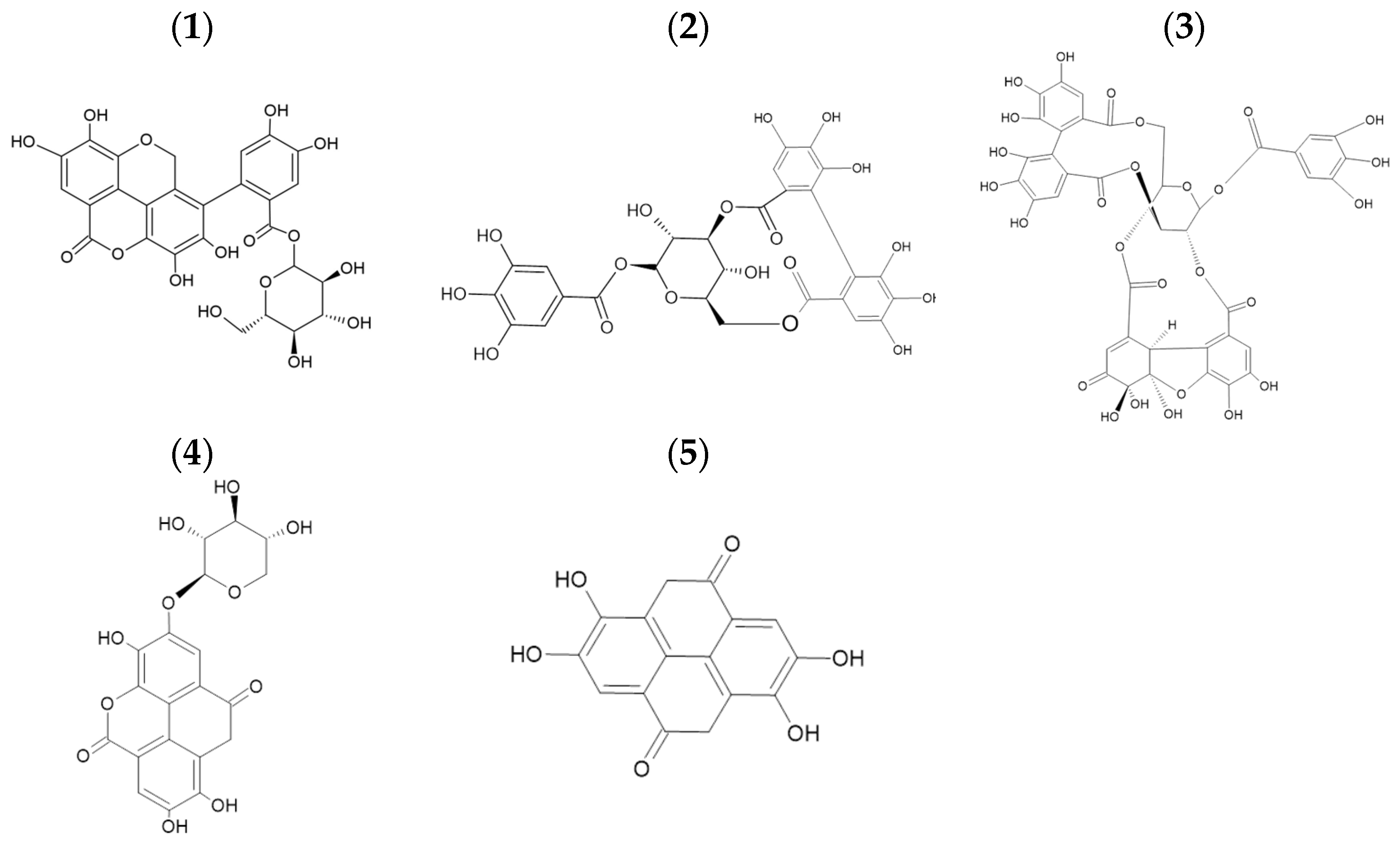
| ID | Retention Time (min) | Compounds | Mass (m/z) | MS2 | Group/Family |
|---|---|---|---|---|---|
| 1 | 25.92 | Dimmers of tergallagic-O-hexoside | 630.9 | Ellagitannin | |
| 2 | 26.21 | Corilagin | 633.0 | 481, 301, 275 | Ellagitannin |
| 3 | 28.12 | Geraniin | 950.9 | 933, 301, 169 | Ellagitannin |
| 4 | 32.13 | Ellagic Acid pentoside | 433.0 | 299, 300, 287, 125 | Ellagitannin |
| 5 | 33.71 | Ellagic Acid | 300.9 | 257, 229, 185 | Hydroxybenzoic Acid Dimmers |
Disclaimer/Publisher’s Note: The statements, opinions and data contained in all publications are solely those of the individual author(s) and contributor(s) and not of MDPI and/or the editor(s). MDPI and/or the editor(s) disclaim responsibility for any injury to people or property resulting from any ideas, methods, instructions or products referred to in the content. |
© 2025 by the authors. Licensee MDPI, Basel, Switzerland. This article is an open access article distributed under the terms and conditions of the Creative Commons Attribution (CC BY) license (https://creativecommons.org/licenses/by/4.0/).
Share and Cite
Hernández-Hernández, C.; Estrada-Gil, L.E.; Lozano-Sepúlveda, S.A.; Rivas-Estilla, A.M.; Govea-Salas, M.; Morlett-Chávez, J.; Aguilar, C.N.; Ascacio-Valdés, J.A. Antiviral Activity of Rambutan Peel Polyphenols Obtained Using Green Extraction Technology and Solvents. Sustain. Chem. 2025, 6, 14. https://doi.org/10.3390/suschem6020014
Hernández-Hernández C, Estrada-Gil LE, Lozano-Sepúlveda SA, Rivas-Estilla AM, Govea-Salas M, Morlett-Chávez J, Aguilar CN, Ascacio-Valdés JA. Antiviral Activity of Rambutan Peel Polyphenols Obtained Using Green Extraction Technology and Solvents. Sustainable Chemistry. 2025; 6(2):14. https://doi.org/10.3390/suschem6020014
Chicago/Turabian StyleHernández-Hernández, Christian, Luis E. Estrada-Gil, Sonia A. Lozano-Sepúlveda, Ana M. Rivas-Estilla, Mayela Govea-Salas, Jesús Morlett-Chávez, Cristóbal N. Aguilar, and Juan A. Ascacio-Valdés. 2025. "Antiviral Activity of Rambutan Peel Polyphenols Obtained Using Green Extraction Technology and Solvents" Sustainable Chemistry 6, no. 2: 14. https://doi.org/10.3390/suschem6020014
APA StyleHernández-Hernández, C., Estrada-Gil, L. E., Lozano-Sepúlveda, S. A., Rivas-Estilla, A. M., Govea-Salas, M., Morlett-Chávez, J., Aguilar, C. N., & Ascacio-Valdés, J. A. (2025). Antiviral Activity of Rambutan Peel Polyphenols Obtained Using Green Extraction Technology and Solvents. Sustainable Chemistry, 6(2), 14. https://doi.org/10.3390/suschem6020014